biology curriculum ms life - national park service curriculum ... vocabulary: cell, cell organelles,...
TRANSCRIPT

Life Science
Middle School Biology
A curriculum and activity guide for Carlsbad Caverns National Park

Life Science Biology Curriculum
Characteristics of Life ............................................................................................. 2
1. Wiggly Jiggly Cells? An introductory activity that allows students to build 3D cell models in order to have a concrete example of this concept. ................... 3
2. What Makes You Tick? Teaching portion of the unit focusing on the characteristics of plant and animal cells. ......................................................... 6
3. Here’s Looking at You The lab portion of the unit where students collect and observe plant and animals cells under a microscope............................. 12
4. What do You Really Look Like? A hands on activity that enhances the 3D characteristics of plant and animal cells. ....................................................... 17

2
Characteristics of Life In order to gain a basic understanding of all living things scientists must first understand the cellular structure of living organisms. All living things (plants, animals, and bacteria) are made up of cells. Living cells are divided into two basic types: prokaryotic and eukaryotic. The cells studied in this unit are eukaryotic. The eukaryotic cells of higher plants and animals are highly structured. These cells have developed internal organelles that help them survive. In addition, there is mention of on-going microbial research in Lechuguilla Cave in the Carlsbad Caverns National Park.
This unit focuses on the similarities and differences in plant and animal cells. In the first activity, students create 3D cell models in order to have a concrete example of this concept. The second activity is the teaching portion of the unit that focuses on the characteristics of plant and animal cells. The third activity is the lab portion of the unit where students collect and observe plant and animal cells under a microscope. The last activity is a hands-on activity that enhances the 3D characteristics of plant and animal cells.
3
Wiggly, Jiggly Cells? 3D cells are a hard concept for students to grasp. When viewed under
a microscope they appear 2D. We are going to create 3D cells.
Summary: Students will observe the 3D models of plant and animal cells and describe their appearance. Duration: 2 class periods Setting: Classroom Vocabulary: cell, cell organelles, mitochondria, chloroplasts, cell membrane, cell wall, nucleus, ribosomes Standards/Benchmarks Addressed: SC1-E2, SC2-E3, SC3-E1, SC6-E2, SC6-E6, SC6-E7, SC10-E1, SC10-E2 Objectives Students will:
• create a 3D plant and a 3D animal cell following the directions given. • describe the cell as a 3D object rather than a 2D object as observed under a
microscope. • describe the appearance and location of various cell organelles within a cell. • compare and contrast the 3D plant cell and the 3D animal cell.
Background A cell is the basic unit of life. All living things are made up of cells (plants, animals, and bacteria). These organisms can be either one-celled or multicellular. Most cells are so small that they cannot be seen without a microscope. In multicellular organisms, cells are specialized to carry out different functions to sustain life. In one-celled organisms the cell carries out all the functions to sustain life within itself.
Living cells are divided into two types: prokaryotic and eukaryotic. This division is based on internal complexity.
The eukaryotic cells of protozoa, higher plants, and animals are highly structured. These cells tend to be larger than cells of bacteria, and have developed specialized packaging and transport mechanisms that may be necessary to support their larger size.
Prokaryotic cells are simple in structure, with no recognizable organelles. They have an outer cell wall that gives them shape. Just under the rigid cell wall is the more fluid cell membrane. The cytoplasm enclosed within the cell membrane does not exhibit much structure when viewed by electron microscopy.
Animal and plant cells are eukaryotic. Every animal and plant cell has a nucleus that contains chromosomes. The nuclear envelope surrounding the nucleus separates the chromosomes from the cytoplasm. Chromosomes carry genes (these are bits of DNA, the heredity material).
Animal and plant cells also contain cytoplasm. Perhaps the most important things to be found in cytoplasm are mitochondria. A mitochondrion contains all the enzymes to obtain energy from
4
glucose. They can be seen in detail with an electron microscope. Mitochondria also contain a bit of DNA, which controls how they work. Some people think that mitochondria look like bacteria.
Animal and plant cells also have a cell membrane around them. Cell membranes are very thin; nevertheless they are able to control what can get in or out of a cell.
Plant cells are surrounded by a cell wall made of cellulose. The cell wall is not living. The only thing the cell wall does is to allow very high pressure to build up inside the cell because of osmosis. Since cells have semi-permeable cell membranes, water can enter or leave by osmosis. When plant cells are put in distilled water they start to swell up, but they do not burst. Animal cells are different; they do not have cell walls. If one of your body cells is placed in distilled water it will swell up and burst. That means that animals have to excrete excess water. Some plant cells have an organelle called chloroplast that takes energy from the sun and converts it into sugar.
Materials Plastic sacks Twist ties Lemon Jell-o Boiling water Large mixing bowls Spoons Square plastic food container Plums Mandarin oranges Grapes Refrigerator
Procedure
Warm up: Ask, “What is a cell and what do you think it looks like?” Make a list of the students’ answers and discuss. Discuss with students that cells are 3D objects but when we study them under microscopes we only see them as 2D objects. Before beginning our study of cells we will be creating 3D cells.
Activity 1. Students, in groups or individually, will select 2 plastic sacks. They will put one plastic
sack in a small square plastic container so that the plastic sack completely lines the container. The other plastic sack should be open on the desk.
2. Each student or group should put the same amount of warm lemon Jell-o into the two plastic sacks so that they are nearly full. Then the required cell organelles should be put into the Jell-o: 1 plum representing the nucleus, 2-4 mandarin oranges representing the mitochondria, 2-4 grapes to represent chloroplasts (to be put only in the square plastic container). The plastic sacks represent cell membranes while the plastic container represents a cell wall. The cells should then be closed with a twist tie and refrigerated until the next day.
3. The completed cells should be discussed in depth and compared for structure and appearance. Students should be able to differentiate between the plant cell (in the square container) and the animal cell. Be sure that the students understand that cell walls and chloroplasts are only found in plant cells.

5
Wrap Up: Student can draw a picture of both the cells being sure to draw everything that they see and labeling each part. Eat your cells and enjoy!
Assessment Draw a picture of both the cells and label the parts of the cells accurately.
Extension This lesson lends itself to discussions of tissue and how cells are grouped together to make tissue. You may want to stack the cells together and show how tissue is formed.
6
What Makes You Tick? Cells are full of organelles. This lesson describes the location
and function of the organelles in plant and animal cells.
Summary: This lesson is designed to teach the students about cell organelles and their functions within plant and animal cells. Duration: 1 class period Setting: Classroom Vocabulary: cell membrane, centrosome, cytoplasm, Golgi body, lysosome, mitochondrion, nuclear membrane, nucleolus, nucleus, ribosome, rough endoplasmic reticulum (rough ER), smooth endoplasmic reticulum (smooth ER), vacuole, chloroplast, cell wall, prokaryotic, eukaryotic Standards/Benchmarks Addressed: SC1-E2, SC3-E1, SC5-E2, SC5-E3, SC6-E2, SC6-E3, SC6-E5, SC6-E6, SC10-E1, SC10-E2 Objectives Students will:
• define vocabulary associated with cell structure. • label the parts of plant and animal cells correctly.
Background A cell is the basic unit of life. All living things are made up of cells (plants, animals, and bacteria). These organisms can be either one-celled or multicellular. Most cells are so small that they cannot be seen without a microscope. In multicellular organisms, cells are specialized to carry out different functions to sustain life. In one-celled organisms the cell carries out all the functions to sustain life within itself.
Living cells are divided into two types: prokaryotic and eukaryotic. This division is based on internal complexity.
The eukaryotic cells of protozoa, higher plants, and animals are highly structured. These cells tend to be larger than cells of bacteria, and have developed specialized packaging and transport mechanisms that may be necessary to support their larger size.
Prokaryotic cells are simple in structure, with no recognizable organelles. They have an outer cell wall that gives them shape. Just under the rigid cell wall is the more fluid cell membrane. The cytoplasm enclosed within the cell membrane does not exhibit much structure when viewed by electron microscopy.
Animal and plant cells are eukaryotic. Every animal and plant cell has a nucleus that contains chromosomes. The nuclear envelope surrounding the nucleus separates the chromosomes from the cytoplasm. Chromosomes carry genes (these are bits of DNA, the heredity material).
Animal and plant cells also contain cytoplasm. Perhaps the most important things to be found in cytoplasm are mitochondria. A mitochondrion contains all the enzymes to obtain energy from glucose. They can be seen in detail with an electron microscope. Mitochondria also contain a bit of DNA, which controls how they work. Some people think that mitochondria look like bacteria.
7
Animal and plant cells also have a cell membrane around them. Cell membranes are very thin; nevertheless they are able to control what can get in or out of a cell.
Plant cells are surrounded by a cell wall made of cellulose. The cell wall is not living. The only thing the cell wall does is to allow very high pressure to build up inside the cell because of osmosis. Since cells have semi-permeable cell membranes, water can enter or leave by osmosis. When plant cells are put in distilled water they start to swell up, but they do not burst. Animal cells are different; they do not have cell walls. If one of your body cells is placed in distilled water it will swell up and burst. That means that animals have to excrete excess water. Some plant cells have an organelle called chloroplast that takes energy from the sun and converts it into sugar.
Materials Science textbook Student handouts of plant and animal cells
Procedure Warm up: Ask students to recall what they already know about cells and cell structure. Write the students’ responses on the board.
Activity 1. Students will be given the vocabulary words to look up in their science textbook or other
resource (dictionary, Internet, or encyclopedia). Students must write the definition of each word.
2. Students will then work in pairs to complete the worksheets for plant and animal cells. Students should be allowed to use any resources needed to correctly label the worksheets.
3. Discuss the cell organelles. Describe their location and function within the cell.
Wrap Up: Discuss the similarities and differences in plant and animal cells.
Assessment Participation, completed worksheets and vocabulary words.
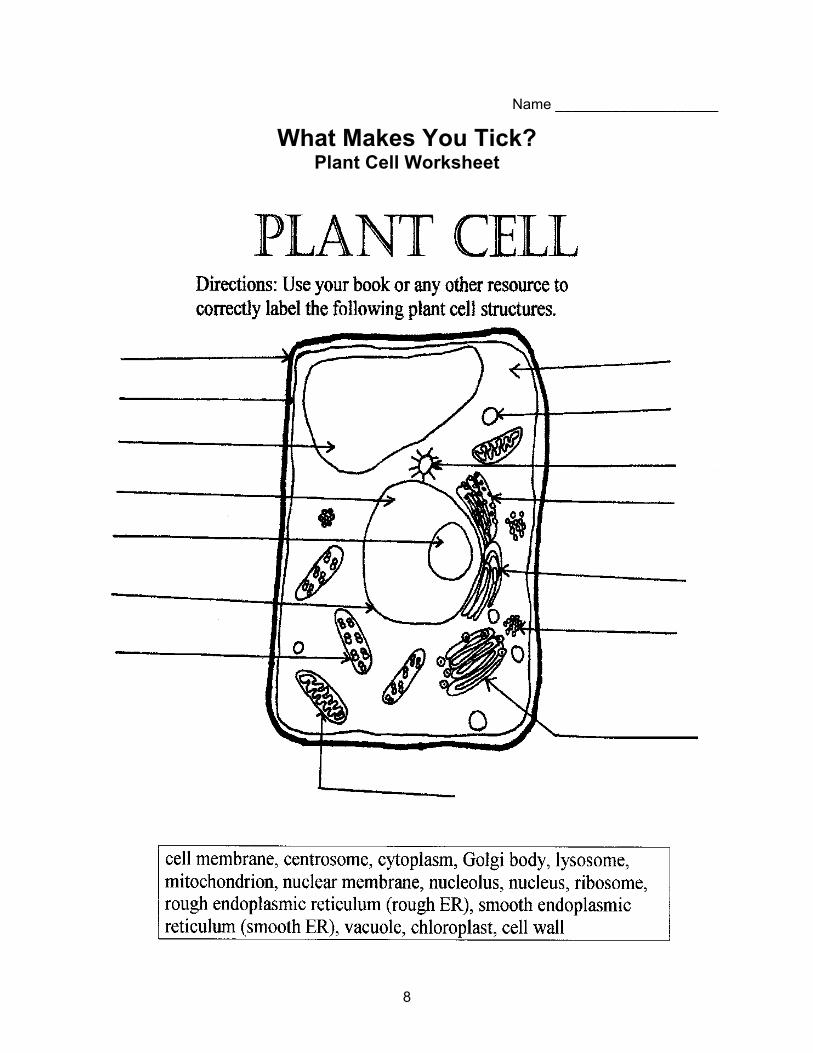
8
Name ____________________
What Makes You Tick? Plant Cell Worksheet
9
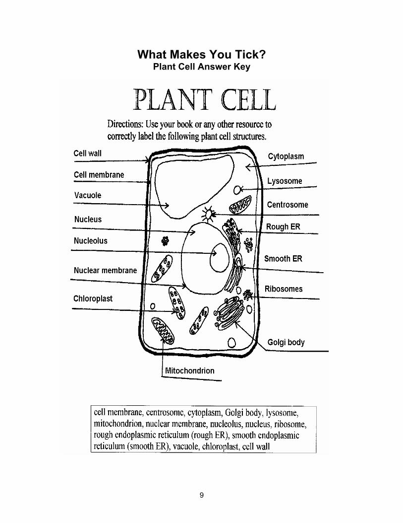
What Makes You Tick? Plant Cell Answer Key

10
Name ____________________
What Makes You Tick? Animal Cell Worksheet
11
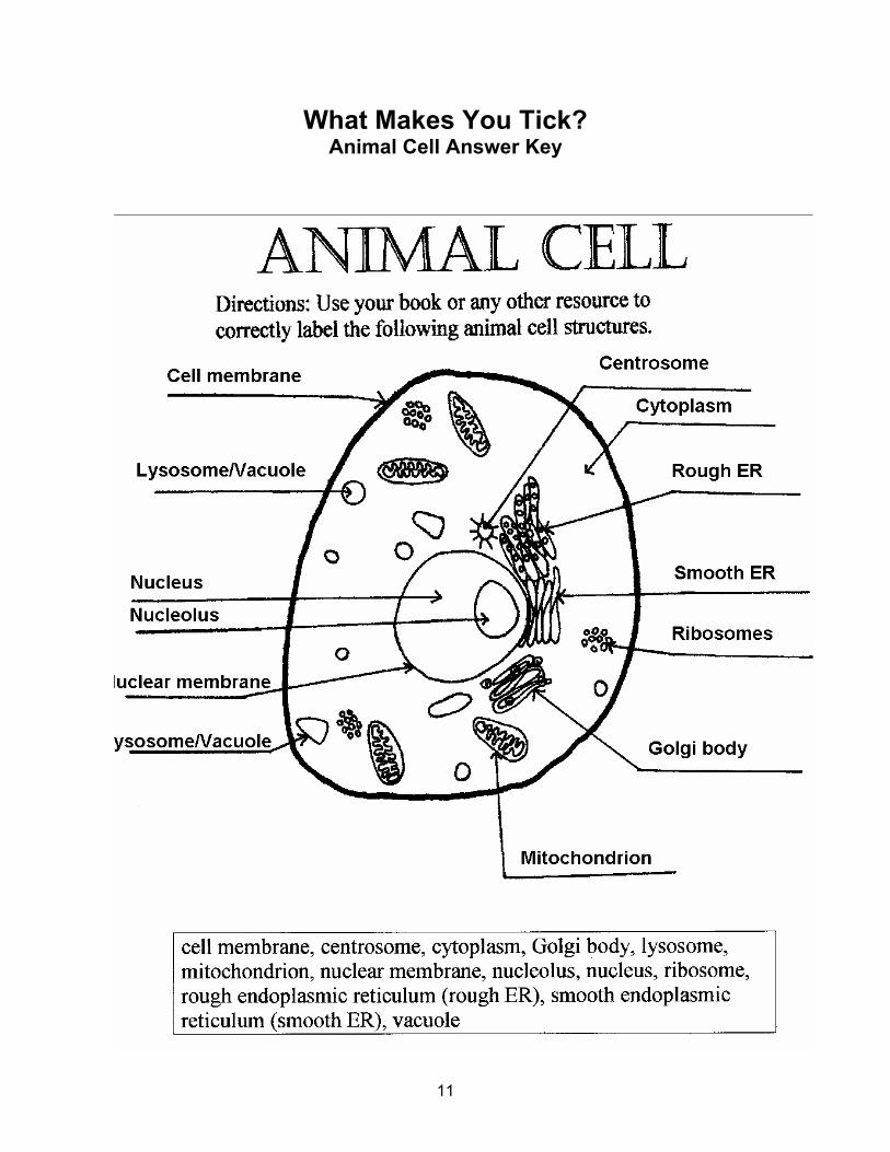
What Makes You Tick? Animal Cell Answer Key

12
Here’s Looking At You! Under the microscope you will be able to see the basic organelles in plant
and animal cells. However, these cells appear 2D but are actually 3D.
Summary: This lab is designed so that students can observe a real plant and animal cell under the microscope. Duration: 1 class period Setting: Lab Vocabulary: bacteria, fungi, chemolithotrophs, biothems Standards/Benchmarks Addressed: SC1-E2, SC3-E1, SC4-E3, SC5-E1, SC5-E2, SC5-E3, SC6-E1, SC6-E2, SC6-E3, SC6-E5, SC6-E6, SC6-E7, SC6-E8, SC10-E1, SC10-E2 Objectives Students will:
• use a microscope to observe plant and animal cells. • describe in writing what they observed about plant and animal cells.
Background In April of 1989, an expedition went down to the deepest known cave pool in Lechuguilla Cave in the Carlsbad Caverns National Park. The team was going to check the hydrogen sulfide level of the cave pool. Cavers had reported smelling a rotten egg odor in the cave. The scientists thought this could mean they were nearing the water table, a first for a Guadalupe cave. A geologist, Kiym Cunningham, was on the expedition. When Cunningham submerged his hands into the cave pool he felt a loose piece of rock. He brought the rock out and took it to his lab to see what the areas of unusual staining were.
After looking at the stains for several hours, under the microscope, he noticed some fungi, which feed on bacteria. He questioned how something could be living so deep in the Earth with no organic food. When he went back to the cave for more research he started to think about rock-eating bacteria, called chemolithotrophs. These organisms are only found in deep aquifers where they form rich microbial ecosystems. Scientists have discovered a strong connection between geology and biology. Nowhere is it more noticeable than in limestone.
These bacteria live throughout Lechuguilla Cave in various cave pools. The bacteria that live in the cave today are direct descendants of the sulfur-loving ones in the oil fields when the cave was formed. That means that the bacteria in Lechuguilla are living relics of an ancient underground ecosystem. With further research scientists have discovered hundreds of types of bacteria in Lechuguilla’s cave pools. Many of these bacteria are still being studied.
The bacterial cells found in Lechuguilla’s cave pools have been extensively studied. Some of these organisms seemed to show the ability to kill cells associated with a particular type of breast cancer without harming healthy cells. How can these cave bugs be capable of this? It has been suggested that in order for these bacteria to protect its underground food source from invading fungi, the organisms have developed powerful toxins to attack an enzyme associated with a particular fungal growth phase. Like humans, fungi are highly evolved eukaryotes. It

13
seems quite possible for some unicellular cave fungi to employ a growth mechanism similar to the ones that cause malignancies to blossom.
Cave bacteria have become adept at sticking tightly to surfaces in places where food will come to them. It turns out that sticking tightly to surfaces is an attribute that researchers look for in anti-cancer drugs. A few of the Lechuguilla samples are very proficient at killing any type of adjoining cell-- animal, plant, fungal, diseased, or healthy. Research has shown that three of the bacteria found in Lechuguilla cave target breast cancer cells.
There is continuing research on the bacteria and microbes found in cave pools. Scientists study microbes using a similar but more complex process than we are using in our lab today.
Materials Iodine solution Dropper bottles Microscope slides Cover slips Onion Tweezers Microscope Dropper Pond water Student lab worksheets
Prep 1. Collect or order pond water for use in this lab. 2. Make a diluted iodine solution mixing just enough iodine to color the water gold; put this
solution into the dropper bottles. 3. Cut the onion into squares. 4. Have all lab supplies out and ready before the class starts.
Procedure Warm up: Review with students the parts of the cells. What is the major difference between plant and animal cells? Today we are going to look at real plant and animal cells under the microscope.
Activity 1. Detail all safety reminders: Iodine is toxic so do not drink it. Wear safety goggles at all
times. Clean up any spills immediately. Wash your hands when you are finished. 2. Show students how to make a wet mount: show them how to add a cover slip to the
microscope slide by holding it over the specimen at an angle to the slide (Be sure the bottom is touching the slide) and gently drop the cover slip on top of the specimen.
3. Students will be paired up and given the lab worksheets that detail the steps for the lab.
Wrap Up: Students will answer questions and do a lab write-up discussing what they did, why they did it, and how it connects to the unit of study.
Assessment Completed worksheets and lab write-up.

14
Extension You may want to collect water from other outdoor sources such as river water, rain water, and puddle water to compare with the pond water. You may also want to bring in a variety of other vegetable peelings such as potato and carrot peelings to be compared with the onion peeling.
Have students view materials from other living things under the microscope. Some suggestions include a strand of hair, a flower petal, and a piece of a leaf.
*For: pond water: Carolina Biological Supply Co. 2700 York Road Burlington, NC 27215 1-800-334-5551
15
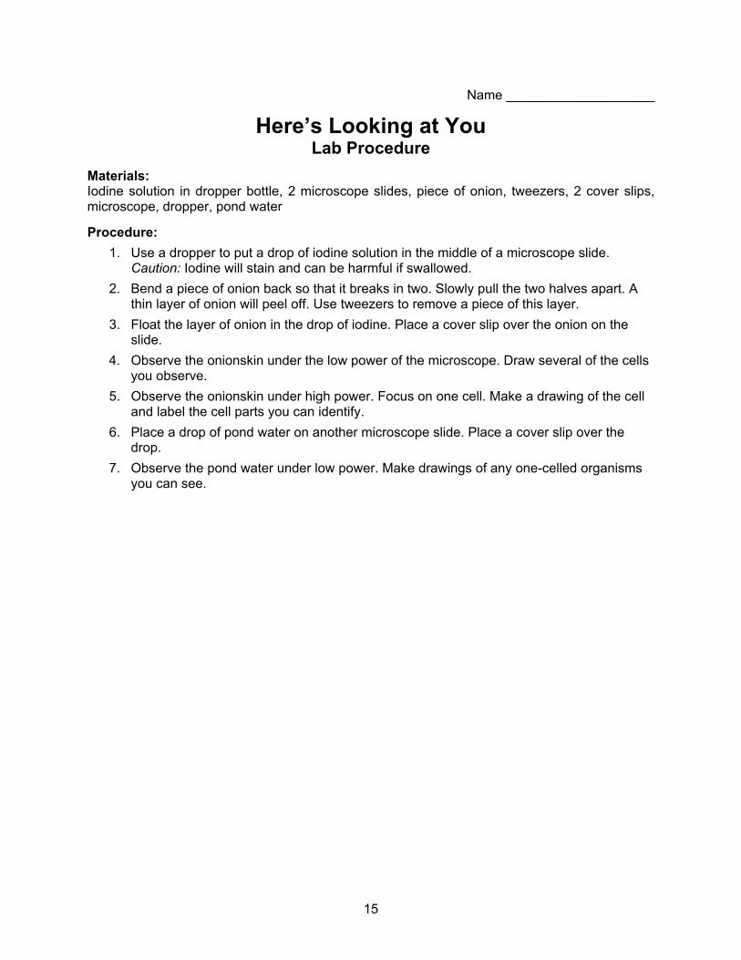
Name ____________________
Here’s Looking at You Lab Procedure
Materials: Iodine solution in dropper bottle, 2 microscope slides, piece of onion, tweezers, 2 cover slips, microscope, dropper, pond water
Procedure: 1. Use a dropper to put a drop of iodine solution in the middle of a microscope slide.
Caution: Iodine will stain and can be harmful if swallowed. 2. Bend a piece of onion back so that it breaks in two. Slowly pull the two halves apart. A
thin layer of onion will peel off. Use tweezers to remove a piece of this layer. 3. Float the layer of onion in the drop of iodine. Place a cover slip over the onion on the
slide. 4. Observe the onionskin under the low power of the microscope. Draw several of the cells
you observe. 5. Observe the onionskin under high power. Focus on one cell. Make a drawing of the cell
and label the cell parts you can identify. 6. Place a drop of pond water on another microscope slide. Place a cover slip over the
drop. 7. Observe the pond water under low power. Make drawings of any one-celled organisms
you can see.
16
Name ____________________
Here’s Looking at You Lab Results
Onion cells under low power Onion cell under high power One-celled organisms in pond water Write the answers to these questions using what you learned from the lab.
1. What do onion cells look like?
2. List the cell parts you observed. You may refer to your book for help.
3. Did you observe any one-celled organisms in the pond water?
17
What Do You Really Look Like? Cells may appear as 2D objects when viewed under the microscope;
however, they are actually 3D objects. We are going to be making 3D cells that include the major components of cell structure.
Summary: This lesson is designed to extend the students’ knowledge of cellular structure, organelles, and their functions within plant and animal cells. Duration: 1 class period Setting: Classroom Vocabulary: cell membrane, cytoplasm, lysosome, mitochondrion, nuclear membrane, nucleus, ribosome, rough endoplasmic reticulum (rough ER), smooth endoplasmic reticulum (smooth ER), vacuole, chloroplast, cell wall, chromosomes Standards/Benchmarks Addressed: SC1-E2, SC2-E2, SC2-E3, SC3-E1, SC6-E1, SC6-E2, SC6-E3, SC6-E4, SC6-E5, SC6-E6, SC6-E7, SC10-E1, SC10-E2 Objectives Students will:
• create 3D plant and animal cells. • compare and contrast plant and animal cell structure. • demonstrate and understand the 3 dimensional aspect of cell structure. • identify the various parts of plant and animal cells.
Background A cell is the basic unit of life. All living things are made up of cells (plants, animals, and bacteria). These organisms can be either one-celled or multicellular. Most cells are so small that they cannot be seen without a microscope. In multicellular organisms, cells are specialized to carry out different functions to sustain life. In one-celled organisms the cell carries out all the functions to sustain life within itself.
Living cells are divided into two types: prokaryotic and eukaryotic. This division is based on internal complexity.
The eukaryotic cells of protozoa, higher plants, and animals are highly structured. These cells tend to be larger than cells of bacteria, and have developed specialized packaging and transport mechanisms that may be necessary to support their larger size.
Prokaryotic cells are simple in structure, with no recognizable organelles. They have an outer cell wall that gives them shape. Just under the rigid cell wall is the more fluid cell membrane. The cytoplasm enclosed within the cell membrane does not exhibit much structure when viewed by electron microscopy.
Animal and plant cells are eukaryotic. Every animal and plant cell has a nucleus that contains chromosomes. The nuclear envelope surrounding the nucleus separates the chromosomes from the cytoplasm. Chromosomes carry genes (these are bits of DNA, the heredity material).
Animal and plant cells also contain cytoplasm. Perhaps the most important things to be found in cytoplasm are mitochondria. A mitochondrion contains all the enzymes to obtain energy from

18
glucose. They can be seen in detail with an electron microscope. Mitochondria also contain a bit of DNA, which controls how they work. Some people think that mitochondria look like bacteria.
Animal and plant cells also have a cell membrane around them. Cell membranes are very thin; nevertheless they are able to control what can get in or out of a cell.
Plant cells are surrounded by a cell wall made of cellulose. The cell wall is not living. The only thing the cell wall does is to allow very high pressure to build up inside the cell because of osmosis. Since cells have semi-permeable cell membranes, water can enter or leave by osmosis. When plant cells are put in distilled water they start to swell up, but they do not burst. Animal cells are different; they do not have cell walls. If one of your body cells is placed in distilled water it will swell up and burst. That means that animals have to excrete excess water. Some plant cells have an organelle called chloroplast that takes energy from the sun and converts it into sugar.
Materials Play-doh Food coloring or tempura paint (red, purple, green, blue) Disposable gloves Yarn Peppercorns Plastic bubble packing Aluminum foil Plastic wrap Pencil shavings Scissors Large knife Glue
Prep Make or buy play-doh in these colors—red, purple, green, blue
Recipe: makes enough for 3 groups 1 cup of baking soda 1 cup flour 1 cup corn starch 4 teaspoons cream of tartar 2 tablespoons oil 1 3/4 cup water A few drops of food coloring (red, purple, green, blue)
Stovetop method: Mix and cook until dough leaves the sides of the pan. Cool on a plate with a wet cloth on top.
Oven method: Bake at 1500F overnight.
*** To color play-doh use either food coloring or tempura paint. This is where the disposable gloves would be handy.
Procedure Warm up: Have students quickly write the major differences in plant and animal cells.
Activity 1. Divide class into groups of 2-4 students.

19
2. Have materials gathered and laid out according to the number of students. Hand out the materials and lists of cell structures to each group.
3. Tell students they will be making two cells (one plant and one animal). The first portion of the lab will focus on creating the cell structures. Students are to fold, cut, and paste until the cell structure is simulated. The students should look at pictures and lists of the cell structures in order to make them as accurate as possible. Tell the students not to put the cells together until you give them directions to do so.
4. Students are to cut the large piece of plastic in half and place each half on the table. 5. Roll the plain play-doh into 2 equal balls. Lay 1 ball on each piece of plastic wrap and
press each into a pancake about 6 inches around. 6. Have the students designate one pancake “animal cell” and the other “plant cell.” 7. Have the students place their finished cell structures (except the cell wall) in a pile on the
center of the appropriate pancake. 8. When all cell parts are in place gather up the pancake, carefully cupping it around its
toppings, and seal all the edges together forming a ball of “cytoplasm.” Now wrap the plastic wrap around the cytoplasm of both cells to form the cell membrane. Then wrap the aluminum foil around the plant cell to form the cell wall.
9. Cells may then be set aside for the next class period or each may be cut in half for observation right away.
Wrap Up: Ask the students: 1. What did you do? 2. What did you learn from this? 3. How can you use this information again?
Assessment Teacher observation, cell quiz.
20
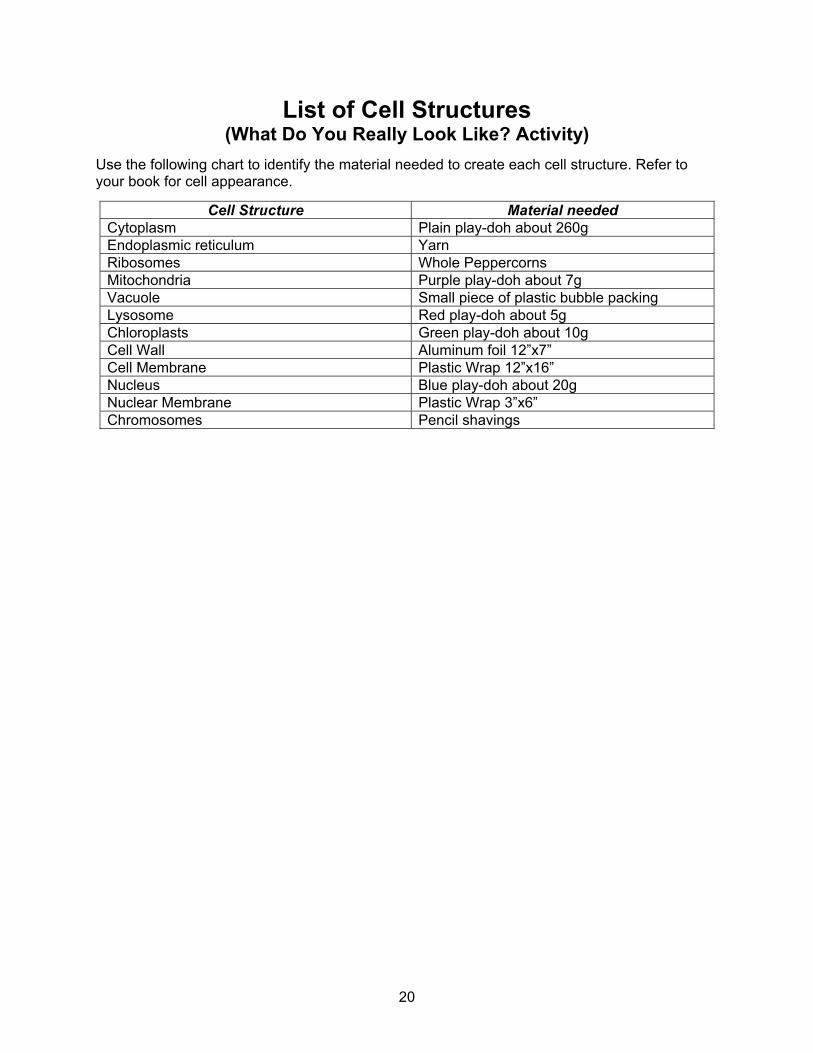
List of Cell Structures (What Do You Really Look Like? Activity)
Use the following chart to identify the material needed to create each cell structure. Refer to your book for cell appearance.
Cell Structure Material needed Cytoplasm Plain play-doh about 260g Endoplasmic reticulum Yarn Ribosomes Whole Peppercorns Mitochondria Purple play-doh about 7g Vacuole Small piece of plastic bubble packing Lysosome Red play-doh about 5g Chloroplasts Green play-doh about 10g Cell Wall Aluminum foil 12”x7” Cell Membrane Plastic Wrap 12”x16” Nucleus Blue play-doh about 20g Nuclear Membrane Plastic Wrap 3”x6” Chromosomes Pencil shavings

21
Name ____________________
Plant and Animal Cell Quiz (What Do You Really Look Like Activity)
Matching: use the words in the box below and write them next to the correct definition.
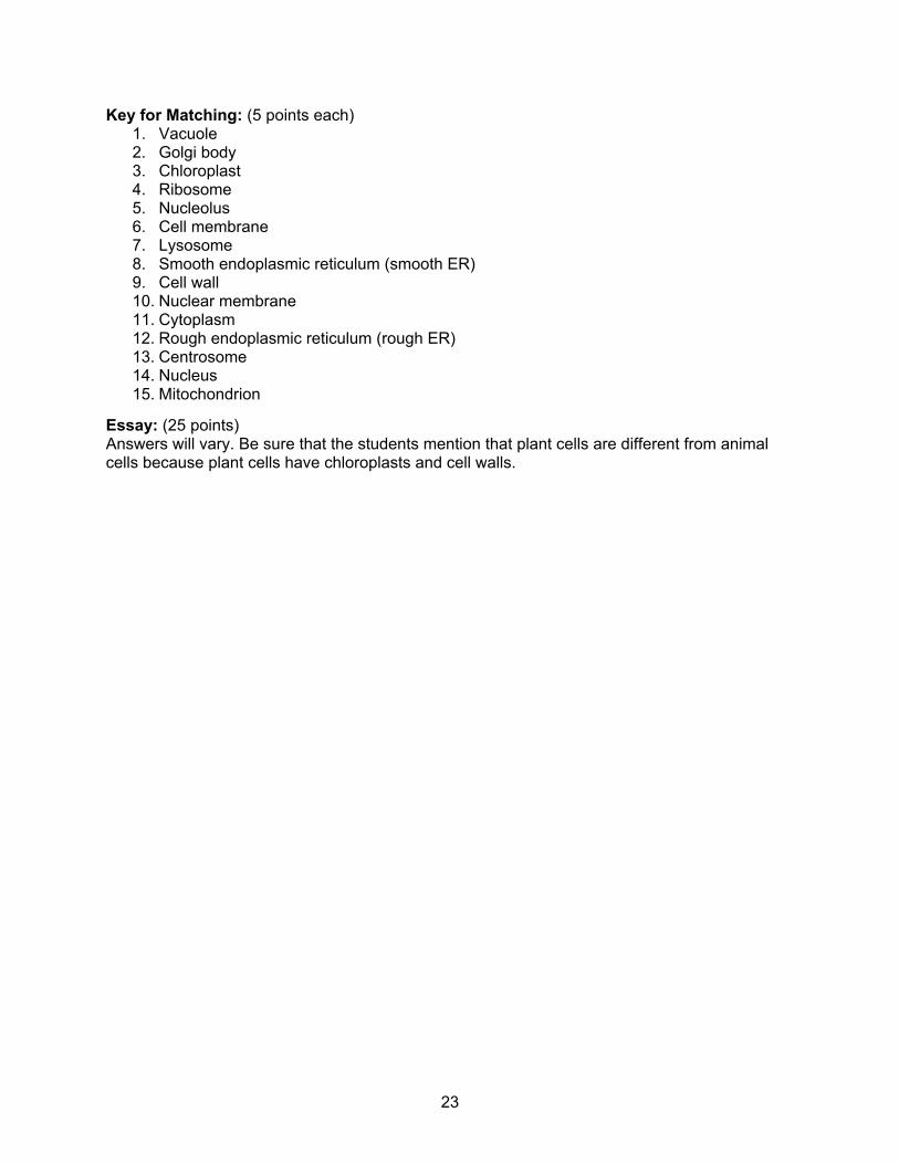
cell membrane, centrosome, cytoplasm, Golgi body, lysosome, mitochondrion, nuclear membrane, nucleolus, nucleus, ribosome, rough endoplasmic reticulum (rough ER), smooth endoplasmic reticulum (smooth ER), vacuole, chloroplast, cell wall
1. This is a fluid-filled, membrane-surrounded cavity located inside a cell. It fills with food being digested and waste material that is on its way out of the cell. _________________
2. This is a flattened, layered, sac-like organelle that looks like a stack of pancakes and is located near the nucleus. It produces the membranes that surround the lysosomes. It packages proteins and carbohydrates for “export” from the cell. ____________________
3. This is an elongated or disc-shaped organelle containing chlorophyll. Photosynthesis takes place here. _____________________
4. These are small organelles composed of RNA rich cytoplasmic granules that are sites of protein synthesis. _____________________
5. This is an organelle within the nucleus. It is where ribosomal RNA is produced. ____________________
6. This is the thin layer of protein and fat that surrounds the cell. It is semipermeable, allowing some substances to pass into the cell and blocking others. ____________________
7. These are spherical organelles surrounded by a membrane; they contain digestive enzymes. This is where the digestion of cell nutrients takes place. ____________________
8. This is a vast system of interconnected, membranous, infolded, and convoluted tubes that are located in the cell’s cytoplasm. It transports materials throughout the cell. It contains enzymes and produces and digests lipids (fats) and membrane proteins. It buds off the rough ER, moving the newly made proteins and lipids to the Golgi body, lysosomes, and membranes. ________________________
9. This is a thick, rigid membrane that surrounds a plant cell. This layer of cellulose fiber gives the cell most of its support and structure. _____________________
10. This is the membrane that surrounds the nucleus. _______________________
11. This is a jelly-like material outside the cell nucleus in which the organelles are located. _____________________
12. This is a vast system of interconnected, membranous, infolded, and convoluted sacks that are located in the cell’s cytoplasm. It is covered with ribosomes that give it a rough appearance. It transports materials through the cell and produces proteins in sacks called cisternae. ______________________
22
13. This is a small body located near the nucleus. It has a dense center and radiating tubules. It is where microtubules are made. __________________________
14. This is a spherical body containing many organelles, including the nucleolus. It controls many of the functions of the cell. _____________________
15. This is a spherical to rod-shaped organelle with a double membrane. The inner membrane is infolded many times, forming a series of projections. It converts the energy stored in glucose into ATP (adenosine triphosphate) for the cell. It is often referred to as the powerhouse of the cell. _________________________
Essay: Please answer the following question in a paragraph using complete sentences. Compare and contrast a plant and animal cell. How are they alike and how are they different?
____________________________________________________________________________
____________________________________________________________________________
____________________________________________________________________________
____________________________________________________________________________
____________________________________________________________________________
____________________________________________________________________________
____________________________________________________________________________
____________________________________________________________________________
____________________________________________________________________________
____________________________________________________________________________
____________________________________________________________________________
____________________________________________________________________________
____________________________________________________________________________
____________________________________________________________________________
____________________________________________________________________________
____________________________________________________________________________
____________________________________________________________________________
____________________________________________________________________________
____________________________________________________________________________
____________________________________________________________________________
____________________________________________________________________________
____________________________________________________________________________
____________________________________________________________________________
____________________________________________________________________________
____________________________________________________________________________
23
Key for Matching: (5 points each) 1. Vacuole 2. Golgi body 3. Chloroplast 4. Ribosome 5. Nucleolus 6. Cell membrane 7. Lysosome 8. Smooth endoplasmic reticulum (smooth ER) 9. Cell wall 10. Nuclear membrane 11. Cytoplasm 12. Rough endoplasmic reticulum (rough ER) 13. Centrosome 14. Nucleus 15. Mitochondrion
Essay: (25 points) Answers will vary. Be sure that the students mention that plant cells are different from animal cells because plant cells have chloroplasts and cell walls.