patterning of human cord blood-derived stem cells on
TRANSCRIPT
Research paper Acta Neurobiol Exp 2012, 72: 325–336
© 2012 by Polish Neuroscience Society - PTBUN, Nencki Institute of Experimental Biology
INTRODUCTION
The ability to control stem cell fate decisions is cru-cial for developing new strategies for clinical applica-tions of tissue engineering. This involves learning about cells interactions with designed microenviron-ments, such as cell-cell contacts, cell-extracellular matrix (ECM) interactions and effect of soluble/para-crine signals. Besides this strictly biological interface there are other factors that may contribute to the dif-ferentiation of stem cells or maintaining self-renewal potential. These include intrinsic properties of the cel-
lular environment, such as matrix structure and orga-nization, chemistry and mechanical properties of the substrate (Reilly and Engler 2010, Szymczak et al. 2010). It has been shown that in vivo neural cells respond to the orientation of extracellular matrix guid-ing the neuronal outgrowth and migration in the brain (Vu and Werb 2000). Similarly in vitro cells are able to distinguish geometric topography of a substrate and hance cell growth can be regulated by the patterns on the surface (Recknor et al. 2006).
Neural stem cells are found in the adult brain in two neurogenic regions: the subventricular zone (SVZ) of the lateral ventricle and subgranular zone (SGZ) of the hip-pocampus (Doetsch 2003). These two regions create spe-cific microenvironment where neural stem cells can sus-tain their undifferentiated, self-renewal state or undergo
Patterning of human cord blood-derived stem cells on single cell posts and lines: implications for neural commitment
Marzena Zychowicz1, Dora Mehn2, Ana Ruiz2, Małgorzata Frontczak-Baniewicz3, François Rossi2, and Leonora Buzanska1*
1NeuroRepair Department, Mossakowski Medical Research Centre, Polish Academy of Sciences, Warsaw, Poland, *Email: [email protected]; 2Nanobiosciences Unit, Institute for Health and Consumer Protection, European
Commission, Joint Research Centre, Ispra, Italy; 3Electron Microscopy Platform, Mossakowski Medical Research Centre, Polish Academy of Sciences, Warsaw, Poland
Using stem cells (SC) in new strategies for clinical treatment requires control of stem cell fate decision and the ability to govern their patterning and commitment in tissue engineering. Neural stem cells and other adult SC can respond to the different components of the microenvironment and their spatial arrangement in the stem cell niche. It has been shown previously by our group that different composition and architecture of patterned bioactive domains influence the developmental response of neural stem cells. In the present report we verify the commitment and differentiation of neural stem cells derived from human cord blood (HUCB-NSC) by a single cell patterning system. Microcontact printing technology was used to generate single cell positioning areas of different geometry: 10 µm-thin lines and 10 µm-width one cell posts. The commitment and differentiation of HUCB-NSC cells cultured on different surfaces were dependent on the geometry and the type of biomaterial present in bioactive domains. Fibronectin promoted neuronal protrusion outgrowth (β-tubulin III and MAP-2 positive cells) and gap junction development (Cx43 marker) between cells growing on lines while poly-L-lysine promoted HUCB-NSC differentiation into astrocytic, glial fibrillary acidic protein expressing (GFAP positive) cell phenotype. Here we also demonstrate by scanning electron microscopy that morphology of cells on the patterned surface is highly dependent upon the type of biomolecules used for printing. These kinds of platforms can be used for investigating the influence of spatial organization of environment on the SC fate decision and for studying the molecular processes occurring in a single cell.
Key words: neural stem cells, microcontact printing, single cell positioning, fibronectin, poly-L-lysine, neural commitment
Correspondence should be addressed to L. Buzanska Email: [email protected]
Received 05 September 2012, accepted 28 December 2012
326 M. Zychowicz et al.
differentiation by mutual interaction with components of the endogenous stem cell niche (Riquelme et al. 2008). The ECM can influence neural stem cell fate for example by the cell-generated physical forces/mechanical factors – stress, substrate elasticity (Leipzig and Schoichet 2009) and nanotopography (Wang et al. 2010), ECM geometry and stiffness (Saha et al. 2007). Changes in tension or stiffness alter the conformation of ECM molecules, thus altering integrin binding site accessibility, activation and releasing of growth factors, and ultimately can modify cytoskeleton structure and mechano- and osmosensitive ion channels (Gerecht et al. 2007). Mesenchymal stem cells cultured on a soft matrix with stiffness similar to the one of the brain (i.e. Young’s modulus between 0.1 and 1 kPa) differentiate into neuronal cells (showing β-tubulin III expression). In contrast, culturing of these cells on tougher matrix (8–17 kPa and 25–40 kPa) acted as a myo-genic and osteogenic inducing factor, respectively, while adding an inhibitor of myosin II, blebbistatin, completely reversed these effects (Engler et al. 2006). Ruiz and Chen (2008) have shown that besides the ECM’s having chemi-cal properties governing the fate of stem cells, differentia-tion of human mesenchymal stem cells can be depend on the mechanical forces the cells exert on each other. Using microcontact printing of domains with different shape, they have shown that spatial differentials of multicellular organization generate gradient of traction forces. The regions of high stress resulted in osteogenic differentia-tion while adipocytes originated in region of low stress of the domains (Ruiz and Chen 2008). The type of differen-tiation was also dependent on the size of islands the cells were cultured on. It has been also reported that different dimensions of aligned micro and nanostructures may influence neuronal differentiation of human embryonic and adult stem cells (Lee et al. 2010).
In this study, we investigate the influence of differ-ent patterns immobilizing single cells, on neural com-mitment and differentiation of stem cells derived from human cord blood (HUCB-NSC, Buzanska et al. 2006). Specifically, we assess: (1) different geometry of cell patterning (posts or lines) and (2) different adhesion-promoting compounds using fibronectin (integrin receptor mediated) and poly-L-lysine (elec-trostatic, non-specific adhesion).
For this purpose we have prepared single cell patterns with lines and posts, microcontact-printed on a cell repel-lent surface with extracellular matrix protein fibronectin or synthetic poly-L-lysine (PLL) (Ruiz et al. 2009, Zychowicz et al. 2011). The patterns with fibronectin and
PLL, but immobilizing group of cells and with different geometry, have been already shown to guide and commit human umbilical cord blood neural stem cells (HUCB-NSC) towards a neuronal fate (Buzanska et al. 2009). In order to identify the cell commitment and differentiation on different patterns, immunostaining against: β-tubulin-III (an early neuronal marker), GFAP (glial fibrillar acidic protein expressed in cells committed to astrocytic lin-eage), MAP-2 (microtubule associated protein-2, advanced neuronal marker localized in dendritic protrusions) and connexin 43 (a gap junction protein) was performed.
METHODS
Cell culture
A neural stem cell line, obtained from non-he-matopoietic fraction of human umbilical cord blood (HUCB-NSC, Buzanska et al. 2006) was maintained as mixed population composed of the adherent neural pro-genitors and loosely attached in DMEM/F12, 2% fetal bovine serum (FBS, Gibco), supplemented with insulin-transferrin-selenium (ITS 1:100, Gibco), antibiotic-anti-mycotic solution (AAS, 1:100, Gibco). A non-differenti-ated population of free floating cells was cultured in F12/DMEM + B27 (1:50) and EGF (20 ng/ml). The cells were propagated by trypsinization every two weeks and cultured at 37°C, 5% CO2 and 95% humidity.
Preparation of microcontact printed patterns
Petri dishes were coated with a cell-repellent plasma polymerized polyethylene oxide-like (PEO). Microstructured stamps for patterning were made of polidymetylsiloxane (PDMS), were coated with either FITC-labeled poly-L-lysine (25 µg/ml, Sigma Aldrich) diluted in carbonate buffer (100 mM NaHCO3, pH 8.5) or fibronectin (42 µg/ml, Sigma Aldrich) diluted in printing buffer (100 mM acetate at pH 5.0, 5mM EDTA, 0.01% Triton-X 100, and 0.1% glycerol). After ultrasonication for 5 minutes and cleaning in mild O2 plasma (200 W, 1.2 torr, 30 seconds), PDMS stamps were inked with PLL or fibronectin for 15 min and 45, respectively, and driven into conformal contact with the anti-adhesive PEO sur-face of the Petri dish. More detailed information of the entire procedure can be found in Bretagnol and coau-thors (2006) and Ruiz and colleagues (2008). The pat-terned Petri dishes were UV sterilized for 15 min before seeding cells.
Single cell positioning of neural stem cells 327
Cell culture on pattered surface and phenotypic analysis
Cells were seeded on the patterned Petri dish at 2 × 10⁴ cells/cm2
density in two culture conditions: serum free and in the presence of 2% serum. After incubating the cells through the night the excess was removed by changing the culture medium. Adherent cells were cultured for 1 or 7 days. At this time points cells were analysed using immunocytochemistry or scanning electron microscopy methods.
For immunocytochemical analysis, after washing with PBS, samples were fixed with 4% PFA for 15 min-utes, permeabilized with 0.1% Triton X-100 for 15 minutes and blocked with NGS 10% for 1 hour at room temperature. The primary mouse monoclonal antibod-ies for β-tubulin III (IgG2b, 1:1 000, Sigma), MAP-2 (IgG1, 1:750, Sigma), Connexin 43 (IgG1, 1:500, Chemicon), rabbit antibody for GFAP (IgG, 1:500, Cappel) were applied overnight at 4°C. After washing with PBS the secondary goat anti-mouse IgG2b (Alexa 488, Invitrogen) and IgG1 (Alexa 488 and 546, Invitrogen) and goat anti rabbit IgG H+L (Alexa 546, Invitrogen) antibodies were applied for 1 hour at room temperature. Following the Hoechst staining for 15 minutes the samples were closed in mounting medium (FluoroMount, Invitrogen) and imaged using an Axioscope microscope with AxioCamMRC5 camera (Carl Zeiss GmBH, Jena, Germany). Counting and cell measurement were performed using AxioVision Rel 4.8 (Carl Zeiss) software.
For scanning electron microscopy the cells were fixed in 1% PFA and 1.25% glutaraldehyde in cacodylate buffer for 2 h, stained in 1% OsO4 and gradient series of ethyl alcohol was used for dehydration. After drying in a criti-cal point the samples were coated with gold and observed by scanning electron microscopy (JEOL JSM-6390LV). The studies were performed in the Electron Microscopy Platform, Mossakowski Medical Research Centre Polish Academy of Sciences Warsaw, Poland.
Statistical analysis
Data are presented as mean ±SE. At least 300 cells per experiment were measured. Statistical comparisons were made using One-Way Anova Bonferroni multiple com-parison test (GraphPad Prism 5.0), from three independent experiments, where difference at P-value <0.05 was estab-lished as statistically significant.
RESULTS
Phenotype of the HUCB-NSC adhered to the patterns with lines and single cell posts
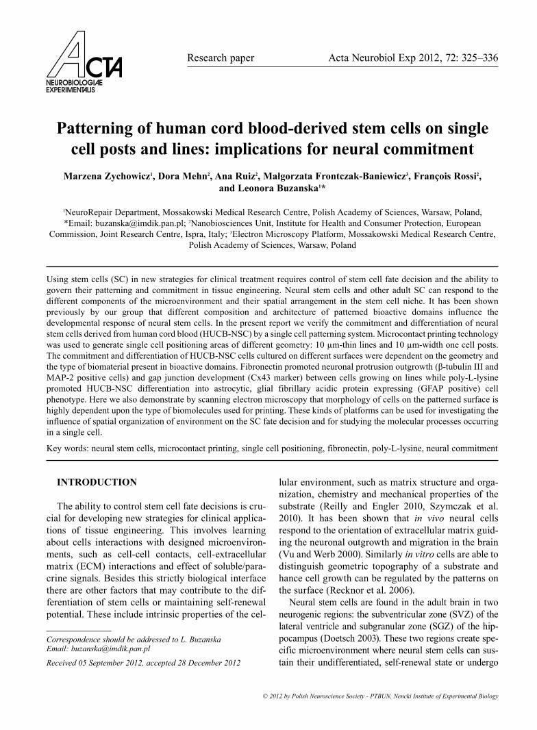
The experiment was performed on poly-L-lysine or fibronectin microprinted patterns of 10 µm wide lines (Fig. 1) and on 10 µm square posts designed to capture a single cell (Fig. 2). One hour after plating, the cells attached firmly to the fibronectin patterns while strong adhesion on PLL was visible only after 24 hours (data not shown). The cells adhered robustly to fibronectin lines, with their morphology illustrating a tendency to spread along the patterned substrate (Fig. 1C, D). The cells on PLL had a more rounded morphology, indicating that their adhesion was not that robust though they nevertheless respect the underlying patterning geometry (Fig. 1A, B).
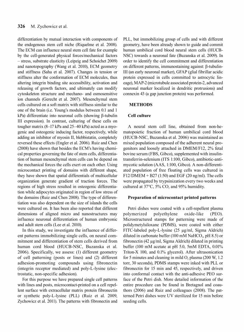
The cells seeded on the PLL single square patterns were restricted to the domains, remained rounded, without a tendency to cross the non-adhesive surface (Fig. 2A, B); while the on FN cells tended to spread beyond the 10 µm square boundary, creating protru-sions and interconnections between the neighboring domains (Fig. 2C, D).
Neural commitment and differentiation of the HUCB-NSC on the pattern with lines
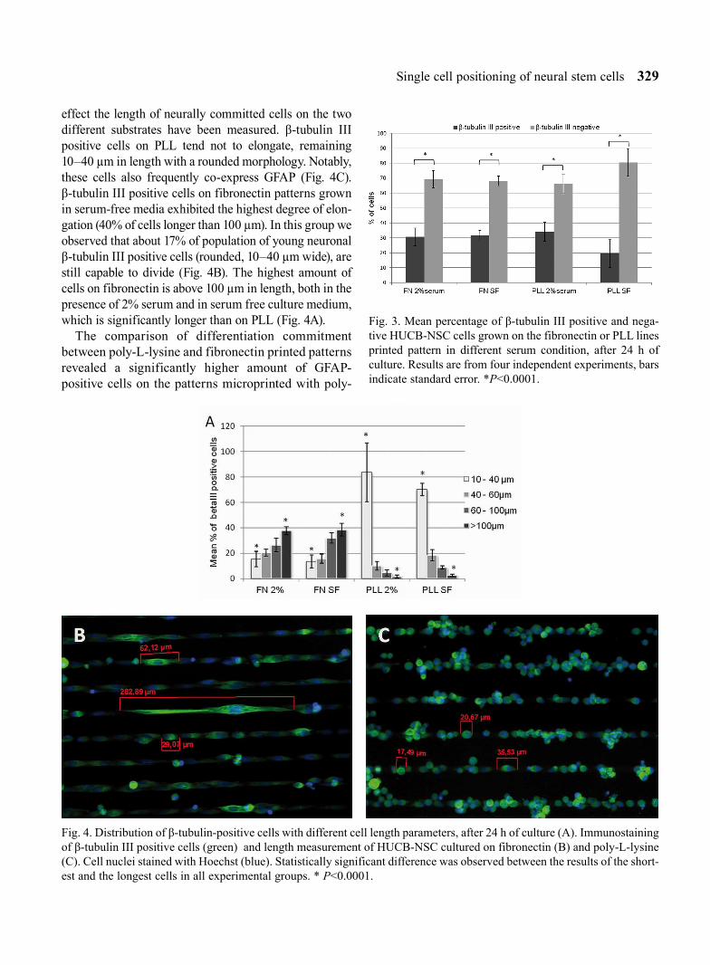
After culturing the cells for one day they were fixed and stained in order to visualise the influence of micro-contact printed pattern on commitment of the HUCB-NSC cells. We have observed the highest number of non-neuronally-commited cells on poly-L-lysine printed pat-terns in serum free culture, though this difference was not statistically significant. However, in all the tested experimental conditions the number of undifferentiated cells was statistically higher than the amount of β-tubulin positive cells (Fig. 3). There was no statistically signifi-cant difference between the proportion of neuronally commited cells, as measured by the presence β-tubulin III positive cells in the four tested experimental groups. Although the proportion of β-tubulin III positive cells does not vary across groups, there are distinct differences in morphology (Fig. 4B and C). Neuronal differentiation is usually associated with a higher degree of cell elonga-tion, characterized by forming filopodial protrusions with adhesive contacts in growth cones, and accompa-nied by establishing cytoskeletal filaments orientation (Fass and Odde 2003, Dent et al. 2007). To quantify this
328 M. Zychowicz et al.
Fig. 1. SEM micrographs showing the adhesion of HUCB-NSC after 24 hours culture on poly-L-lysine (A, B) and on fibronectin (C, D) microprinted lines. Culture was performed in the presence of 2% serum. Note loosely adherent and rounded cells on PLL (B) and very firmly attached cells on fibronectin (D).
Fig. 2. SEM images showing the adhesion of HUCB-NSC after 24 hours culture on poly-L-lysine (A, B) and on fibronectin (C, D) microprinted single squares. Culture was performed in the presence of 2% serum. On the PLL cells remain rounded (B) while on fibronectin start to grow over the pattern (D).
Single cell positioning of neural stem cells 329
effect the length of neurally committed cells on the two different substrates have been measured. β-tubulin III positive cells on PLL tend not to elongate, remaining 10–40 µm in length with a rounded morphology. Notably, these cells also frequently co-express GFAP (Fig. 4C). β-tubulin III positive cells on fibronectin patterns grown in serum-free media exhibited the highest degree of elon-gation (40% of cells longer than 100 µm). In this group we observed that about 17% of population of young neuronal β-tubulin III positive cells (rounded, 10–40 µm wide), are still capable to divide (Fig. 4B). The highest amount of cells on fibronectin is above 100 µm in length, both in the presence of 2% serum and in serum free culture medium, which is significantly longer than on PLL (Fig. 4A).
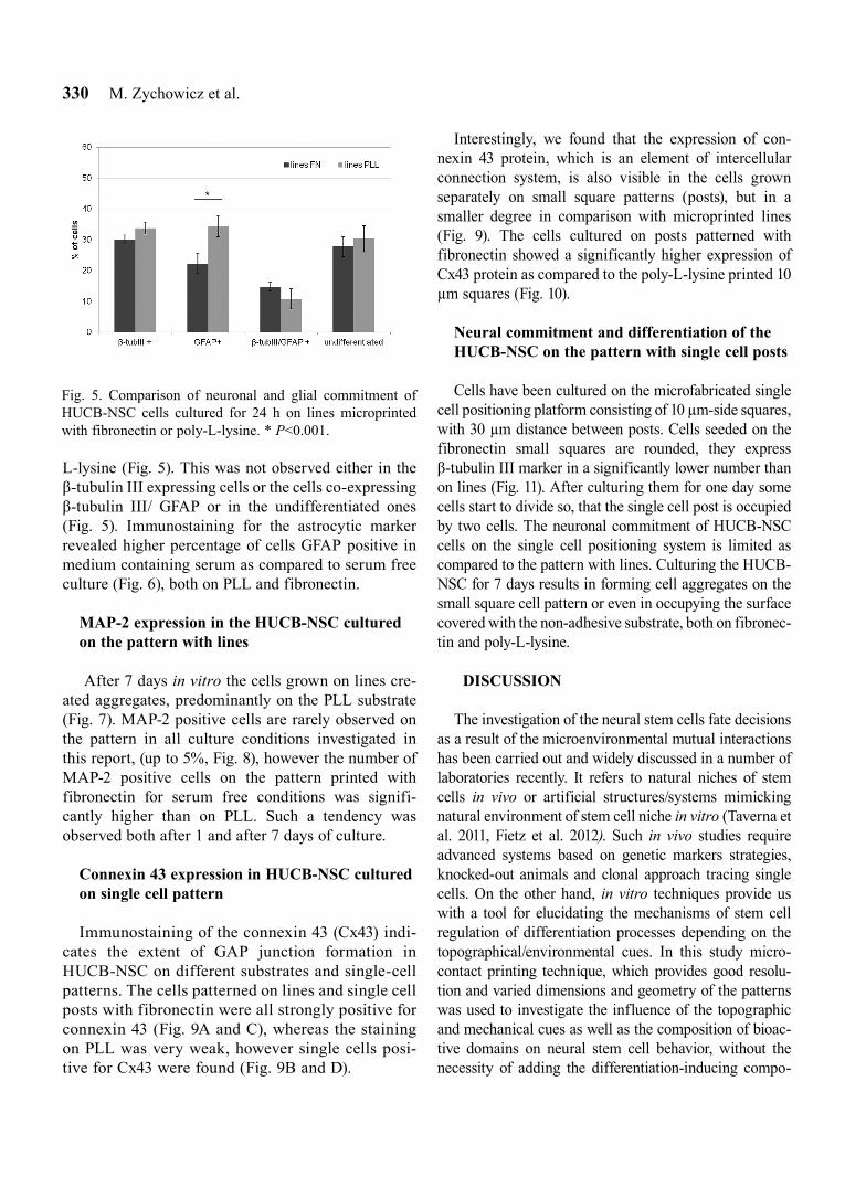
The comparison of differentiation commitment between poly-L-lysine and fibronectin printed patterns revealed a significantly higher amount of GFAP-positive cells on the patterns microprinted with poly-
Fig. 4. Distribution of β-tubulin-positive cells with different cell length parameters, after 24 h of culture (A). Immunostaining of β-tubulin III positive cells (green) and length measurement of HUCB-NSC cultured on fibronectin (B) and poly-L-lysine (C). Cell nuclei stained with Hoechst (blue). Statistically significant difference was observed between the results of the short-est and the longest cells in all experimental groups. * P<0.0001.
Fig. 3. Mean percentage of β-tubulin III positive and nega-tive HUCB-NSC cells grown on the fibronectin or PLL lines printed pattern in different serum condition, after 24 h of culture. Results are from four independent experiments, bars indicate standard error. *P<0.0001.
330 M. Zychowicz et al.
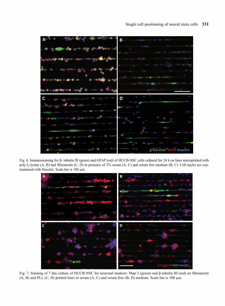
L-lysine (Fig. 5). This was not observed either in the β-tubulin III expressing cells or the cells co-expressing β-tubulin III/ GFAP or in the undifferentiated ones (Fig. 5). Immunostaining for the astrocytic marker revealed higher percentage of cells GFAP positive in medium containing serum as compared to serum free culture (Fig. 6), both on PLL and fibronectin.
MAP-2 expression in the HUCB-NSC cultured on the pattern with lines
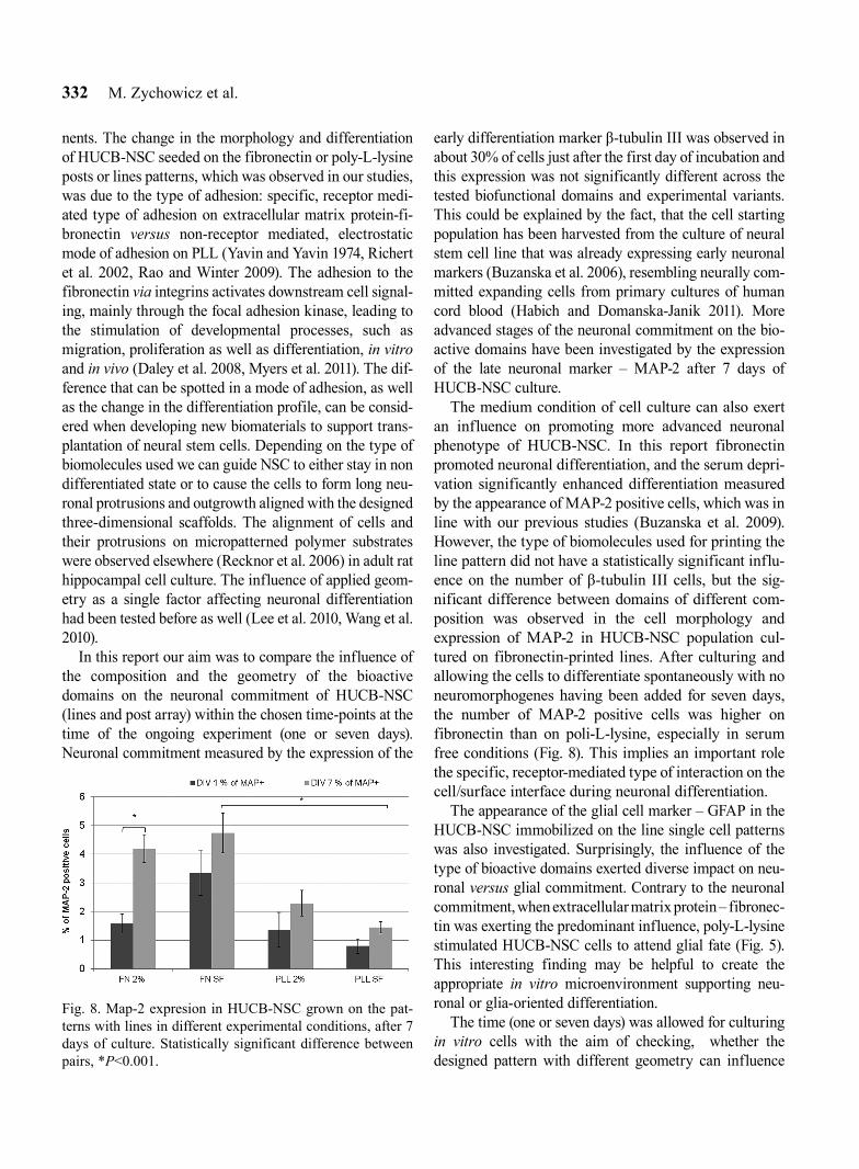
After 7 days in vitro the cells grown on lines cre-ated aggregates, predominantly on the PLL substrate (Fig. 7). MAP-2 positive cells are rarely observed on the pattern in all culture conditions investigated in this report, (up to 5%, Fig. 8), however the number of MAP-2 positive cells on the pattern printed with fibronectin for serum free conditions was signifi-cantly higher than on PLL. Such a tendency was observed both after 1 and after 7 days of culture.
Connexin 43 expression in HUCB-NSC cultured on single cell pattern
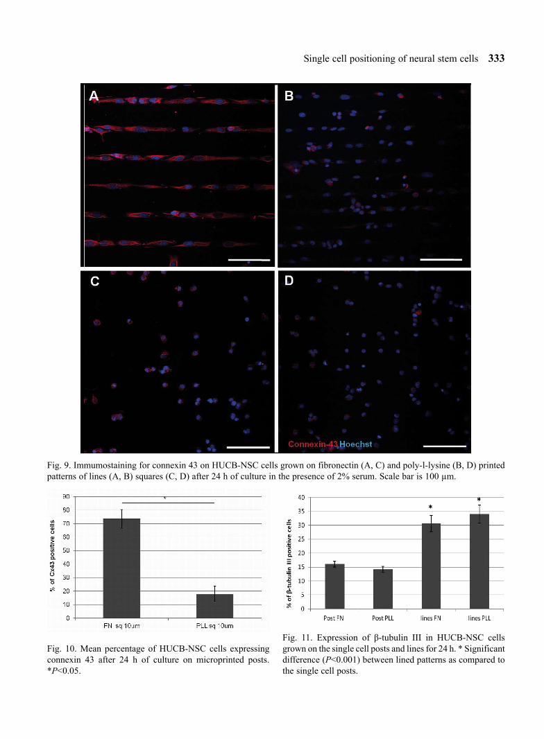
Immunostaining of the connexin 43 (Cx43) indi-cates the extent of GAP junction formation in HUCB-NSC on different substrates and single-cell patterns. The cells patterned on lines and single cell posts with fibronectin were all strongly positive for connexin 43 (Fig. 9A and C), whereas the staining on PLL was very weak, however single cells posi-tive for Cx43 were found (Fig. 9B and D).
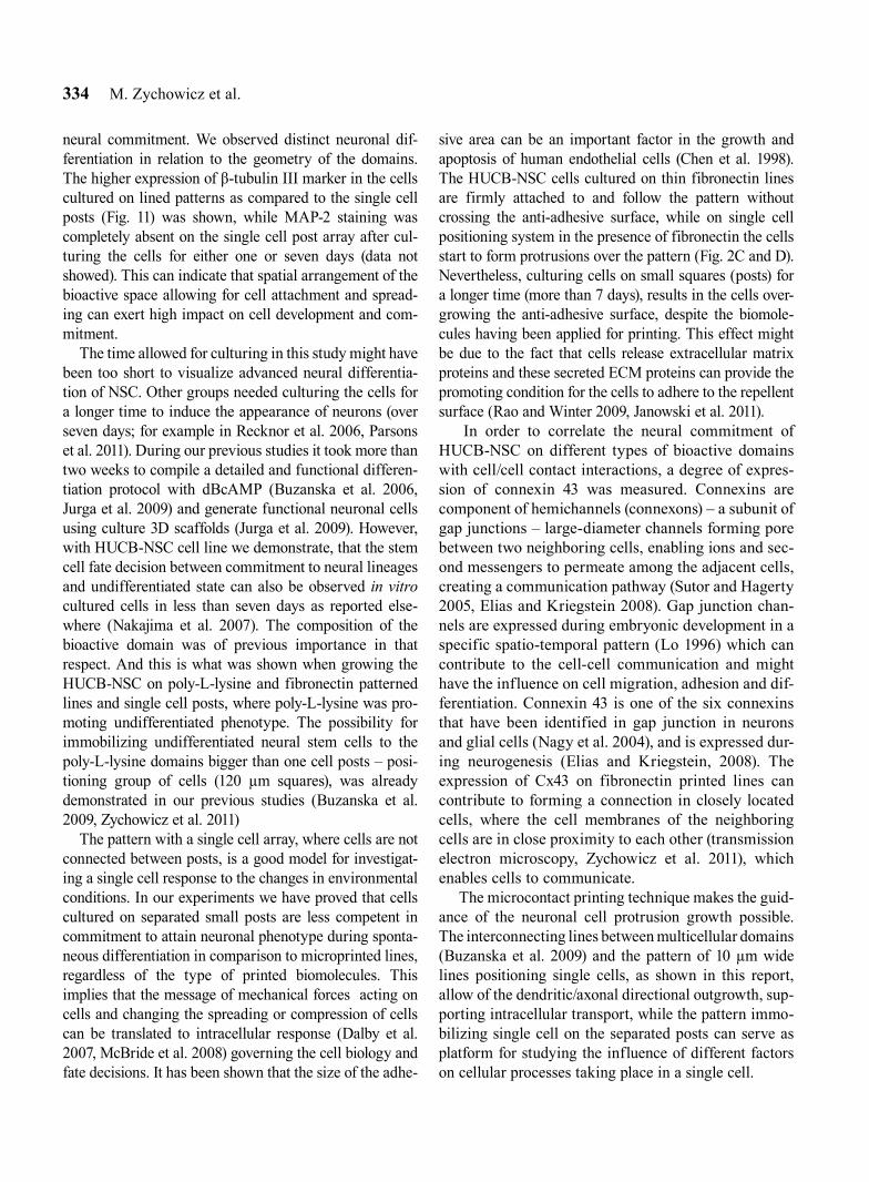
Interestingly, we found that the expression of con-nexin 43 protein, which is an element of intercellular connection system, is also visible in the cells grown separately on small square patterns (posts), but in a smaller degree in comparison with microprinted lines (Fig. 9). The cells cultured on posts patterned with fibronectin showed a significantly higher expression of Cx43 protein as compared to the poly-L-lysine printed 10 µm squares (Fig. 10).
Neural commitment and differentiation of the HUCB-NSC on the pattern with single cell posts
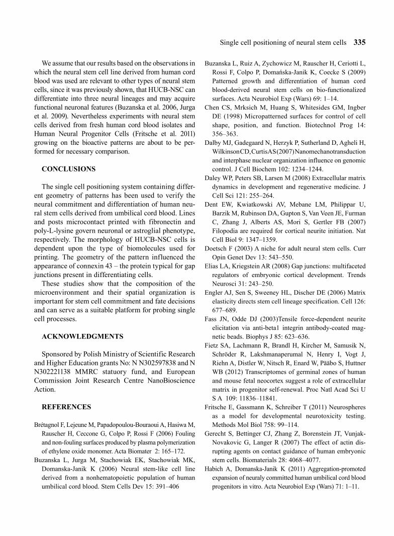
Cells have been cultured on the microfabricated single cell positioning platform consisting of 10 µm-side squares, with 30 µm distance between posts. Cells seeded on the fibronectin small squares are rounded, they express β-tubulin III marker in a significantly lower number than on lines (Fig. 11). After culturing them for one day some cells start to divide so, that the single cell post is occupied by two cells. The neuronal commitment of HUCB-NSC cells on the single cell positioning system is limited as compared to the pattern with lines. Culturing the HUCB-NSC for 7 days results in forming cell aggregates on the small square cell pattern or even in occupying the surface covered with the non-adhesive substrate, both on fibronec-tin and poly-L-lysine.
DISCUSSION
The investigation of the neural stem cells fate decisions as a result of the microenvironmental mutual interactions has been carried out and widely discussed in a number of laboratories recently. It refers to natural niches of stem cells in vivo or artificial structures/systems mimicking natural environment of stem cell niche in vitro (Taverna et al. 2011, Fietz et al. 2012). Such in vivo studies require advanced systems based on genetic markers strategies, knocked-out animals and clonal approach tracing single cells. On the other hand, in vitro techniques provide us with a tool for elucidating the mechanisms of stem cell regulation of differentiation processes depending on the topographical/environmental cues. In this study micro-contact printing technique, which provides good resolu-tion and varied dimensions and geometry of the patterns was used to investigate the influence of the topographic and mechanical cues as well as the composition of bioac-tive domains on neural stem cell behavior, without the necessity of adding the differentiation-inducing compo-
Fig. 5. Comparison of neuronal and glial commitment of HUCB-NSC cells cultured for 24 h on lines microprinted with fibronectin or poly-L-lysine. * P<0.001.
Single cell positioning of neural stem cells 331
Fig. 7. Staining of 7 day culture of HUCB-NSC for neuronal markers: Map-2 (green) and β-tubulin III (red) on fibronectin (A, B) and PLL (C, D) printed lines in serum (A, C) and serum free (B, D) medium. Scale bar is 100 µm.
Fig. 6. Immunostaining for β- tubulin III (green) and GFAP (red) of HUCB-NSC cells cultured for 24 h on lines microprinted with poly-L-lysine (A, B) and fibronectin (C, D) in presence of 2% serum (A, C) and serum free medium (B, C). Cell nuclei are con-trastained with Hoechst. Scale bar is 100 µm.
332 M. Zychowicz et al.
nents. The change in the morphology and differentiation of HUCB-NSC seeded on the fibronectin or poly-L-lysine posts or lines patterns, which was observed in our studies, was due to the type of adhesion: specific, receptor medi-ated type of adhesion on extracellular matrix protein-fi-bronectin versus non-receptor mediated, electrostatic mode of adhesion on PLL (Yavin and Yavin 1974, Richert et al. 2002, Rao and Winter 2009). The adhesion to the fibronectin via integrins activates downstream cell signal-ing, mainly through the focal adhesion kinase, leading to the stimulation of developmental processes, such as migration, proliferation as well as differentiation, in vitro and in vivo (Daley et al. 2008, Myers et al. 2011). The dif-ference that can be spotted in a mode of adhesion, as well as the change in the differentiation profile, can be consid-ered when developing new biomaterials to support trans-plantation of neural stem cells. Depending on the type of biomolecules used we can guide NSC to either stay in non differentiated state or to cause the cells to form long neu-ronal protrusions and outgrowth aligned with the designed three-dimensional scaffolds. The alignment of cells and their protrusions on micropatterned polymer substrates were observed elsewhere (Recknor et al. 2006) in adult rat hippocampal cell culture. The influence of applied geom-etry as a single factor affecting neuronal differentiation had been tested before as well (Lee et al. 2010, Wang et al. 2010).
In this report our aim was to compare the influence of the composition and the geometry of the bioactive domains on the neuronal commitment of HUCB-NSC (lines and post array) within the chosen time-points at the time of the ongoing experiment (one or seven days). Neuronal commitment measured by the expression of the
early differentiation marker β-tubulin III was observed in about 30% of cells just after the first day of incubation and this expression was not significantly different across the tested biofunctional domains and experimental variants. This could be explained by the fact, that the cell starting population has been harvested from the culture of neural stem cell line that was already expressing early neuronal markers (Buzanska et al. 2006), resembling neurally com-mitted expanding cells from primary cultures of human cord blood (Habich and Domanska-Janik 2011). More advanced stages of the neuronal commitment on the bio-active domains have been investigated by the expression of the late neuronal marker – MAP-2 after 7 days of HUCB-NSC culture.
The medium condition of cell culture can also exert an influence on promoting more advanced neuronal phenotype of HUCB-NSC. In this report fibronectin promoted neuronal differentiation, and the serum depri-vation significantly enhanced differentiation measured by the appearance of MAP-2 positive cells, which was in line with our previous studies (Buzanska et al. 2009). However, the type of biomolecules used for printing the line pattern did not have a statistically significant influ-ence on the number of β-tubulin III cells, but the sig-nificant difference between domains of different com-position was observed in the cell morphology and expression of MAP-2 in HUCB-NSC population cul-tured on fibronectin-printed lines. After culturing and allowing the cells to differentiate spontaneously with no neuromorphogenes having been added for seven days, the number of MAP-2 positive cells was higher on fibronectin than on poli-L-lysine, especially in serum free conditions (Fig. 8). This implies an important role the specific, receptor-mediated type of interaction on the cell/surface interface during neuronal differentiation.
The appearance of the glial cell marker – GFAP in the HUCB-NSC immobilized on the line single cell patterns was also investigated. Surprisingly, the influence of the type of bioactive domains exerted diverse impact on neu-ronal versus glial commitment. Contrary to the neuronal commitment, when extracellular matrix protein – fibronec-tin was exerting the predominant influence, poly-L-lysine stimulated HUCB-NSC cells to attend glial fate (Fig. 5). This interesting finding may be helpful to create the appropriate in vitro microenvironment supporting neu-ronal or glia-oriented differentiation.
The time (one or seven days) was allowed for culturing in vitro cells with the aim of checking, whether the designed pattern with different geometry can influence
Fig. 8. Map-2 expresion in HUCB-NSC grown on the pat-terns with lines in different experimental conditions, after 7 days of culture. Statistically significant difference between pairs, *P<0.001.
Single cell positioning of neural stem cells 333
Fig. 9. Immumostaining for connexin 43 on HUCB-NSC cells grown on fibronectin (A, C) and poly-l-lysine (B, D) printed patterns of lines (A, B) squares (C, D) after 24 h of culture in the presence of 2% serum. Scale bar is 100 µm.
Fig. 10. Mean percentage of HUCB-NSC cells expressing connexin 43 after 24 h of culture on microprinted posts. *P<0.05.
Fig. 11. Expression of β-tubulin III in HUCB-NSC cells grown on the single cell posts and lines for 24 h. * Significant difference (P<0.001) between lined patterns as compared to the single cell posts.
334 M. Zychowicz et al.
neural commitment. We observed distinct neuronal dif-ferentiation in relation to the geometry of the domains. The higher expression of β-tubulin III marker in the cells cultured on lined patterns as compared to the single cell posts (Fig. 11) was shown, while MAP-2 staining was completely absent on the single cell post array after cul-turing the cells for either one or seven days (data not showed). This can indicate that spatial arrangement of the bioactive space allowing for cell attachment and spread-ing can exert high impact on cell development and com-mitment.
The time allowed for culturing in this study might have been too short to visualize advanced neural differentia-tion of NSC. Other groups needed culturing the cells for a longer time to induce the appearance of neurons (over seven days; for example in Recknor et al. 2006, Parsons et al. 2011). During our previous studies it took more than two weeks to compile a detailed and functional differen-tiation protocol with dBcAMP (Buzanska et al. 2006, Jurga et al. 2009) and generate functional neuronal cells using culture 3D scaffolds (Jurga et al. 2009). However, with HUCB-NSC cell line we demonstrate, that the stem cell fate decision between commitment to neural lineages and undifferentiated state can also be observed in vitro cultured cells in less than seven days as reported else-where (Nakajima et al. 2007). The composition of the bioactive domain was of previous importance in that respect. And this is what was shown when growing the HUCB-NSC on poly-L-lysine and fibronectin patterned lines and single cell posts, where poly-L-lysine was pro-moting undifferentiated phenotype. The possibility for immobilizing undifferentiated neural stem cells to the poly-L-lysine domains bigger than one cell posts – posi-tioning group of cells (120 µm squares), was already demonstrated in our previous studies (Buzanska et al. 2009, Zychowicz et al. 2011)
The pattern with a single cell array, where cells are not connected between posts, is a good model for investigat-ing a single cell response to the changes in environmental conditions. In our experiments we have proved that cells cultured on separated small posts are less competent in commitment to attain neuronal phenotype during sponta-neous differentiation in comparison to microprinted lines, regardless of the type of printed biomolecules. This implies that the message of mechanical forces acting on cells and changing the spreading or compression of cells can be translated to intracellular response (Dalby et al. 2007, McBride et al. 2008) governing the cell biology and fate decisions. It has been shown that the size of the adhe-
sive area can be an important factor in the growth and apoptosis of human endothelial cells (Chen et al. 1998). The HUCB-NSC cells cultured on thin fibronectin lines are firmly attached to and follow the pattern without crossing the anti-adhesive surface, while on single cell positioning system in the presence of fibronectin the cells start to form protrusions over the pattern (Fig. 2C and D). Nevertheless, culturing cells on small squares (posts) for a longer time (more than 7 days), results in the cells over-growing the anti-adhesive surface, despite the biomole-cules having been applied for printing. This effect might be due to the fact that cells release extracellular matrix proteins and these secreted ECM proteins can provide the promoting condition for the cells to adhere to the repellent surface (Rao and Winter 2009, Janowski et al. 2011).
In order to correlate the neural commitment of HUCB-NSC on different types of bioactive domains with cell/cell contact interactions, a degree of expres-sion of connexin 43 was measured. Connexins are component of hemichannels (connexons) – a subunit of gap junctions – large-diameter channels forming pore between two neighboring cells, enabling ions and sec-ond messengers to permeate among the adjacent cells, creating a communication pathway (Sutor and Hagerty 2005, Elias and Kriegstein 2008). Gap junction chan-nels are expressed during embryonic development in a specific spatio-temporal pattern (Lo 1996) which can contribute to the cell-cell communication and might have the influence on cell migration, adhesion and dif-ferentiation. Connexin 43 is one of the six connexins that have been identified in gap junction in neurons and glial cells (Nagy et al. 2004), and is expressed dur-ing neurogenesis (Elias and Kriegstein, 2008). The expression of Cx43 on fibronectin printed lines can contribute to forming a connection in closely located cells, where the cell membranes of the neighboring cells are in close proximity to each other (transmission electron microscopy, Zychowicz et al. 2011), which enables cells to communicate.
The microcontact printing technique makes the guid-ance of the neuronal cell protrusion growth possible. The interconnecting lines between multicellular domains (Buzanska et al. 2009) and the pattern of 10 µm wide lines positioning single cells, as shown in this report, allow of the dendritic/axonal directional outgrowth, sup-porting intracellular transport, while the pattern immo-bilizing single cell on the separated posts can serve as platform for studying the influence of different factors on cellular processes taking place in a single cell.
Single cell positioning of neural stem cells 335
We assume that our results based on the observations in which the neural stem cell line derived from human cord blood was used are relevant to other types of neural stem cells, since it was previously shown, that HUCB-NSC can differentiate into three neural lineages and may acquire functional neuronal features (Buzanska et al. 2006, Jurga et al. 2009). Nevertheless experiments with neural stem cells derived from fresh human cord blood isolates and Human Neural Progenitor Cells (Fritsche et al. 2011) growing on the bioactive patterns are about to be per-formed for necessary comparison.
CONCLUSIONS
The single cell positioning system containing differ-ent geometry of patterns has been used to verify the neural commitment and differentiation of human neu-ral stem cells derived from umbilical cord blood. Lines and posts microcontact printed with fibronectin and poly-L-lysine govern neuronal or astroglial phenotype, respectively. The morphology of HUCB-NSC cells is dependent upon the type of biomolecules used for printing. The geometry of the pattern influenced the appearance of connexin 43 – the protein typical for gap junctions present in differentiating cells.
These studies show that the composition of the microenvironment and their spatial organization is important for stem cell commitment and fate decisions and can serve as a suitable platform for probing single cell processes.
ACkNOwLEDgMENTS
Sponsored by Polish Ministry of Scientific Research and Higher Education grants No: N N302597838 and N N302221138 MMRC statuory fund, and European Commission Joint Research Centre NanoBioscience Action.
REFERENCES
Brétagnol F, Lejeune M, Papadopoulou-Bouraoui A, Hasiwa M, Rauscher H, Ceccone G, Colpo P, Rossi F (2006) Fouling and non-fouling surfaces produced by plasma polymerization of ethylene oxide monomer. Acta Biomater 2: 165–172.
Buzanska L, Jurga M, Stachowiak EK, Stachowiak MK, Domanska-Janik K (2006) Neural stem-like cell line derived from a nonhematopoietic population of human umbilical cord blood. Stem Cells Dev 15: 391–406
Buzanska L, Ruiz A, Zychowicz M, Rauscher H, Ceriotti L, Rossi F, Colpo P, Domańska-Janik K, Coecke S (2009) Patterned growth and differentiation of human cord blood-derived neural stem cells on bio-functionalized surfaces. Acta Neurobiol Exp (Wars) 69: 1–14.
Chen CS, Mrksich M, Huang S, Whitesides GM, Ingber DE (1998) Micropatterned surfaces for control of cell shape, position, and function. Biotechnol Prog 14: 356–363.
Dalby MJ, Gadegaard N, Herzyk P, Sutherland D, Agheli H, Wilkinson CD, Curtis AS (2007) Nanomechanotransduction and interphase nuclear organization influence on genomic control. J Cell Biochem 102: 1234–1244.
Daley WP, Peters SB, Larsen M (2008) Extracellular matrix dynamics in development and regenerative medicine. J Cell Sci 121: 255–264.
Dent EW, Kwiatkowski AV, Mebane LM, Philippar U, Barzik M, Rubinson DA, Gupton S, Van Veen JE, Furman C, Zhang J, Alberts AS, Mori S, Gertler FB (2007) Filopodia are required for cortical neurite initiation. Nat Cell Biol 9: 1347–1359.
Doetsch F (2003) A niche for adult neural stem cells. Curr Opin Genet Dev 13: 543–550.
Elias LA, Kriegstein AR (2008) Gap junctions: multifaceted regulators of embryonic cortical development. Trends Neurosci 31: 243–250.
Engler AJ, Sen S, Sweeney HL, Discher DE (2006) Matrix elasticity directs stem cell lineage specification. Cell 126: 677–689.
Fass JN, Odde DJ (2003)Tensile force-dependent neurite elicitation via anti-beta1 integrin antibody-coated mag-netic beads. Biophys J 85: 623–636.
Fietz SA, Lachmann R, Brandl H, Kircher M, Samusik N, Schröder R, Lakshmanaperumal N, Henry I, Vogt J, Riehn A, Distler W, Nitsch R, Enard W, Pääbo S, Huttner WB (2012) Transcriptomes of germinal zones of human and mouse fetal neocortex suggest a role of extracellular matrix in progenitor self-renewal. Proc Natl Acad Sci U S A 109: 11836–11841.
Fritsche E, Gassmann K, Schreiber T (2011) Neurospheres as a model for developmental neurotoxicity testing. Methods Mol Biol 758: 99–114.
Gerecht S, Bettinger CJ, Zhang Z, Borenstein JT, Vunjak-Novakovic G, Langer R (2007) The effect of actin dis-rupting agents on contact guidance of human embryonic stem cells. Biomaterials 28: 4068–4077.
Habich A, Domanska-Janik K (2011) Aggregation-promoted expansion of neuraly committed human umbilical cord blood progenitors in vitro. Acta Neurobiol Exp (Wars) 71: 1–11.
336 M. Zychowicz et al.
Janowski M, Lukomska B, Domanska-Janik K (2011) Migratory capabilities of human umbilical cord blood-derived neural stem cells (HUCB-NSC) in vitro. Acta Neurobiol Exp (Wars) 71: 24–35.
Jurga M, Lipkowski AW, Lukomska B, Buzanska L, Kurzepa K, Sobanski T, Habich A, Coecke S, Gajkowska B, Domanska-Janik K (2009) Generation of functional neu-ral artificial tissue from human umbilical cord-blood stem cells. Tissue Eng Part C Methods 15: 365–372.
Lee MR, Kwon KW, Jung H, Kim HN, Suh KY, Kim K, Kim KS (2010) Direct differentiation of human embry-onic stem cells into selective neurons on nanoscale ridge/groove pattern arrays. Biomaterials 31: 4360–4366.
Leipzig ND, Shoichet MS (2009) The effect of substrate stiffness on adult neural stem cell behavior. Biomaterials 30: 6867–6878.
Lo CW (1996) The role of gap junction membrane channels in development. J Bioenerg Biomembr 8: 379–385.
McBride SH, Falls T, Knothe Tate ML (2008) Modulation of stem cell shape and fate B: mechanical modulation of cell shape and gene expression. Tissue Eng Part A 14: 1573–1580.
Myers JP, Santiago-Medina M, Gomez TM (2011) Regulation of axonal outgrowth and pathfinding by integrin-ECM interactions. Dev Neurobiol 71: 901–923.
Nagy JI, Dudek FE, Rash JE (2004) Update on connexins and gap junctions in neurons and glia in the mammalian nervous system. Brain Res Brain Res Rev 47: 191–215.
Nakajima M, Ishimuro T, Kato K, Ko IK, Hirata I, Arima Y, Iwata H (2007) Combinatorial protein display for the cell-based screening of biomaterials that direct neural stem cell differentiation. Biomaterials 28: 1048–1060.
Parsons XH, Teng YD, Parsons JF, Snyder EY, Smotrich DB, Moore DA (2011) Efficient derivation of human neuronal progenitors and neurons from pluripotent human embryonic stem cells with small molecule induction. J Vis Exp 56: e3273.
Rao SS, Winter JO (2009) Adhesion molecule-modified biomaterials for neural tissue engineering. Front Neuroeng 2: 6.
Recknor JB, Sakaguchi DS, Mallapragada SK (2006) Directed growth and selective differentiation of neural progenitor cells on micropatterned polymer substrates. Biomaterials 27: 4098–4108.
Reilly GC, Engler AJ (2010) Intrinsic extracellular matrix properties regulate stem cell differentiation. J Biomech 43: 55–62.
Richert L, Lavalle P, Vautier D, Senger B, Stoltz JF, Schaaf P, Voegel JC, Picart C (2002) Cell interactions with poly-
electrolyte multilayer films. Biomacromolecules 3: 1170–1178.
Riquelme PA, Drapeau E, Doetsch F (2008) Brain micro-ecologies: neural stem cell niches in the adult mam-malian brain. Philos Trans R Soc Lond B Biol Sci 12: 123–137.
Ruiz SA, Chen CS (2008) Emergence of patterned stem cell differentiation within multicellular structures. Stem Cells 26: 2921–2927.
Ruiz A, Buzanska L, Gilliland D, Rausher H, Sirghi L, Sobanski T, Zychowicz M, Ceriotti L, Bretagnol F, Coecke S, Colpo P, Rossi F (2008) Micro-stamped sur-faces for the patterned growth of neural stem cells. Biomaterials 29: 4766–4774
Ruiz A, Zychowicz M, Buzanska L, Mehn D, Mills CA, Martinez E, Coecke S, Samitier J, Colpo P, Rossi F (2009) Single stem cell positioning on polylysine and fibronectin microarrays. Micro and Nanosystems 1: 50–56.
Saha K, Pollock JF, Schaffer DV, Healy KE (2007) Designing synthetic materials to control stem cell phenotype. Curr Opin Chem Biol 11: 381–387.
Sutor B, Hagerty T (2005) Involvement of gap junctions in the development of the neocortex. Biochim Biophys Acta 1719: 59–68.
Szymczak P, Wojcik-Stanaszek L, Sypecka J, Sokolowska A, Zalewska T (2010) Effect of matrix metalloproteinases inhibition on the proliferation and differentiation of HUCB-NSCs cultured in the presence of adhesive sub-strates. Acta Neurobiol Exp (Wars) 70: 325–336.
Taverna E, Haffner C, Pepperkok R, Huttner WB (2011) A new approach to manipulate the fate of single neural stem cells in tissue. Nat Neurosci 15: 329–337
Vu TH, Werb Z (2000) Matrix metalloproteinases: effectors of development and normal physiology. Genes Dev 14: 2123–2133.
Wang G, Ao Q, Gong K, Wang A, Zheng L, Gong Y, Zhang X (2010) The effect of topology of chitosan biomaterials on the differentiation and proliferation of neural stem cells. Acta Biomater 6: 3630–3639.
Yavin E, Yavin Z (1974) Attachment and culture of dissoci-ated cells from rat embryo cerebral hemispheres on polylysine-coated surface. J Cell Biol 62: 540–546.
Zychowicz M , Mehn D, Ana Ruiz A, Colpo P, Francois Rossi F, Frontczak-Baniewicz M, Domanska-Janik K and Buzanska L (2011) Proliferation capacity of cord blood derived neural stem cell line on different micro-scale biofunctional domains. Acta Neurobiol Exp (Wars) 71: 12–23.