patrick s hosford abeba habtetion - ucl...
TRANSCRIPT

Ammonia mediates cortical hemichannel dysfunction in rodent models of chronic
liver disease
Anna Hadjihambi1,2 ([email protected])
Francesco De Chiara1 ([email protected])
Patrick S Hosford2 ([email protected])
Abeba Habtetion1 ([email protected])
Anastassios Karagiannis3 ([email protected])
Nathan Davies1 ([email protected])
Alexander V Gourine2 ([email protected])
Rajiv Jalan1 ([email protected])
1 UCL Institute for Liver and Digestive Health, Division of Medicine, UCL Medical
School, Royal Free Hospital, Rowland Hill Street, NW3 2PF, London, UK
2 Centre for Cardiovascular and Metabolic Neuroscience, Neuroscience, Physiology
and Pharmacology, University College London, WC1E 6BT, London, UK
3 Neurocentre Magendie, Inserm U1215, Bordeaux, France, University of Bordeaux,
Bordeaux, France
Keywords: astrocyte, hepatic encephalopathy, neuron, lactate, ornithine
phenylacetate
This article has been accepted for publication and undergone full peer review but has not beenthrough the copyediting, typesetting, pagination and proofreading process which may lead todifferences between this version and the Version of Record. Please cite this article asdoi: 10.1002/hep.29031
This article is protected by copyright. All rights reserved.

2
A.V.G. and R.J. are joint last authors.
Abbreviations:
HE: Hepatic encephalopathy
BDL: Bile duct ligation
HA: Hyperammonemia
OP: Ornithine phenylacetate
ALF: Acute liver failure
CLD: Chronic liver disease
aCSF: Artificial cerebrospinal fluid
RT-qPCR: Quantitative real-time PCR
CBX: Carbenoxolone
NPPB: 5-Nitro-2-(3-phenylpropylamino) benzoic acid
CBF: Carboxyfluorescein
ROI: Region of interest
SEM: Standard error mean
Contact information: Rajiv Jalan, Professor of Hepatology, Liver Failure Group
ILDH, Division of Medicine, UCL Medical School, Royal Free Campus, Rowland Hill
Street, London, NW3 2PF
Phone: +442074332795
Page 2 of 44
Hepatology
Hepatology
This article is protected by copyright. All rights reserved.

3
Financial support: This work was supported by Grand challenges UCL and The
Wellcome Trust (A.V.G.). A.V.G is a Wellcome Trust Senior Research Fellow
(Ref: 095064).
Potential Conflict of Interest:
Rajiv Jalan has research collaborations with Ocera and Takeda and consults with
Ocera and has received speaking fees from Norgine. Rajiv Jalan is the inventor of
OCR-002, which has been patented by UCL and licensed to Ocera Therapeutics. He
is also the founder of Yaqrit limited, a spin out company from University College
London.
Page 3 of 44
Hepatology
Hepatology
This article is protected by copyright. All rights reserved.

4
Abstract
The pathogenesis of hepatic encephalopathy (HE) in cirrhosis is multifactorial and
ammonia is thought to play a key role. Astroglial dysfunction is known to be present
in HE. Astrocytes are extensively connected by gap junctions formed of connexins,
which also exist as functional hemichannels allowing exchange of molecules
between the cytoplasm and the extracellular milieu. The astrocyte-neuron lactate
shuttle hypothesis suggests that neuronal activity is fuelled (at least in part) by
lactate provided by neighbouring astrocytes. We hypothesised that in HE, astroglial
dysfunction could impair metabolic communication between astrocytes and neurons.
In this study we determined whether hyperammonemia leads to hemichannel
dysfunction and impairs lactate transport in the cerebral cortex using rat models of
HE (bile duct ligation [BDL] and induced-hyperammonemia [HA]) and also evaluated
the effect of ammonia-lowering treatment (ornithine phenylacetate, OP). Plasma
ammonia concentration in BDL rats was indeed significantly reduced by OP
treatment. Biosensor recordings demonstrated that HE is associated with a
significant reduction in both tonic and hypoxia-induced lactate release in the cerebral
cortex, which was normalized by OP treatment. Cortical dye loading experiments
revealed hemichannel dysfunction in HE with improvement following OP treatment,
while the expression of key connexins was unaffected. Conclusion. The results of
the present study demonstrate that HE is associated with CNS hemichannel
dysfunction, with ammonia playing a key role. The data provide evidence of a
potential neuronal energy deficit due to impaired hemichannel-mediated lactate
transport between astrocytes and neurons as a possible mechanism underlying
pathogenesis of HE.
Page 4 of 44
Hepatology
Hepatology
This article is protected by copyright. All rights reserved.
5
Hepatic encephalopathy (HE) is a serious neuropsychiatric complication associated
with liver dysfunction, diagnosed when other known brain disorders are excluded(1).
HE comprises a range of symptoms including sleep disturbances, alterations in
cognitive, behavioural, fine motor and psychomotor functions, with coma and death
occurring at the late stages(2). Several hypotheses regarding the pathogenesis of
HE have been proposed and numerous factors have been suggested as key players
in HE, including: inflammation(3), oxidative stress(4), impaired brain energy
metabolism(5) and (the most common one) the neurotoxic effects of ammonia(6).
Recent evidence demonstrated that astroglial lactate production and release in
cortical cultures and in the somatosensory cortex of anesthetised rats is facilitated in
the presence of ammonia(7). This appears to be due to acidification of the
mitochondrial matrix resulting in a direct inhibition of mitochondrial pyruvate uptake.
Increased brain lactate levels have also been reported in hyperammonemic
conditions such as in acute liver failure (ALF), which is thought to be due to inhibition
of the tricarboxylic acid cycle enzyme α-ketoglutarate dehydrogenase, suggesting a
reduction in oxidative metabolism (8). It remains unknown whether significant
changes in brain lactate metabolism develop in conditions of long-term CNS
exposure to increased ammonia concentration, such as that seen during chronic liver
disease (CLD) or HE.
Astrocytes, the most numerous type of glial cell in the CNS, are thought to play an
important role in HE pathogenesis. The astrocytic dysfunction developing during the
progression of the disease could precipitate neuronal pathology, leading to
Page 5 of 44
Hepatology
Hepatology
This article is protected by copyright. All rights reserved.
6
neurological impairment. Astrocytes are extensively connected by gap junctions
formed of connexins, which also exist as functional hemichannels allowing effective
transfer of ions, metabolic substrates and signaling molecules across the plasma
membrane(9). Under normal physiological conditions, hemichannels are either
closed(10) or in a flickering state(11). In certain pathological conditions, such as
epilepsy and ischaemia, significant changes in astroglial structure and function may
occur, which are associated with changes in connexin hemichannel function,
affecting coupling within the astroglial networks and their communication with other
brain cells(12). We hypothesised, therefore, that in HE, connexin hemichannel
dysfunction contributes to the development of its neurological features.
There is recent evidence that hemichannels may function as a conduit of lactate
transport across the membrane(13). In this study we first investigated whether
connexin hemichannel expression and hemichannel-mediated release of lactate are
altered in animal models of HE. Ornithine phenylacetate (OP, OCR-002; Ocera
Therapeutics, CA, USA) has been shown to reduce ammonia levels in animal
models of cirrhosis and ALF. OP treatment was found to be associated with a
significant reduction in the severity of brain swelling(14), improvement in
neurophysiological function(15) and reduction in intracranial pressure(16). Therefore,
we applied OP in this study as an experimental ammonia-lowering treatment. The
data obtained demonstrate that, in HE, ammonia mediates cortical hemichannel
dysfunction associated with a significant reduction in hemichannel-mediated lactate
release.
Page 6 of 44
Hepatology
Hepatology
This article is protected by copyright. All rights reserved.

7
Materials and Methods
All the experiments were performed in accordance with the Animals (Scientific
Procedures) Act 1986 (ASPA) revised according to the European Directive
2010/63/EU. All animals received humane care according to the criteria outlined in
the “Guide for the Care and Use of Laboratory Animals” prepared by the National
Academy of Sciences and published by the National Institutes of Health (NIH
publication 86-23 revised 1985).
Animal models
Male Sprague-Dawley rats (body weight ~350-400g) were obtained from a
commercial supplier (Charles Rivers Laboratories, Inc.).
Bile duct ligation (BDL) surgery: Under general anaesthesia (5% isoflurane in 100%
oxygen for induction, 2% isofluorane in air for maintenance) 30 rats underwent triple
ligation of the bile duct via a small laparotomy to induce chronic liver injury and were
studied 28 days after the surgery (17).
Non-cirrhotic hyperammonemia condition: 32 rats were administered high
ammoniagenic diet (HA). The amino acid recipe used for a stock of ~100g was: 15g
leucine, 7.7g phenylalanine, 7g glutamate, 10g alanine, 4.4g proline, 5.8g threonine,
11g aspartate, 5g serine, 4.8g glycine, 3.3g arginine, 9.6g lysine, 8.4g histidine, 3g
tyrosine, 1.5g tryptophan and 10.6g valine. 25 g of this mix (mixed 1:5 with standard
rodent chow powder) was freshly prepared daily with water in a mash form and rats
Page 7 of 44
Hepatology
Hepatology
This article is protected by copyright. All rights reserved.
8
were given free access to it for 5 days. The recipe approximates the amino acid
composition of a rodent haemoglobin(18), mimicking the effect of gastrointestinal
bleeding, which is a known to result in systemic hyperammonemia (19).
Ornithine Phenylacetate (OP) treatment: 3 weeks after the surgery, 24 BDL-operated
rats were given twice daily intraperitoneal injections of combined L-ornithine and
phenylacetate (0.3g/kg; OP) ~7 hours apart for 5 days – a dosing regimen that has
previously been shown to effectively reduce plasma ammonia concentration(20). The
rats were studied on day 28 post BDL surgery, within 3 hours of the last OP injection.
Blood and brain tissue were collected under terminal isoflurane anaesthesia. Plasma
biochemistry was performed using a Cobas Integra II system (Roche Diagnostics).
In vitro slice preparation
Rats were humanely sacrificed by isoflurane inhalation overdose. After cardiac
perfusion with chilled (4°C) artificial cerebrospinal fluid (aCSF, 124mM NaCl,
3mM KCl, 2mM CaCl2, 26mM NaHCO3, 1.25mM NaH2PO4, 1mM MgSO4, 10mM D-
glucose saturated with 95% O2, 5% CO2, pH 7.5, PCO2 35 mmHg), with an
additional 9mM Mg2+, the brain was rapidly removed and placed in a bath of chilled
(4-6°C) aCSF. 300 µm coronal cortical slices were cut using a vibrating microtome.
The slices were recovered in oxygenated (95% O2, 5% CO2) aCSF at room
temperature for 30 min.
Page 8 of 44
Hepatology
Hepatology
This article is protected by copyright. All rights reserved.

9
Western blot
Proteins (30 µg) extracted from the cortices of 5 sham-operated, 6 BDL, 6 HA and 5
BDL-OP-treated rats were separated by sodium dodecyl sulphate-polyacrylamide gel
electrophoresis on a 4% to 12% Bis-Tris NuPAGE gel (Invitrogen) and transferred to
nitrocellulose membranes. Membranes were blocked with 5% Bovine Serum
Albumin and incubated with antibodies against connexin-43 (Cell Signaling
Technology, 1:1000), connexin-36 (Santa Cruz, 1:1000), connexin-30 (Invitrogen, 1
µg/mL) and connexin-26 (Thermo Fisher Scientific, 1 µg/mL). Detection of actin
(Santa Cruz, 1:1000) was used to control for protein loading. Binding of antibody was
detected using a horseradish peroxidase-conjugated secondary antibody (Goat anti-
rabbit or goat anti-mouse IgG-HPR, Santa Cruz, 1:10000) where appropriate and the
SuperSignal Chemiluminescence Substrate for detection of horseradish peroxidase
(Pierce). Densitometric analysis was performed using Kodak 1D image analysis
software (Kodak, Rochester, NY).
Measurements of lactate release using microelectrode biosensors
Amperometric enzymatic biosensors were obtained from Sarissa Biomedical
(Coventry, UK). The design and operation of the biosensors were described in detail
previously (21, 22). All sensors were operating against a reference electrode
(Ag/AgCl) and had a linear response to lactate within the concentration range
recorded in this study (22, 23). Further information on the principle of operation and
response time is given in the online data supplement (Supplementary Figure 1A-C).
To control for the release of non-specific electroactive interferants, a dual recording
configuration was used. In every recording, a “null” sensor, lacking enzymes but
Page 9 of 44
Hepatology
Hepatology
This article is protected by copyright. All rights reserved.

10
otherwise identical, was used to measure current changes not associated with
lactate oxidase activity, which were then subtracted from the current recorded by the
lactate biosensor (24) (Figure 1).
The sensors were calibrated directly in the slice chamber immediately before and
after every recording by application of 100 µM of lactate (Figure 1). To convert
changes in the biosensor current to changes in lactate concentration, an average of
sensor calibrations before and after the recording were used. For each of the
recordings, a slice was transferred into the recording chamber and superfused with
aCSF at 35°C (3 ml min-1). Sensors were initially placed in the chamber having no
contact with the brain slice. Once a steady-state baseline was achieved, the sensors
were laid flat in direct contact with the surface of the slice (Figure 1), revealing tonic
lactate release which stabilised within ~15 min. Hypoxic conditions, known to
increase both lactate production due to inhibition of oxidative phosphorylation and
the opening probability of astroglial connexin hemichannels (23, 25), were induced
for 2-4 min by replacement of oxygen in the medium with nitrogen (perfusion of the
chamber with aCSF saturated with 95% N2/5% CO2)(26, 27). As detection of lactate
by the biosensors requires oxygen (Supplementary Figure 1B) (28), the effect of
hypoxia was determined by measuring the peak lactate release upon re-oxygenation
(Figure 1) as described in detail previously(23, 28). Once the baseline was restored,
Ca2+-free aCSF (with the addition of 1 mM EGTA) was applied for 20 min as the
second stimulus known to increase the opening probability of certain membrane
channels, including connexin hemichannels(29). There is no prior evidence that Ca2+
- mediated increases in mitochondrial NADH influence cytosolic NAD+/NADH
Page 10 of 44
Hepatology
Hepatology
This article is protected by copyright. All rights reserved.

11
homeostasis and therefore lactate production(30). These stimuli were reapplied in
the presence of connexin hemichannel blockers carbenoxolone (CBX, 100 µM;
Sigma) or 5-Nitro-2-(3-phenylpropylamino)benzoic acid (NPPB, 200 µM, Sigma).
CBX and NPPB were previously shown to have no effect on lactate biosensor
detection system(23).
Assessment of hemichannel function by dye loading
For the assessment of hemichannel functionality (effectiveness of channel opening
and closing) we used a fluorescent dye carboxyfluorescein (CBF: 376 Da). Connexin
hemichannels are permeable to CBF and can act as conduits of CBF transfer across
the membrane in accord with the concentration gradient of the dye. Cortical slices
from sham-operated, BDL, HA and BDL-OP-treated rats were exposed to control
aCSF with an addition of CBF (200 µM) for 9 min resulting in background connexin-
mediated dye loading (Figure 5A-B), followed by perfusion with Ca2+-free aCSF
without CBF for 9 min resulting in CBF unloading. Ca2+-free aCSF with CBF was
then applied for 4 min increasing the permeability of hemichannels and therefore
resulting in dye loading. Hypoxic conditions (without CBF) were next applied to
unload the slice and also to demonstrate bidirectional permeability of the channel to
CBF(27). The same hypoxic stimulus was then re-applied in the presence of CBF,
resulting in dye loading. After application of each stimulus in the presence of CBF, a
further 5 min perfusion with aCSF containing CBF, followed by a 10 min wash with
normal aCSF was applied, enabling the channels to return to their physiological
state. Images were taken using MiCAM-02 imaging system (SciMedia). Using
ImageJ software, regions of interest (ROI) were drawn around an area of the
Page 11 of 44
Hepatology
Hepatology
This article is protected by copyright. All rights reserved.

12
cerebral cortex (~3cm2) across layers I-III and the mean pixel intensities for the ROI
were calculated. Background fluorescence was subtracted.
Statistical analysis
Western blot data were normalised using the protocol of LI-COR Biosciences
(Normalization Accuracy for Western Blotting) and group data were compared using
two-way ANOVA with Tukey post hoc test. Data obtained using biosensor recordings
were analysed and presented non-parametrically using box and whisker plots
(Figures 2A, 3A, 4A). For the comparisons between the experimental groups, Mann
Whitney U test was applied. The peak hypoxia- or 0 Ca2+-induced lactate releases
are presented as changes in release from the baseline (Figure 1). The effects of
connexin blockers are presented as % changes from the control responses recorded
in the absence of the blockers and Wilcoxon signed rank test was applied for
comparison. P values in Figures 2-4B,C indicate significance level of differences
between the control responses and the responses recorded in the presence of the
drugs.
Data obtained in dye loading experiments were analysed using two-way ANOVA
(Figure 5B, data normally distributed) followed by Tukey post hoc test or Wilcoxon
signed rank test (Figures 5C-D, data not normally distributed), as appropriate. The
biochemistry data was analysed using one-way ANOVA. Data are reported as
mean±SEM. Differences with p value of <0.05 were considered to be significant. In
all the experiments the ‘n’ number represents the number of animals. Sample sizes
were calculated using Gpower 3 v3.1.9.2 (http://www.gpower.hhu.de/en.html)(31);
Page 12 of 44
Hepatology
Hepatology
This article is protected by copyright. All rights reserved.

13
using a ‘means: Wilcoxon-Mann-Whitney test (two groups)’ test, with a desired
power of 90% and a significance level of 5%. The effect size varied between groups
according to the preliminary data acquired during the study. Statistical analysis was
performed using OriginPro 9.1 (OriginLab).
Page 13 of 44
Hepatology
Hepatology
This article is protected by copyright. All rights reserved.

14
Results
Biochemistry
Plasma biochemistry and ammonia concentration was assessed in all groups of
animals (Supplementary Table 1). Compared to sham surgery, BDL resulted in a
significant increase in plasma ALT and bilirubin (p<0.001), indicating impaired liver
function, while albumin and total protein concentrations were significantly decreased
(p<0.001). Treatment of BDL rats with OP had no effect on these parameters. Rats
fed with HA diet had similar plasma biochemistry to control rats.
Plasma ammonia concentrations were significantly higher in BDL and HA rats when
compared to sham-operated animals (p<0.001). Treatment of BDL animals with OP
lowered plasma ammonia concentration, which was similar to that measured in
sham-operated animals (p=0.3) (Supplementary Table 1).
Release of lactate in the cerebral cortex in animal models of HE
In cortical slices of sham-operated animals, enzymatic amperometric biosensors
detected tonic lactate efflux of 335±10 µM (n=18). Recordings from cortical slices of
BDL and HA rats showed lower tonic release of lactate of 203±6 µM (p=0.03, n=18)
and 178±8 µM (p=0.005, n=16), respectively (Figure 2A). Increasing the permeability
of connexin hemichannels by lowering [Ca2+]e (0 Ca2+ conditions) triggered similar
increases in the release of lactate in sham-operated rats (by 43±3µM, n=15), BDL
(by 38±4 µM; p=0.07, n=17) and HA animals (by 54±4 µM; p=0.5, n=16) (Figure 3A).
Hypoxia facilitated release of lactate in cortical slices of sham-operated rats (43±3
µM, n=18), but had no effect on lactate release in slices of BDL (1±0.8 µM; p<0.001,
Page 14 of 44
Hepatology
Hepatology
This article is protected by copyright. All rights reserved.
15
n=18) and HA animals (5±2 µM; p<0.001, n=16) (Figure 4A). These results
demonstrated impaired tonic and hypoxia-induced release of lactate in both animal
models of HE.
Ammonia lowering treatment restores cortical lactate release
Daily OP treatments had been shown to decrease systemic and brain ammonia
concentrations in BDL animals(20). We next found that in our experiments, OP
treatment of BDL rats restored tonic (374±11 µM; p=0.4, n=14) and hypoxia-induced
(32±3 µM; p=0.6, n=14) lactate release similar to that recorded in cortical slices of
sham-operated animals (Figure 2A-4A). Direct application of OP on cortical slices of
sham-operated and BDL rats had no effect on lactate release (Supplementary Figure
1D). These results strongly suggest that high ammonia levels are responsible for the
reduction in lactate release in the cerebral cortex of BDL animals.
Impaired hemichannel function underlies reduced cortical lactate release in
animal models of HE
In cortical slices of sham-operated animals, application of hemichannel blockers
CBX (n=9) or NPPB (n=9) resulted in a significant reduction in lactate tone (Figure
2B-C). Hemichannel blockade had no effect on lactate tone in cortical slices of BDL
animals (Figure 2B-C). However, hemichannel blockade had an effect on lactate
tone recorded in cortical slices of HA animals (CBX: n=10; NPPB: n=8) (Figure 2B-
C). CBX and NPPB reduced lactate tone in cortical slices of BDL animals treated
with OP (CBX: n=10; NPPB: n=10) (Figure 2B-C).
Page 15 of 44
Hepatology
Hepatology
This article is protected by copyright. All rights reserved.
16
In cortical slices of sham-operated (CBX: n=8; NPPB: n=6) and HA animals (CBX:
n=10; NPPB: n=8), hemichannel blockade using CBX or NPPB abolished or
significantly reduced the amount of lactate release facilitated in Ca2+-free conditions
(Figure 3B-C). Smaller effect of hemichannel blockers on 0 Ca2+-induced release of
lactate was observed in cortical slices of BDL rats (CBX: n=11; NPPB: n=9) (Figure
3B-C). In conditions of OP treatment, when basal and evoked lactate release were
restored in cortical slices of BDL rats, CBX (n=7) and NPPB (n=7) abolished the
release of lactate triggered by 0 Ca2+ (CBX: n=7; NPPB: n=7); an effect similar to
that observed after application of hemichannel blockers in cortical slices of sham-
operated animals (Figure 3B-C).
Hypoxia-induced release of lactate recorded in cortical slices of sham-operated
animals was also abolished or dramatically reduced by connexin blockade (CBX:
n=9; NPPB: n=7) (Figure 4B-C). CBX and NPPB had no significant effect on the
release of lactate induced by hypoxia in cortical slices of BDL (CBX: n=13; NPPB:
n=9) and HA rats (CBX: n=10; NPPB: n=12) (Figure 4B-C). In cortical slices of OP-
treated BDL rats, the effects of CBX and NPPB were restored. CBX (n=8) and NPPB
(n=7) effectively abolished hypoxia-induced lactate release in cortical slices of BDL
rats treated with OP (Figure 4B-C).
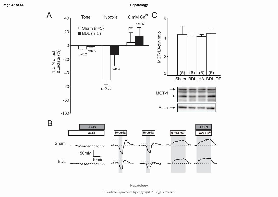
Application of MCT blocker, α-Cyano-4-hydroxycinnamic acid (4-CIN) had no
significant effect on tonic release of lactate in cortical slices of sham-operated (n=5)
and BDL rats (n=5) (Supplementary Figure 2A-B). Hypoxia-induced lactate release
recorded in sham-operated rats (n=5) was significantly reduced by 4-CIN, as
Page 16 of 44
Hepatology
Hepatology
This article is protected by copyright. All rights reserved.

17
reported previously(13). In cortical slices of BDL rats (n=5) the effect of 4-CIN on
hypoxia-induced release of lactate was found to be smaller (Supplementary Figure
2A-B).
These results demonstrate that connexin hemichannel blockade has no effect on the
release of lactate in the cerebral cortex of BDL rats. This implies that the function of
hemichannels, which act as conduits of lactate release(23), is already compromised
in the brains of these animals. These data also suggest that cortical hemichannel
dysfunction in the BDL animals is likely to be due to the actions of ammonia.
Hemichannel-mediated dye loading in animal models of HE
Membrane channel-mediated dye loading experiments were next performed in
cortical slices to confirm hemichannel dysfunction in HE.
In cortical slices of sham-operated rats (n=10), significant background loading
(14.3±0.4 A.U) was observed in control conditions when slices were perfused with
aCSF containing CBF (Figure 5A-B). Increasing the permeability of hemichannels by
lowering [Ca2+]e in the absence of CBF reduced slice fluorescence by 6.4±0.2 A.U.
The same stimulus applied in the presence of CBF significantly increased
fluorescence by 16.9±0.6 A.U. Hypoxia-induced opening of hemichannels in the
absence of CBF resulted in dye unloading with fluorescence decreasing by 12.7±0.4
A.U. Addition of CBF in conditions of hypoxia increased slice fluorescence by
13.9±0.4 A.U (Figure 5B).
Page 17 of 44
Hepatology
Hepatology
This article is protected by copyright. All rights reserved.
18
Cortical slices from HA rats (n=10) displayed background loading (Figure 5A-B) and
0 Ca2+ -induced unloading of 11.2±0.2 A.U (p=0.04) and 4.7±0.08 A.U (p=0.4)
respectively (Figure 5B). Addition of CBF in 0 Ca2+ conditions increased fluorescence
by 11.6±0.2 A.U (p=0.004), similar to that observed in slices of sham-operated
animals. However, the effect of hypoxia was significantly reduced in slices of HA
animals (unloading by 7.6±0.1 A.U, p<0.001; loading by 7.8±0.2 A.U, p<0.001)
(Figure 5B).
In cortical slices of BDL rats (n=10) efficacy of CBF dye loading and unloading was
markedly reduced under all the conditions (Figure 5A-B). In cortical slices of BDL
animals treated with OP, hemichannel-mediated CBF dye loading and unloading
were similar to that observed in slices of sham-operated animals (Figure 5A-B).
Hemichannel blockade with CBX or NPPB effectively abolished CBF dye loading and
unloading in cortical slices of sham-operated animals (Figure 5C-D). In cortical slices
of BDL and HA rats, CBX and NPPB had no significant effect on dye loading and
unloading (Figure 5C-D). In cortical slices of OP-treated BDL rats, the effects of CBX
and NPPB on CBF dye loading and unloading were similar to that observed in sham-
operated animals. (Figure 5C-D). Figures 5C-D illustrate changes in fluorescence
(∆Fluorescence) following application of the hemichannel blockers, compared to the
respective changes in fluorescence recorded in the absence of blockers in slices of
the same animal. Negative values show decreases in fluorescence while positive
values illustrate higher fluorescence levels compared to the controls.
Page 18 of 44
Hepatology
Hepatology
This article is protected by copyright. All rights reserved.

19
These data show that the background activity and stimuli-evoked opening and
closure of connexin hemichannels is impaired in BDL and HA rats. The efficacy of
CBF dye loading is restored by OP treatment of BDL animals suggesting that the
actions of ammonia are responsible for cortical hemichannel dysfunction in HE.
Cortical connexin expression in animal models of HE
We next evaluated the expression of main astrocytic and neuronal connexins in
animal models of HE used in this study. Western blots were performed on proteins
extracted from the cerebral cortices of sham-operated, BDL, HA and BDL-OP
animals Figure 6). No differences in cortical connexin-43, connexin-36 and connexin-
30 expression was observed between sham-operated, BDL, HA and OP-treated BDL
animals. An increase in connexin-26 expression (p=0.03) was observed in BDL-OP
rats compared to the BDL animals (Figure 6). Expression of MCT-1 was similar in all
the experimental groups (Supplementary Figure 2C)
Page 19 of 44
Hepatology
Hepatology
This article is protected by copyright. All rights reserved.
20
Discussion
Brain information processing requires constant and sufficient supply of oxygen and
metabolic substrates. Astrocytes represent an important source of lactate which
contributes to the extracellular pool of readily available metabolic substrates taken
up by neurons to fuel their activity(32). While previously, lactate transport across the
cell membranes was thought to be achieved solely via operation of monocarboxylate
transporters (MCTs), a recent study(23) demonstrated the role of connexin
hemichannels as equally important conduits of lactate release.
In animal models of ALF and patients, an increase in brain lactate concentration has
consistently been reported(33). Concentrations of lactate in the cerebrospinal fluid
were also found to be elevated in cirrhotic patients but only in severe cases of
HE(34). The BDL and HA rats used in our experiments are models of minimal
HE(35). In contrast to the existing evidence suggesting that brain lactate
concentrations are increased in patients with ALF(36), our experiments
demonstrated that the development of HE in rats is associated with a significant
reduction in tonic and stimulated release of lactate in the cerebral cortex. Blockade
of connexin hemichannels was found to be effective in reducing lactate release in
sham-operated and HA animals but was ineffective in BDL rats, suggesting that the
reduction in hemichannel-mediated lactate release in BDL animals is due to a
combination of pathological factors (e.g. inflammation, oxidative stress).
Increased lactate production by astrocytes appears to be essential for the recovery
of synaptic function during re-oxygenation after hypoxia(37). We found that hypoxia-
Page 20 of 44
Hepatology
Hepatology
This article is protected by copyright. All rights reserved.
21
induced lactate release was significantly lower in the cerebral cortex of BDL and HA
rats compared to control animals and this was unaffected by the hemichannel
blockers indicating hemichannel dysfunction. The observed decrease in extracellular
lactate is likely due to impaired release from astrocytes although increased neuronal
activity and therefore lactate consumption cannot be excluded. The observation of
Bosoi et al.,(38) showing higher total brain lactate of BDL rats using NMR
spectroscopy seems to contradict our data. Bosoi and colleagues suggested that
increased lactate contributes to the pathogenesis of brain edema (cytotoxic), and
may imply that the observed increase in total brain lactate is due to its intracellular
accumulation. If the rate of lactate production and glymphatic clearance (39) are not
affected, intracellular retention of lactate would explain higher concentration of this
metabolite as measured by NMR spectroscopy (40) and would be in full agreement
with our data showing impairment of hemichannel-mediated release in HE.
High concentration of ammonia can potentially generate significant pH changes,
which can have various effects on many pH sensitive membrane channels, including
hemichannels. The pH sensitivity of hemichannels is known as the chemical gate,
which is the phenomenon of hemichannel blockade when intracellular pH (pHi)
decreases(40). This provides one potential mechanism which might be responsible
for impaired hemichannel-mediated lactate release in HE.
We also examined hemichannel functionality using the dye loading method. Some
differences between the data obtained using biosensor recordings and this technique
could be due to the fact that CBF is not identical to the molecular structure and size
Page 21 of 44
Hepatology
Hepatology
This article is protected by copyright. All rights reserved.

22
of lactate. Additionally, lactate could be released through specific connexin
hemichannels, whereas CBF is small enough to pass through the majority of
hemichannels expressed by both astrocytes and neurons. While induction of hypoxia
targets predominantly astroglial hemichannels, the rest of the conditions are not cell
specific and conclusions on affected cell types cannot be drawn from the results
obtained using dye loading experiments.
The experimental stimuli used (0 Ca2+ and hypoxia) are known to increase the
permeability of hemichannels, possibly by affecting various protein bonds resulting in
conformational changes(29). Dye loading experiments clearly demonstrated a
marked reduction in fluorescent dye uptake and release in cortical slices of BDL and
HA rats compared to sham-operated animals (the differences were more profound
when hypoxia was used as a stimulus), suggesting reduced bidirectional
permeability of hemichannels in these animal models of HE. Hemichannel blockade
had no effect on fluorescent dye uptake in BDL and HA rats providing additional
evidence that the function of these channels is already compromised in HE.
Ammonium ions may cause structural alterations to the connexin proteins, by
interacting with various amino acid side chains, which could affect gating of the
channel. However, since hemichannels have a relatively short life cycle and recycled
frequently, the observed changes in hemichannel functionality appear to be
reversible with OP treatment.
In CLD, hyperammonemia is believed to impair mitochondrial function and induce
astroglial dysfunction, which is associated with altered neurotransmitter recycling
Page 22 of 44
Hepatology
Hepatology
This article is protected by copyright. All rights reserved.

23
leading to neuronal damage(41). Ammonia may also interfere with cell energy
metabolism in several ways. There is recent evidence that in astrocytes ammonia
may divert the flux of pyruvate to lactate production, contributing to the net aerobic
lactate production(7). However, the effects of chronic ammonia exposure on
astrocytes are unknown. We investigated the role of ammonia by treating BDL rats
with OP, a drug known to lower systemic and brain ammonia(20). OP treatment
improved the neurochemical phenotype of BDL animals by restoring the tonic and
stimulated connexin hemichannel-mediated lactate release. Furthermore,
hemichannel blockade became effective following OP treatment, suggesting that
ammonia is indeed responsible for hemichannel dysfunction observed in this model.
Cytotoxic brain edema observed in BDL animals is attenuated by ammonia-lowering
treatments such as the one used in this study(20, 42). The effect of cell swelling on
hemichannel function is poorly understood. Ye et al.,(43) showed that astrocytes
obtained from connexin-43 knock-out animals developed cell swelling as efficiently
as the wild type animals when exposed to a hypotonic solution suggesting that
hemichannels do not play a significant role in this process although evidence to the
contrary exists (44).
We also examined the expression profile of key astroglial and neuronal connexins in
the animal models of HE used in this study. No significant differences in connexin
hemichannel protein expression profile were observed suggesting that HE is
associated with altered hemichannel function but not with changes in connexin
expression. The upregulated expression of connexin-26 observed in the BDL-OP
Page 23 of 44
Hepatology
Hepatology
This article is protected by copyright. All rights reserved.
24
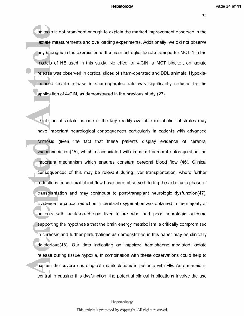
animals is not prominent enough to explain the marked improvement observed in the
lactate measurements and dye loading experiments. Additionally, we did not observe
any changes in the expression of the main astroglial lactate transporter MCT-1 in the
models of HE used in this study. No effect of 4-CIN, a MCT blocker, on lactate
release was observed in cortical slices of sham-operated and BDL animals. Hypoxia-
induced lactate release in sham-operated rats was significantly reduced by the
application of 4-CIN, as demonstrated in the previous study (23).
Depletion of lactate as one of the key readily available metabolic substrates may
have important neurological consequences particularly in patients with advanced
cirrhosis given the fact that these patients display evidence of cerebral
vasoconstriction(45), which is associated with impaired cerebral autoregulation, an
important mechanism which ensures constant cerebral blood flow (46). Clinical
consequences of this may be relevant during liver transplantation, where further
reductions in cerebral blood flow have been observed during the anhepatic phase of
transplantation and may contribute to post-transplant neurologic dysfunction(47).
Evidence for critical reduction in cerebral oxygenation was obtained in the majority of
patients with acute-on-chronic liver failure who had poor neurologic outcome
supporting the hypothesis that the brain energy metabolism is critically compromised
in cirrhosis and further perturbations as demonstrated in this paper may be clinically
deleterious(48). Our data indicating an impaired hemichannel-mediated lactate
release during tissue hypoxia, in combination with these observations could help to
explain the severe neurological manifestations in patients with HE. As ammonia is
central in causing this dysfunction, the potential clinical implications involve the use
Page 24 of 44
Hepatology
Hepatology
This article is protected by copyright. All rights reserved.

25
of ammonia lowering treatments as the main therapeutic strategy, as well as
attempts to increase cerebral oxygenation in order to preserve the neuronal function.
In conclusion, the results of the present study suggest that HE is associated with
CNS hemichannel dysfunction, with ammonia playing a key role. The data provide
evidence of a potential neuronal energy deficit due to impaired hemichannel-
mediated lactate transport between astrocytes and neurons as a possible
mechanism underlying pathogenesis of HE.
Page 25 of 44
Hepatology
Hepatology
This article is protected by copyright. All rights reserved.

26
References
1. Ciecko-Michalska I, Szczepanek M, Slowik A, Mach T. Pathogenesis of hepatic
encephalopathy. Gastroenterol Res Pract 2012;2012:642108.
2. Prakash R, Mullen KD. Mechanisms, diagnosis and management of hepatic
encephalopathy. Nat Rev Gastroenterol Hepatol 2010;7:515-525.
3. Jalan R, Olde Damink SW, Hayes PC, Deutz NE, Lee A. Pathogenesis of intracranial
hypertension in acute liver failure: inflammation, ammonia and cerebral blood flow. J Hepatol
2004;41:613-620.
4. Bosoi CR, Rose CF. Oxidative stress: a systemic factor implicated in the pathogenesis of
hepatic encephalopathy. Metab Brain Dis 2013;28:175-178.
5. Rao KV, Norenberg MD. Cerebral energy metabolism in hepatic encephalopathy and
hyperammonemia. Metab Brain Dis 2001;16:67-78.
6. Shawcross D, Jalan R. The pathophysiologic basis of hepatic encephalopathy: central role
for ammonia and inflammation. Cell Mol Life Sci 2005;62:2295-2304.
7. Lerchundi R, Fernandez-Moncada I, Contreras-Baeza Y, Sotelo-Hitschfeld T, Machler P,
Wyss MT, Stobart J, et al. NH4+ triggers the release of astrocytic lactate via mitochondrial
pyruvate shunting. Proc Natl Acad Sci U S A 2015;112:11090-11095.
8. Rose C, Ytrebo LM, Davies NA, Sen S, Nedredal GI, Belanger M, Revhaug A, et al.
Association of reduced extracellular brain ammonia, lactate, and intracranial pressure in pigs
with acute liver failure. Hepatology 2007;46:1883-1892.
9. Bennett MV, Contreras JE, Bukauskas FF, Saez JC. New roles for astrocytes: gap junction
hemichannels have something to communicate. Trends Neurosci 2003;26:610-617.
10. Contreras JE, Saez JC, Bukauskas FF, Bennett MV. Gating and regulation of connexin 43
(Cx43) hemichannels. Proc Natl Acad Sci U S A 2003;100:11388-11393.
11. Bukauskas FF, Peracchia C. Two distinct gating mechanisms in gap junction channels:
CO2-sensitive and voltage-sensitive. Biophys J 1997;72:2137-2142.
12. Giaume C, Koulakoff A, Roux L, Holcman D, Rouach N. Astroglial networks: a step further
in neuroglial and gliovascular interactions. Nat Rev Neurosci 2010;11:87-99.
13. Karagiannis A, Sylantyev S, Hadjihambi A, Hosford PS, Kasparov S, Gourine AV.
Hemichannel-mediated release of lactate. J Cereb Blood Flow Metab 2015.
14. Jalan R, Wright G, Davies NA, Hodges SJ. L-Ornithine phenylacetate (OP): a novel
treatment for hyperammonemia and hepatic encephalopathy. Med Hypotheses 2007;69:1064-
1069.
15. Oria M, Romero-Gimenez J, Arranz JA, Riudor E, Raguer N, Cordoba J. Ornithine
phenylacetate prevents disturbances of motor-evoked potentials induced by intestinal blood in
rats with portacaval anastomosis. J Hepatol 2012;56:109-114.
16. Ytrebo LM, Kristiansen RG, Maehre H, Fuskevag OM, Kalstad T, Revhaug A, Cobos MJ, et
al. L-ornithine phenylacetate attenuates increased arterial and extracellular brain ammonia and
prevents intracranial hypertension in pigs with acute liver failure. Hepatology 2009;50:165-
174.
17. Harry D, Anand R, Holt S, Davies S, Marley R, Fernando B, Goodier D, et al. Increased
sensitivity to endotoxemia in the bile duct-ligated cirrhotic Rat. Hepatology 1999;30:1198-
1205.
18. Riggs A. The Amino Acid Composition of Some Mammalian Hemoglobins: Mouse, Guinea
Pig, and Elephant. J Biol Chem 1963;238:2983-2987.
19. Balata S, Olde Damink SW, Ferguson K, Marshall I, Hayes PC, Deutz NE, Williams R, et al.
Induced hyperammonemia alters neuropsychology, brain MR spectroscopy and magnetization
transfer in cirrhosis. Hepatology 2003;37:931-939.
20. Davies NA, Wright G, Ytrebo LM, Stadlbauer V, Fuskevag OM, Zwingmann C, Davies DC,
et al. L-ornithine and phenylacetate synergistically produce sustained reduction in ammonia
and brain water in cirrhotic rats. Hepatology 2009;50:155-164.
Page 26 of 44
Hepatology
Hepatology
This article is protected by copyright. All rights reserved.

27
21. Llaudet E, Botting NP, Crayston JA, Dale N. A three-enzyme microelectrode sensor for
detecting purine release from central nervous system. Biosens Bioelectron 2003;18:43-52.
22. Tian F, Gourine AV, Huckstepp RT, Dale N. A microelectrode biosensor for real time
monitoring of L-glutamate release. Anal Chim Acta 2009;645:86-91.
23. Anastassios Karagiannis SS, Anna Hadjihambi, Patrick S Hosford, Sergey Kasparov,
Alexander V Gourine. Hemichannel-mediated release of lactate. Journal of cerebral blood flow &
Metabolism 2015;0:1-10.
24. Gourine AV, Llaudet E, Thomas T, Dale N, Spyer KM. Adenosine release in nucleus
tractus solitarii does not appear to mediate hypoxia-induced respiratory depression in rats. J
Physiol 2002;544:161-170.
25. Orellana JA, Hernandez DE, Ezan P, Velarde V, Bennett MV, Giaume C, Saez JC. Hypoxia in
high glucose followed by reoxygenation in normal glucose reduces the viability of cortical
astrocytes through increased permeability of connexin 43 hemichannels. Glia 2010;58:329-343.
26. Huckstepp RT, Eason R, Sachdev A, Dale N. CO2-dependent opening of connexin 26 and
related beta connexins. J Physiol 2010;588:3921-3931.
27. Meigh L, Greenhalgh SA, Rodgers TL, Cann MJ, Roper DI, Dale N. CO(2)directly
modulates connexin 26 by formation of carbamate bridges between subunits. Elife
2013;2:e01213.
28. Turovsky E, Karagiannis A, Abdala AP, Gourine AV. Impaired CO2 sensitivity of
astrocytes in a mouse model of Rett syndrome. J Physiol 2015;593:3159-3168.
29. Muller DJ, Hand GM, Engel A, Sosinsky GE. Conformational changes in surface structures
of isolated connexin 26 gap junctions. EMBO J 2002;21:3598-3607.
30. Marcu R, Wiczer BM, Neeley CK, Hawkins BJ. Mitochondrial matrix Ca(2)(+)
accumulation regulates cytosolic NAD(+)/NADH metabolism, protein acetylation, and sirtuin
expression. Mol Cell Biol 2014;34:2890-2902.
31. Faul F, Erdfelder E, Buchner A, Lang AG. Statistical power analyses using G*Power 3.1:
tests for correlation and regression analyses. Behav Res Methods 2009;41:1149-1160.
32. Pellerin L, Magistretti PJ. Glutamate uptake into astrocytes stimulates aerobic glycolysis:
a mechanism coupling neuronal activity to glucose utilization. Proc Natl Acad Sci U S A
1994;91:10625-10629.
33. Rose CF. Increase brain lactate in hepatic encephalopathy: cause or consequence?
Neurochem Int 2010;57:389-394.
34. Yao H, Sadoshima S, Fujii K, Kusuda K, Ishitsuka T, Tamaki K, Fujishima M. Cerebrospinal
fluid lactate in patients with hepatic encephalopathy. Eur Neurol 1987;27:182-187.
35. Butterworth RF, Norenberg MD, Felipo V, Ferenci P, Albrecht J, Blei AT, Members of the
ICoEMoHE. Experimental models of hepatic encephalopathy: ISHEN guidelines. Liver Int
2009;29:783-788.
36. Bernal W. Lactate is important in determining prognosis in acute liver failure. J Hepatol
2010;53:209-210.
37. Schurr A, Payne RS, Miller JJ, Rigor BM. Brain lactate is an obligatory aerobic energy
substrate for functional recovery after hypoxia: further in vitro validation. J Neurochem
1997;69:423-426.
38. Bosoi CR, Zwingmann C, Marin H, Parent-Robitaille C, Huynh J, Tremblay M, Rose CF.
Increased brain lactate is central to the development of brain edema in rats with chronic liver
disease. J Hepatol 2014;60:554-560.
39. Lundgaard I, Lu ML, Yang E, Peng W, Mestre H, Hitomi E, Deane R, et al. Glymphatic
clearance controls state-dependent changes in brain lactate concentration. J Cereb Blood Flow
Metab 2016.
40. Duffy HS, Sorgen PL, Girvin ME, O'Donnell P, Coombs W, Taffet SM, Delmar M, et al. pH-
dependent intramolecular binding and structure involving Cx43 cytoplasmic domains. J Biol
Chem 2002;277:36706-36714.
41. Bosoi CR, Rose CF. Identifying the direct effects of ammonia on the brain. Metab Brain
Dis 2009;24:95-102.
Page 27 of 44
Hepatology
Hepatology
This article is protected by copyright. All rights reserved.
28
42. Bosoi CR, Parent-Robitaille C, Anderson K, Tremblay M, Rose CF. AST-120 (spherical
carbon adsorbent) lowers ammonia levels and attenuates brain edema in bile duct-ligated rats.
Hepatology 2011;53:1995-2002.
43. Ye ZC, Oberheim N, Kettenmann H, Ransom BR. Pharmacological "cross-inhibition" of
connexin hemichannels and swelling activated anion channels. Glia 2009;57:258-269.
44. Quist AP, Rhee SK, Lin H, Lal R. Physiological role of gap-junctional hemichannels.
Extracellular calcium-dependent isosmotic volume regulation. J Cell Biol 2000;148:1063-1074.
45. Guevara M, Bru C, Gines P, Fernandez-Esparrach G, Sort P, Bataller R, Jimenez W, et al.
Increased cerebrovascular resistance in cirrhotic patients with ascites. Hepatology 1998;28:39-
44.
46. Larsen FS, Olsen KS, Ejlersen E, Hansen BA, Paulson OB, Knudsen GM. Cerebral blood
flow autoregulation and transcranial Doppler sonography in patients with cirrhosis. Hepatology
1995;22:730-736.
47. Philips BJ, Armstrong IR, Pollock A, Lee A. Cerebral blood flow and metabolism in
patients with chronic liver disease undergoing orthotopic liver transplantation. Hepatology
1998;27:369-376.
48. Sawhney R, Holland-Fischer P, Rosselli M, Mookerjee RP, Agarwal B, Jalan R. Role of
ammonia, inflammation, and cerebral oxygenation in brain dysfunction of acute-on-chronic
liver failure patients. Liver Transpl 2016;22:732-742.
Page 28 of 44
Hepatology
Hepatology
This article is protected by copyright. All rights reserved.

LEGENDS TO FIGURES
Figure 1: Measuring release of lactate using microelectrode biosensors.
Representative example of changes in the net lactate biosensor current
(difference in current between lactate and null sensors) during calibration (100
µM lactate), after biosensor placement in a direct contact with the surface of
the cortical slice (recording tonic lactate release), in response to a hypoxic
challenge (perfusion with aCSF saturated with 95% N2/5% CO2), and in
response to lowering extracellular [Ca2+]. Peak hypoxia-induced lactate
release is measured upon re-oxygenation. Inset: Schematic drawing of the
dual recording configuration of lactate and null (control) biosensors placed on
the surface of the slice.
Figure 2: HE is associated with a reduction in hemichannel-mediated
tonic release of lactate in the cerebral cortex. A) Summary data illustrating
tonic release of lactate in cortical slices of sham-operated, BDL, HA and BDL-
OP treated rats. p values indicate differences from sham-operated group. B)
Left: Summary data illustrating the effect of hemichannel blocker
carbenoxolone (CBX, 100 µM) on tonic release of lactate (expressed as %
change from the baseline) in cortical slices of sham-operated, BDL, HA and
BDL-OP treated rats. Right: Representative recordings of lactate biosensor
current showing changes in tonic release of lactate in response to CBX
application. p values indicate the level of significant differences from the
respective baseline. C) Left: Summary data illustrating the effect of 5-Nitro-2-
(3-phenylpropylamino) benzoic acid (NPPB, 200 µM) on tonic release of
Page 29 of 44
Hepatology
Hepatology
This article is protected by copyright. All rights reserved.

lactate (expressed as % change from the baseline) in cortical slices of sham-
operated, BDL, HA and BDL-OP treated rats. Right: Representative
recordings of lactate biosensor current showing changes in tonic release of
lactate in response to NPPB application. p values indicate the level of
significant differences from the respective baseline.
Figure 3: HE is associated with a reduction of lactate release in
response to lowering extracellular [Ca2+], which promotes hemichannel
opening. A) Summary data illustrating peak changes in lactate release in
response to lowering [Ca2+]e in cortical slices of sham-operated, BDL, HA and
BDL-OP-treated rats. p values indicate differences from the responses in
sham-operated animals. B) Left: Summary data illustrating the effect of CBX
(100 µM) on the release of lactate facilitated in response to 0 [Ca2+]e
(expressed as the % of the amount of lactate released in response to 0
[Ca2+]e in the absence of CBX) in cortical slices of sham-operated, BDL, HA
and BDL-OP treated rats. Right: Representative recordings of lactate
biosensor current showing the effect of CBX on 0 [Ca2+]e -induced release of
lactate. p values indicate differences between the responses recorded in the
absence and presence of CBX. C) Left: Summary data illustrating the effect of
NPPB (200 µM) on the release of lactate facilitated in response to 0 [Ca2+]e
(expressed as the % of the amount of lactate released in response to 0
[Ca2+]e in the absence of NPPB) in cortical slices of sham-operated, BDL, HA
and BDL-OP treated rats. Right: Representative recordings of lactate
biosensor current showing the effect of NPPB on 0 [Ca2+]e -induced release of
Page 30 of 44
Hepatology
Hepatology
This article is protected by copyright. All rights reserved.

lactate. p values indicate differences between the responses recorded in the
absence and presence of NPPB.
Figure 4: HE is associated with a reduction of hypoxia-induced release
of lactate. A) Summary data illustrating peak changes in lactate release in
response to hypoxia (aCSF saturated with 95% N2/5% CO2) in cortical slices
of sham-operated, BDL, HA and BDL-OP-treated rats. p values indicate
differences from the responses in sham-operated animals. B) Left: Summary
data illustrating the effect of CBX (100 µM) on the release of lactate facilitated
in response to tissue hypoxia (expressed as the % of the amount of lactate
released in response to hypoxia in the absence of CBX) in cortical slices of
sham-operated, BDL, HA and BDL-OP treated rats. Right: Representative
recordings of lactate biosensor current showing the effect of CBX on hypoxia-
induced release of lactate. Decrease in O2 availability reduces biosensor
current followed by a positive signal upon re-oxygenation, which is used to
estimate hypoxia-induced lactate release. p values indicate differences
between the responses recorded the absence and presence of CBX. C) Left:
Summary data illustrating the effect of NPPB (200 µM) on the release of
lactate facilitated in response to hypoxia (expressed as the % of the amount
of lactate released in response to hypoxia in the absence of NPPB) in cortical
slices of sham-operated, BDL, HA and BDL-OP-treated rats. Right:
Representative recordings of lactate biosensor current showing the effect of
NPPB on the hypoxia-induced release of lactate. p values indicate differences
between the responses recorded in the absence and presence of NPPB.
Page 31 of 44
Hepatology
Hepatology
This article is protected by copyright. All rights reserved.

Figure 5: Impaired hemichannel-mediated dye loading reveals cortical
hemichannel dysfunction in HE. A) Representative images of background
loading with carboxyfluorescein (CBF) dye in cortical slices of sham-operated,
BDL, HA and BDL-OP-treated rats. White dashed line depicts the edge of the
cortical slice. B) Fluorescence intensity changes in cortical slices of sham-
operated, BDL, HA and BDL-OP treated rats in response to 0 [Ca2+]e and
hypoxia in the absence and presence of CBF in the medium. Application of 0
[Ca2+]e aCSF or hypoxia in the presence of CBF results in dye loading and
increase in fluorescence, while application of these stimuli in the absence of
CBF results in dye unloading and decrease in fluorescence. Insets: Schematic
drawings of connexin hemichannel mediated dye loading and unloading. *
p<0.05, **p<0.001 significant differences from the sham-operated group. C)
Summary data illustrating the effect of CBX (100 µM) on fluorescence
intensity changes (∆Fluorescence) in cortical slices of sham-operated, BDL,
HA and BDL-OP treated rats induced by 0 [Ca2+]e and hypoxia in the absence
and presence of CBF in the medium. The data are presented as differences in
fluorescence following CBX application compared to the respective
fluorescence recorded in the absence of CBX. * p<0.05 significant effect of
CBX on CBF loading and unloading. D) Summary data illustrating the effect of
NPPB (200 µM) on fluorescence intensity changes (∆Fluorescence) in cortical
slices of sham-operated, BDL, HA and BDL-OP treated rats induced by 0
[Ca2+]e and hypoxia in the absence and presence of CBF in the medium. The
data are presented as differences in fluorescence following NPPB application
Page 32 of 44
Hepatology
Hepatology
This article is protected by copyright. All rights reserved.

compared to the respective fluorescence recorded in the absence of NPPB. *
p<0.05 significant effect of NPPB on CBF loading and unloading.
Figure 6: Cortical connexin expression is not affected in HE. Summary
data illustrating means ± SE of the densitometry of connexin-43, connexin-36,
connexin-30 and connexin-26 protein expression normalized to the
expression of Actin, in cell lysates of the cerebral cortices of sham-operated,
BDL, HA and BDL-OP treated rats. Bottom: Representative immunoblots
showing connexin-43, connexin-36, connexin-30 and connexin-26 protein
expression in cerebral cortices of sham-operated, BDL, HA and BDL-OP
treated rats. p value indicates difference in expression level between BDL and
BDL-OP groups.
Page 33 of 44
Hepatology
Hepatology
This article is protected by copyright. All rights reserved.
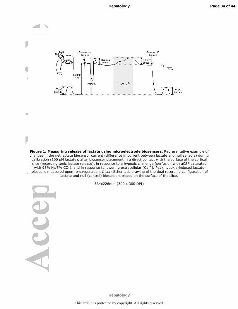
Figure 1: Measuring release of lactate using microelectrode biosensors. Representative example of changes in the net lactate biosensor current (difference in current between lactate and null sensors) during calibration (100 µM lactate), after biosensor placement in a direct contact with the surface of the cortical
slice (recording tonic lactate release), in response to a hypoxic challenge (perfusion with aCSF saturated with 95% N2/5% CO2), and in response to lowering extracellular [Ca
2+]. Peak hypoxia-induced lactate release is measured upon re-oxygenation. Inset: Schematic drawing of the dual recording configuration of
lactate and null (control) biosensors placed on the surface of the slice.
334x226mm (300 x 300 DPI)
Page 34 of 44
Hepatology
Hepatology
This article is protected by copyright. All rights reserved.

Figure 2: HE is associated with a reduction in hemichannel-mediated tonic release of lactate in the cerebral cortex. A) Summary data illustrating tonic release of lactate in cortical slices of sham-
operated, BDL, HA and BDL-OP treated rats. p values indicate differences from sham-operated group. B)
Left: Summary data illustrating the effect of hemichannel blocker carbenoxolone (CBX, 100 µM) on tonic release of lactate (expressed as % change from the baseline) in cortical slices of sham-operated, BDL, HA and BDL-OP treated rats. Right: Representative recordings of lactate biosensor current showing changes in tonic release of lactate in response to CBX application. p values indicate the level of significant differences
from the respective baseline. C) Left: Summary data illustrating the effect of 5-Nitro-2-(3-phenylpropylamino) benzoic acid (NPPB, 200 µM) on tonic release of lactate (expressed as % change from the baseline) in cortical slices of sham-operated, BDL, HA and BDL-OP treated rats. Right: Representative recordings of lactate biosensor current showing changes in tonic release of lactate in response to NPPB
application. p values indicate the level of significant differences from the respective baseline.
Page 35 of 44
Hepatology
Hepatology
This article is protected by copyright. All rights reserved.

Figure 3: HE is associated with a reduction of lactate release in response to lowering extracellular [Ca2+], which promotes hemichannel opening. A) Summary data illustrating peak
changes in lactate release in response to lowering [Ca2+]e in cortical slices of sham-operated, BDL, HA and
BDL-OP-treated rats. p values indicate differences from the responses in sham-operated animals. B) Left: Summary data illustrating the effect of CBX (100 µM) on the release of lactate facilitated in response to 0 [Ca2+]e (expressed as the % of the amount of lactate released in response to 0 [Ca2+]e in the absence of
CBX) in cortical slices of sham-operated, BDL, HA and BDL-OP treated rats. Right: Representative recordings of lactate biosensor current showing the effect of CBX on 0 [Ca2+]e -induced release of lactate. p values
indicate differences between the responses recorded in the absence and presence of CBX. C) Left: Summary data illustrating the effect of NPPB (200 µM) on the release of lactate facilitated in response to 0 [Ca2+]e (expressed as the % of the amount of lactate released in response to 0 [Ca2+]e in the absence of NPPB) in cortical slices of sham-operated, BDL, HA and BDL-OP treated rats. Right: Representative recordings of lactate biosensor current showing the effect of NPPB on 0 [Ca2+]e -induced release of lactate. p values
Page 37 of 44
Hepatology
Hepatology
This article is protected by copyright. All rights reserved.

indicate differences between the responses recorded in the absence and presence of NPPB.
225x314mm (300 x 300 DPI)
Page 38 of 44
Hepatology
Hepatology
This article is protected by copyright. All rights reserved.

Figure 4: HE is associated with a reduction of hypoxia-induced release of lactate. A) Summary data illustrating peak changes in lactate release in response to hypoxia (aCSF saturated with 95% N2/5% CO2) in cortical slices of sham-operated, BDL, HA and BDL-OP-treated rats. p values indicate differences from the
responses in sham-operated animals. B) Left: Summary data illustrating the effect of CBX (100 µM) on the release of lactate facilitated in response to tissue hypoxia (expressed as the % of the amount of lactate released in response to hypoxia in the absence of CBX) in cortical slices of sham-operated, BDL, HA and
BDL-OP treated rats. Right: Representative recordings of lactate biosensor current showing the effect of CBX on hypoxia-induced release of lactate. Decrease in O2 availability reduces biosensor current followed by a positive signal upon re-oxygenation, which is used to estimate hypoxia-induced lactate release. p values indicate differences between the responses recorded the absence and presence of CBX. C) Left: Summary data illustrating the effect of NPPB (200 µM) on the release of lactate facilitated in response to hypoxia (expressed as the % of the amount of lactate released in response to hypoxia in the absence of NPPB) in cortical slices of sham-operated, BDL, HA and BDL-OP-treated rats. Right: Representative recordings of
Page 39 of 44
Hepatology
Hepatology
This article is protected by copyright. All rights reserved.

lactate biosensor current showing the effect of NPPB on the hypoxia-induced release of lactate. p values indicate differences between the responses recorded in the absence and presence of NPPB.
211x297mm (300 x 300 DPI)
Page 40 of 44
Hepatology
Hepatology
This article is protected by copyright. All rights reserved.

Figure 5: Impaired hemichannel-mediated dye loading reveals cortical hemichannel dysfunction in HE. A) Representative images of background loading with carboxyfluorescein (CBF) dye in cortical slices
of sham-operated, BDL, HA and BDL-OP-treated rats. White dashed line depicts the edge of the cortical
slice. B) Fluorescence intensity changes in cortical slices of sham-operated, BDL, HA and BDL-OP treated rats in response to 0 [Ca2+]e and hypoxia in the absence and presence of CBF in the medium. Application of 0 [Ca2+]e aCSF or hypoxia in the presence of CBF results in dye loading and increase in fluorescence, while application of these stimuli in the absence of CBF results in dye unloading and decrease in fluorescence. Insets: Schematic drawings of connexin hemichannel mediated dye loading and unloading. * p<0.05,
**p<0.001 significant differences from the sham-operated group. C) Summary data illustrating the effect of CBX (100 µM) on fluorescence intensity changes (∆Fluorescence) in cortical slices of sham-operated, BDL, HA and BDL-OP treated rats induced by 0 [Ca2+]e and hypoxia in the absence and presence of CBF in the medium. The data are presented as differences in fluorescence following CBX application compared to the respective fluorescence recorded in the absence of CBX. * p<0.05 significant effect of CBX on CBF loading
Page 41 of 44
Hepatology
Hepatology
This article is protected by copyright. All rights reserved.

and unloading. D) Summary data illustrating the effect of NPPB (200 µM) on fluorescence intensity changes (∆Fluorescence) in cortical slices of sham-operated, BDL, HA and BDL-OP treated rats induced by 0 [Ca2+]e and hypoxia in the absence and presence of CBF in the medium. The data are presented as differences in
fluorescence following NPPB application compared to the respective fluorescence recorded in the absence of NPPB. * p<0.05 significant effect of NPPB on CBF loading and unloading.
239x337mm (300 x 300 DPI)
Page 42 of 44
Hepatology
Hepatology
This article is protected by copyright. All rights reserved.

Figure 6: Cortical connexin expression is not affected in HE. Summary data illustrating means ± SE of
the densitometry of connexin-43, connexin-36, connexin-30 and connexin-26 protein expression normalized to the expression of Actin, in cell lysates of the cerebral cortices of sham-operated, BDL, HA and BDL-OP treated rats. Bottom: Representative immunoblots showing connexin-43, connexin-36, connexin-30 and connexin-26 protein expression in cerebral cortices of sham-operated, BDL, HA and BDL-OP treated rats. p
value indicates difference in expression level between BDL and BDL-OP groups.
376x286mm (300 x 300 DPI)
Page 43 of 44
Hepatology
Hepatology
This article is protected by copyright. All rights reserved.
SUPPLEMENTARY MATERIAL
Supplementary Figure 1: Response time, principles of lactate biosensor
operation and the effect of OP on lactate detection system. A) A
schematic drawing of the lactate biosensor assembly showing the enzymatic
biolayer, which surrounds the tip of the platinum (Pt) wire. B) Enzymatic
reaction taking place in the biolayer of the sensor. C) Expanded portion of the
lactate calibration trace illustrating the response characteristics of the lactate
biosensor. Note that the sensor responds immediately when lactate solution is
starting to enter the calibration chamber. D) Raw traces illustrating lactate
biosensor current responses to lactate (100 µM) when calibrated in control
artificial cerebrospinal fluid (aCSF), and in the presence of ornithine
phenylacetate (OP, 1:50 dilution of the stock solution of 0.1g/ml; concentration
applied is similar to the concentration estimated to be reached in a 300 g rat
following IP injections). Bottom: Representative trace from the recordings
obtained in the cerebral cortex of a BDL rat showing the effect of OP on
lactate tone. Grey shading indicates period of drug application.
Supplementary Figure 2: Monocarboxylate transporter (MCT) protein
expression and functionality in cortical slices of sham-operated and
BDL rats. A) Summary data illustrating the effect of MCT blocker, 4-CIN (250
Μm, Sigma) on tonic release of lactate (expressed as % change from the
baseline) and release of lactate facilitated in response to tissue hypoxia and 0
[Ca2+]e (expressed as the % of the amount of lactate released in response to
hypoxia/0 [Ca2+]e in the absence of 4-CIN) in cortical slices of sham-operated,
Page 44 of 44
Hepatology
Hepatology
This article is protected by copyright. All rights reserved.

BDL, HA and BDL-OP treated rats. p values indicate differences between
responses recorded in the absence and presence of 4-CIN. B) Representative
recordings of lactate biosensor current showing the effect of 4-CIN on tonic as
well as hypoxia and 0 [Ca2+]e -induced release of lactate. C) Summary data
illustrating means ± SE of the densitometry of MCT-1 protein levels, mainly
expressed in astrocytes (Chemicon International, 0.5 µg/mL) normalized to
the expression of Actin, in cell lysates of the cerebral cortices of sham-
operated, BDL, HA and BDL-OP treated rats. Bottom: Representative
immunoblots showing MCT-1 protein expression in cerebral cortices of sham-
operated, BDL, HA and BDL-OP treated rats.
Supplementary Table 1: Plasma biochemistry.
Bile duct ligated (BDL), hyperammonemic (HA), ornithine phenylacetate
treated BDL rats (BDL-OP); ALT, alanine aminotransferase.
Data expressed as means±SEM, ** p<0.001 compared to sham group using
one-way ANOVA.
Page 45 of 44
Hepatology
Hepatology
This article is protected by copyright. All rights reserved.

Page 46 of 44
Hepatology
Hepatology
495051525354555657585960
This article is protected by copyright. All rights reserved.
Page 47 of 44
Hepatology
Hepatology
5051525354555657585960
This article is protected by copyright. All rights reserved.

Parameters Sham BDL HA BDL+OP
N numbers 22 26 22 14
Ammonia, μmol/L 56±3 141±4** 121±9** 60±2
Albumin, g/L 35±4 23±0.4** 30±0.8 23±1
Total protein, g/L 50±0.3 38±0.5** 51±3 40±1
Bilirubin, μmol/L 5±0.5 205±3** 4±0.6 161±6
ALT, U/L 11±0.4 130±2** 17±4 87±4
Page 48 of 44
Hepatology
Hepatology
This article is protected by copyright. All rights reserved.