patient 1 – acute abdomen 19 year old male – no prior medical history just back from a trip to...
TRANSCRIPT

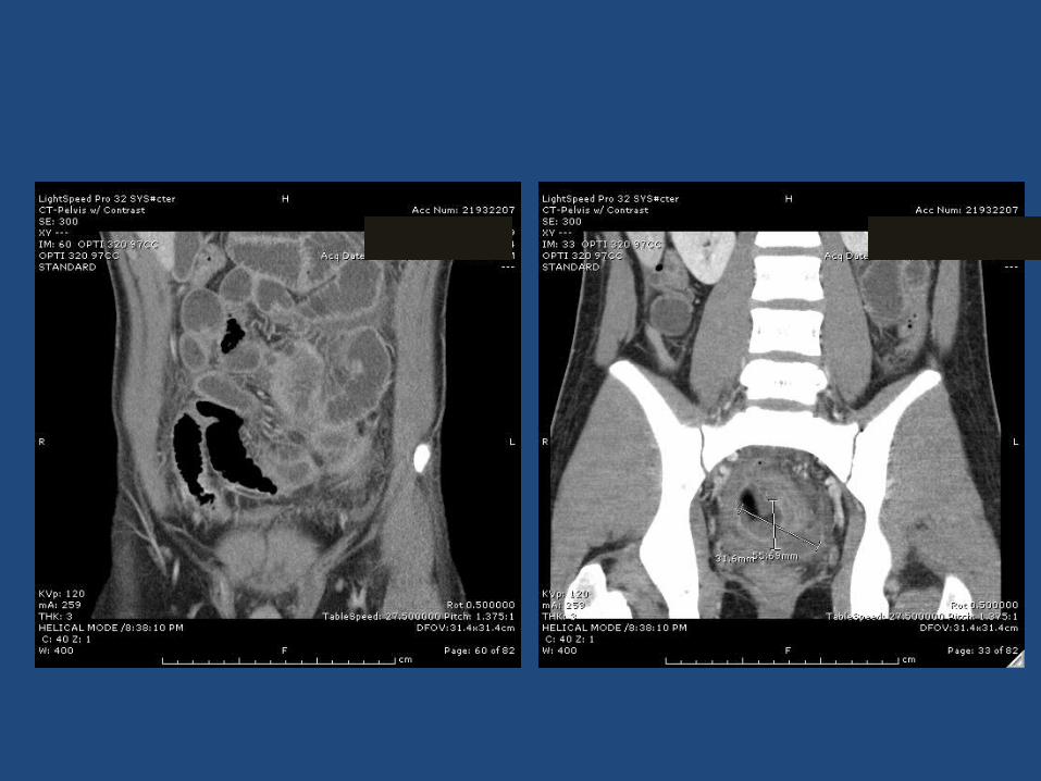
Patient 1 – acute abdomen
• 19 year old male – no prior medical history• Just back from a trip to Israel• Two days of “writhing in pain”, fever to 102.5– Similar episode about a week ago in Israel
resolved• Pediatrician – acute abdomen, wbc 30,000• CT – pelvic abscess, inflamed small and large
bowel, inflamed Ileum
Questions• In an acute presentation, how does one
differentiate between:– Perforated appendix– Newly presenting Crohn’s with perforation– Other causes of pelvic abscess
• How do you determine which patients warrant emergency surgery, vs. which should be “cooled off” medically?
• What is your protocol for medical management prior to surgery?
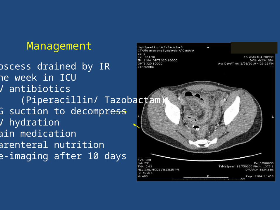
Management
1. Abscess drained by IR2. One week in ICU3. IV antibiotics (Piperacillin/ Tazobactam)3. NG suction to decompress4. IV hydration5. Pain medication 6. Parenteral nutrition7. Re-imaging after 10 days
Questions
• What additional evaluation would you perform at this time to establish a Crohn’s diagnosis?
• Would you consider treating this patient medically? – Short term vs. long term?– With what agent?
• If you choose surgery, what is the appropriate time to intervene?– Laparoscopic vs. open?
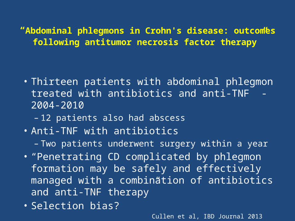
“Abdominal phlegmons in Crohn's disease: outcomes following antitumor necrosis factor therapy”
• Thirteen patients with abdominal phlegmon treated with antibiotics and anti-TNF - 2004-2010– 12 patients also had abscess
• Anti-TNF with antibiotics – Two patients underwent surgery within a year
• “Penetrating CD complicated by phlegmon formation may be safely and effectively managed with a combination of antibiotics and anti-TNF therapy”
• Selection bias? Cullen et al, IBD Journal 2013

Management and outcome
• Hospitalized with PN, NPO, decompression and medical management for approximately 3 weeks.
• EGD/Colonoscopy – no gastric or colonic disease• Laparoscopic ileocecectomy – Fibrotic ileum and cecum– Residual abdominal abscesses– 30 cm of ileum plus cecum resected
• Uneventful recovery
Postoperative management
• How would you monitor patient postoperatively?– Clinical follow-up and labs only?– Small bowel imaging?– Colonoscopy (timing)
• What (if any) postoperative medical therapy would you utilize ?– Aminosalicylate– Thiopurine– Infliximab
Postoperative management and outcome
• Discussion with family and patient• Opted for mercaptopurine– Transaminitis developed– Changed to low dose 6MP and allopurinol
• Surveillance 1 year after initial presentation– Normal MRI, normal colonoscopy
• No clinical or laboratory recurrence 3 years after his initial surgery.– Follow up colonoscopy planned
Presenting history
• 7 year old female• Presented with Serratia osteomyelitis at 3
months of age• History of multiple GI issues in infancy– Poor weight gain requiring NG tube– Diarrhea– Rectal bleeding as infant while breast-fed,
responsive to mother eliminating milk from diet.• Diagnosis of CGD by dihydrorhodamine test

Chronic granulomatous diseaseWinkelstein et al 2000; Medicine 79:155
• Primary immunodeficiency characterized by inability of cells to kill bacteria and fungi.– Staphylococcus, Aspergillus cause serious infection– Catalase positive organisms– Pneumonia most common infection (80%)
• 75% of patients present under age 5 years• Autosomal recessive or X-linked• GI manifestations
– Colitis present in approximately 20%– Perianal abscess– Gastric outlet obstruction
• Diagnosis – defective NADPH oxidase– Dihydrorhodamine test
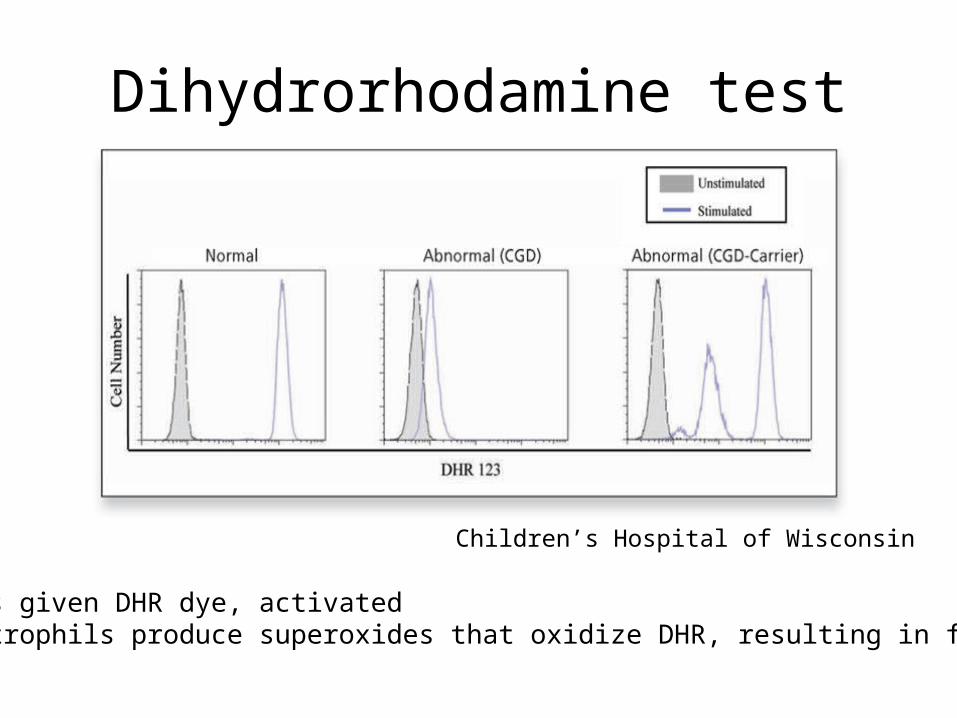
Dihydrorhodamine test
Children’s Hospital of Wisconsin
Neutrophils given DHR dye, activatedNormal neutrophils produce superoxides that oxidize DHR, resulting in fluorescence.

Chronic granulomatous disease• Mimics Crohn disease on endoscopy and histology• Granulomas in only 34 % (Levine, Histopathology 2005)• Paucity of neutrophils compared to UC (Shappi JPGN 2003)• Decreased CD68+ macrophages (Liu et al, IBD Journal 2009)• Therapy – gamma-IFN, steroids, thalidomide, SCT
Clinical course
• Continued with diarrhea, anemia for over a year despite therapy.– Labial abscess grew E. coli
• By age 4:– 3 BM daily, no bleeding– Height at 25% ile, but falling off– Colonoscopy with ileal and colonic granulomas– Therapies included
• Alpha - Interferon• Bactrim and fungal prophylaxis
Medical options for this colitis
• Aminosalicylates• Antibiotics• Probiotics• Corticosteroids• Azathioprine• GM-CSF• Anti-TNF agents
Medical options for this colitis
• Aminosalicylates - intolerant, got diarrhea• Antibiotics – vancomycin for C. difficile• Probiotics – VSL 3• Corticosteroids – recurrent courses • Azathioprine – tried for over 6 months– unable to wean off steroids without worsening
• GM-CSF• Anti-TNF agents?
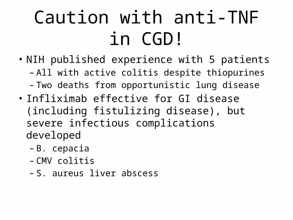
Caution with anti-TNF in CGD!
• NIH published experience with 5 patients– All with active colitis despite thiopurines– Two deaths from opportunistic lung disease
• Infliximab effective for GI disease (including fistulizing disease), but severe infectious complications developed– B. cepacia– CMV colitis– S. aureus liver abscess

Potential options
• GM-CSF– Wang et al, J . Allergy Clinical Immunology 2005
• Anakinra– Rationale – upregulation of IL-1 beta pathway– van de Veerdonk et al, Netherland Journal of
Medicine 2011• Stem cell transplant– Controversial in CGD– No matched siblings in this case.

Current outcome
• Anakinra 200 mg daily for 3 weeks– Azathioprine stopped
• Diarrhea improving with 2 stools daily• Prednisone weaned to 6 mg daily• CRP improved from – 2.2 mg/dL to 0.9 mg/dL
• Plan for 3-6 months of Anakinra, the re-evaluate with colonoscopy
Fever in CD Patient on 6-MP
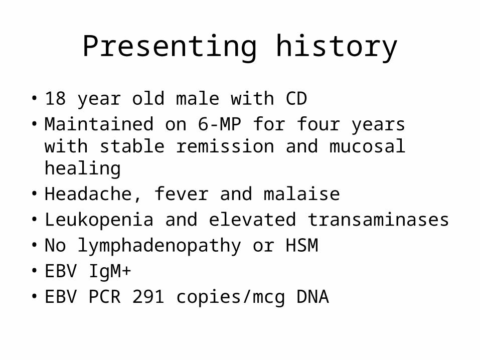
Presenting history
• 18 year old male with CD• Maintained on 6-MP for four years with stable
remission and mucosal healing• Headache, fever and malaise• Leukopenia and elevated transaminases• No lymphadenopathy or HSM• EBV IgM+• EBV PCR 291 copies/mcg DNA
Options
• Additional diagnostic tests?• What to do with 6-MP?• Follow-up?
Clinical course
• Initially improved• Within a few weeks fevers & chills returned• Became jaundiced

Options
• Additional diagnostic tests?• Differential diagnosis?
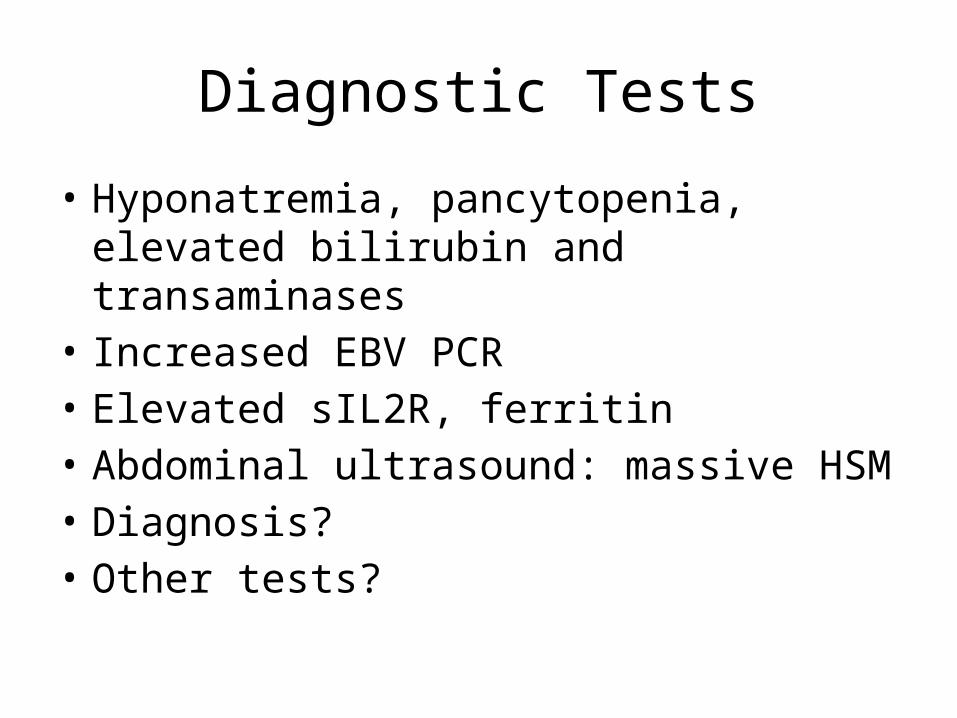
Diagnostic Tests
• Hyponatremia, pancytopenia, elevated bilirubin and transaminases
• Increased EBV PCR• Elevated sIL2R, ferritin • Abdominal ultrasound: massive HSM • Diagnosis?• Other tests?

Diagnostic Tests• EGD/colon: multiple gastric polypoid lesions
with path consistent with EBV+ lymphoma• Liver biopsy: EBV+ lymphoma infiltrate• Bone marrow: hemophagocytosis• PET-CT: widespread
Discussion
• What to do with 6MP with febrile illness/mono?
• RISK of EBV associated HLH or lymphoma with 6-MP?
• Different for primary EBV infection versus reactivation?
• Monitoring for this?