pathophysiology of adrenal disorders and clinical use of...
TRANSCRIPT

Pathophysiology of Adrenal Disorders and Clinical Use of
Corticosteroids
Wayne Kradjan

Adrenal Glands• Located on upper poles of both kidneys
• Adrenal cortex (90% gland wt)– Zona Glomerulosa: Aldosterone– Zona Fasciculata: Cortisol– Zona Reticularis: Testosterone and estradiol production
from cholesterol• Adrenal medulla: Catecholamines

Corticosteroid PhysiologyHypothalamus
(Corticotropin releasing factor)
Pituitary(Corticotropin/ACTH)
Adrenals
Cortisol(Hydrocortisone)
(12-25 mg/day; 300 mg with stress)(Plasma level = 5-20 μg/dL; >60 μg/dL with stress
Aldosterone(50-250 mg/24 hr)
Regulatevia
negativefeedback

Diurnal Cycle of Release
• ACTH peaks midnight to 2 AM• Cortisol peaks at 6-8 AM• Varies with sleep cycle
– Night shift workers– Consider consequence of multiple shift or
changing shift workers
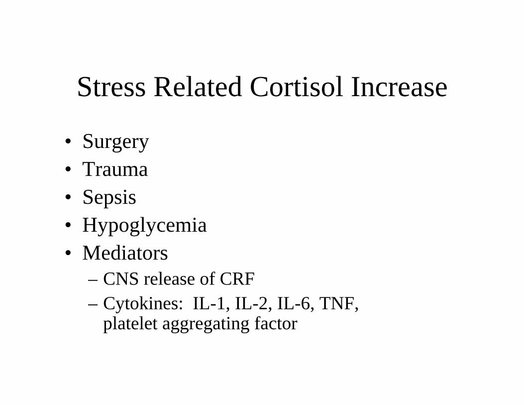
Stress Related Cortisol Increase
• Surgery• Trauma• Sepsis• Hypoglycemia• Mediators
– CNS release of CRF– Cytokines: IL-1, IL-2, IL-6, TNF,
platelet aggregating factor

Prostaglandins and LeukotrienesCell Membrane Destruction
Arachidonic Acid
Leukotrienes Prostaglandins
Lipooxygenase cyclooxygenase
InflammationBronchospasm
VasodilateEdema
BronchodilatePain sensitization
VasodilateInhibit platelets
VasoconstrictPlatelet
aggregation
Vaso-constrict
PGE Prostacyclin(vascular)
Thromboxane(platelets)
PGF
phospholipase A2

Glucocorticoid Actions• Decrease inflammatory response
– Inhibit phospholipase A2 production of arachidonic acid indirect inhibition of both prostaglandins and leukotrienes.
– Inhibit activity of T lymphocytes (especially TH2)– Suppress IL-1, IL-3, IL-4, IL-5;
chemotraction of eosinophils and macrophages– Vasoconstrict, edema, fever– Stabilize neutrophilic (granulocyte) lysosomes to
lysosomal enzyme release
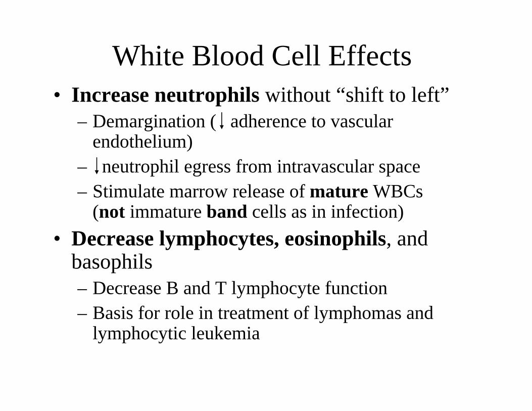
White Blood Cell Effects• Increase neutrophils without “shift to left”
– Demargination ( adherence to vascular endothelium)
– neutrophil egress from intravascular space– Stimulate marrow release of mature WBCs
(not immature band cells as in infection)• Decrease lymphocytes, eosinophils, and
basophils– Decrease B and T lymphocyte function– Basis for role in treatment of lymphomas and
lymphocytic leukemia

Other Glucocorticoid effects– protein synthesis and protein movement
out of vessels– Gluconeogenesis (hyperglycemia)– Fat Redistribution: suppress lipolysis and
lipogenesis via insulin inhibition.– beta adrenergic responses (note value in
asthma)
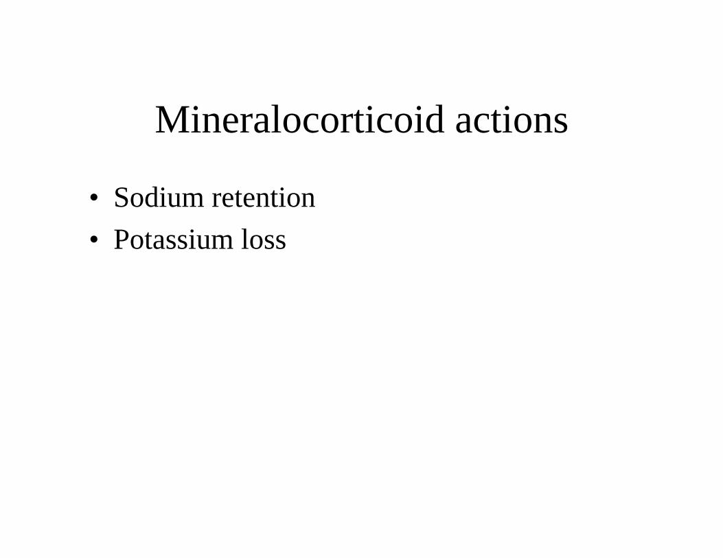
Mineralocorticoid actions
• Sodium retention• Potassium loss

Clinical Use of Corticosteroids• Goal of treatment: symptomatic, not curative
– Topical or oral: contact dermatitis or allergic rxn– Allergic rhinitis and asthma– Arthritis– Psoriasis– Autoimmune (lupus, polymyalgia rheumatica, non-viral
hepatitis)– Inflammatory bowel disease– Lymphoma, leukemia– Transplant surgery: post-op/maintenance, rejection prevention– Prophylaxis of “dry socket” with dental surgery– Brain and spinal cord tumors (“cerebral edema)– PCP infection in AIDS patients, ARDS, sepsis– Hypercalcemia- multiple myeloma, bone mets, sarcoid
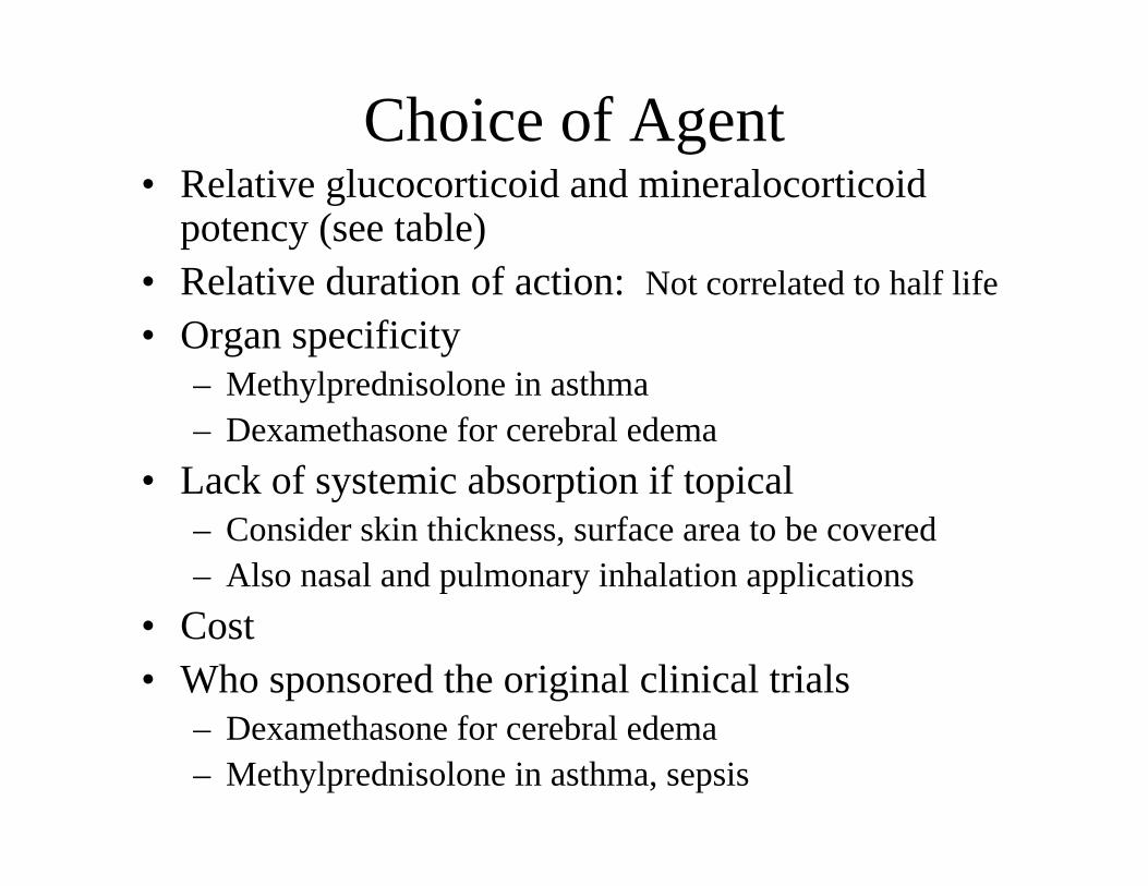
Choice of Agent• Relative glucocorticoid and mineralocorticoid
potency (see table)• Relative duration of action: Not correlated to half life• Organ specificity
– Methylprednisolone in asthma– Dexamethasone for cerebral edema
• Lack of systemic absorption if topical– Consider skin thickness, surface area to be covered– Also nasal and pulmonary inhalation applications
• Cost• Who sponsored the original clinical trials
– Dexamethasone for cerebral edema– Methylprednisolone in asthma, sepsis

Relative potencies
125Fludro
36-54 hr0300.75 mgDexa
18-36 hr0.5+54 mgMethylpred
18-36 hr1+3.55 mgPred
8-12 hr2+120-25 mgCortisol
DurationNa retention
Anti-inflam
Eqivalntdose
Name

Dosing• Acute:
– Moderate to high dose for rapid resolution of symptoms; high dose “bursts” x 7-14 days
– E.g., 60-80 mg prednisone or equivalent “burst therapy” for asthma in ER
– 60-120 mg methylprednisolone QID for hospitalized patient
– 4 mg QID dexamethasone for brain tumor• Chronic (maintenance)
– Minimum dose for shortest duration possible. Prefer to avoid all together.
– Morning doses preferred– Every other day in some cases (next slide)

Every other day dosing• Theory: use drug that works 36 hours
(prednisone, methylprednisolone) every other day to allow HPA to function every other night– Fewer side effect and HPA suppression
possible– Concern over loss of therapeutic effect on
evening of day 2– E.g. 10 mg QD slowly converted to 20 mg
QOD

Tapering Principles• Less than 10 – 14 days, with no prior exposure
– Rapid taper acceptable, even with high doses– Major considerations are disease exacerbation, mild flu like
symptoms, mild depression• Longer term exposure, even with low doses
– Slow taper mandatory, especially as approach physiologic dose equivalent.
– Physiologic withdrawal effects may be observed (see next slide)• Short term exposure to high doses in patient with chronic
use of high doses. – Rapid taper from high dose may be acceptable, but do not go
below the chronic dose – E.g. patient on 10 mg prednisone per day for 3 months.
Physiologic for this person is 10 mg. Then even slower taper iftrying to remove drug entirely.

Physiologic Withdrawal Signs• Time and dose dependent.
– Unlikely if duration less than 10-14 days– Avoid night time doses if >5-7 days
• CNS depression• Flu-like symptoms• Muscle and joint pain• Tremor• Hypotension (not necessarily hyponatremic)• Hyperkalemia, arrhythmias

Timing considerations• Takes 7-14 days, even with very high doses to
suppress pituitary ACTH release and adrenal cortisol release.
• With prolonged dosing (>30 days?) HPA suppression is evident, but dose dependent.– 9-12 months to fully restore HPA axis after slow
withdrawal and complete removal.– Pituitary response recovers before adrenal gland– May need to cover with prednisone bursts during times of
stress after complete withdrawal.
Example tapering dose• Methylprednisolone 60 mg IV QID x 3 days• Day 4: change to prednisone 60-80 mg/ day
– QD or split into 2-3 doses?– Compare to physiologic dose of 5 mg prednisone
or 4 mg methylprednisolone• Then 60 mg x 3 days, 40 mg x 3 days, 30 mg x 3
days, 20 mg x 3 days, 15 mg x 3 days, 10 mg x 5 days, 5 mg x 5 days, 2.5 mg x 5 days, then stop
• Increase dose or slow taper rate if symptoms worsen• What if patient was taking 10 mg per day at home
before exacerbation?• What if patient is using inhaled steroids?

Another example• Steroid naïve patient receives 80 mg of
prednisone in the emergency room– Should IV drug have been used instead of oral?– Assuming the symptoms resolve over 4 hours,
why does the patient need a prednisone prescription for outpatient use?
– How long should treatment continue?– Should the dose be tapered?
• E.g 20 mg qd x 7-14 days without taper• E.g. 40 mg x 2-3 days, 30 mg x 2-3 days, 20 mg x 2-
3 days, 10 mg x 2-3days, 5 mg x 2-3 days

Acute side effects• Dose dependent, low risk• Endocrine: hyperglycemia. Diabetic?• Elevated white count: demargination vs. infection• GI: Bleeding, “stress ulcers”
– mucous production, local vasoconstriction
• Na retention (caution re edema, HTN, CHF)• Hypokalemia, metabolic acidosis• Jitteriness, euphoria, confusion (steroid psychosis)

Longer term side effects• Continuation of short term side effects• HPA axis suppression after 2 weeks• Cushingoid features: fat redistribution to face and
back, striae• Muscle weakness, myopathy, protein wasting• Thinning of skin, capillary fragility with petechiae,
bruising, acne• Osteoporosis in adults with compression fractures;
aseptic necrosis of hip, growth retardation in children• Cataracts, glaucoma• Decreased immune response; TB activation, poor
wound healing

Buffalo Hump:Accumulation of faton back of neck and upper back
Moon Facies:fat deposition in face
Central obesity and striae

Striae (stretch marks)

Drug Interactions
• Steroids increase aspirin clearance. Risk of ASA toxicity when steroids stopped.
• Barbiturates, phenytoin, rifampin increase steroid clearance/ metabolism
• Cimetidine: decreased steroid metabolism?• Ketoconazole: decreased cortisol production• Hypoglycemics: steroid induced glucose increase• Additive hypokalemia to potassium wasting
diuretics• Additive ulcerogenic property to NSAIDS?

Precautions
• Sodium retention in patients with hypertension or heart failure
• Effects on electrolytes in patients with arrhythmias or renal disease
• Hepatic failure: lack of conversion of prednisone to prednisolone.– Clinical significance debated
HyperfunctioningAdrenal Gland
• Cushing’s Disease (60-70%)– Overactive pituitary gland (85% via adenomas)– Excess ACTH production with secondary bilateral
adrenal hyperplasia– Primary (via pituitary) or secondary (via hypothalamus)
• Cushing’s Syndrome– Adrenal adenoma (benign)– Adrenal carcinoma– Ectopic ACTH syndrome (e.g., oat cell lung carcinoma,
pancreas)• Iatrogenic: overuse of exogenous corticosteroids. Mimics
hyperfunctioning gland, but actually underactive gland

Clinical Features of Cushing’s Disease
• Central obesity and facial rounding –90%– “moon face”, “buffalo hump”, central obesity
• Hypertension-75-80% via Na retention• Glucose intolerance-80%• Menstrual dysfunction, hirsutism -76%
– Androgen excess• Abdominal striae (red-purple)• Muscle weakness, myopathy, compression
fractures–50-60%;osteoporosis 20%
• Anxiety, tremor, mood elevation, psychosis- 50%
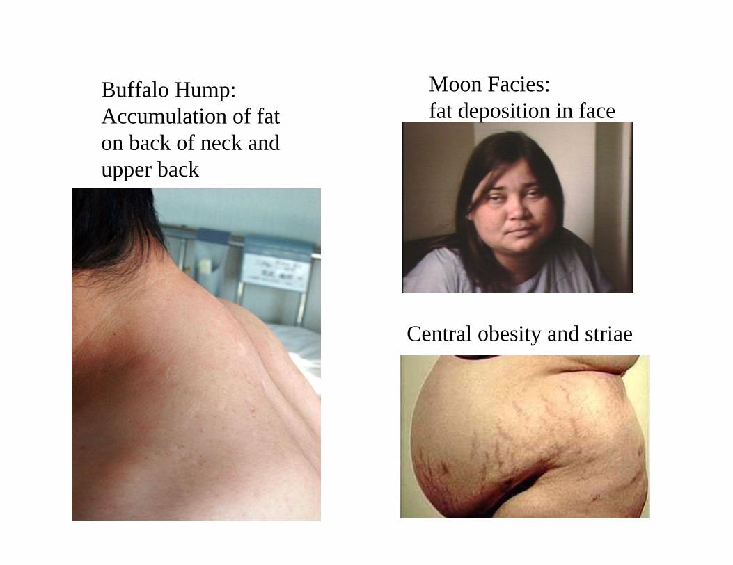
Buffalo Hump:Accumulation of faton back of neck and upper back
Moon Facies:fat deposition in face
Central obesity and striae

Workup of patient• Suspect Cushing’s based on symptoms• Screening serum or urine cortisol level
– Should be high. If not, look for other disease.– Exception: low if taking exogenous steroids as
cause of Cushing’s syndrome• If cortisol level is high, measure ACTH level
to differentiate pituitary vs adrenal cause. (see next slide)
• To differentiate pituitary origin vs adrenal origin: conduct dexamethasone suppression test. (details on another slide)

Adrenal Lab Tests• 24 hour urine cortisol
– Nl: 20-90 mcg per 24 hr.– Increase 2-3x with Cushings
• Plasma cortisol– Nl: 12-20 mcg/ 100 ml @ 8AM;
4-8 mcg/100 mL @ 11 PM– Cushings: higher (up to 50 mcg/100 mL) and not
circadian– Low if taking exogenous steroids
• Plasma ACTH– Nl 150 pcg/mL– High (up to 500 pcg/mL) if pituitary or hypothalamic
origin– Low (<50 pcg/mL) if adrenal adenoma or carcinoma

More Adrenal Tests
• Dexamethasone suppression test– 1 mg dexamethsone @ 11 PM– Nl: suppressed plasma cortisol at 8 AM; < 5 mcg/100
mL– Cushings: fail to suppress– May repeat times 2 days– May use higher doses
(4 mg Q 11 PM times 2 days)
• Nuclear scanning, CT scans, MRI

Treatment of Cushings• Surgical removal or radiation of adrenal, pituitary, or
hypothalamus– Replace cortisol and fludrocortisone (Florinef) 0.1 mg QD
x (6-12 months if one adrenal only involved)– Replace other pituitary hormones as indicated (thyroid, sex
hormones)• Ketoconazole (Nizoral) for refractory cases
– Inhibits 11-hydroxylase and 17-hydroxylase• Metapyrone (Metopirone) and/or aminoglutethamide
(Cytadren) for ectopic ACTH syndrome• Mitotane (o,p’DD, Lysodren) for adrenal carcinoma.
Inhibits 11 hydroxylation• RU 486 (mifepristone): progesterone and
glucocorticosteroid receptor antagonist

Hormone Synthetic Pathways
Cholesterol Pregnenolone Progesterone 17-hydroxyprogesterone
11-deoxycortisol Cortisol
11-desoxycorticosterone
Corticosterone 18-hydroxycorticosterone
Aldosterone
18-hydroxypregnenolone
Dehydroepiandrosterone
Androstenedione Testosterone
Metyrapone,Mitotane
From figure 76-2, Chapter 76, Pharmacotherapy
Aminoglutethimide Ketoconazole
Mitotane

HypofunctioningAdrenal Gland
• Addison’s Disease• Primary
– Autoimmune-70% (may involve other organs: thyroid, ovary, pancreas)
– Tuberculosis, AIDS, other infections– Vascular obstruction, bleeds
• Secondary (low ACTH)– Hypopituitarism– Corticosteroid administration

Clinical Features of Addison’s• Weakness-100%• Weight loss-100%• *Increased pigmentation-95%
(via ACTH stimulation of melanocyte stimulating hormone; absent in secondary forms)
• Hypotension-90%• Vitiligo-20%• Diuresis (analogy to spironolactone)
Hyponatremia/hyperkalemianote: aldosterone (mineralocorticoid function) may be spared in secondary cases

Vitiligo

Vitiligo
Workup of patient• Suspect Addison’s based on symptoms• Screening serum or urine cortisol level
– Should be low. If not, look for other disease.• If cortisol level is low, measure ACTH level
to differentiate pituitary vs adrenal cause. (see next slide)
• To differentiate pituitary origin vs adrenal origin: conduct ACTH or Cosyntropin stimulation test. (details on another slide)

Lab Tests forAdrenal Insufficiency
• Serum cortisol low in both primary and secondary Addison’s
• Serum ACTH above normal in primary Addison's, but low or absent in secondary

ACTH Stimulation Test• Administer 25-40 units ACTH (Acthar) in early
morning.– Measure serum cortisol pre-ACTH and 30 minutes post– Cortisol level should double.
Lack of response if Addison’s• Cosyntropin
– Synthetic polypeptide of ACTH(24 of 39 amino acids present)
– Dose = 0.25 mg– 1 mg cosyntropin = 100 mg ACTH
• Lack of cortisol response in primary, possibly in secondary.

Metyrapone Test
• To verify secondary Addison’s(hypopituitarism)
• Metyrapone blocks conversion of desoxycortisol to cortisol
• As cortisol levels fall, there should be a reflex increase in ACTH production.
• ACTH levels do not rise if pituitary is hypofunctioning
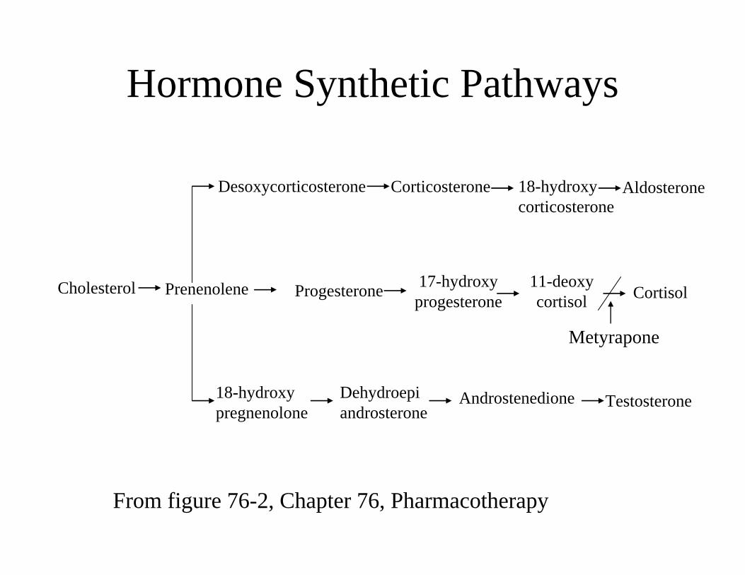
Hormone Synthetic Pathways
Cholesterol Prenenolene Progesterone 17-hydroxyprogesterone
11-deoxycortisol Cortisol
Desoxycorticosterone Corticosterone 18-hydroxycorticosterone
Aldosterone
18-hydroxypregnenolone
Dehydroepiandrosterone
Androstenedione Testosterone
Metyrapone
From figure 76-2, Chapter 76, Pharmacotherapy
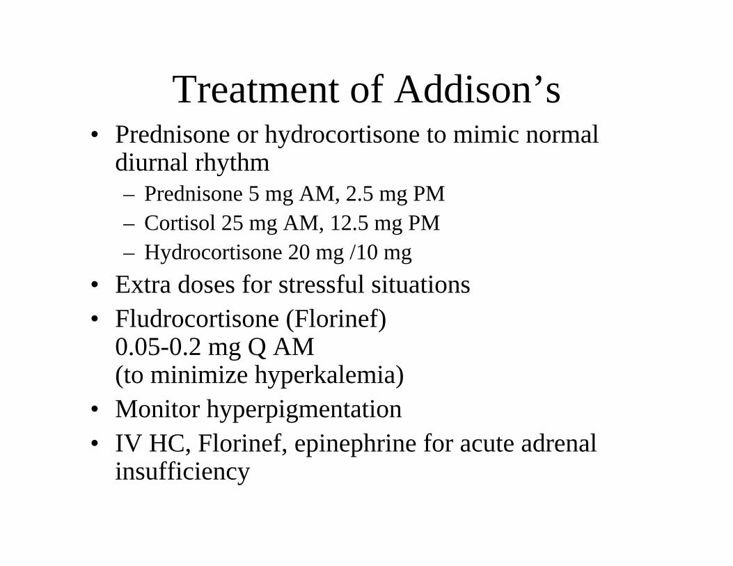
Treatment of Addison’s• Prednisone or hydrocortisone to mimic normal
diurnal rhythm– Prednisone 5 mg AM, 2.5 mg PM– Cortisol 25 mg AM, 12.5 mg PM– Hydrocortisone 20 mg /10 mg
• Extra doses for stressful situations• Fludrocortisone (Florinef)
0.05-0.2 mg Q AM(to minimize hyperkalemia)
• Monitor hyperpigmentation• IV HC, Florinef, epinephrine for acute adrenal
insufficiency