pathophysiology: left to right shunts - columbia university · 2006-12-18 · 1 pathophysiology:...
TRANSCRIPT

1
Pathophysiology:
Left To Right Shunts
Daphne T. Hsu, MD
Learning Objectives
• Learn the relationships between
pressure, blood flow, and resistance
• Review the transition from fetal to
mature circulation
• Determine the effects of the
transitional circulation on the
physiology of left to right shunts
• Correlate clinical signs and
symptoms with cardiac physiology

2
Pressure, Flow, Resistance
• Perfusion Pressure: Pressuregradient across vascular bed– Mean Arterial - Venous pressure
• Flow: Velocity of flow acrossvascular bed
• Resistance: Opposition to flow– Vessel diameter
– Vessel structure and organization
– Physical characteristics of blood
Hemodynamics
PressureFlow =
Resistance
PressureResistance =
Flow

3
Fetal Circulation
Placenta supplies
oxygenated blood via
ductus venosus
Pulmonary blood flow
minimal (10%)
Foramen ovale directs
ductus venous blood to
left atrium (40%)
Ductus arteriosus
allows flow from PA to
descending aorta (50%)
Fetal Pulmonary Vascular Bed
• Pulmonary Pressure
– Vasoconstriction
– Medial wall hypertrophy
– Pulmonary = Aortic Pressure (DA)
• Pulmonary blood flow
– Blood bypasses the lungs via theductus arteriosus to the aorta
– Flow: Minimal
• Pulmonary resistance: High-Infinite
– Resistance = Pressure
Flow
4
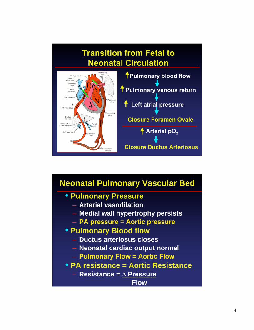
Transition from Fetal to
Neonatal Circulation
Pulmonary blood flow
Pulmonary venous return
Left atrial pressure
Arterial pO2
Closure Foramen Ovale
Closure Ductus Arteriosus
Neonatal Pulmonary Vascular Bed
• Pulmonary Pressure– Arterial vasodilation
– Medial wall hypertrophy persists
– PA pressure = Aortic pressure
• Pulmonary Blood flow– Ductus arteriosus closes
– Neonatal cardiac output normal
– Pulmonary Flow = Aortic Flow
• PA resistance = Aortic Resistance– Resistance = Pressure
Flow

5
Regulation of Pulmonary Vascular Tone
• Vascoconstriction
– Hypoxia/acidosis
– High blood flow and pressure
– Failure of vessel maturation (noregression of medial hypertrophy)
• Vasodilation
– Improved oxygenation
– Prostaglandin inhibition
– Thinning of vessel media(regression of medial hypertrophy)
Pulmonary Vascular Bed:
Transition from Fetal to Adult
PR=
F
6
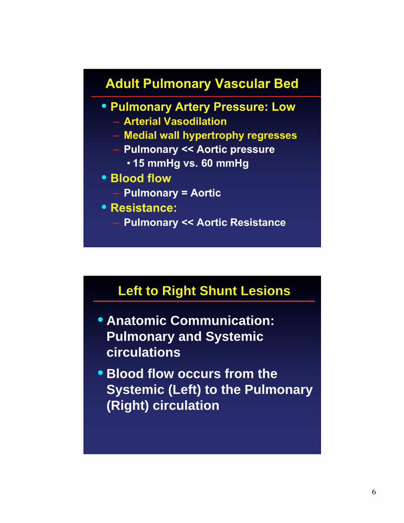
Adult Pulmonary Vascular Bed
• Pulmonary Artery Pressure: Low
– Arterial Vasodilation
– Medial wall hypertrophy regresses
– Pulmonary << Aortic pressure
• 15 mmHg vs. 60 mmHg
• Blood flow
– Pulmonary = Aortic
• Resistance:
– Pulmonary << Aortic Resistance
Left to Right Shunt Lesions
• Anatomic Communication:
Pulmonary and Systemic
circulations
• Blood flow occurs from the
Systemic (Left) to the Pulmonary
(Right) circulation

7
• Ventricular Septal Defect (VSD)– Left ventricle to Right ventricle
• Persistent Patent Ductus Arteriosus(PDA)– Aorta to Pulmonary artery
• Endocardial Cushion Defect (ECD)– Left ventricle to Right ventricle
– Left atrium to Right atrium
• Atrial Septal Defect (ASD)– Left atrium to Right atrium
“Top 4” Left to Right Shunt Lesions
Ventricular Septal Defect

8
VSD: 2/1000 live births
Determinants of L to R shunt Flow
• Size of Defect
• Maturity of
Pulmonary Bed
• Pressure difference
between RV and LV
• Relative resistance
between Pulmonary
and Systemic
circulations
Determinants of Left to Right Shunt
• Small (restrictive) VSD: L to R
shunt flow limited by size of hole
• Large (unrestrictive) VSD: L to R
shunt flow is determined by:
– RV vs. LV pressure
• If RV < LV, L to R shunt occurs
– If RV = LV
• If pulmonary < aortic resistance, L
to R shunt occurs
9
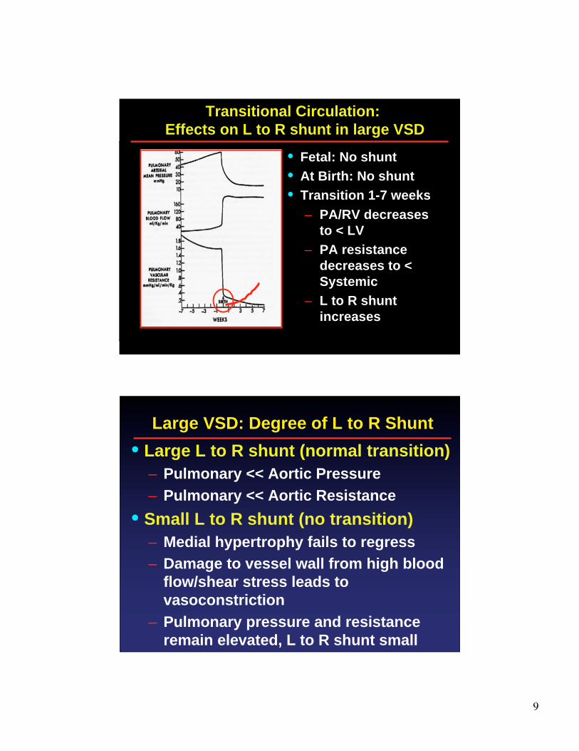
Transitional Circulation:
Effects on L to R shunt in large VSD
• Fetal: No shunt
• At Birth: No shunt
• Transition 1-7 weeks
– PA/RV decreases
to < LV
– PA resistance
decreases to <
Systemic
– L to R shunt
increases
Large VSD: Degree of L to R Shunt
• Large L to R shunt (normal transition)
– Pulmonary << Aortic Pressure
– Pulmonary << Aortic Resistance
• Small L to R shunt (no transition)
– Medial hypertrophy fails to regress
– Damage to vessel wall from high blood
flow/shear stress leads to
vasoconstriction
– Pulmonary pressure and resistance
remain elevated, L to R shunt small
10
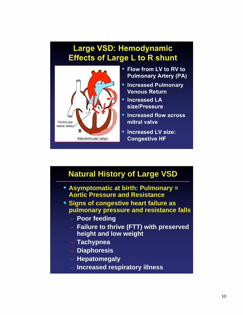
Large VSD: Hemodynamic
Effects of Large L to R shunt
• Flow from LV to RV to
Pulmonary Artery (PA)
• Increased Pulmonary
Venous Return
• Increased LA
size/Pressure
• Increased LV size:
Congestive HF
• Increased flow across
mitral valve
Natural History of Large VSD
• Asymptomatic at birth: Pulmonary =Aortic Pressure and Resistance
• Signs of congestive heart failure aspulmonary pressure and resistance falls
– Poor feeding
– Failure to thrive (FTT) with preservedheight and low weight
– Tachypnea
– Diaphoresis
– Hepatomegaly
– Increased respiratory illness

11
• Holosystolic murmur loudest LLSB
radiating to apex and back
• Mid-Diastolic rumble: Increased flow
across the mitral valve
• LV heave: LV dilation
• Precordial Thrill: turbulent blood flow
across VSD
• Heart failure: Gallop rhythm (S3),Hepatomegaly, Rales
• Second heart sound: elevated PA
pressure
VSD: Clinical Findings
• EKG: LV dilatation ± RVH (if
pulmonary artery pressure high)
• Chest x-ray: Large heart, PVM
• Echo: Gold Standard
– Location/Size of lesion
– LA/LV size
– Estimation RV pressure
• Catheterization: only in cases
when high PVR suspected
Laboratory Findings: VSD
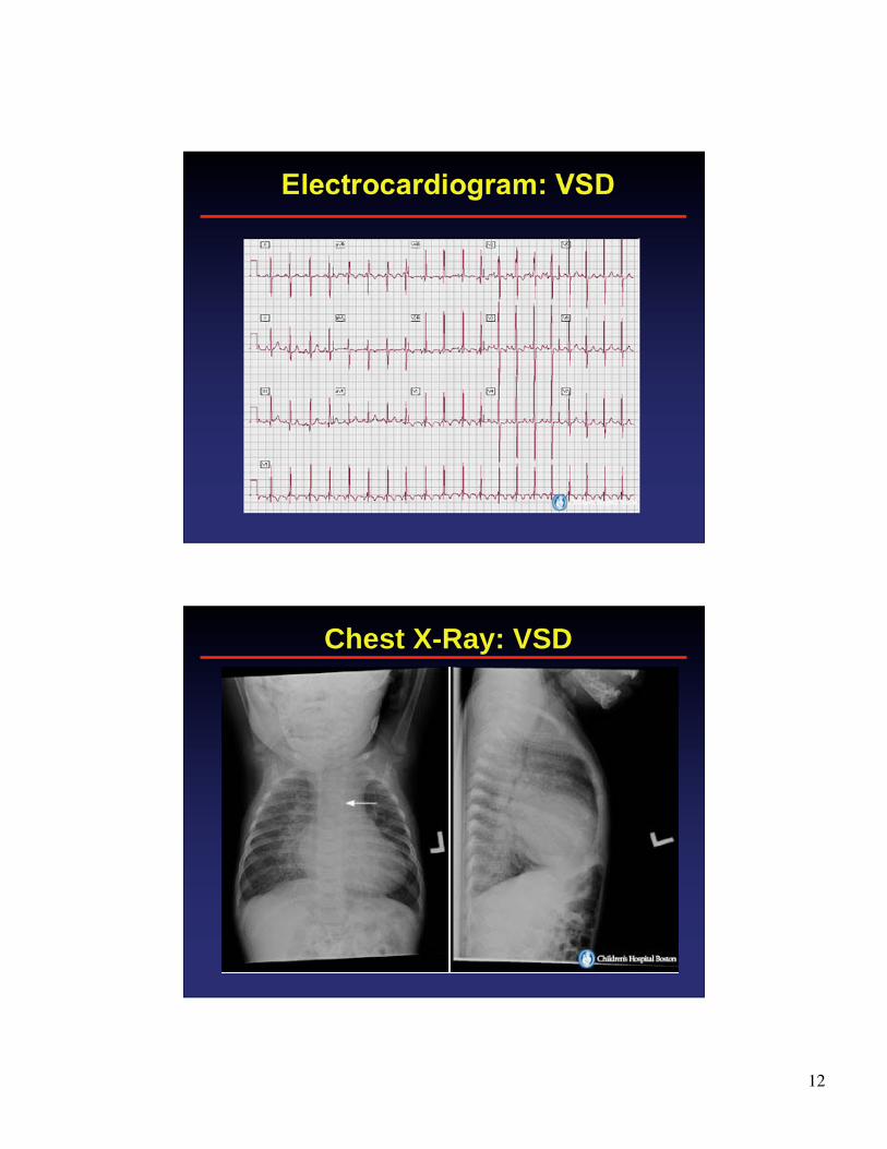
12
Electrocardiogram: VSD
Chest X-Ray: VSD

13
Echocardiogram: Membranous VSD
Angiogram: VSD

14
Treatment of Large VSD
• Medical: Anticongestive Therapy
– Digoxin
– Lasix
– Increased caloric intake
• VSD size decreases
– Resolution of CHF without surgery(50%)
• Indications for VSD closure
– Persistent CHF with failure to thrive orother symptoms
– Increasing pulmonary vascularresistance
– Within first two years of life
Eisenmenger’s Syndrome
• Dr. Victor Eisenmenger, 1897
• Pathophysiology
– Medial hypertrophy of pulmonaryarteries
– Perivascular necrosis
– Replacement of normal vasculararchitecture
• High pulmonary vascular resistance
– Right to left shunt via VSD
– Severe cyanosis
15
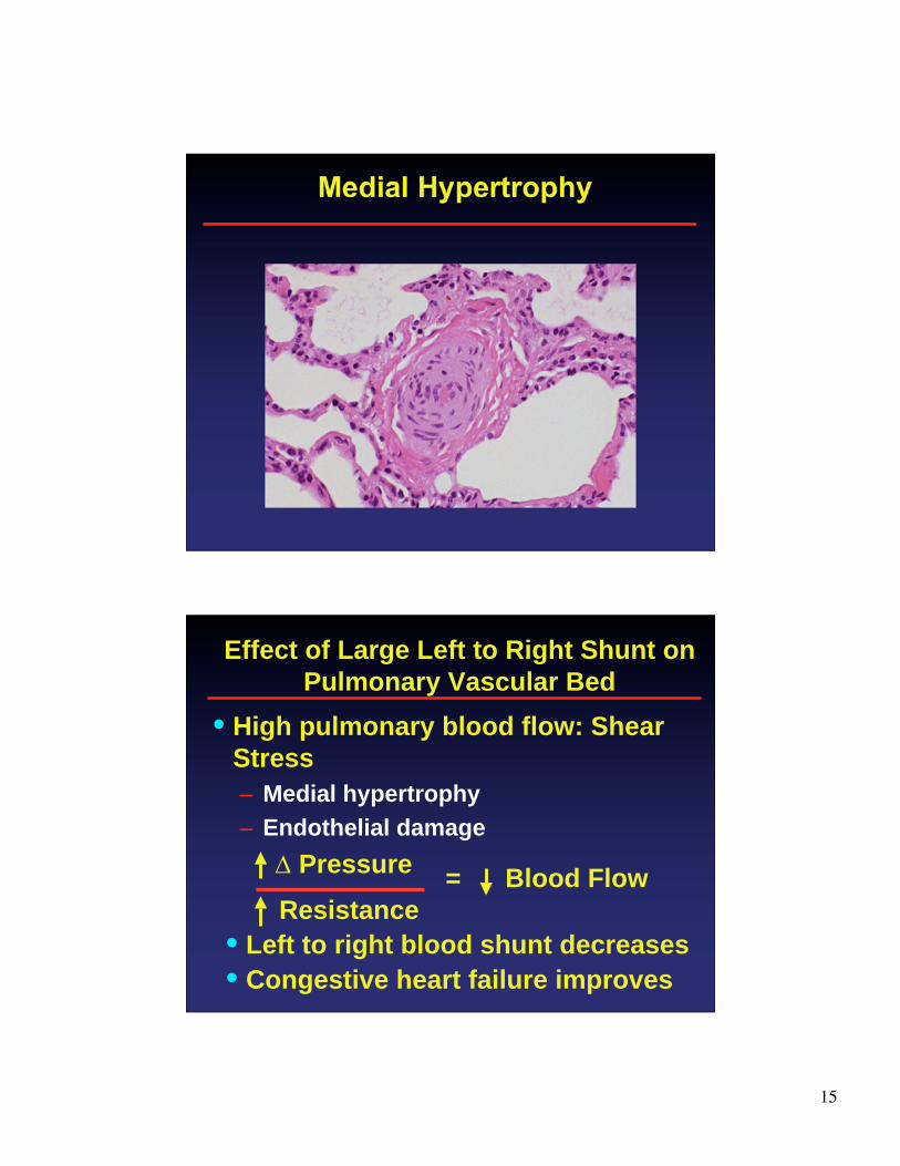
Medial Hypertrophy
Effect of Large Left to Right Shunt on
Pulmonary Vascular Bed
• High pulmonary blood flow: Shear
Stress
– Medial hypertrophy
– Endothelial damage
Pressure
Resistance
• Left to right blood shunt decreases
• Congestive heart failure improves
= Blood Flow

16
Eisenmenger’s Syndrome
R to L flow via VSD
• Pressure:
– Pulmonary = Aortic
• Resistance
– Pulmonary > Aorta
• Blood flow: RV to LV
• Normal LA/LV size
• RV hypertrophy
• Cyanosis
Clinical Picture: Eisenmenger’s• Rare disease in modern era
• Clinical improvement of heart failure in infancydue to decreased left to right shunt
• Clinical presentation: young adulthood
– Exercise Intolerance
– Cyanosis
– Clubbing
– No systolic murmur
• Elevated PA pressure/resistance
– Second heart sound increased
– RV heave (RV hypertension)
– Pulmonary insufficiency murmur

17
Lab findings: Eisenmenger’s
• No LV volume overload
• High RV pressure overload
• EKG: RVH ± strain
• Echo: RV hypertrophy, right to
left shunt at VSD
• Chest x-ray: Clear lung fields,
prominent PA segment, small
heart
EKG: Eisenmenger Syndrome

18
Chest X-Ray: Eisenmenger Syndrome
Management
• Do NOT close VSD
– No left to right shunt: No heart failure
– Shunt is right to left through VSD
– VSD must stay open to decompress highpressure RV and prevent RV failure
• Pulmonary vasodilators
– Calcium channel blocker
– PGI2, Sidenafil
• Inotropic support
– Right heart failure
• Transplant
– Heart-Lung
– Lung transplant, heart repair
19
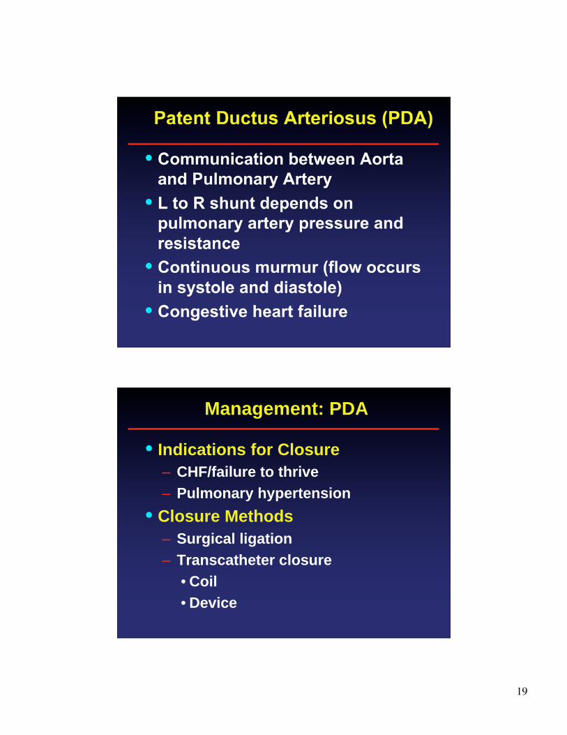
Patent Ductus Arteriosus (PDA)
• Communication between Aorta
and Pulmonary Artery
• L to R shunt depends on
pulmonary artery pressure and
resistance
• Continuous murmur (flow occurs
in systole and diastole)
• Congestive heart failure
Management: PDA
• Indications for Closure
– CHF/failure to thrive
– Pulmonary hypertension
• Closure Methods
– Surgical ligation
– Transcatheter closure
• Coil
• Device

20
PDA Coil Closure
Endocardial Cushion Defect
• Atrial Septal
Defect (Primum)
• VSD
• Common
Atrioventricular
Valve

21
Management: ECD
• Closure always indicated
• Timing of surgery (elective by 6 mos.)
– Heart Failure
• Large left to right shunt
• Mitral insufficiency
– Pulmonary hypertension
• Surgical repair
– ASD, VSD closure
– Repair of AV-Valves
Summary: VSD, PDA and ECD
• Asymptomatic in fetus and neonate
• Progressive increase in L to R shunt
from 3-8 weeks of life as pulmonary
pressure and vascular resistance
decreases
• Indications for intervention
– Congestive heart failure: FTT
– Pulmonary vascular disease
• End stage: Eisenmenger’s syndrome
22
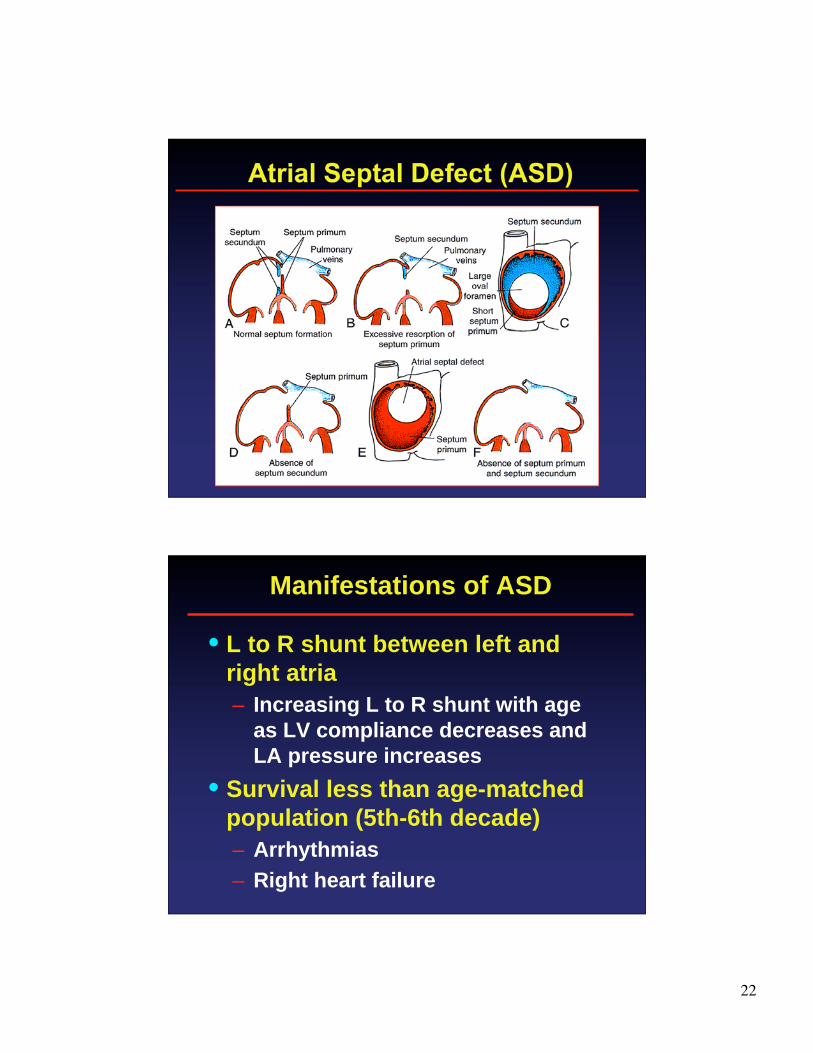
Atrial Septal Defect (ASD)
Manifestations of ASD
• L to R shunt between left and
right atria
– Increasing L to R shunt with age
as LV compliance decreases and
LA pressure increases
• Survival less than age-matched
population (5th-6th decade)
– Arrhythmias
– Right heart failure
23
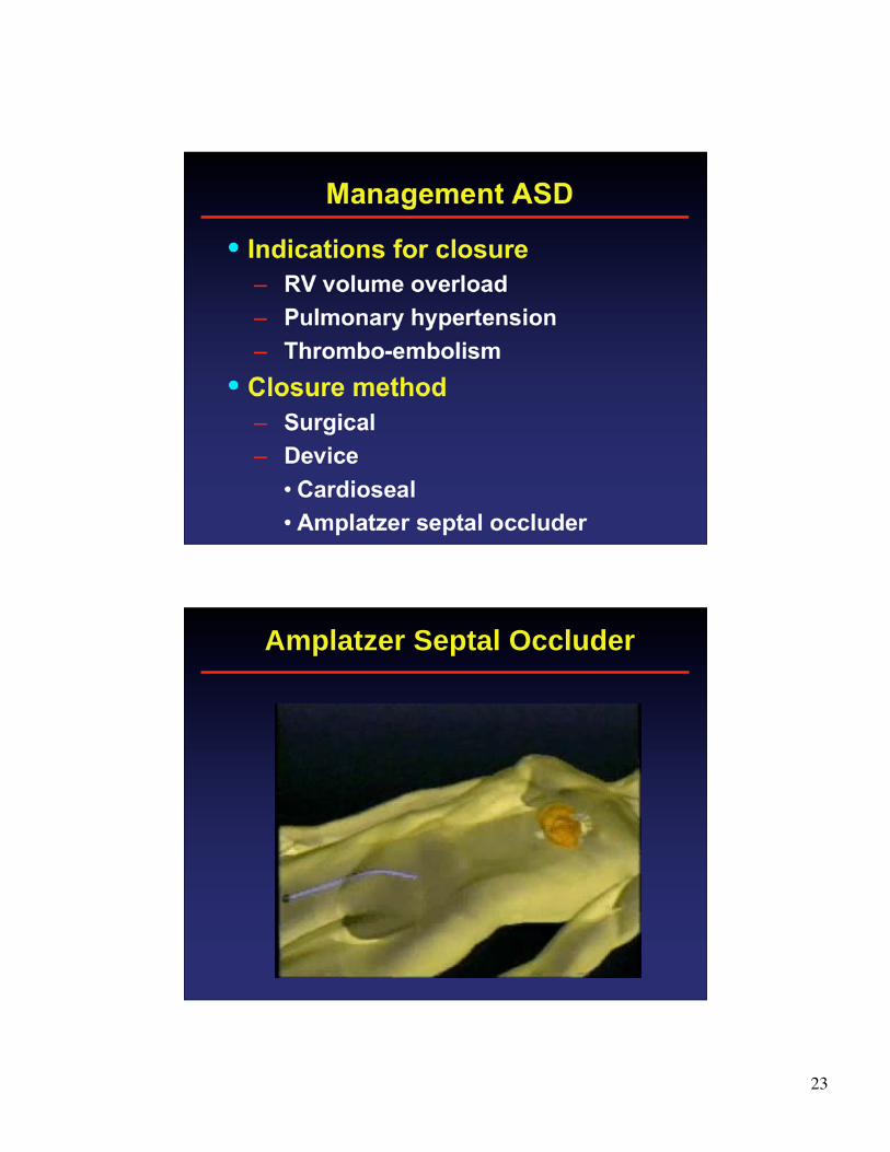
Management ASD
• Indications for closure
– RV volume overload
– Pulmonary hypertension
– Thrombo-embolism
• Closure method
– Surgical
– Device
• Cardioseal
• Amplatzer septal occluder
Amplatzer Septal Occluder