pathology patient report · pdf filepathology patient report examples. robert bush, md 701...
TRANSCRIPT

Orchard Pathology Sample Patient Reports
PATHOLOGY PATIENT REPORT EXAMPLES

Robert Bush, MD 701 Congressional Blvd. Phone: (800) 555-1234Laboratory Director Carmel, Indiana 46032 Fax: (317) 573-2528
Prostate Biopsy Pathology Report Final
PATIENT INFORMATION PHYSICIAN INFORMATION ACCESSION NUMBER
Patient Name: Patient, JohnM, Age 34 | DOB: 4/12/1979
Phone: (123) 555-1234
EMR: (123) 555-1234
CLINICAL INFORMATION:8.6; Oct. 2012 / Elevated PSA
12XX0002COLLECTION DATE: 2/15/2013
RECEIVED DATE: 2/15/2013
REPORT DATE: 2/17/2013
TAT: [26 hours]
James Provider, MDABC Medical
400 Royal Drive
Anytown USA, 12345
Phone: (123) 555-4321
Fax: (999) 555-4322
PROSTATE BIOPSY DIAGRAM AND MICROSCOPIC IMAGES
Site Gleason Score Diagnosis Tumor % Tumor Length (mm) Core Length (mm) # Cores
1 Left Base 3+4=7 ADENOCARCINOMA 60 6 10 1
2 Left Mid Benign 0 10 1
3 Left Apex Benign 0 10 1
4 Right Base Benign 0 10 1
5 Right Mid Benign 0 10 1
6 Right Apex HGPIN 0 10 1
FINAL DIAGNOSIS
CLIA ID#: 01DO000XXX000 END OF REPORT (FINAL) Page 1 of 1
Electronically Signed on 2/17/2013 at 11:42 am
John Pathologist

Orchard Pathology Laboratories, Inc.Robert Bush, MD, Laboratory DirectorCLIA ID#: 01D000XXX000CAP #: 123456
701 Congressional Blvd.Carmel, Indiana 46032Phone: (800) 555-1234
Fax: (317) 573-2528
Patient Name: Patient, Jane A.Sex: FDOB: 06/22/1955Patient ID: 12345-6
Collection Date: 10/02/2013 08:57Received Date: 10/02/2013 08:57Reported: 10/03/2013 10:02
Provider: James Provider, MDAccount Number: Client: ABC MedicalClient Address: 1234 Anystreet Anytown, USA 12345
Telephone: (123) 555-1234Accession #: S-01-012345-6
Surgical Pathology Report
FINAL DIAGNOSIS
A. Soft Tissue, Left Supraclavicular, Core Needle Biopsy: MALIGNANT LYMPHOMA, LARGE CELL ANAPLASTIC TYPE, ALK-1 NEGATIVE
B. Skin, Upper Back, Incisional Biopsy: MALIGNANT LYMPHOMA, LARGE CELL ANAPLASTIC TYPE, ALK-1 NEGATIVE
Specimen: Left Supraclavicular Skin, Upper Back
Preoperative Diagnosis Probable malignancy. History of recentleft supraclavicular mass of rapid onset with an upper back skinnodule. CT scan shows mediastinal adenopathy without other organmasses.
Gross ExaminationReceived are two formalin filled containers labeled “Jane Patient”
A. Container A is labeled “Needle biopsy left supraclavicular area” and holds 2cylindrical shaped fragments measuring 2 mm in diameter and 10 and 13 mm inlength. The specimen is poured into a filter bag and entirely submitted in cassetteA.
B. Container B is labeled “Skin Upper Back” and holds a wedge-shaped fragment ofskin measuring 8.0 x 10.0 x 6.0 cm. The specimen is dissected and entirelysubmitted in cassette B.
Performed by: A. Tech
Microscopic ExaminationThe Final Diagnosis for each specimen is based on a microscopic examination of the tissues or preparation from these tissues.
S-01-012345-6
S-01-012345-6
S-01-012345-6
Case number: S-01-012345-6 This report continues.... (Preliminary) Reviewed by: _______
Orchard Pathology Laboratories, Inc.Robert Bush, MD, Laboratory DirectorCLIA ID#: 01D000XXX000CAP #: 123456
701 Congressional Blvd.Carmel, Indiana 46032Phone: (800) 555-1234
Fax: (317) 573-2528
Patient Name: Patient, Jane A.Sex: FDOB: 06/22/1955Patient ID: 12345-6
Collection Date: 10/02/2013 08:57Received Date: 10/02/2013 08:57Reported: 10/03/2013 10:02
Provider: James Provider, M.D.Account Number: Client: ABC MedicalClient Address: 1234 Anystreet Anytown, USA 12345
Telephone: (888) 876-5432Accession #: S-01-012345-6
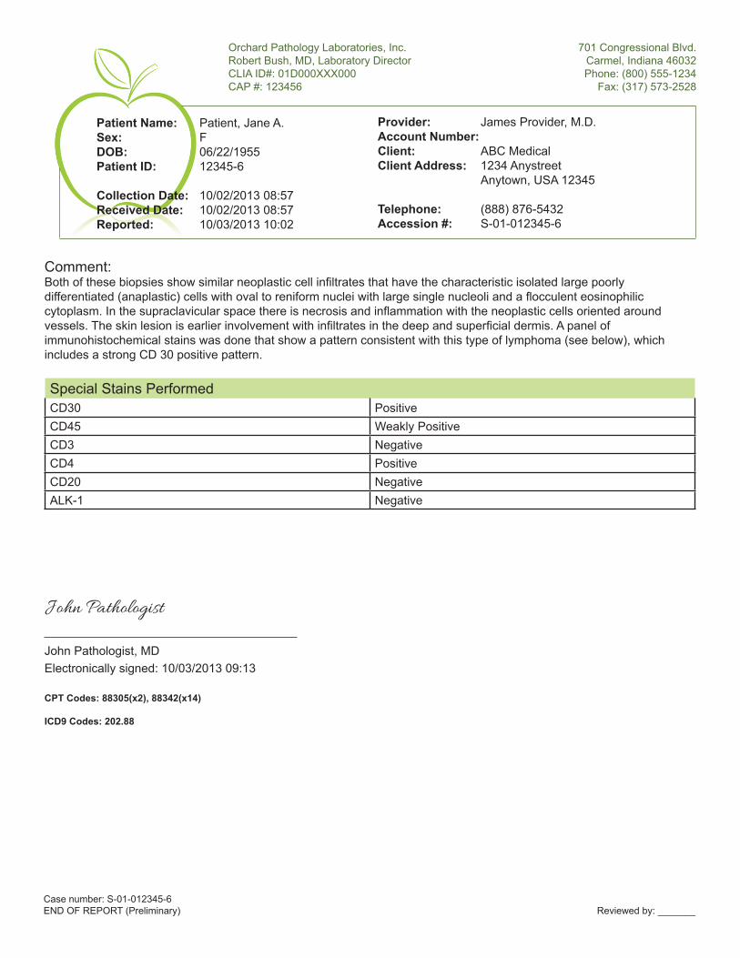
Comment:Both of these biopsies show similar neoplastic cell infiltrates that have the characteristic isolated large poorlydifferentiated (anaplastic) cells with oval to reniform nuclei with large single nucleoli and a flocculent eosinophiliccytoplasm. In the supraclavicular space there is necrosis and inflammation with the neoplastic cells oriented aroundvessels. The skin lesion is earlier involvement with infiltrates in the deep and superficial dermis. A panel ofimmunohistochemical stains was done that show a pattern consistent with this type of lymphoma (see below), whichincludes a strong CD 30 positive pattern.
Special Stains PerformedCD30 PositiveCD45 Weakly PositiveCD3 NegativeCD4 PositiveCD20 NegativeALK-1 Negative
John Pathologist, MDElectronically signed: 10/03/2013 09:13
CPT Codes: 88305(x2), 88342(x14)
ICD9 Codes: 202.88
John Pathologist
Case number: S-01-012345-6 END OF REPORT (Preliminary) Reviewed by: _______

Orchard Pathology Laboratories, Inc.Robert Bush, MD, Laboratory DirectorCLIA ID#: 01D000XXX000CAP #: 123456
701 Congressional Blvd.Carmel, Indiana 46032Phone: (800) 555-1234
Fax: (317) 573-2528
Patient Name: Patient, John C.Sex: MDOB: 04/12/1979Patient ID: 54321-6
Collection Date: 10/15/2013 17:45Received Date: 10/15/2013 18:52Reported: 10/16/2013 10:44
Provider: James Provider, MDAccount Number: Client: ABC MedicalClient Address: 1234 Anystreet Anytown, USA 12345
Telephone: (123) 555-1234Accession #: S-01-543210-6
FINAL DIAGNOSIS
A. Ascending Colon SESSILE SERRATED ADENOMA (POLYP) WITH LOW-GRADE ADENOMATOUS DYSPLASIA.
B. Sigmoid Colon TUBULAR ADENOMA
COMMENT:Patients with sessile serrated adenomas, especially with cytologic dysplasia,are at increased risk for the development of adenocarcinoma showingmicrosatellite instability. This progression may occur at a more rapid rate thanwith traditional adenomas. Complete endoscopic excision is recommended ifclinically appropriate. If unresectable, repeat colonoscopy at a shortenedinterval (1 year), with sampling of suspicious areas or surgical resectionpossibly warranted.
Specimen: 2 cm polyp ascending colon 2 mm polyp in sigmoid colon
Clinical History: Screening colonoscopy. Maternal hx of adenocarcinoma ofcolon age 57
Gross ExaminationA. The first container is labeled “ascending colon.” It contains a polypoid piece of tan mucosal tissue measuring 2.0 cm in greatest dimension. The polyp margin is inked, sectioned, and submitted in cassettes Al and A2.
B. The second container is labeled “sigmoid colon.” It contains one piece of light tan mucosal tissue 0.2 cm in greatest dimension. Entirely submitted in cassette B.
Microscopic ExaminationMicroscopic Examination performed supportive of the Final Diagnosis above.
Case number: S-01-543210-6 END OF REPORT Reviewed by: _______
Surgical Pathology Report
John Pathologist, MDElectronically signed: 10/16/2013 10:44
John Pathologist
S-01-543210-6
S-01-543210-6
Orchard Pathology Laboratories, Inc.700 Congressional Blvd.Carmel, Indiana 46032Phone: (800) 555-1234
Fax: (317) 573-2528 Robert Bush, MD, Laboratory DirectorCLIA ID#: 01D000XXX000
Patient Name: Patient, Jane A.Patient ID: 12345-6Date of Birth: 06/22/1955 Age: 58 Sex: F
Provider: James Provider, MD
Case number: B08-0006Collection Date: 08/12/2013 08:40Received Date: 08/12/2013 08:40Reported: 08/16/2013 10:02
Bone Marrow Pathology Report
FINAL DIAGNOSIS
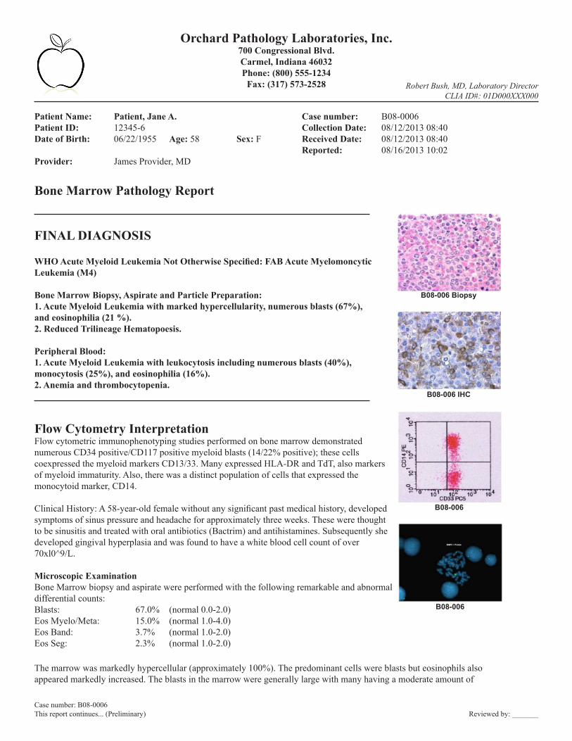
WHO Acute Myeloid Leukemia Not Otherwise Specified: FAB Acute MyelomoncyticLeukemia (M4)
Bone Marrow Biopsy, Aspirate and Particle Preparation:1. Acute Myeloid Leukemia with marked hypercellularity, numerous blasts (67%),and eosinophilia (21 %).2. Reduced Trilineage Hematopoesis.
Peripheral Blood:1. Acute Myeloid Leukemia with leukocytosis including numerous blasts (40%),monocytosis (25%), and eosinophilia (16%).2. Anemia and thrombocytopenia.
Flow Cytometry InterpretationFlow cytometric immunophenotyping studies performed on bone marrow demonstratednumerous CD34 positive/CD117 positive myeloid blasts (14/22% positive); these cellscoexpressed the myeloid markers CD13/33. Many expressed HLA-DR and TdT, also markersof myeloid immaturity. Also, there was a distinct population of cells that expressed themonocytoid marker, CD14.
Clinical History: A 58-year-old female without any significant past medical history, developedsymptoms of sinus pressure and headache for approximately three weeks. These were thoughtto be sinusitis and treated with oral antibiotics (Bactrim) and antihistamines. Subsequently shedeveloped gingival hyperplasia and was found to have a white blood cell count of over70xl0^9/L.
Microscopic ExaminationBone Marrow biopsy and aspirate were performed with the following remarkable and abnormaldifferential counts:Blasts: 67.0% (normal 0.0-2.0)Eos Myelo/Meta: 15.0% (normal 1.0-4.0)Eos Band: 3.7% (normal 1.0-2.0)Eos Seg: 2.3% (normal 1.0-2.0)
The marrow was markedly hypercellular (approximately 100%). The predominant cells were blasts but eosinophils alsoappeared markedly increased. The blasts in the marrow were generally large with many having a moderate amount of
Case number: B08-0006This report continues... (Preliminary) Reviewed by: _______
B08-006 Biopsy
B08-006 IHC
B08-006
B08-006
Orchard Pathology Laboratories, Inc.700 Congressional Blvd.Carmel, Indiana 46032Phone: (800) 555-1234
Fax: (317) 573-2528 Robert Bush, MD, Laboratory DirectorCLIA ID#: 01D000XXX000
Patient Name: Patient, Jane A.Patient ID: 12345-6Date of Birth: 06/22/1955 Age: 58 Sex: F
Provider: James Provider, MD
Case number: B08-0006Collection Date: 08/12/2013 08:40Received Date: 08/12/2013 08:40Reported: 08/16/2013 10:02
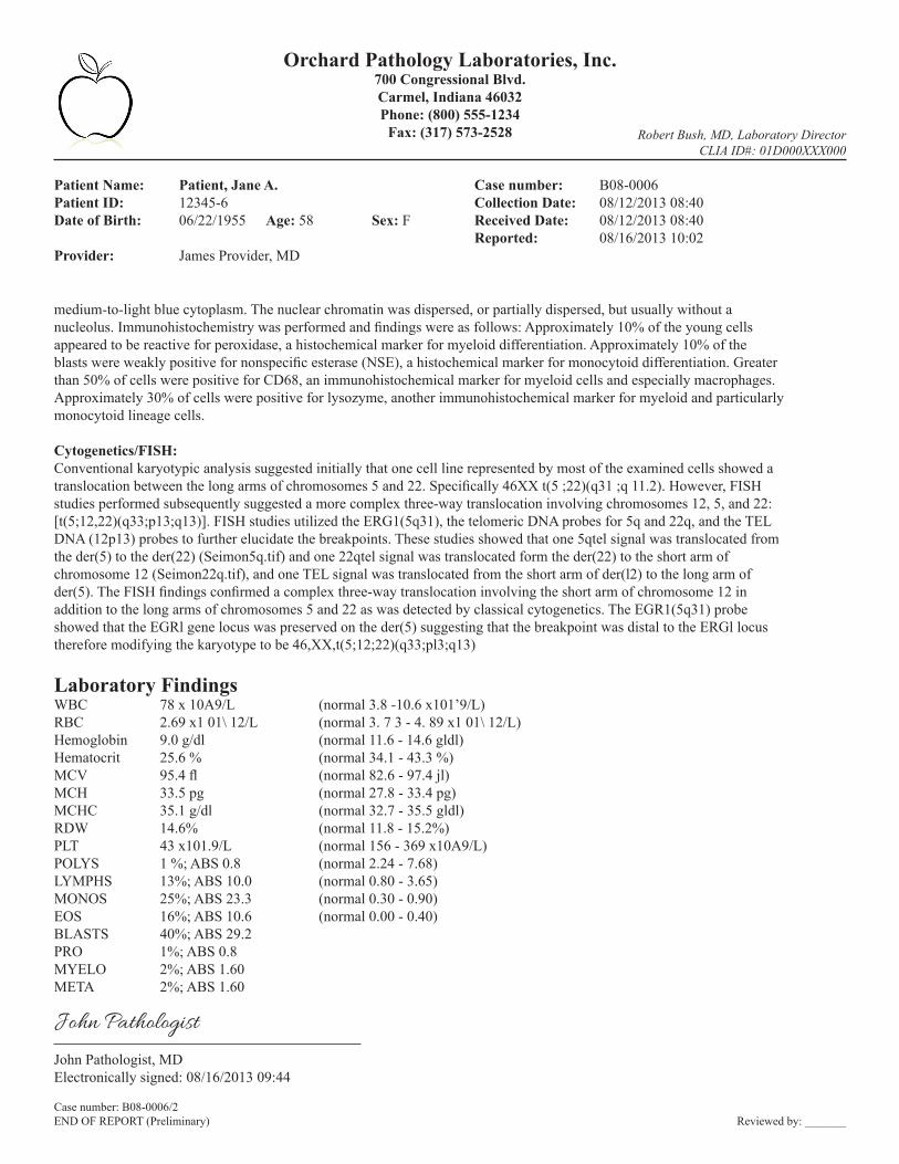
medium-to-light blue cytoplasm. The nuclear chromatin was dispersed, or partially dispersed, but usually without anucleolus. Immunohistochemistry was performed and findings were as follows: Approximately 10% of the young cellsappeared to be reactive for peroxidase, a histochemical marker for myeloid differentiation. Approximately 10% of theblasts were weakly positive for nonspecific esterase (NSE), a histochemical marker for monocytoid differentiation. Greaterthan 50% of cells were positive for CD68, an immunohistochemical marker for myeloid cells and especially macrophages.Approximately 30% of cells were positive for lysozyme, another immunohistochemical marker for myeloid and particularlymonocytoid lineage cells.
Cytogenetics/FISH:Conventional karyotypic analysis suggested initially that one cell line represented by most of the examined cells showed atranslocation between the long arms of chromosomes 5 and 22. Specifically 46XX t(5 ;22)(q31 ;q 11.2). However, FISHstudies performed subsequently suggested a more complex three-way translocation involving chromosomes 12, 5, and 22:[t(5;12,22)(q33;p13;q13)]. FISH studies utilized the ERG1(5q31), the telomeric DNA probes for 5q and 22q, and the TELDNA (12p13) probes to further elucidate the breakpoints. These studies showed that one 5qtel signal was translocated fromthe der(5) to the der(22) (Seimon5q.tif) and one 22qtel signal was translocated form the der(22) to the short arm ofchromosome 12 (Seimon22q.tif), and one TEL signal was translocated from the short arm of der(l2) to the long arm ofder(5). The FISH findings confirmed a complex three-way translocation involving the short arm of chromosome 12 inaddition to the long arms of chromosomes 5 and 22 as was detected by classical cytogenetics. The EGR1(5q31) probeshowed that the EGRl gene locus was preserved on the der(5) suggesting that the breakpoint was distal to the ERGl locustherefore modifying the karyotype to be 46,XX,t(5;12;22)(q33;pl3;q13)
Laboratory FindingsWBC 78 x 10A9/L (normal 3.8 -10.6 x101’9/L)RBC 2.69 x1 01\ 12/L (normal 3. 7 3 - 4. 89 x1 01\ 12/L)Hemoglobin 9.0 g/dl (normal 11.6 - 14.6 gldl)Hematocrit 25.6 % (normal 34.1 - 43.3 %)MCV 95.4 fl (normal 82.6 - 97.4 jl)MCH 33.5 pg (normal 27.8 - 33.4 pg)MCHC 35.1 g/dl (normal 32.7 - 35.5 gldl)RDW 14.6% (normal 11.8 - 15.2%)PLT 43 x101.9/L (normal 156 - 369 x10A9/L)POLYS 1 %; ABS 0.8 (normal 2.24 - 7.68)LYMPHS 13%; ABS 10.0 (normal 0.80 - 3.65)MONOS 25%; ABS 23.3 (normal 0.30 - 0.90)EOS 16%; ABS 10.6 (normal 0.00 - 0.40)BLASTS 40%; ABS 29.2PRO 1%; ABS 0.8MYELO 2%; ABS 1.60META 2%; ABS 1.60
John Pathologist, MDElectronically signed: 08/16/2013 09:44
John Pathologist
Case number: B08-0006/2END OF REPORT (Preliminary) Reviewed by: _______
Robert Bush, MD 701 Congressional Blvd. Phone: (800) 555-1234Laboratory Director Carmel, Indiana 46032 Fax: (317) 573-2528
Molecular Pathology Report Final
PATIENT INFORMATION PHYSICIAN INFORMATION ACCESSION NUMBER
Patient Name: Patient, JosieF, Age 62 | DOB: 8/9/1951Phone: (123) 123-1234EMR: (123) 123-1234
M07-0134COLLECTION DATE: 2/20/2013RECEIVED DATE: 2/20/2013REPORT DATE: 2/21/2013
James Provider, MDABC Medical400 Royal DriveAnytown USA, 12345Phone: (888) 876-5432Fax: (999) 876-5432
Molecular Pathology Report
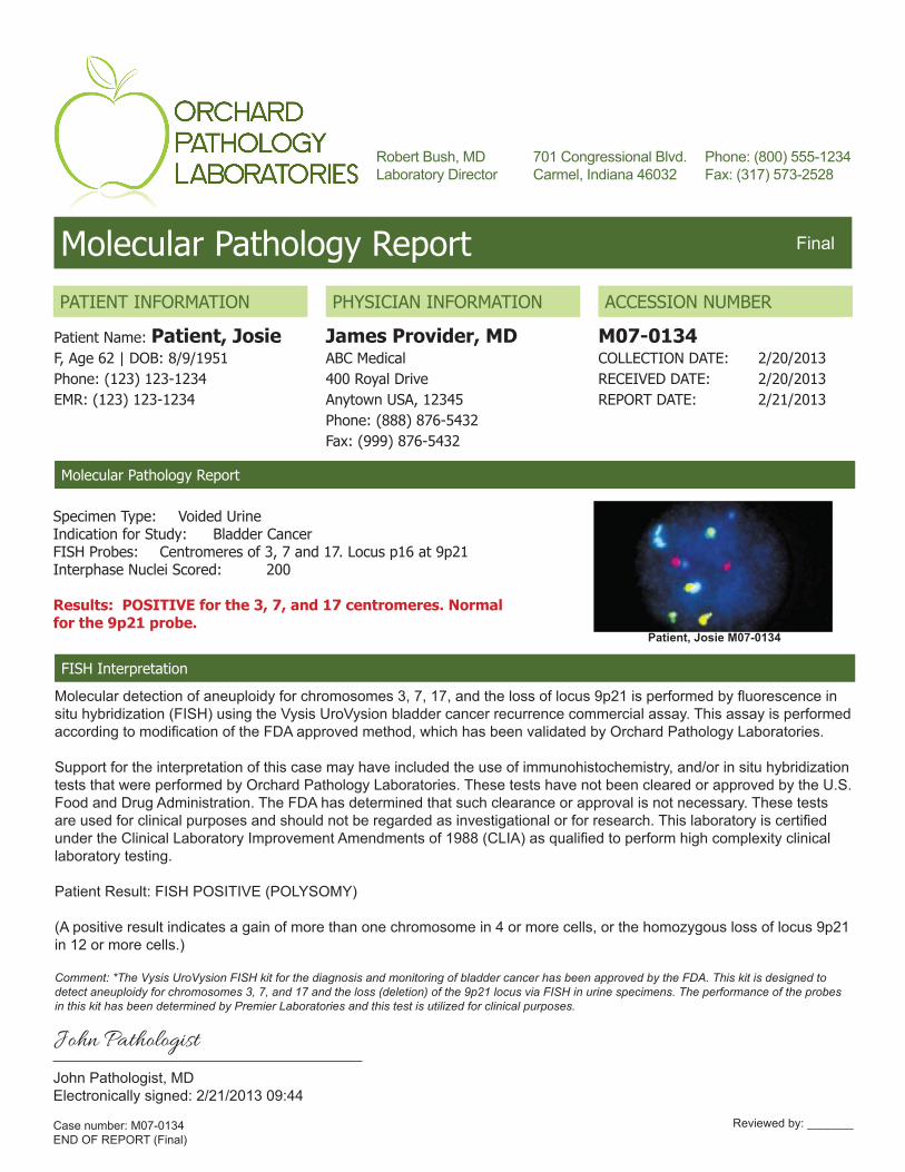
Specimen Type: Voided UrineIndication for Study: Bladder CancerFISH Probes: Centromeres of 3, 7 and 17. Locus p16 at 9p21Interphase Nuclei Scored: 200
Results: POSITIVE for the 3, 7, and 17 centromeres. Normalfor the 9p21 probe.
Patient, Josie M07-0134
FISH Interpretation
Molecular detection of aneuploidy for chromosomes 3, 7, 17, and the loss of locus 9p21 is performed by fluorescence insitu hybridization (FISH) using the Vysis UroVysion bladder cancer recurrence commercial assay. This assay is performedaccording to modification of the FDA approved method, which has been validated by Orchard Pathology Laboratories.
Support for the interpretation of this case may have included the use of immunohistochemistry, and/or in situ hybridizationtests that were performed by Orchard Pathology Laboratories. These tests have not been cleared or approved by the U.S. Food and Drug Administration. The FDA has determined that such clearance or approval is not necessary. These tests are used for clinical purposes and should not be regarded as investigational or for research. This laboratory is certified under the Clinical Laboratory Improvement Amendments of 1988 (CLIA) as qualified to perform high complexity clinical laboratory testing.
Patient Result: FISH POSITIVE (POLYSOMY)
(A positive result indicates a gain of more than one chromosome in 4 or more cells, or the homozygous loss of locus 9p21in 12 or more cells.)
Comment: *The Vysis UroVysion FISH kit for the diagnosis and monitoring of bladder cancer has been approved by the FDA. This kit is designed todetect aneuploidy for chromosomes 3, 7, and 17 and the loss (deletion) of the 9p21 locus via FISH in urine specimens. The performance of the probes in this kit has been determined by Premier Laboratories and this test is utilized for clinical purposes.
John Pathologist, MDElectronically signed: 2/21/2013 09:44
John Pathologist
Case number: M07-0134END OF REPORT (Final)
Reviewed by: _______

Orchard Pathology Laboratories, Inc.700 Congressional Blvd.Carmel, Indiana 46032Phone: (800) 555-1234
Fax: (317) 573-2528 Robert Bush, MD, Laboratory DirectorCLIA ID#: 01D000XXX000
Patient Name: Patient, Michael E.Patient ID: 555333Date of Birth: 12/14/1957 Age: 56 Sex: M
Provider: James Provider, MD
Case number: S07-0110Collection Date: 03/29/2013 09:42Received Date: 03/29/2013 09:42Reported: 03/30/2013 11:26
Surgical Pathology Report
FINAL DIAGNOSIS
A. GE Junction: ADENOCARCINOMA of the esophagus.
B. Antrum: Non-neoplastic gastric antral mucosa, negative forinflammation. Stain for Helicobacter pylori microorganisms is negative.
Gross ExaminationA. Received in formalin and labeled “Patient, Michael E.” and “EGJunction” are multiple fragments of tan tissue measuring 0.7 x 0.4 x0.2 cm in aggregate. The specimen is entirely submitted in onecassette labeled “A.”
B. Received in formalin and labeled, “Patient, Michael E.” and “Antrum”are two fragments of pink-tan tissue measuring 0.2 x 0.2 x 0.2 em.The specimen is entirely submitted in one cassette labeled “B.”
John Pathologist, MDElectronically signed: 03/30/2013 09:44
John Pathologist
Case number: S07-0110END OF REPORT (Preliminary) Reviewed by: _______
Patient, Michael E. S07-0110
Orchard Pathology Laboratories, Inc.700 Congressional Blvd.Carmel, Indiana 46032Phone: (800) 555-1234
Fax: (317) 573-2528 Robert Bush, MD, Laboratory DirectorCLIA ID#: 01D000XXX000
Patient Name: Patient, Julie B.Patient ID: 444222Date of Birth: 09/05/1968 Age: 45 Sex: F
Provider: James Provider, MD
Case number: G07-0062Collection Date: 06/13/2013 21:14Received Date: 06/13/2013 21:14Reported: 06/14/2013 02:43
Final Cytology GYN Report
Interpretation/Result: Epithelial Cell Abnormalities:ASCH - Atypical squamous cells cannot exclude HSIL
Organisms: Normal Findings
Specimen Adequacy: Satisfactory for Evaluation - Presence ofEndocervicali Transformation Zone Component
CLINICAL INFORMATION
Date Last Pap Feb 2012GYN Source: Cervical/endocervicalDate LMP/Menopause: 05/22/2013Clinical Impressions: Oral contraceptivesPrevious Treatment: NonePrevious PAP: Atypical; ASCUS
*The Pap smear is a screening test, not a diagnostic procedure and should not be used as the sole means of detecting cervical cancer.Both false-positive and false-negative reports do occur.
Human Papilloma Virus HPV Assay by in-situ hybridization16, 18, 31, 33, 35, 39, 45, 51, 52, 56, 58, 66 ....... NOT DETECTED
Laboratory FindingsHistorical Diagnosis:Thin Prep PAP Test Case Number: G06-42-01 Issued: 06/12/07INTERPRETATION/RESULT: Epithelial Cell Abnormalities: ASCUS-Atypical squamous cells of undetermined significance
Screened by: M Jones, CT
John Doctor, MDElectronically signed: 06/14/2013 02:31
John Doctor
Case number: G07-0062END OF REPORT (Final) Reviewed by: _______
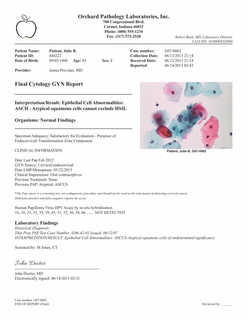
Patient, Julie B. G07-0062

Orchard Pathology Laboratories, Inc.Robert Bush, MD, Laboratory DirectorCLIA ID#: 01D000XXX000CAP #: 123456
701 Congressional Blvd.Carmel, Indiana 46032Phone: (800) 555-1234
Fax: (317) 573-2528
Patient Name: Patient, Julie B.Sex: FDOB: 09/05/1968Patient ID: 444222
Collection Date: 09/15/2013 17:45Received Date: 09/15/2013 18:52Reported: 09/16/2013 10:44
Provider: James Provider, MDAccount Number: Client: ABC MedicalClient Address: 1234 Anystreet Anytown, USA 12345
Telephone: (123) 555-1234Accession #: P07-6824
Cytology GYN Report
General Categorization:Epithelial cell abnormality (See Interpretation)
Interpretation:Atypical squamous cells of undetermined significance (ASCUS)Atypical koilocytosis, cytoplasmic and nuclear change consistent with HPV effectHPV, High Risk - Positive; HPV, Type 16 - Positive; HPV, Type 18 - NegativeChlamydia (CT) - Positive; Gonorrhoeae (GC) - Negative
The Pap smear is a cancer screening test that has an overall1 5-25% false negative rate. For this reason, an annual Pap smear is recommended.Please discuss this with your patients.
CLINICAL INFORMATIONType of test: High Risk ScreeningDate LMP: 5/5/10Clinical diagnosis: None givenClinical history: None givenPrevious Smears: Unknown
Specimen type: Liquid basedSpecimen source: Cervical, EndocervicalSpecimen Adequacy: Smear is satisfactory for evaluation
Comment: The Thin Prep Imaging System from Hologic, Inc. was used to pre-screen this Pap smear.Primary screening reported Pap abnormalities. Pathologist review was required to interpret the primary screener review.
Pap screening performed by: Breakthrough Cytology Services, 527 Joanne Lane, Dekalb, IL 60115.Interpretation required by undersigned pathologist at Orchard Pathology Laboratories, 701 Congressional Blvd, Carmel, IN 46032.
09/16/2013Cy Totech, CT, ASCP
09/16/2013 Authenticated by:
John Doctor, MDElectronically signed: 06/14/2013 02:31
John Doctor
Case number: P07-6824END OF REPORT (Final) Reviewed by: _______

Orchard Pathology Laboratories, Inc.Robert Bush, MD, Laboratory DirectorCLIA ID#: 01D000XXX000CAP #: 123456
701 Congressional Blvd.Carmel, Indiana 46032Phone: (800) 555-1234
Fax: (317) 573-2528
Patient Name: Patient, Julie B.Sex: FDOB: 09/05/1968Patient ID: 444222
Collection Date: 09/15/2013 17:45Received Date: 09/15/2013 18:52Reported: 09/16/2013 10:44
Provider: James Provider, MDAccount Number: Client: ABC MedicalClient Address: 1234 Anystreet Anytown, USA 12345
Telephone: (123) 555-1234Accession #: S07-6825
DERMATOPATHOLOGY REPORT
FINAL DIAGNOSIS
1) SKIN, RIGHT ANTERIOR MEDIAL SHOULDER, EXCISION: -INVASIVE MALIGNANT MELANOMA, NODULAR TYPE WITH ULCERATION, BRESLOW’S DEPTH 7 MM, CLARK’S LEVEL IV, MARGINS NARROWLY CLEAR (SEE MELANOMA SUMMARY AND COMMENT).
GROSS EXAMINATIONSKIN, RIGHT ANTERIOR MEDIAL SHOULDER: Labeled “right ant med shoulder” is a 2.6 x 2.0 cm ovoid gray-tan irregular skin excised to a depth of 0.8 cm. The skin surface displays an eccentric 1.5 x 1.4 cm white-tan to dark brown nodule. No orienta-tion provided. Inked, sectioned. ES (4) as follows:
1A: Tips1B-1D: Remainder of Specimen
Specimen: Right shoulder
MELANOMA CANCER SUMMARY[MACROSCOPIC]SPECIMEN TYPE: Excision, ellipseMACROSCOPIC TUMOR: PresentTUMOR SITE: Right anterior medial shoulderLESION SIZE: 1.5 x 1.4 cm in greatest dimensionSATELLITE NODULES: Absent[MICROSCOPIC]HISTOLOGIC TYPE: Nodular melanomaULCERATION: PresentDEPTH OF INVATION/BRESLOW’S DEPTH: 7mmCLARK’S LEVEL: IVGROWTH PHASE: VerticalREGRESSION: AbsentMITOTIC INDEX: High (Greater than 20 mitotic figures per mm squared)ANGIOLYMPHATIC INVASION: AbsentNEUROTROPISM: AbsentTUMOR INFILTRATING LYMPHOCYTES: Present (non-brisk)MICROSCOPIC SATELLITES: Not present in tissue submittedMARGINS: Invasive melanoma within 2 mm of peripheral marginLYMPH NODES: Not submittedPATHOLOGIC STAGING (PTNM) pT4b, pNX, pMX
Case number: S07-6825/1This report continues... (Preliminary) Reviewed by: _______

Orchard Pathology Laboratories, Inc.Robert Bush, MD, Laboratory DirectorCLIA ID#: 01D000XXX000CAP #: 123456
701 Congressional Blvd.Carmel, Indiana 46032Phone: (800) 555-1234
Fax: (317) 573-2528
IMMUNOHISTOCHEMISTRYHMB45 PositiveS100 PositiveMELAN A Positive
Patient Name: Patient, Julie B.Sex: FDOB: 09/05/1968Patient ID: 444222
Collection Date: 09/15/2013 17:45Received Date: 09/15/2013 18:52Reported: 09/16/2013 10:44
Provider: James Provider, MDAccount Number: Client: ABC MedicalClient Address: 1234 Anystreet Anytown, USA 12345
Telephone: (123) 555-1234Accession #: S07-6825
John Pathologist, M.D.Board Certified in Anatomic and Clinical PathologyAuthenticated by Pathologist: 09/16/2013 10:31
John Pathologist
Case number: S07-6825/2END OF REPORT (Preliminary) Reviewed by: _______

701 Congressional Blvd., Suite 360 • Carmel, Indiana 46032General Information: (800) 856-1948 • Technical Support: (800) 571-5835
www.orchardsoft.com
Sample reports generated from Orchard Software test database.
Note that these are sample reports generated with tools available in Orchard Software’s laboratory solutions.Specific report formatting assistance from Orchard Software may require a professional services agreement.
All patient and provider names and data contained within these sample reportsare fictitious and do not represent real names or data.
© Copyright 2015 Orchard Software Corporation. All rights reserved.