pathology of the hematopoietic system. - usmf
TRANSCRIPT
Pathology of the hematopoietic system.
Pathology of the hematopoietic system.
I. Microspecimens:
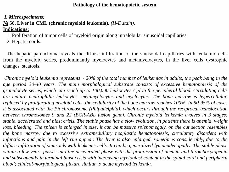
№ 56. Liver in CML (chronic myeloid leukemia). (H-E stain).
Indications:
1. Proliferation of tumor cells of myeloid origin along intralobular sinusoidal capillaries.
2. Hepatic cords.
The hepatic parenchyma reveals the diffuse infiltration of the sinusoidal capillaries with leukemic cells
from the myeloid series, predominantly myelocytes and metamyelocytes, in the liver cells dystrophic
changes, steatosis.
Chronic myeloid leukemia represents ~ 20% of the total number of leukemias in adults, the peak being in the
age period 30-40 years. The main morphological substrate consists of excessive hematopoiesis of the
granulocyte series, which can reach up to 100,000 leukocytes / µl in the peripheral blood. Circulating cells
are mature neutrophilic leukocytes, metamyelocytes and myelocytes. The bone marrow is hypercellular,
replaced by proliferating myeloid cells, the cellularity of the bone marrow reaches 100%. In 90-95% of cases
it is associated with the Ph chromosome (Phipadelphia), which occurs through the reciprocal translocation
between chromosomes 9 and 22 (BCR-ABL fusion gene). Chronic myeloid leukemia evolves in 3 stages:
stable, accelerated and blast crisis. The stable phase has a slow evolution, in patients there is anemia, weight
loss, bleeding. The spleen is enlarged in size, it can be massive splenomegaly, on the cut section resembles
the bone marrow due to excessive extramedullary neoplastic hematopoiesis, circulatory disorders with
infarctions and pain in the left rim appear. The liver is also enlarged, sometimes considerably, due to the
diffuse infiltration of sinusoids with leukemic cells. It can be generalized lymphadenopathy. The stable phase
within a few years passes into the accelerated phase with the progression of anemia and thrombocytopenia
and subsequently in terminal blast crisis with increasing myeloblast content in the spinal cord and peripheral
blood; clinical-morphological picture similar to acute myeloid leukemia.

№ 56. Liver in CML (chronic myeloid leukemia). (H-E stain).
2
1

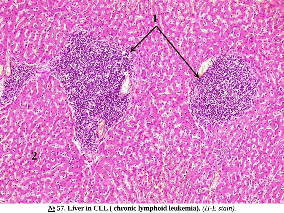
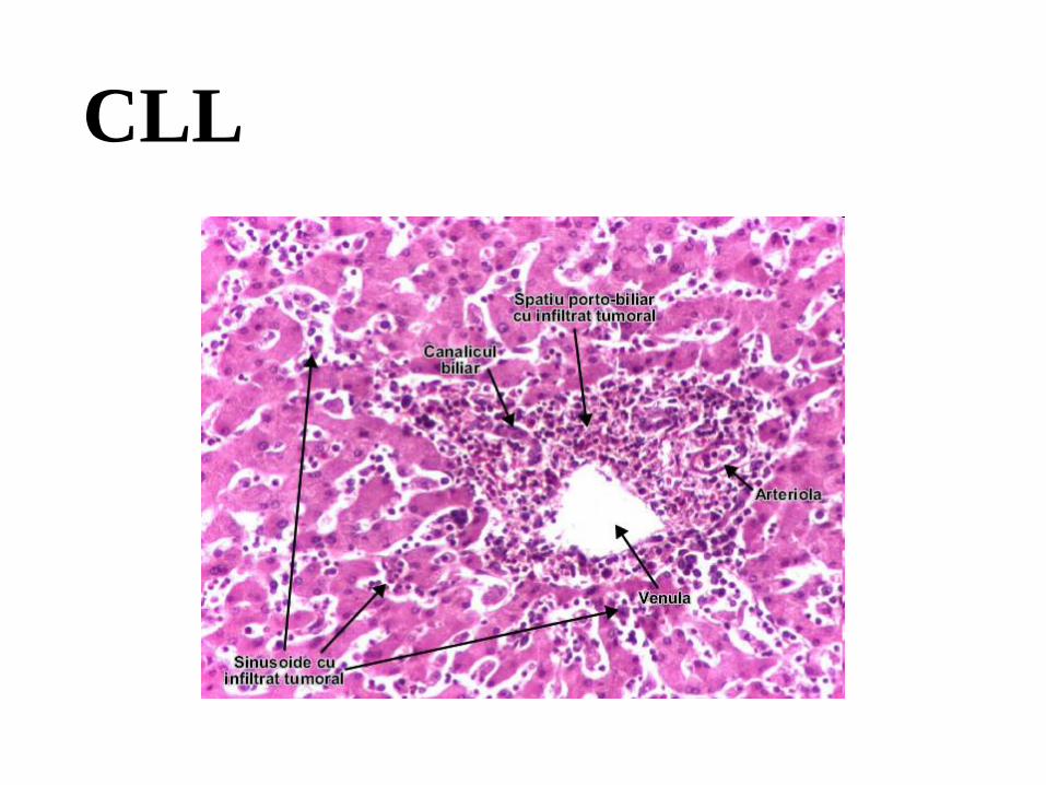
№ 57. Liver in CLL ( chronic lymphoid leukemia). (H-E stain).
Indications:
1. Foci of proliferation of tumor cells of lymphoid origin along portal tracts (triads).
2. Hepatic lobule.
With the naked eye in the microspecimen the motley appearance of the section is observed, the
microscopic examination reveals multiple focal cell agglomerations, consisting of small
lymphocytes, with round nucleus, intensely colored basophilic, with little cytoplasm, located
along the interlobular fibroconjunctival septa, in some places in the region of triads, hepatocytes
with dystrophic changes, steatosis; leukemic infiltration does not extend into the sinusoidal
capillaries.
Chronic lymphocytic leukemia (CLL) is the most common form of leukemia in adults and is
identical to small cell lymphocytic lymphoma (CLL). In LCC, leukemic cells predominate in the
peripheral blood, and in LLCM - in lymphoid tissues. These lymphoid neoplasms can pass into
each other. Most of them are of B-lymphocyte origin. Absolute lymphocytosis of up to 200,000
cells / µ is detected in peripheral blood in CLL, with small mature lymphocytes constituting more
than 90% of leukocytes. Morphological lesions: 1) in the bone marrow there is a diffuse
proliferation of small lymphocytes, which replace the normal hematopoietic tissue, causing
anemia, neutropenia, thrombocytopenia; 2) generalized lymphadenopathy; 3) splenomegaly,
sometimes massive, with proliferation of small lymphocytes in the white pulp; 4) hepatomegaly,
which can be also massive. In patients with CLL, there are autoimmune reactions with
autoantibodies against erythrocytes and platelets, bleeding, distorted immune response and
increased susceptibility to infections.
№ 57. Liver in CLL ( chronic lymphoid leukemia). (H-E stain).
1
2

№ 145. Plasmocytoma. (H-E stain).
Indications:
1. Polymorphic tumoral cells of plasmo cytic origin.
2. Tumoral stroma with blood vessels.
3. Hemorrhagic foci.
The biopsy was taken from a solitary tumoral node from the region of the skull vault. Microscopically there
is a cell mass, composed predominantly of tumoral plasma cells, most of them similar to normal plasma cells,
with eccentric nucleus, hyperchrome, chromatin arranged "in wheel spokes", rich cytoplasm, basophilic, with
perinuclear halo, absent nucleoli, larger plasmoblasts are observed, the nucleus with a well-defined nucleoli,
the tumor stroma is poor, there are foci of plasmorrhagia and hemorrhage.
Solitary plasmacytoma (localized) and multiple myeloma are the main diseases in the group of plasma cell
neoplasms, the morphological substrate of which is the excessive, neoplastic proliferation of plasma cells.
The lesions begin in 95% of cases in the medullary cavity of the bones and gradually erode the spongy bone
tissue, and later the compact one, causing pathological fractures. In solitary plasmacytoma, a single bone is
affected, and in multiple myeloma the lesions are multifocal, involving bones with active hematopoiesis:
spine, ribs, skull, pelvic bones and a. Solitary plasmacytoma is an early stage of multiple myeloma,
progressing within 5-10 years from monoosal to polyosal lesions. The affected bones take on a "moth-eaten"
appearance, with defects having a diameter of 1-4 cm. The cellularity of the bone marrow is increased, over
30% being the plasma cells. Tumor cells secrete an immunoglobulin, usually IgG (monoclonal secretion) or
light chains of immunoglobulins, which are excreted in the urine - the Bence-Jonce protein. Very important is
myelomatous nephropathy, which is manifested by deposits of protein cylinders in the distal tubules and
collecting ducts, necrosis of the epithelium of the twisted tubules, metastatic calcinosis, bacterial
pyelonephritis, AL amyloidosis. In the terminal stage, the multiple plasmacytoma / myeloma acquires a
leukemic appearance. Complications: bone fractures, anemia. Causes of death: renal failure, infectious
complications.

№ 145. Plasmocytoma. (H-E stain).
1
3
2

№ 58. Lymph node in Hodgkin’s disease (nodular sclerosis type). (H-E stain).
Indications:
1. Giant polynuclear Reed-Sternberg cells.
2. Lymphocytes.
3. Bundles of newly formed connective tissue.
The microspecimen reveals tumor nodules, consisting of different cellular elements: 1) giant Reed-
Sternberg cells, up to 45µ in diameter, binucleated, with 2 nuclei arranged symmetrically as in a mirror,
with prominent nucleoli with a clear perinucleolar halo "owl eyes ”, 2) large mononuclear Hodgkin
cells, 3) lacunar cells (mononuclear cells, with multilobate nucleus, multiple nucleoli and abundant,
pale cytoplasm), 4) non-tumoral inflammatory infiltrate with lymphocytes, histiocytes, eosinophils,
neutrophils and plasmocytes in various proportions; tumor nodules are separated by collagen bundles
of different thickness.
LH is a neoplasm that develops from B lymphocytes in germinal centers. It constitutes on average ~
30% of the total number of lymphomas. There are 4 classic histological forms (subtypes) of LH: 1) with
nodular sclerosis, 2) with mixed cellularity, 3) with lymphocyte predominance and 4) with lymphocyte
depletion. The most common are the first 2 - with nodular sclerosis ~ 65-75% and with mixed
cellularity ~ 25%. The morphological substrate consists in the proliferation of pathognomonic tumor
cells - Reed-Sternberg (RS) cells and their derivatives: lacunar cells, which are a particular form of RS
cells and Hodgkin cells, which are the precursors of RS cells. These tumor cells represent only 1-5% of
the total cell mass, the other cellular elements are of reactive, inflammatory origin.
Immunohistochemical studies have demonstrated with certainty the B lymphocyte origin of RS cells.
Although the number of specific tumor cells is so small, the definite diagnosis of LH is established only
on the basis of the identification of RS cells or their variants in the biopsy or necropsy material.

№ 58. Lymph node in Hodgkin’s disease (nodular sclerosis type). (H-E stain).
1
3
2
II. Macrospecimens:
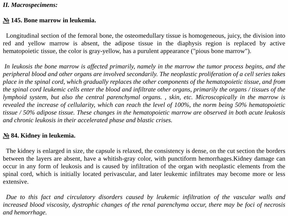
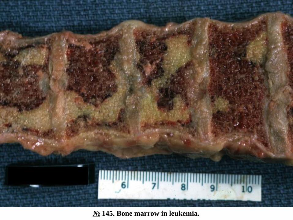
№ 145. Bone marrow in leukemia.
Longitudinal section of the femoral bone, the osteomedullary tissue is homogeneous, juicy, the division into
red and yellow marrow is absent, the adipose tissue in the diaphysis region is replaced by active
hematopoietic tissue, the color is gray-yellow, has a purulent appearance ("pious bone marrow").
In leukosis the bone marrow is affected primarily, namely in the marrow the tumor process begins, and the
peripheral blood and other organs are involved secondarily. The neoplastic proliferation of a cell series takes
place in the spinal cord, which gradually replaces the other components of the hematopoietic tissue, and from
the spinal cord leukemic cells enter the blood and infiltrate other organs, primarily the organs / tissues of the
lymphoid system, but also the central parenchymal organs. , skin, etc. Microscopically in the marrow is
revealed the increase of cellularity, which can reach the level of 100%, the norm being 50% hematopoietic
tissue / 50% adipose tissue. These changes in the hematopoietic marrow are observed in both acute leukosis
and chronic leukosis in their accelerated phase and blastic crises.
№ 84. Kidney in leukemia.
The kidney is enlarged in size, the capsule is relaxed, the consistency is dense, on the cut section the borders
between the layers are absent, have a whitish-gray color, with punctiform hemorrhages.Kidney damage can
occur in any form of leukosis and is caused by infiltration of the organ with neoplastic elements from the
spinal cord, which is initially located perivascular, and later leukemic infiltrates may become more or less
extensive.
Due to this fact and circulatory disorders caused by leukemic infiltration of the vascular walls and
increased blood viscosity, dystrophic changes of the renal parenchyma occur, there may be foci of necrosis
and hemorrhage.
№ 145. Bone marrow in leukemia.

№ 84. Kidney in leukemia.
№ 142. Spleen in CML (chronic myeloid leukemia).
The spleen is considerably enlarged in size, sometimes 20-30 times, the mass reaching a few kg (norm ~
180 gr), per section reddish-gray color, homogeneous, dense consistency, there may be foci of ischemic
infarction and hemorrhage.
Massive splenomegaly, which is revealed in chronic myeloid leukosis is caused by intense leukemic
infiltration, diffuse with cells from the myeloid series, predominantly with myelocytes and metamyelocytes;
Outbreaks of infarction are caused by increased blood viscosity, which can sometimes lead to leukemic
thrombi. Fibrin deposits (perisplenitis), cracks may be deposited on the spleen capsule, it is possible to
rupture the capsule with lethal intraperitoneal hemorrhage.
№ 143. Mesenterial lymph node in CLL (chronic lymphoid leukemia).
The lymph nodes are uniformly enlarged in size, dense-elastic consistency, whitish color, form tumor
conglomerates, which compress the adjacent organs.
Generalized lymphadenopathy is the predominant clinical-morphological sign of chronic lymphoid leukosis.
The lymph nodes are symmetrically enlarged in size, microscopically reveals diffuse infiltration with small
mature, uniform lymphocytes, without atypia; foci of proliferation are also observed with larger, mitotically
active lymphocytes, without precise limits. 80% of chronic lymphocytic leukosis comes from B-lymphocytes.
Although the number of neoplastic lymphocytes is considerably increased, they are immunologically inactive,
leading to hypogamaglobulinemia, decreased humoral immunity with infectious complications, and
autoimmune reactions, primarily hemolytic anemias and autoimmune thrombocytopenias.

№ 142. Spleen in CML (chronic myeloid leukemia).
30cm
infarct

№ 143. Mesenterial lymph node in CLL (chronic lymphoid leukemia).
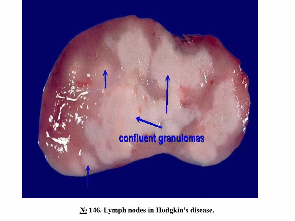
№ 146. Lymph nodes in Hodgkin’s disease.
The lymph nodes are unevenly enlarged in size, of dense consistency, light-whitish color, adhere to each
other due to infiltration of perinodular connective tissue, on a motley-looking section, white-yellow foci of
necrosis and fibrosis.
Hodgkin's lymphoma begins in a single lymph node or in a group of lymph nodes, usually cervical,
supraclavicular, or axillary. Subsequently, the tumor process progresses, gradually involving other groups
of lymph nodes on the same side of the diaphragm, on both sides of the diaphragm or extralymph
(extranodal) tissues / organs. At first the lymph nodes are separated, and later they become adherent,
forming tumor conglomerates, which compress the adjacent tissues / organs.
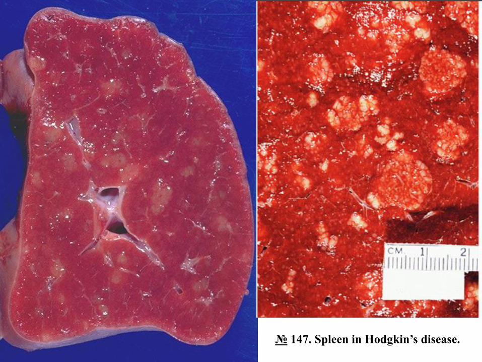
№ 147. Spleen in Hodgkin’s disease.
The spleen is enlarged in size 3-5 times, the mass reaching up to 1 kg, dense consistency, on a motley-
looking section due to the alternation of white-yellowish proliferative foci and necrosis with whitish
sclerosis foci on the background of the red pulp, which gives the spinal tissue an appearance similar to
porphyry granite ('porphyry spleen') [the motley appearance is poorly pronounced due to the action of
formalin].
Splenomegaly in Hodgkin's lymphoma is an expression of tumor progression, in the first stage being
affected lymph nodes, and later other extranodal organs, primarily the spleen. Spleen damage is observed
in about half of patients, being a process of metastasis from the primary focus of the lymph nodes.
Histologically, tumor nodules consisting of a mixture of Reed-Sternberg cells and reactive cells
(eosinophils, plasma cells, neutrophilic leukocytes, macrophages), foci of necrosis, sometimes caseous, and
fibrosis are revealed.
№ 146. Lymph nodes in Hodgkin’s disease.
№ 147. Spleen in Hodgkin’s disease.

AML - with gingival manifestations.
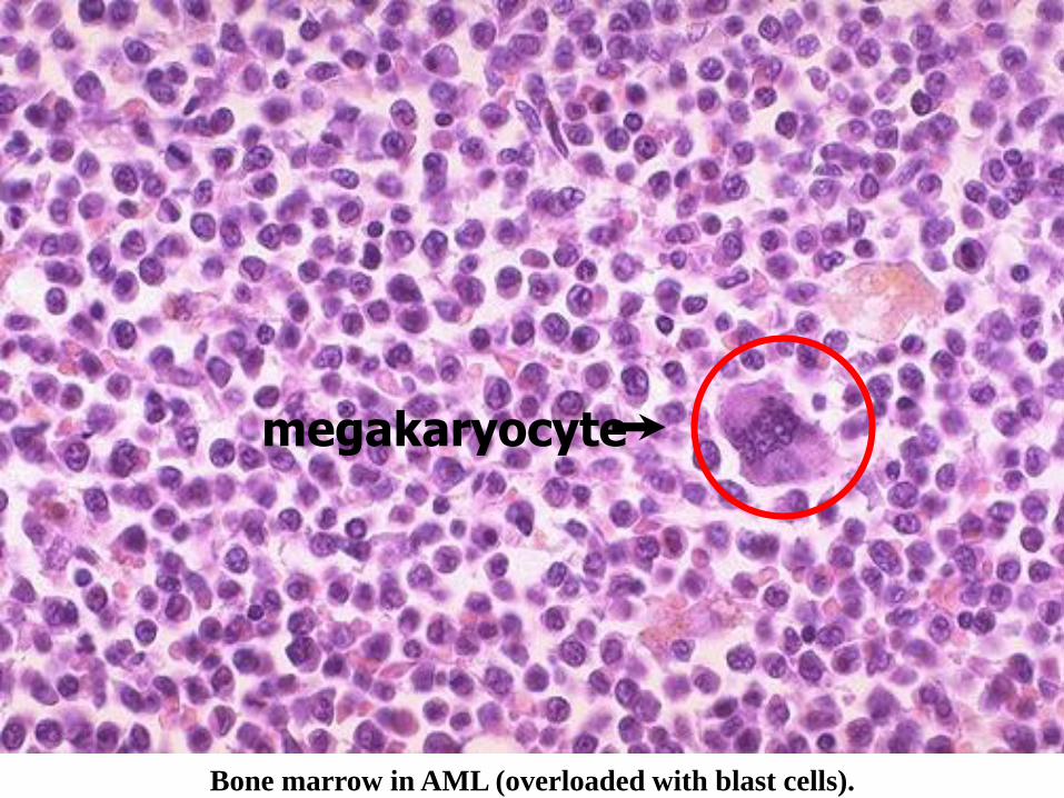
Bone marrow in AML (overloaded with blast cells).
megakaryocyte

ALL - cervical lymphadenopathy.
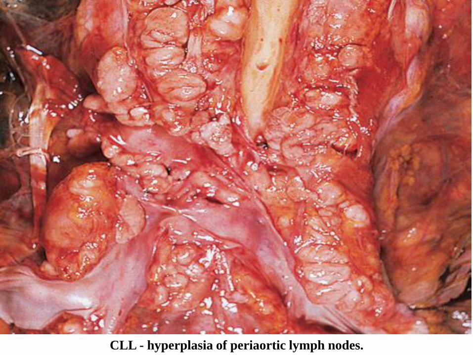
CLL - hyperplasia of periaortic lymph nodes.

Hyperplasia of the spleen and intestinal lymph follicles in CLL.

Liver in CLL
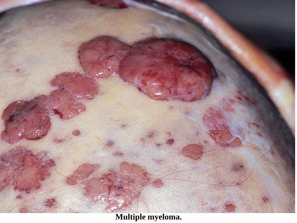
Multiple myeloma.

Myeloma kidney and normal kidneys.

Lymph node in Hodgkin's lymphoma.
(nodular surface).

Reed - Sternberg cell.

Lymph node in Hodgkin's disease (mixed-cellularity type). (H-E stain).
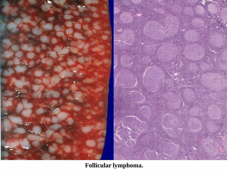
Follicular lymphoma.
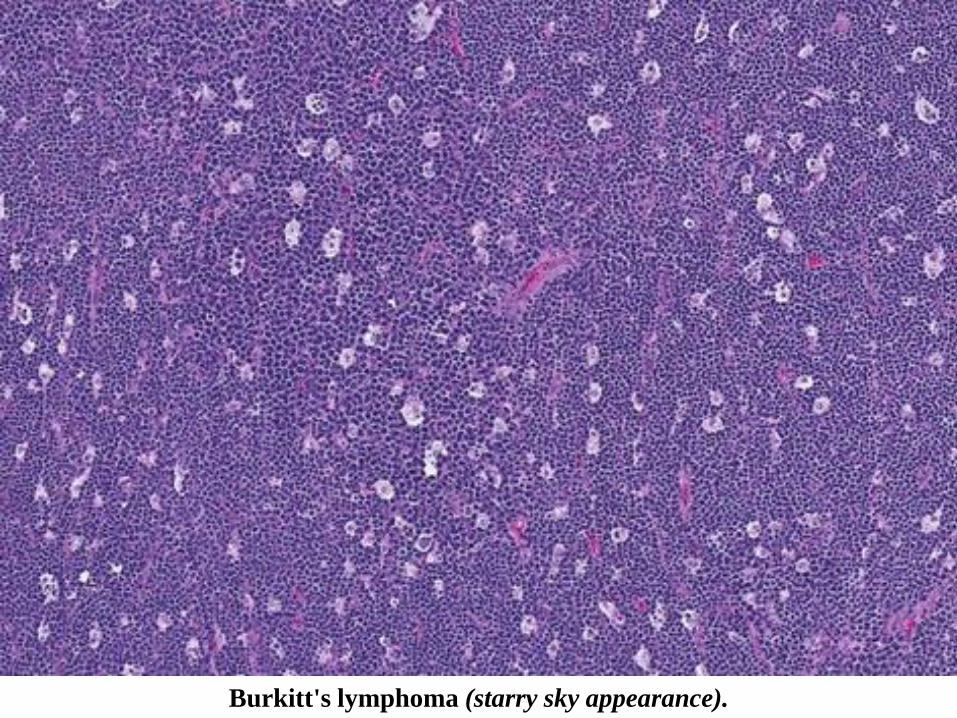
Burkitt's lymphoma (starry sky appearance).
Blood and lymphatic disorders

Structure of bone marrow

WHERE is MARROW?
• Yolk Sac: very early embryo
• Liver, Spleen: NEWBORN
• BONE• CHILDHOOD: AXIAL SKELETON & APPENDICULAR SKELETON BOTH HAVE RED
(active) MARROW
• ADULT: AXIAL SKELETON RED MARROW, APPENDICULAR SKELETON YELLOW MARROW
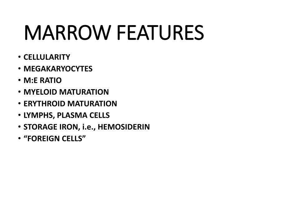
MARROW FEATURES• CELLULARITY
• MEGAKARYOCYTES
• M:E RATIO
• MYELOID MATURATION
• ERYTHROID MATURATION
• LYMPHS, PLASMA CELLS
• STORAGE IRON, i.e., HEMOSIDERIN
• “FOREIGN CELLS”

MARROW “DIFFERENTIATION”

Bone marrow


ANEMIASA good definition would be a decrease in OXYGEN
CARRYING CAPACITY, rather than just a decrease in red blood cells, because you need to have enough blood cells THAT FUNCTION, and not just
enough blood cells.
• BLOOD LOSS
1.acute
2.chronic
• IN-creased destruction
(HEMOLYTIC)
• DE-creased production

Features of ALL anemias
•Pallor, where?•Tiredness•Weakness•Dyspnea, why?•Palpitations•Heart Failure (high output), why?

Blood LossAcute: trauma
Chronic: lesions of gastrointestinal tract, gynecologic disturbances. The features of chronic blood loss anemia are the same as iron deficiency anemia, and is defined as a situation in which the production cannot keep up with the loss

HEMOLYTIC• HEREDITARY
• MEMBRANE disorders: e.g., spherocytosis
• ENZYME disorders: e.g., G6PD deficciency
• HGB disorders (hemoglobinopathies)
• ACQUIRED• MEMBRANE disorders (PNH)
• ANTIBODY MEDIATED, transfusion
• or autoantibodies
• MECHANICAL TRAUMA
• INFECTIONS
• DRUGS, TOXINS
• HYPERSPLENISM
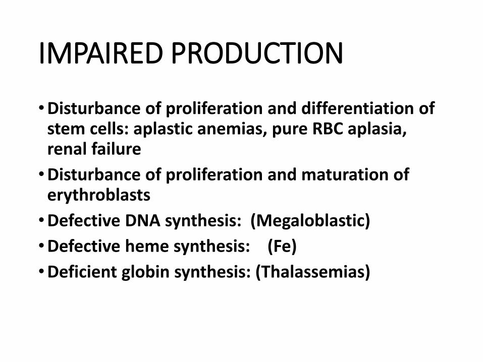
IMPAIRED PRODUCTION
•Disturbance of proliferation and differentiation of stem cells: aplastic anemias, pure RBC aplasia, renal failure
•Disturbance of proliferation and maturation of erythroblasts
•Defective DNA synthesis: (Megaloblastic)
•Defective heme synthesis: (Fe)
•Deficient globin synthesis: (Thalassemias)
MODIFIERS
•MCV, microcytosis, macrocytosis
•MCH
•MCHC, hypochromic
•RDW, anisocytosis

HEMOLYTIC ANEMIAS
•Life span LESS than 120 days
•Marrow hyperplasia (M:E), EPO+
•Increased catabolic products, e.g., bilirubin, serum HGB, hemosiderin

HEMOLYSIS
•INTRA-vascular (vessels)
•EXTRA-vascular (spleen)
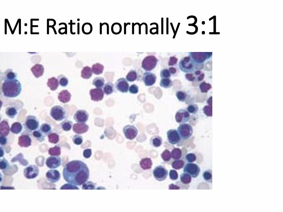
M:E Ratio normally 3:1

HEREDITARY SPHEROCYTOSIS
Genetic defects affecting
ankyrin, spectrin, usually
autosomal dominant
Children, adults
Anemia, hemolysis,
jaundice, splenomegaly,
gallstones (what kind?)

NON-Hemolytic Anemias:i.e., DE-creased Production
• “Megaloblastic” Anemias
• B12 Deficiency (Pernicious Anemia)
• Folate Deficiency
• Iron Deficiency
• Anemia of Chronic Disease
• Aplastic Anemia
• “Pure” Red Cell Aplasia
• OTHER forms of Marrow Failure

MEGALOBLASTIC ANEMIAS•Differentiating megaloblasts
(marrow) from macrocytes (peripheral smear, MCV>94)
• Impaired DNA synthesis
•For all practical purposes, also called the anemias of B12 and FOLATE deficiency

etiologyDecreased intakeInadequate diet, vegetarianismImpaired absorptionIntrinsic factor deficiencyPernicious anemiaGastrectomyMalabsorption statesDiffuse intestinal disease, e.g., lymphoma, systemic
sclerosisIleal resection, ileitisCompetitive parasitic uptakeFish tapeworm infestationBacterial overgrowth in blind loops and diverticula of
bowelIncreased requirementPregnancy, hyperthyroidism, disseminated cancer

Vit-B12 Physiology
•Oral ingestion
•Combines with INTRINSIC FACTOR in the gastric mucosa
•Absorbed in the terminal ileum
•DEFECTS at ANY of these sites can produce a MEGALOBLASTIC anemia

Please remember that ALL
megaloblastic anemias are also
MACROCYTIC (MCV>94 or
MCV~100), and that not only are
the RBC’s BIG, but so are the
neutrophils, and neutrophilic
precursors in the bone marrow
too, and even more so,
HYPERSEGMENTED!!!

FOLATE DEFICIENCY MEGALOBLASTIC AMEMIAS
• Decreased Intake: diet, etoh-ism, infancy
• Impaired Absorption: intestinal disease
• DRUGS: anticonvulsants, BCPs, CHEMO
• Increased Loss: Hemodialysis
• Increased Requirement: Pregnancy, infancy
• Impaired Usage

Clinical Fe-Defic-Anemia
•Adult men: GI Blood Loss
•PRE menopausal women: menorrhagia
•POST menopausal women: GI Blood Loss

Clinical Fe-Defic-Anemia

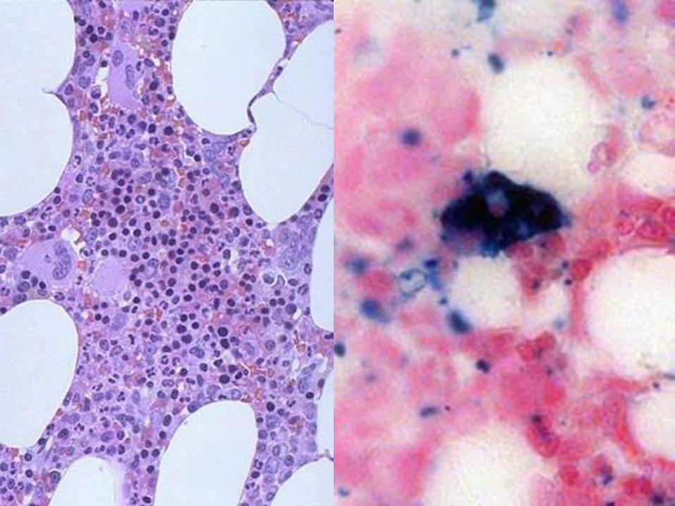
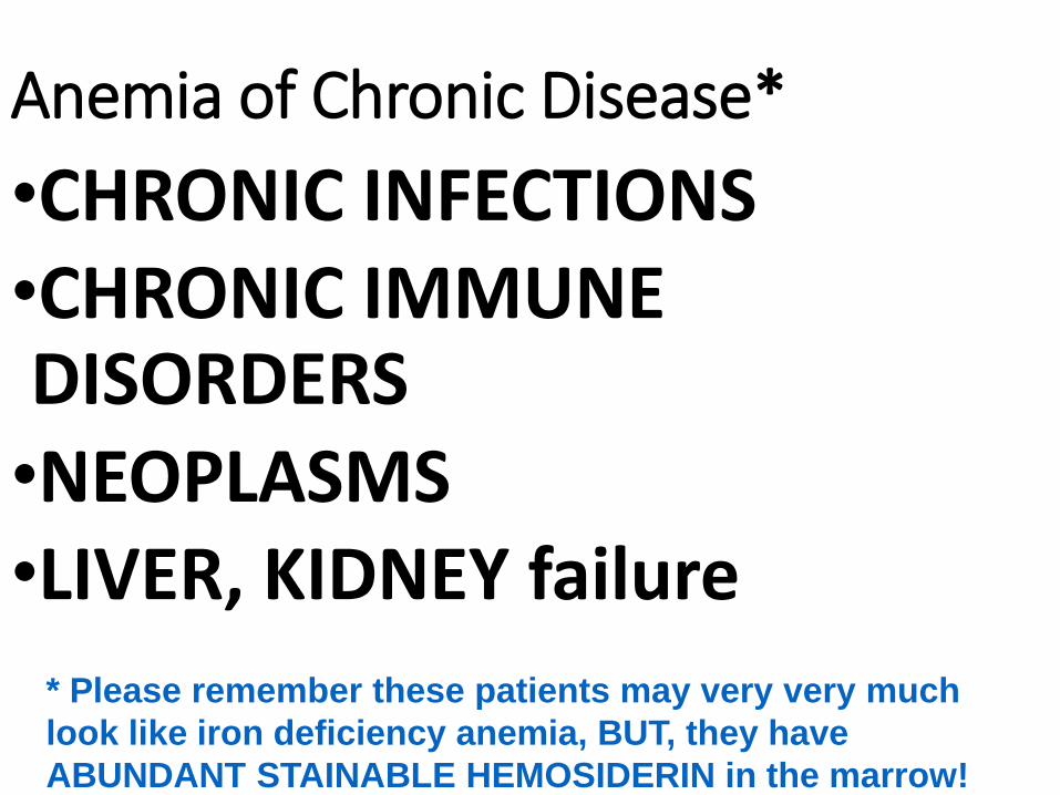
Anemia of Chronic Disease*
•CHRONIC INFECTIONS•CHRONIC IMMUNE DISORDERS•NEOPLASMS•LIVER, KIDNEY failure
* Please remember these patients may very very much
look like iron deficiency anemia, BUT, they have
ABUNDANT STAINABLE HEMOSIDERIN in the marrow!

APLASTIC ANEMIAS
•ALMOST ALWAYS involve platelet and WBC suppression as well•Some are idiopathic, but MOST are related to drugs, radiation•FANCONI’s ANEMIA is the only one that is inherited, and NOT acquired•Act at STEM CELL level, except for “pure” red cell aplasia

APLASTIC ANEMIAS

APLASTIC ANEMIAS•CHLORAMPHENICOL
•OTHER ANTIBIOTICS
•CHEMO
•INSECTICIDES
•VIRUSES•EBV•HEPATITIS

MYELOPHTHISIC ANEMIAS
•Are anemias caused by metastatic tumor cells replacing the bone marrow extensively

DISEASES of WHITE CELLS and LYMPHOID TISSUE

Myeloid Lymphoid
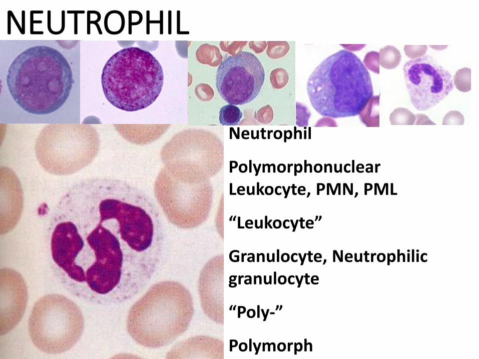
NEUTROPHIL
Neutrophil
Polymorphonuclear Leukocyte, PMN, PML
“Leukocyte”
Granulocyte, Neutrophilic granulocyte
“Poly-”
Polymorph

LEUKO-penia/NEUTRO-peniaNeutropenia/Agranulocytosis
•INADEQUATE PRODUCTION
•INCREASED DESTRUCTION
•500-1000/mm3 is the DANGERzone!

Leukocytosis/Neutrophilia• Marrow and splenic pool size
• Rate of release between pool and circulation
• Marginating pool
• Rate of WBCs (neutrophils/monocytes) leaving the vascular compartment
• NON-vascular pools FIFTY times larger than the vascular pools
• TNF/IL-1/cytokines stimulate T-cells to produce CSF, the WBC equivalent of EPO

NEUTROPHIL INCREASES(e.g., “NEUTROPHILIA”)
• BACTERIA
• TISSUE NECROSIS, e.g., MI
• DÖHLE BODIES (e.r. remnants) and TOXIC GRANULES are often seen with NEUTROPHILIA
• Accompanied by a “LEFT” shift
LEUKEMIAS•MALIGNANT PROLIFERATIONS of WHITE BLOOD CALLS•In the case of neutrophilic precursors, the primary process is marrow and peripheral blood, but can involve any organ or tissue which receives blood
•In the case of lymphocytes, there is an intimate concurrence with malignant lymphomas

LeukemiasThese are composed of two major groups: myeloid (granulocytic) and
lymphoid.
Causes: The cause is unknown but some predisposing factors have been recognized:
1. Myelodysplastic syndromes precede the onset of leukemia
2. Genetic factors may play a role, chromosomal syndromes (Downs, etc.) are associated with increased risk of leukemias.
3. Ionizing radiation; there is increased incidence in those exposed to radiation for treatment or otherwise.
4. Alkylating agents used in chemotherapy are associated with increased risk
5. Viruses: Human T-cell lymphocytic virus-1 (HTLV-1) is an RNA oncogenic virus that causes T-cell leukemias
6. Endogenous oncogenes play a role and are associated with chromosomal breaks, translocations or deletions. The Philadelphia chromosome (translocation of fragments of chromosomes 9 & 22) is associated with the formation of an oncogene (a proto-oncogene on #22 separates from its expression control) and are associated with the development of CML (also ALL & AML to a lesser extent).

Leukemias vs. Lymphomas•All leukemias of lymphocytes have lymphoma
counterparts
•Primary lymphomas can have “leukemic” phases, including multiple myelomas
•Any myeloid leukemia can infiltrate a lymph node, or any other site, but if/when it does it is NOT called a lymphoma, but simply a myeloid infiltrate INTO a lymph node
•ALL lymphomas are malignant proliferations of lymphocytes
•ALL leukemias involve bone marrow changes

LYMPHOMAS•NODAL or EXTRANODAL
•T or B
•SMALL or LARGE CELLS
•FOLLICULAR or DIFFUSE
•Hodgkins or NON-Hodgkins
•“F.A.B. classification” is currently popular
this week (FrenchAmericaBritish), for the NON-Hodgkins lymphomas

LEUKEMIAS• Acute or Chronic
• Myeloid or Lymphocytic
• Childhood or Adult
• All involve marrow
• All ACUTE leukemias suppress normal hematopoesis, i.e., have anemia, thrombocytopenia
• Most have chromosomal aberrations
• Some can respond DRASTICALLY to chemo, most notably ALL in children, even be cured!!!!

Types of Leukemias1. Acute Lymphoblastic leukemia (ALL) ~30% of all leukemias, the most
common among children under 5 years old a second peak occurs after age 60. The marrow contains more than 30% lymphoblasts. The prognosis is inversely proportional to age, responds remarkably well to chemotherapy & marrow transplant, 85% long term survival in 1-10 year olds, ~50% in adults.
2. Acute myelogenous leukemia (AML) ~80% of acute leukemias in adults. Marrow has >20% myeloblasts. Overall prognosis is poor with relapse after chemotherapy and most do not survive more than 5 years after diagnosis. Two forms; acute denovo AML(better prognosis ~70% 5 year (especially acute promyelocytic~90% 5 year) or as an end-stage of CML and myelofibrosis (poorer prognosis~15% 5 year).
AML
ALL

BLAST
Types of Leukemias
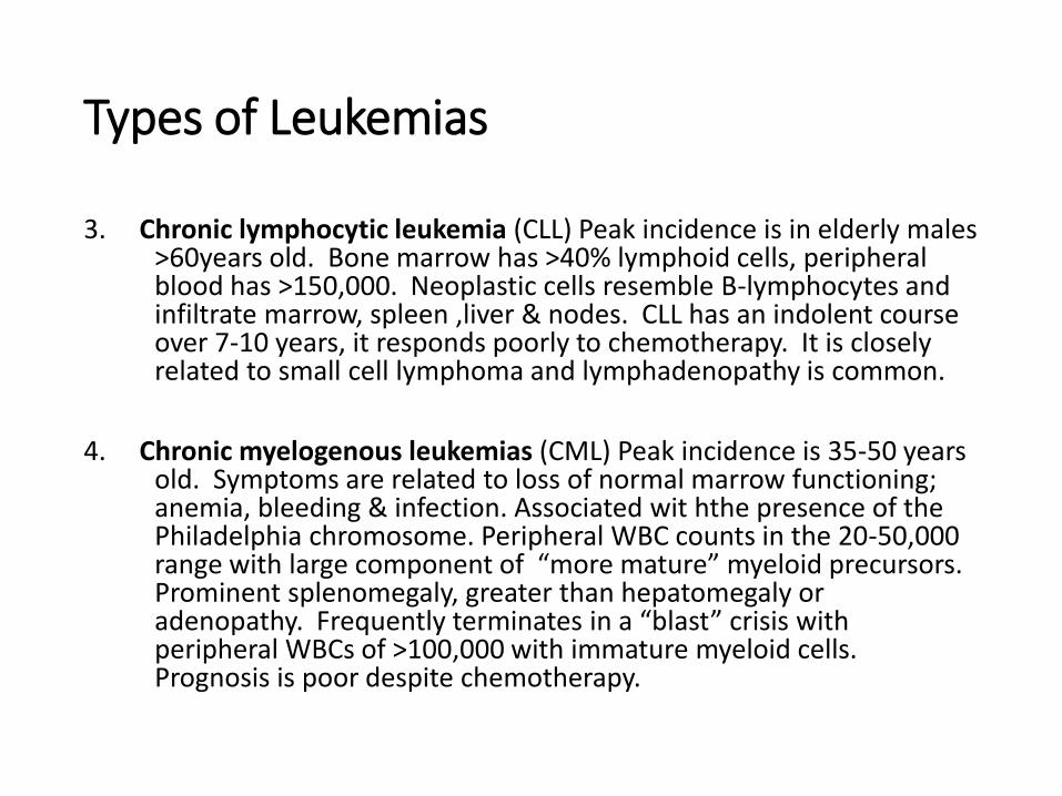
3. Chronic lymphocytic leukemia (CLL) Peak incidence is in elderly males >60years old. Bone marrow has >40% lymphoid cells, peripheral blood has >150,000. Neoplastic cells resemble B-lymphocytes and infiltrate marrow, spleen ,liver & nodes. CLL has an indolent course over 7-10 years, it responds poorly to chemotherapy. It is closely related to small cell lymphoma and lymphadenopathy is common.
4. Chronic myelogenous leukemias (CML) Peak incidence is 35-50 years old. Symptoms are related to loss of normal marrow functioning; anemia, bleeding & infection. Associated wit hthe presence of the Philadelphia chromosome. Peripheral WBC counts in the 20-50,000 range with large component of “more mature” myeloid precursors. Prominent splenomegaly, greater than hepatomegaly or adenopathy. Frequently terminates in a “blast” crisis with peripheral WBCs of >100,000 with immature myeloid cells. Prognosis is poor despite chemotherapy.

Organomegaly

A.L.L.
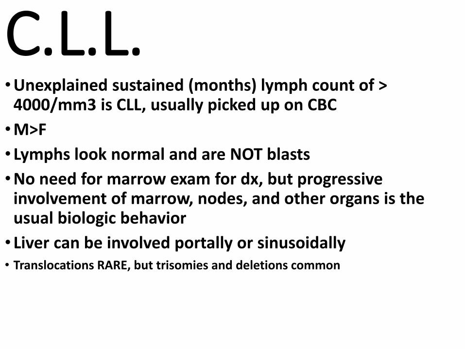
C.L.L.•Unexplained sustained (months) lymph count of >
4000/mm3 is CLL, usually picked up on CBC
•M>F
• Lymphs look normal and are NOT blasts
•No need for marrow exam for dx, but progressive involvement of marrow, nodes, and other organs is the usual biologic behavior
• Liver can be involved portally or sinusoidally• Translocations RARE, but trisomies and deletions common

C.L.L.

A.M.L.• GENETIC ABERRATIONS INHIBIT DIFFERENTIATION
• Many have various TRANSLOCATIONS
• F.A.B. classifies them as M0→M7
• MORE than 20% of BLASTS are needed in the marrow for a diagnosis of acute leukemia!!! (i.e., ANY kind of BLAST
• NORMALLY, a marrow should have only about 1-2 % blasts

M0→M2
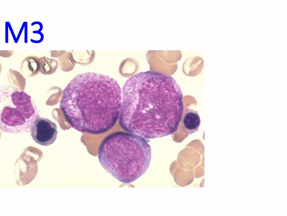
M3
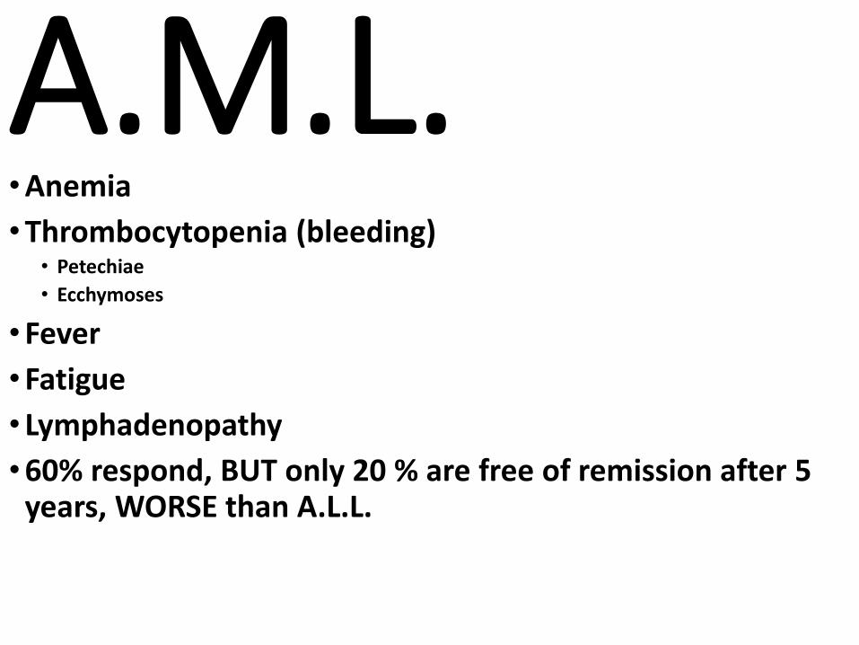
A.M.L.•Anemia
•Thrombocytopenia (bleeding)• Petechiae
• Ecchymoses
•Fever
•Fatigue
• Lymphadenopathy
•60% respond, BUT only 20 % are free of remission after 5 years, WORSE than A.L.L.

CML high-power
blast
mielocite

Massive splenomegaly in CML
30cminfarct
CLL

CML

Plasma cell disordersMain types
• Multiple myeloma• Waldenstrom macroglobulinemia: A malignancy of plasmacytoid
lymphocytes that secrete IgM resulting in a hyperviscosity syndrome with renal, retinal and cerebral ischemia as a result of microvascular occlusion. Infiltration of plasmacytoid cells in the marrow, spleen and nodes.
• Monoclonal gammopathy of unknown significance: often diagnosed in asymptomatic elderly patients. It is present in ~1% of patients over 60 years old and 3% of patients over 70. There is a 1% risk of developing multiple myeloma. The vast majority suffer no ill effects.
Clinical features• Tend to occur in those >45 years old.• Neoplastic plasma cells produce a monoclonal immunoglobulin
component that can be identified by serum electrophoresis• Deposition of light chain immunoglobulin may form amyloid deposits in
the kidneys, vessels and other organs.

PLASMA CELL classic features
•OVAL cytoplasm, ROUND nucleus off to side
•Cartwheel/Clockface chromatin
•Prominent Golgi or “Hoff”
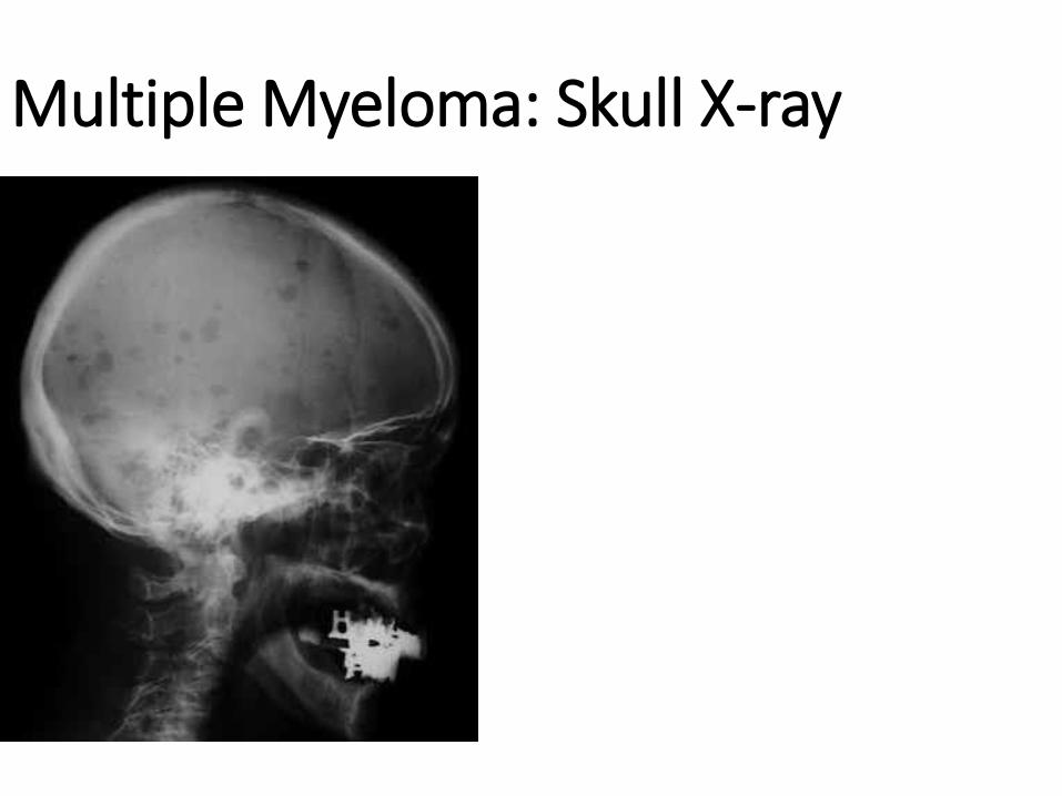
Multiple Myeloma: Skull X-ray

LYMPH NODES• Normal Structure, Function
• Benign enlargement/Benign disease• Acute
• Chronic (follicular vs. “sinus histiocytosis”)
• Lymphomas/Malignant Lymphomas• Adjectives of various classifications
• Features
• STAGING
• Metastatic disease TO lymph nodes


CORTEX
---SUB-capsular Sinus
---Follicles (Pri? Or second.?)
---PARA-follicular zone
MEDULLABlood flow?
Lymph flow?
Definition of TERMS• Lymphadenopathy
• Lymphadenitis
• What to do if a lymph node is enlarged?
• Diffuse/Follicular
• T/B/NK, Small/Large, Cleaved/Non-cleaved
• Precursor/Peripheral
• HD/Non-HD

BENIGN ENLARGEMENT
•Also called LYMPHADENITIS, and HYPERPLASIA
•Can be ACUTE (tender), or CHRONIC (non-tender)
•Usually SUBSIDE in, say, less than 6 weeks
•FOLLICULAR HYPERPLASIA is enlargement of the cortical secondary follicles and increase in number of the cortical secondary follicles
•SINUS HISTIOCYTOSIS is prominence in medullary sinuses (also called “reticular” hyperplasia)


(MALIGNANT) LYMPHOMAS
•Terms in historic classifications:• Diffuse/Follicular, Small/Large, Cleaved/Non-cleaved
• Hodgkins /NON-Hodgkins
• Lukes, Rappaport, etc.
• Working Formulation, WHO, NIH, FAB, Intl., etc.
• B
• T
• PRECURSOR (less mature looking)
• PERIPHERAL (more mature looking)

DIFFUSE LYMPHOMA
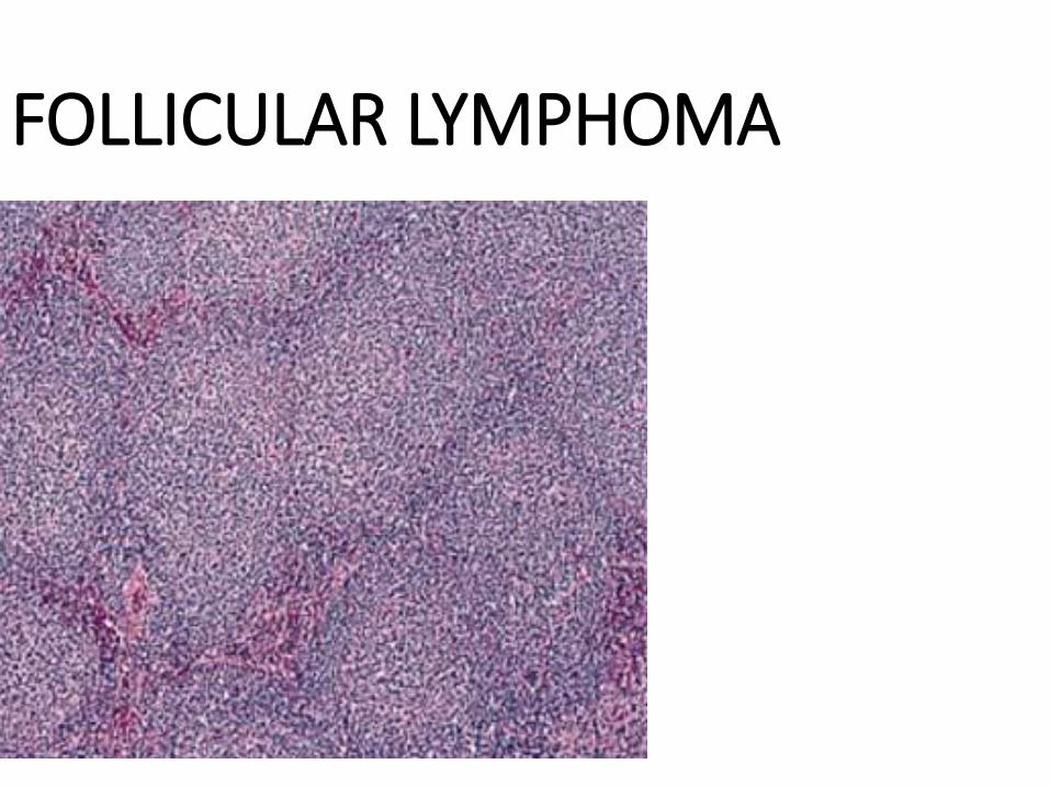
FOLLICULAR LYMPHOMA

LARGE CELL LYMPHOMA

SMALL CELL LYMPHOMA

“CLEAVED” CELL LYMPHOMA

FEATURES of LYMPHOMAS•The Antigen receptor genes re-arrangement PRECEDES
malignant transformation, so the cells are MONOCLONAL, NOT the usual POLYCLONAL
•85% B-cell, 15% T-Cell
•The tumor cells congregate wherever T and B cell congregate normally however
•DISRUPTED or “EFFACED” normal architecture, obliterated subcapsular sinus
•HD/Non-HD staging CRUCIALLY IMPORTANT, esp. HD. Why? HD grows more “linearly”

LATEST CLASSIFICATION•NON-HODGKIN•PRECURSOR B•PERIPHERAL B•PRECURSOR T•PERIPHERAL T
•HODGKIN’S DISEASE (i.e., HODGKINS LYMPHOMA)

HODGKINS DISEASE• NEED R-S (Reed-Sternberg, or Sternberg-Reed) cells for correct diagnosis
•NODULAR SCLEROSIS (Young Women), the R-S cells may
be called “LACUNAR” cells
•MIXED CELLULARITY• Lymphocyte RICH
• Lymphocyte POOR
• Lymphocyte PREDOMONANCE

STERNBERG-REED CELL

METASTATIC SQUAMOUS CELL CARCINOMA

METASTATIC ADENOCARCINOMA

SPLEEN•150 grams POST-LUQ (just like kidney, 1/10 of liver)•Bordered by diaphragm, kidney, pancreas, splenic flexure,
stomach•SMOOTH & GLISTENING capsule•50% RED pulp, 50% WHITE pulp


ABNORMAL SPLEEN
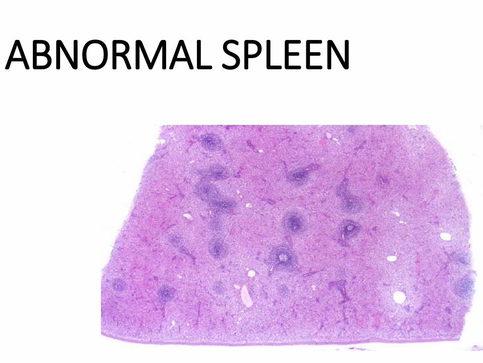
ABNORMAL SPLEEN
SPLENIC FUNCTION
•REMOVE OLD BLOOD CELLS
•MAJOR SECONDARY ORGAN of the IMMUNE SYSTEM
•HEMATOPOIESIS
•SEQUESTER (POOL) BLOOD CELLS
•15% of body’s PHAGOCYTIC activity is in the spleen (liver has >80)

SPLENOMEGALY•CONGESTIVE vs INFILTRATIVE
•HYPERSPLENISM•Anemia•Leukopenia•Thrombocytopenia
•DECISION for SPLENECTOMY

SPLENOMEGALY•INFECTIONS: TB, Mono, Malaria, Fungus
•PORTAL HTN: CHF, CIRRHOSIS, PV Thromb.
•LYMPHOHEMATOGENOUS: Leuk, Lymph
•IMMUNE: RA, SLE
•STORAGE: Gaucher, Niemann-Pick
•MISC: Amyloid, mets (melanoma, lymphoma, Germ cell tumors of testis)
LONG STANDING CONGESTION breeds FIBROSIS
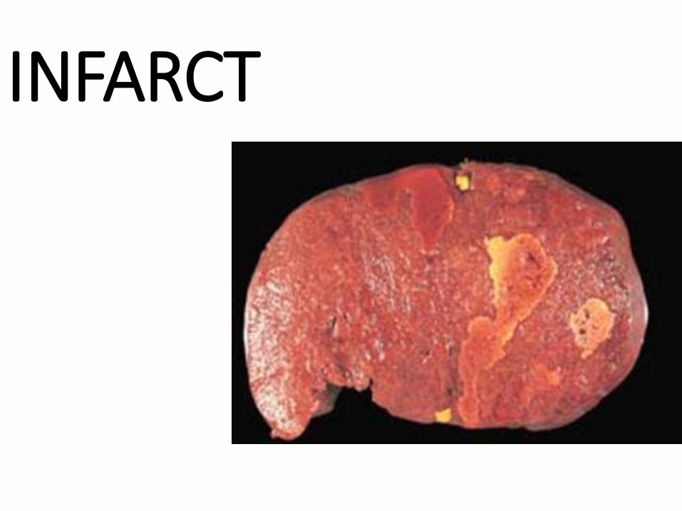
INFARCT

PRIMARY TUMORS (RARE)
•HEMANGIOMA•LYMPHANGIOMA•fibroma
•osteoma
•chondroma

THYMUS•Mother of all T-Cells
•Massive in newborns, virtually absent in the elderly, bilobed
•Under manubrium
•1) Thymocytes
•2) Epithelial Ret. Cells
•3) Hassal’s Corpuscles

HASSAL’s CORPUSCLES

DISEASES•HYPOPLASIA/APLASIA
• DiGeorge Syndrome
•CYSTS (incidental)
•THYMOMAS