pathology of the hematopoietic system - university of...
TRANSCRIPT
Pathology of the Hematopoietic
System
Lecture 3: Spleen and Thymus
Paul Hanna,
Fall 2014

Spleen – Structure and Function
Normal spleen from a cat
1 cm
• present in the left cranial part of the abdomen
• attached to the greater curvature of the stomach by the gastrosplenic ligament
• covered by a fibromuscular capsule and dissected by fibromuscular trabeculae
• varies in size and shape among species

Structure Function
Red Pulp • splenic sinusoids
(vascular spaces)
• splenic cords
• filters blood - removal
of foreign material
(phagocytosis)
• RBC storage
• hematopoiesis (EMH)
White Pulp • PALS - periarteriolar
lymphatic sheaths
(T-cells)
• lymphoid nodules
(B-cells)
• marginal zone
(macrophages)
• immune response
Image:Education.vetmed.ve.edu/…/labs/Lab13.htm
Spleen – Structure and Function

Miscellaneous Diseases
Circulatory diseases of the spleen
Inflammation of the spleen (splenitis)
Adaptations of growth
Primary and secondary splenic neoplasia
Diseases of the spleen

Spleen: Miscellaneous Diseases
Misc. Diseases
Siderofibrosis (Gamna-gandy bodies)
Splenic amyloidosis
Splenic contraction
Splenic lymphoid necrosis
Hemosiderin deposition / hemosiderosis
Dog spleens. Normal spleen (right) vs spleen with large
numbers of siderofibrotic plaques / nodules in the capsule

Normal spleen – with smooth, glistening capsule
Dr E Aburto
Dr E Aburto
1. Siderofibrosis = Gamna-Gandy bodies
• incidental finding / senile change
• likely sequela of prior hemorrhage
Gross: Granular white-yellow deposits in the splenic capsule
Spleen: Miscellaneous Diseases

Spleen: Miscellaneous Diseases
1. Siderofibrosis = Gamna-Gandy bodies
• incidental finding / senile change
• likley sequela of prior hemorrhage
Histo: hematoidin
hemosiderin
mineral deposits
fibrosis
Web Fig. 13-6B (Zachary) Sidero-calcific plaque,
spleen, dog. The sidero-calcific plaque lies in the
fibrous connective tissue of the capsule and consists
chiefly of calcium (blue) and hemosiderin (brown) in
fibrous connective tissue. The yellow material is bilirubin
(hematoidin), resulting from the breakdown of
erythrocytes in a capsular hemorrhage. H&E stain.

Spleen: Miscellaneous Diseases
Fig. 13-57 (Zachary) Amyloid spleen, dog. The
spleen is pale beige, firm and waxy, and uniformly
distended in this advanced case of amyloidosis.
Normal spleen
2. Splenic amyloidosis
• especially 2°amyloidosis – chronic inflammation
• Gross: splenomegaly, beige to orange
• Histology: deposition of amyloid around the splenic arteries (Congo red stain)

3. Splenic contraction
• contraction of the smooth muscle in the capsule / trabeculae
• occurs with catecholamine release, shock, acute splenic rupture
• Gross: small dry spleen with wrinkling of the capsule
Spleen: Miscellaneous Diseases
If contraction is incomplete it can look similar to splenic infarction
splenic infarction

Spleen: Miscellaneous Diseases For information only
4. Splenic lymphoid necrosis
Viral infections
• Panleukopenia virus
• Canine parvovirus
• Bovine viral diarrhea virus
• Equine viral rhinotracheitis
Stress
Toxins
Aging
5. Hemosiderin deposition / Hemosiderosis
• small amount of hemosiderin in splenic macrophages → normal rbc turnover
• hemosiderosis = increased amount due to increased erythrocyte destruction.

Spleen: Circulatory Disturbances
Circulatory
disturbances
Active hyperemia
Passive congestion
Splenic infarction
Splenic rupture
Splenic hematoma
Splenic torsion

Congested vs normal spleen
Spleen: Circulatory Disturbances
2. Passive congestion
• vascular pooling (shock)
• barbiturate administration**
• hemolytic anemia
1. Active hyperemia
• acute systemic infection
Similar grossly:
• splenomegaly
• red to purple
• oozes blood on cut surface

Spleen: Circulatory Disturbances
2. Passive congestion
• vascular pooling (shock)
• barbiturate administration**
• hemolytic anemia
1. Active hyperemia
• acute systemic infection
Similar grossly:
• splenomegaly
• red to purple
• oozes blood on cut surface
note, blood oozing from cut surface
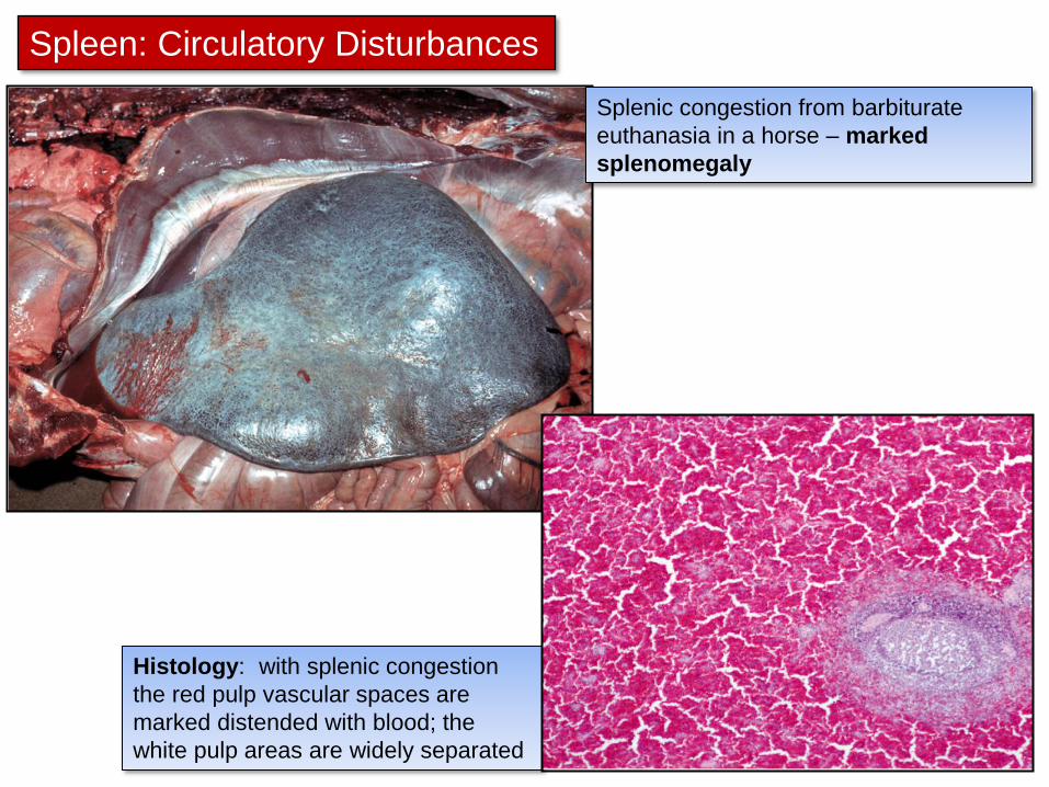
Splenic congestion from barbiturate
euthanasia in a horse – marked
splenomegaly
Spleen: Circulatory Disturbances
Histology: with splenic congestion
the red pulp vascular spaces are
marked distended with blood; the
white pulp areas are widely separated

Spleen: Circulatory Disturbances
Gross Lesions:
• acutely: infarcts are discrete, raised & dark
red – at the margins of the organ
• with time: pale and firm (fibrosis)
3. Splenic infarction
Thrombosis and infarction occurs with:
• diseases causing vascular damage
• hypercoagulable states
• splenomegaly of any cause
• septic thromboemboli

4. Splenic rupture
• fairly common
• primary - trauma
• 2o to splenomegaly, splenic neoplasia
• sequelae: hemoabdomen and splenosis
Spleen: Circulatory Disturbances
Splenic rupture 2° to hemangiosarcoma
in a dog
Splenic rupture and hemoabdomen 2° to
splenomegaly (lymphoma) in a pig
Dr E Aburto

4. Splenic rupture - splenosis
• seeding of splenic explants on peritoneal surfaces (“splenosis”)
Gross: small red nodules within the omentum
Histology: looks like normal spleen
Grossly can be mistaken for
hemangiosarcoma metastases

Spleen: Circulatory Disturbances
5. Splenic hematoma
• common in dogs
• trauma
• 2o to nodular hyperplasia or vascular tumors
Need histology to
rule out underlying
neoplasia!
Gross: red nodular mass; often large, soft & dark red on cut surface

Spleen: Circulatory Disturbances
6. Splenic torsion
• dogs and pigs
• with or without volvulus of the stomach (GDV)
• twists around the gastrosplenic ligament
Gross: splenomegaly, blue to black, often folded back on itself
Dr E Aburto

• Tularemia (Francisella tularensis)
• Yersiniosis (Yersinia pseudotuberculosis)
Spleen: Inflammation
Gross: - multifocal miliary white foci within the spleen
- older lesions resemble granulomas/abscesses
- can see similar lesions in the lymph nodes & liver
Acute splenitis - Multifocal necrosuppurative splenitis
Image courtesy of Dr Daoust
Tularemia, multifocal hepatitis
and splenitis in a beaver
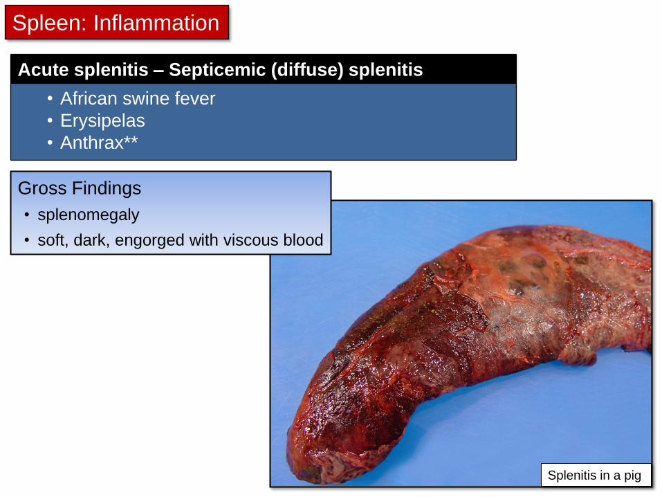
Acute splenitis – Septicemic (diffuse) splenitis
• African swine fever
• Erysipelas
• Anthrax**
Gross Findings
• splenomegaly
• soft, dark, engorged with viscous blood
Spleen: Inflammation
Splenitis in a pig

Anthrax
• zoonosis caused by a gram-positive, spore-forming bacillus: Bacillus anthracis
• in ruminants – septicemic disease
• in horses, pigs & dogs – pharyngeal and enteric disease
Acute splenitis – Septicemic (diffuse) splenitis

Anthrax – Pathogenesis in ruminants:
ingestion / wound contamination / inhalation of spores
lymphangitis and localized lymphadenitis
massive bacteremia (sepsis) and toxemia
increased vascular permeability and impaired coagulation
Sudden Death
• with sepsis, huge numbers of vegetative organisms in blood
• become spores when exposed to air:
• spores very resistant:
survive decades in soil
infections often follow soil excavation

Anthrax - Lesions in ruminants
healthierusonline.co
m
Characteristic Findings:
• bloated autolysed carcass with blood oozing from the orifices

• take a blood smear from the ear!
• methylene blue stain: see short chains of large bacilli with distinct pink capsule and square ends
Anthrax - Lesions in ruminants
Characteristic Findings:
• bloated autolysed carcass with blood oozing from the orifices
• Do Not Necropsy Suspect Cases!
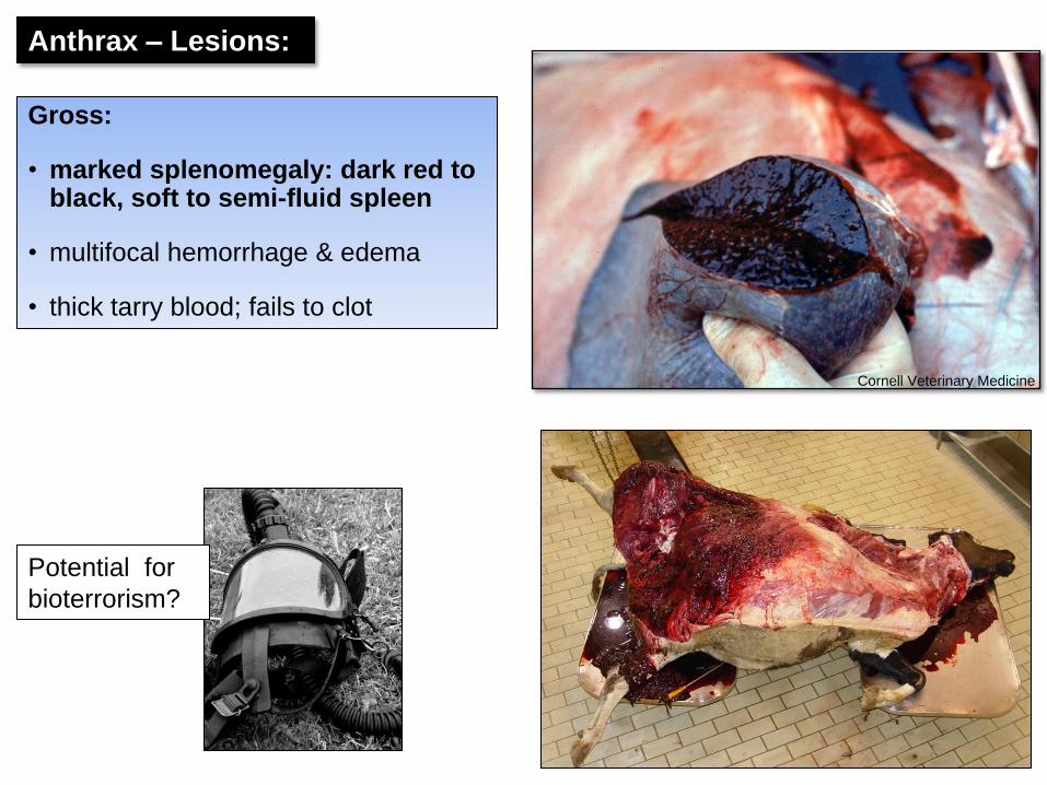
Potential for
bioterrorism?
Cornell Veterinary Medicine
Anthrax – Lesions:
Gross:
• marked splenomegaly: dark red to black, soft to semi-fluid spleen
• multifocal hemorrhage & edema
• thick tarry blood; fails to clot

Diffuse granulomatous splenitis:
• Histoplasmosis, Blastomycosis
Nodular granulomatous splenitis:
• avian & bovine tuberculosis
2. Chronic splenitis: Granulomatous splenitis
Mycobacterium avium
infection in a turkey
Histoplasmosis in a dog
Spleen: Inflammation
Fig. 13-50 Histoplasmosis, spleen, dog. A, There
is uniform splenomegaly and the surface of the
spleen is mottled from the diffuse granulomatous
infiltrate. B, Cross section of spleen. The red pulp
has been almost completely replaced by diffuse
noncaseous granulomatous inflammation.
The Ohio State University
Noah’s arkive

3. Chronic splenitis: Splenic abscesses
• rarely following sepsis with pyogenic bacteria (esp T. pyogenes)
Spleen: Inflammation

Spleen: Disturbances of Growth
Growth disturbances
Aplasia
Atrophy
Benign nodular hyperplasia
Lymphoid hyperplasia
Hyperplasia of the monocyte-
macrophage system
Extramedullary hematopoiesis

Importance rule-out neoplasia
Benign nodular hyperplasia
• common finding in old dogs
• usually incidental
• may predispose to splenic hematomas
Spleen: Disturbances of Growth
Fig. 13-59 (Zachary) Nodular hyperplasia, spleen, dog.
A, A hemispherical 4-cm diameter nodule is protruding from the capsular surface.
B, Cross section of the nodular mass showing intermixed red and white areas composed of red blood cells & proliferating leukocytes.
Gross: gray to red nodular mass(es), composed of lymphoid tissue and red pulp
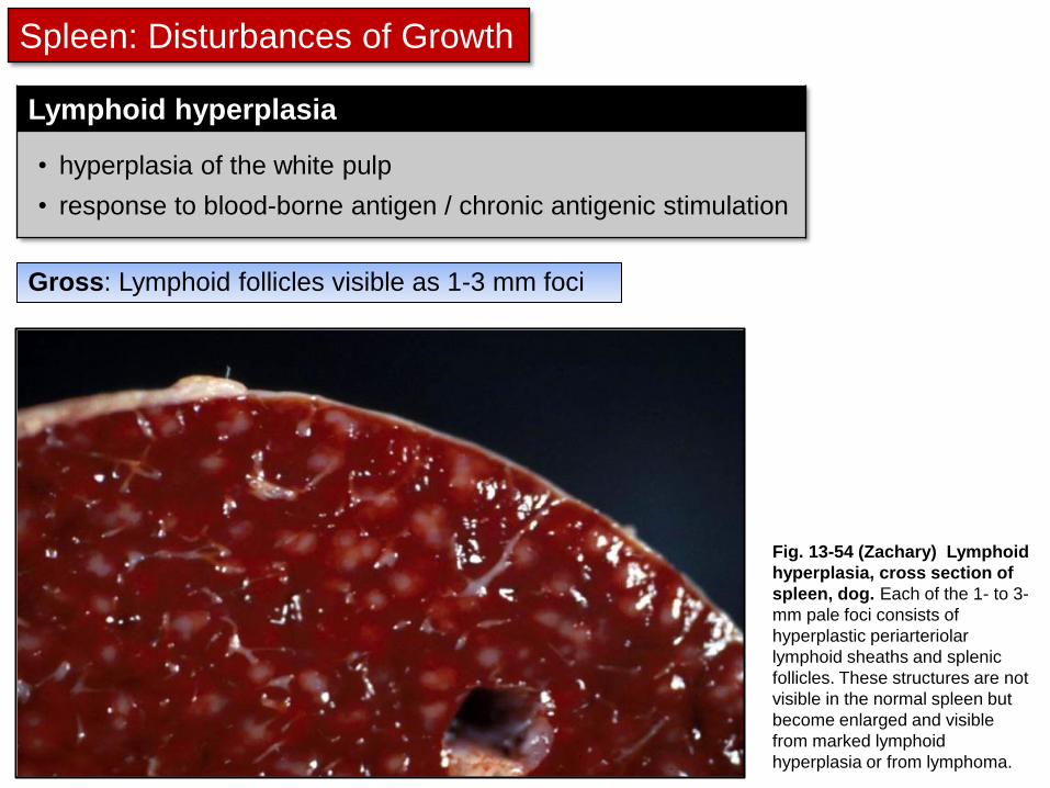
Lymphoid hyperplasia
• hyperplasia of the white pulp
• response to blood-borne antigen / chronic antigenic stimulation
Spleen: Disturbances of Growth
Fig. 13-54 (Zachary) Lymphoid
hyperplasia, cross section of
spleen, dog. Each of the 1- to 3-
mm pale foci consists of
hyperplastic periarteriolar
lymphoid sheaths and splenic
follicles. These structures are not
visible in the normal spleen but
become enlarged and visible
from marked lymphoid
hyperplasia or from lymphoma.
Gross: Lymphoid follicles visible as 1-3 mm foci

Histiocytic sarcoma, dog
Spleen: Neoplasia
* covered in primary hematopoietic neoplasia
Primary splenic neoplasia
Lymphoma/Leukemia*
Myeloproliferative diseases
(eg histiocytic sarcoma)*
Mastocytosis*
Hemangioma
Hemangiosarcoma
Others: Fibrosarcoma,
Fibrohistiocytic nodules, etc
Lymphoma / Leukemia, dog
Lymphoma, cat
Mastocytosis, cat
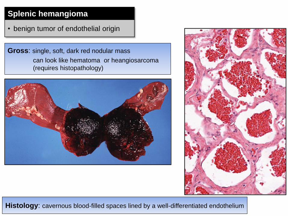
Splenic hemangioma
• benign tumor of endothelial origin
Gross: single, soft, dark red nodular mass
can look like hematoma or heangiosarcoma
(requires histopathology)
Histology: cavernous blood-filled spaces lined by a well-differentiated endothelium

Splenic hemangiosarcoma
• most common malignant tumour of the canine spleen
Gross:
• single to multiple, discrete to coalescing dark red masses
• +/- metastases
Dr E Aburto
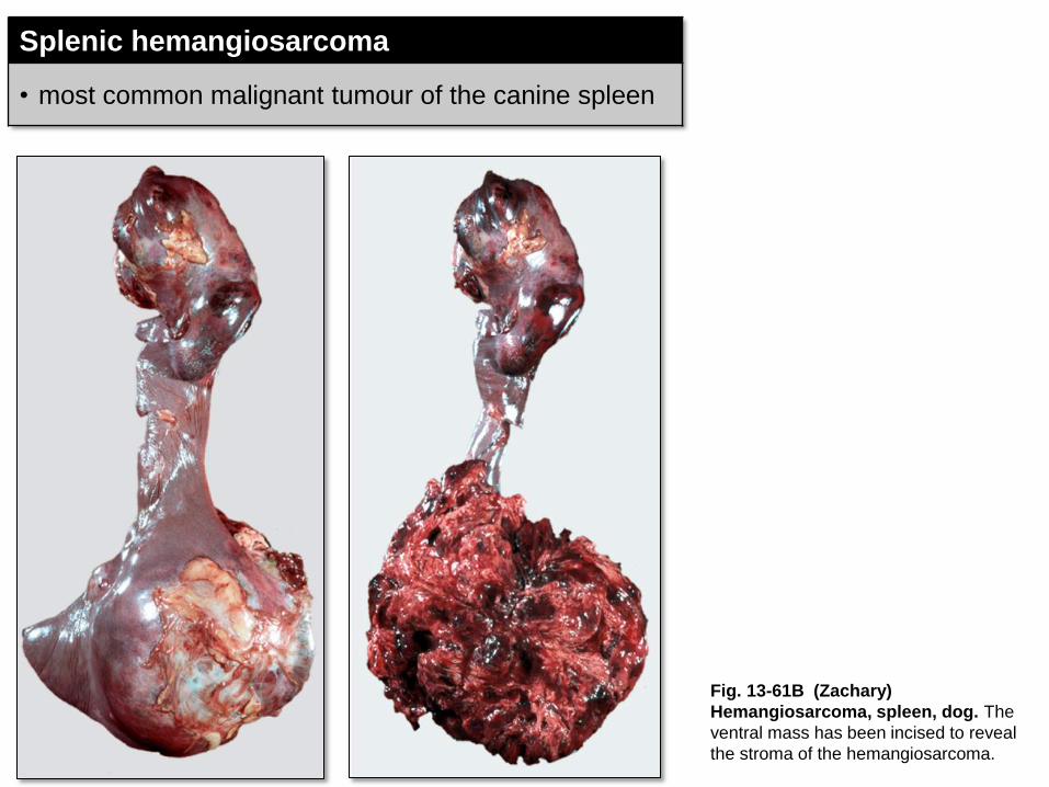
Splenic hemangiosarcoma
• most common malignant tumour of the canine spleen
Fig. 13-61B (Zachary)
Hemangiosarcoma, spleen, dog. The
ventral mass has been incised to reveal
the stroma of the hemangiosarcoma.

Splenic Hemangiosarcoma
Histology: blood-filled vascular spaces lined by anaplastic endothelial cells
Figure 13-57. Hemangiosarcoma, spleen, dog. Note the
haphazardly arranged vascular channels lined by anaplastic
endothelial cells.
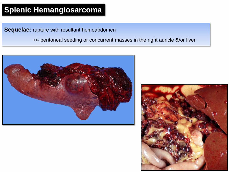
Sequelae: rupture with resultant hemoabdomen
+/- peritoneal seeding or concurrent masses in the right auricle &/or liver
Splenic Hemangiosarcoma

Spleen: Metastatic Neoplasia
Metastatic splenic tumours: dogs with
pancreatic carcinoma (top) and
mammary gland carcinoma (bottom) Dr E Aburto
• metastases to the spleen doesn’t occur as frequently as expected

Splenic Nodules – important differentials
Splenic nodules with a
bloody consistency
Hematoma
Hemangioma
Hemangiosarcoma
Splenic infarcts
Incompletely contracted areas
of the spleen
Splenic nodules with a
firm consistency
Nodular hyperplasia
Primary Neoplasia (eg lymphoma, histiocytic sarcoma)
Metastatic (secondary) Neoplasia
Abscess
Granuloma

Diffuse splenomegaly with a firm
consistency =
Meaty Spleens
Septicemia
• Salmonella
Hemolytic anemia
Neoplasia • eg lymphoma, mast cell tumor,
histiocytic sarcoma
Granulomatous disease
Amyloidosis
Diffuse Splenomegaly – important differentials
Diffuse splenomegaly with a
bloody consistency =
Bloody Spleens
Acute septicemia
Acute hemolytic anemia
Splenic torsion
Barbiturate anesthesia/euthanasia
Vascular pooling

• white to tan lobulated organ within the anterior mediastinum
• ruminants & pigs have a large cervical lobe that extends along the cervical trachea
Thymus: structure and function

Thymus: structure and function
Fig. 13-36 (Zachary) Schematic
illustration of the organization of
the thymus. The thymus consists of
several incomplete lobules. Each
lobule contains an independent outer
cortical region, but the central
medullary region is shared by
adjacent lobules. Trabeculae,
extensions of the capsule down to
the corticomedullary region, form the
boundary of each lobule. The cortex
consists of stromal cells and
developing T lymphocytes
(thymocytes), macrophages, and
cortical epithelial cells. Major
histocompatibility complex class I and
II molecules are present on the
surface of the cortical epithelial cells.
The characteristic deep blue nucleus
staining of the cortex in histological
preparation reflects the predominant
population of T lymphocytes as
compared with the less basophilic
medulla, which contains a lower
number of thymocytes. Hassall's
corpuscles are a characteristic
component of the medulla. Hassall's
corpuscles are not seen in the cortex.
Structure
• composed of epithelial tissue and lymphoid tissue
Function
• proliferation & maturation of T cells

Thymus – Misc. diseases
Lymphocytolysis / thymic atrophy
Thymic aplasia / hypoplasia
Thymic hemorrhage / hematoma
Can lead to acquired immunodeficiency
Congenital immunodeficiency
* Severe combined immunodeficiency (SCID): Foals, mice, dogs (jack Russell terriers & Basset hounds)
Both congential & acquired immunodeficiency make animals more susceptible to opportunistic pathogens & more severe infections
For Information only
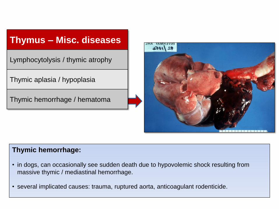
Thymic hemorrhage:
• in dogs, can occasionally see sudden death due to hypovolemic shock resulting from
massive thymic / mediastinal hemorrhage.
• several implicated causes: trauma, ruptured aorta, anticoagulant rodenticide.
Thymus – Misc. diseases
Lymphocytolysis / thymic atrophy
Thymic aplasia / hypoplasia
Thymic hemorrhage / hematoma

Thymus: Primary Neoplasia
Thymoma, goat. Note, it would require histology to differentiate this from a thymic lymphoma.

Thymus: Primary Neoplasia
Thymic (mediastinal) Lymphoma
• neoplastic proliferation of T-cells
• often younger animals
• malignant behavior
Thymoma
• neoplastic proliferation of thymic epithelial cells
• less common (dogs, sheep, goats)
• slow growing, encapsulated, rarely metastasize

Accessory lymphoid organs Lymphoma in the tonsils of a dog
Necrotizing tonsillitis in a pig
• tonsils
• mucosal-associated lymphoid tissue (MALT)
bronchial-associated lymphoid tissue (BALT)
peyer’s patches / GALT
• subjected to similar pathologic processes as the lymph node
