pathology of bronchial asthma dr. maha arafah. at the end of this lecture, the student should be...
TRANSCRIPT

RESPIRATORY BLOCK PATHOLOGY L1
Pathology of bronchial asthma
Dr. Maha Arafah

At the end of this lecture, the student should be capable of:
Understanding asthma as an episodic, reversible bronchoconstriction caused by increased responsiveness of the tracheobronchial tree to various stimuli.
Knowing that asthma is divided into two basic types: extrinsic or atopic allergic and intrinsic asthma.
Understanding the morphological changes seen in the lungs in cases of severe asthma
Listing clinical presentation in cases of asthma Listing the complications of asthma: superimposed
infection, chronic bronchitis and pulmonary emphysema Definition and manifestations of status asthmaticus

Introduction: Anatomy

Chronic Obstructive Pulmonary
Disease
Emphysema
Bronchiectasis
Chronic BronchitisAsthma
Chronic Obstructive Pulmonary Disease

Chronic obstructive pulmonary diseases
Bronchial asthma Chronic relapsing inflammatory disorder
characterized by: Hyperactive airways leading to episodic,
reversible bronchoconstriction Due to increased responsiveness of the
tracheobronchial tree to various stimuli.

bronchial asthma animation
http://link.brightcove.com/services/player/bcpid236059233?bctid=347806802

Primarily targets: the bronchi and terminal bronchioles
Most common chronic respiratory disease in children More common in children than adults Majority (50-80%) develop
symptoms before 5 years of age Two types

Extrinsic Asthma 70%
Initiated by type 1 hypersensivity reaction induced by exposure to extrinsic antigen.
Subtypes include:a. atopic (allergic) asthma.b. occupational asthma.c. allergic bronchopulmonary aspergillosis.
Personal or family history of allergic reaction
Develop early in life
Intrinsic Asthma 30% Initiated by diverse,
non-immune mechanisms, including ingestion of aspirin, pulmonary infections, cold, inhaled irritant, stress and exercise. No personal or family history of allergic reaction. Develop later in life
CLASSIFICATION OF ASTHMA

Extrinsic Asthma
Atopic (allergic) asthma is the most common form, begins in childhood
Other allergic manifestation: allergic rhinitis, urticaria, eczema.
Other family member is also affected Skin test with antigen result in an
immediate wheel and flare reaction

wheel and flare reaction

Pathogenesis of Bronchial Asthma
EXAGGERATED BROCHOCONTRICTION Two components:
1. Chronic airway inflammation.2. Bronchial hyperresponsiveness.
The mechanisms have been best studied in atopic asthma.

Pathogenesis of Atopic Asthma
A classic example of type 1 IgE-mediated hypersensitivity reaction.
In the airway – initial sensitization to antigen (allergen) with stimulation of TH2 type T cells and production of cytokines (IL-4, IL- 5, and IL-13).
Cytokines promote:1 .IgE production by B cell (IL4)
2 .Growth of mast cells (IgE)3 .Growth and activation of eosinophils (IL5)
4 .mucous secretion (IL13)

Extrinsic Asthma
Serum IgE and eosinophil are increased immune related, TH2 subset of CD4+ T
cells
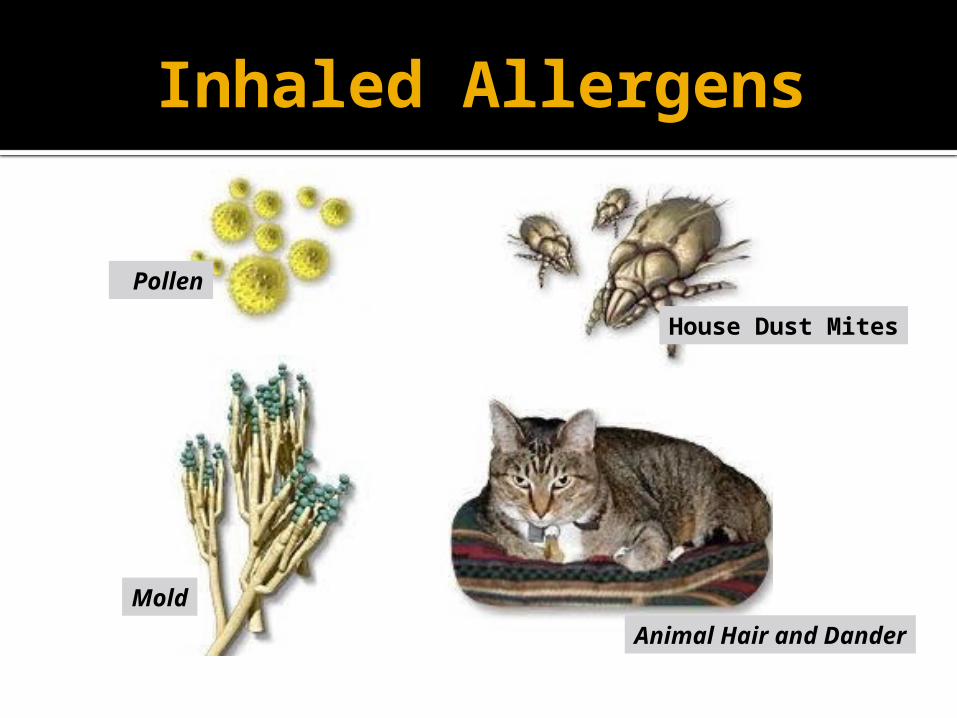
Inhaled Allergens
House Dust Mites
Mold
Pollen
Animal Hair and Dander

Pathogenesis of Atopic Asthma
• IgE-mediated reaction to inhaled allergens elicits: 1. acute response (within minutes) 2. a late phase reaction (after 4-8 hours)

Pathogenesis of Atopic AsthmaAcute-phase response
Begin 30 to 60 minutes after inhalation of antigen. Mast cells on the mucosal surface are activated. Mediator produced are :
Leukotrienes C4, D4 & E4 (induce bronchospasm, vascular permeability & mucous production)
Prostaglandins D2, E2, F2 (induce bronchospasm and vasodilatation) Histamine ( induce bronchospasm and increased vascular
permeability) Platelet-activating factor (cause aggregation of platelets and release
of histamine)
Mast cell tryptase (inactivate normal bronchodilator).
Mediators induce bronchospasm, vascular permeability & mucous production.

Pathogenesis of Atopic Asthma Late phase reaction
Recruitment of leukocytes mediated by product of mast cells including:
1. Eosinophil and neutophil chemotactic factors 2 . IL-4 & IL-5 and induceTH2 subset ofCD4+ T cells
3. Platelet-activating factor 4. Tumor necrosis factor. Other cell types are involved: activated
epithelial cells, macrophages and smooth muscle.

Pathogenesis of Atopic Asthma
Late phase reaction: The arrival of leukocytes at the site of mast cell
degranulation lead to: 1. Release of more mediators to activate more mast cells2. Cause epithelial cell damage
Eosinophils produce major basic protein, eosinophilic cationic protein and eosinophil peroxidase ( toxic to epithelial cells).
These amplify and sustains injury without additional antigen.


Summary of Pathogenesis
Type I hypersensitivity reaction with exposure to extrinsic allergens▪ Typically develops in children with an atopic family history
to allergies
(1) Initial sensitization to an inhaled allergen▪ (a) Stimulate induction of subset 2 helper T cells (CD4 TH2)
that release interleukin (IL) 4 and IL-5▪ (b) IL-4 stimulates isotype switching to IgE production.▪ (c) IL-5 stimulates production and activation of eosinophils.
(2) Inhaled antigens cross-link IgE antibodies on mast cells on mucosal surfaces.▪ (a) Release of histamine and other preformed mediators▪ (b) Functions of mediators:▪ Stimulate bronchoconstriction, mucus production, influx of
leukocytes

Summary of Pathogenesis
(3) Late phase reaction (4-8 hours later)▪ (a) Eotaxin is produced.▪ Chemotactic for eosinophils and activates eosinophils
▪ (b) Eosinophils release major basic protein and cationic protein.▪ Damage epithelial cells and produce airway constriction
Other mediators involved▪ (1) LTC-D-E4 causes prolonged bronchoconstriction.
▪ (2) Acetylcholine causes airway muscle contraction.


Non-Atopic Asthma
Non immune Positive family history is uncommon. Serum IgE – normal. No other associated allergies. Skin test – negative. Hyperirritability of bronchial tree (Stress, exercise,
cigarette smoke) Triggered by respiratory tract infection including
viruses (Examples-rhinovirus, parainfluenza virus, respiratory syncytial virus)
inhaled air pollutants (e.g. sulfur dioxide, ozone) Subtypes:
1. Drug-induced asthma (Aspirin or nonsteroidal drug sensitivity)
2. Occupational asthma ( fumes, dusts, gases)

Morphology of Asthma
Grossly lung over distended (over
inflation) occlusion of bronchi and
bronchioles by thick mucous.

Hyperinflated lung

Morphology of Asthma
Histologic finding: Thick BM. Edema and inflammatory
infiltrate in bronchial wall. Submucosal glands
increased. Hypertrophy of the
bronchial wall muscle.
Mucous contain Curschmann spirals, eosinophil and Charcot-Leyden crystals.
AIRWAY REMODELING

Curschmann spirals
Coiled, basophilic plugs of mucus formed in the lower airways and found in sputum and tracheal washings

Charcot-Leyden crystals.
Eosinophilic needle-shaped crystalline structures from eosinophil proteins

Clinical findings
(1) Episodic expiratory wheezing (inspiratory as well when severe)
(2) Nocturnal cough (3) Increased anteroposterior diameter▪ Due to air trapping and increase in residual
volume
http://www.youtube.com/watch?v=YG0-ukhU1xE&feature=related

Clinical Coarse
Classic asthmatic attack – dyspnea, cough, difficult expiration, progressive hyperinflation of lung and mucous plug in bronchi. This may resolve spontaneously or with Rx
May progress to emphysema or chronic bronchitis
Superimposed bacterial infection may occur

Laboratory findings (1) Initially develop respiratory alkalosis▪ (a) Patients work hard at expelling air through
inflamed airways.▪ (b) May progress into respiratory acidosis if
bronchospasm is not relieved▪ Normal pH or respiratory acidosis is an indication for
intubation and mechanical ventilation.
(2) Eosinophilia, positive skin tests for allergens

Status asthmaticus
Severe cyanosis and persistent dyspnea for days and weeks
Does not respond to therapy Hypercapnia, acidosis, sever hypoxia May be fatal