pathological diagnosis on major pig … · pathological diagnosis on major pig diseases in taiwan...
TRANSCRIPT

Pathological Diagnosis on Major Pig Diseases in Taiwan
103
PATHOLOGICAL DIAGNOSIS ON MAJOR PIG DISEASES IN TAIWAN
Shu-Hwae Lee Animal Drugs Inspection Branch, Animal Health Research Institute,
Council of Agriculture, Taiwan
e-mail: [email protected]
ABSTRACT
The diagnosis reports of pig cases in the Animal Disease Diagnostic Center, Animal Health Research
Institute (AHRI) from1987 to 2012 in Taiwan were analyzed by means of the pathological,
microbiological and epidemiological methods. Results revealed that there were no variations of lesions
in the cases submitted before 1993.However, diseases with respiratory symptoms became dominant in
pig diseases after 1993 because of the invasion of PRRS. In 2000, the invasion of PCV2 and Porcine
teschovirus (PTV) has led to marked increase of respiratory disorders. This phenomenon has
continually presented till now. Results of further analysis of the causative agents of respiratory
diseases are Porcine Reproductive and Respiratory Syndrome (PRRS), Porcine circovirus type 2
(PCV2), and the Porcine Respiratory Disease Complex (PRDC), etc. We also discovered
Pleuropneumonia (Actinobacillus pleuropneumoniae; APP), and Salmonella choleraesuis was
prominent before 1993. The major causative agents of respiratory tract infection were PRRS, PCV2 and
PRDC. We therefore concluded that the profile of Taiwan’s swine disease has shifted from mono -
pathogen to multiple- pathogenic pattern, especially respiratory symptoms. The strategy of prevention
and control of pig diseases should be strengthened based on the biosecurity of pig farm. Correct
immune programs and their practice to increase immunity of pigs and reduce pathogen concentration
in the animal and environment are the vital measures for disease control in pigs
Keywords: Pathology, diagnosis, Pig, Disease Control
INTRODUCTION
Statistics data on the production and number of dead pigs in Taiwan were presented in the Agricultural Statistics
Annual Report 2012, Council of Agriculture. Mortality rate of pigs increased from 6.3% in 1990 to 18.52% in
2012. It is about 2.93 times increasing. Analysis of the causes of the mortality has revealed that the instruction
and spread of PRRS at the end of 1992 to early 1993 has induced genotype changes of classic swine fever virus.
This study was conducted by AHRI. In 1997, the outbreak of FMD type O has led the Taiwan government to
cull more than 400 million pigs. At the time, the mortality rate was only 16.20%. However, following the
detection of porcine teschovirus (PTV) and PCV2 in pigs in 2000, Taiwan's swine diseases have changed from
single pathogen to multiple complex diseases resulting in significant increase of mortality.
Currently, Taiwan's pig production industry is facing many challenges-- the first one is the multiple pathogenic
infections consisting of PRRS, PCV2 and other viruses infection accompanied with secondary pathogens
infection. This type of disease is very difficult to diagnose and prevent. The second is severe mycotoxin
contamination in feeds. The third is chronic swine fever problems. Finally, due to climate change the effects of
which are already being felt today and in years to follow, the changes in climate and its intrinsic anomalies
brought about by weather extremes-- too hot or too cold and high humidity in Taiwan have led the negative
pressure. The Fan and Pad Cooling System is unable to control suitable temperature and humidity in the
animals’ houses. This environmental instability has induced severe complex respiratory diseases in the pig
farms.
Session 3: Swine Health Management
104
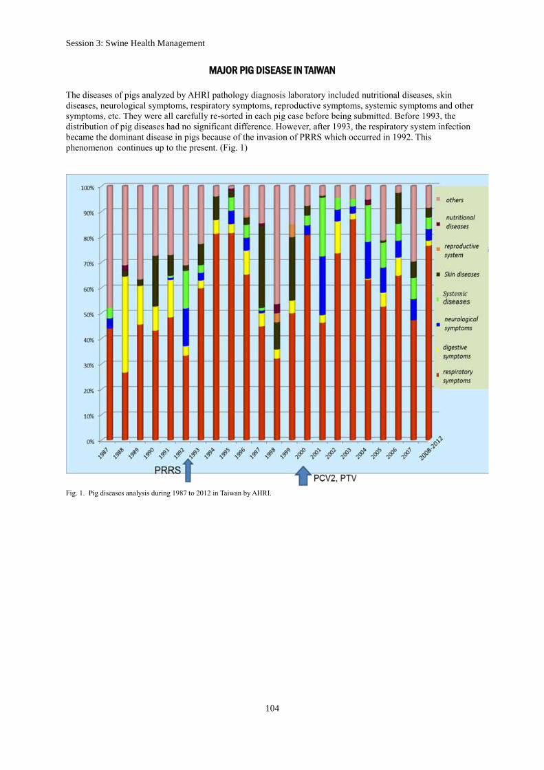
MAJOR PIG DISEASE IN TAIWAN
The diseases of pigs analyzed by AHRI pathology diagnosis laboratory included nutritional diseases, skin
diseases, neurological symptoms, respiratory symptoms, reproductive symptoms, systemic symptoms and other
symptoms, etc. They were all carefully re-sorted in each pig case before being submitted. Before 1993, the
distribution of pig diseases had no significant difference. However, after 1993, the respiratory system infection
became the dominant disease in pigs because of the invasion of PRRS which occurred in 1992. This
phenomenon continues up to the present. (Fig. 1)
Fig. 1. Pig diseases analysis during 1987 to 2012 in Taiwan by AHRI.
Pathological Diagnosis on Major Pig Diseases in Taiwan
105
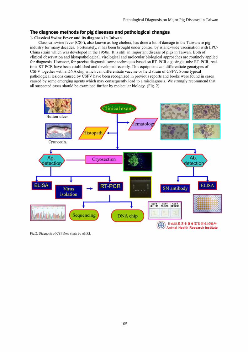
The diagnose methods for pig diseases and pathological changes 1. Classical Swine Fever and its diagnosis in Taiwan
Classical swine fever (CSF), also known as hog cholera, has done a lot of damage to the Taiwanese pig
industry for many decades. Fortunately, it has been brought under control by island-wide vaccination with LPC-
China strain which was developed in the 1950s. It is still an important disease of pigs in Taiwan. Both of
clinical observation and histopathological, virological and molecular biological approaches are routinely applied
for diagnosis. However, for precise diagnosis, some techniques based on RT-PCR e.g. single-tube RT-PCR, real-
time RT-PCR have been established and developed recently. This equipment can differentiate genotypes of
CSFV together with a DNA chip which can differentiate vaccine or field strain of CSFV. Some typical
pathological lesions caused by CSFV have been recognized in previous reports and books were found in cases
caused by some emerging agents which may consequently lead to a misdiagnosis. We strongly recommend that
all suspected cases should be examined further by molecular biology. (Fig. 2)
Fig.2. Diagnosis of CSF flow chats by AHRI.

Session 3: Swine Health Management
106
When CSF virus appears in a herd, it frequently shows clinical signs of disease. Infected pigs show high fever
(higher than 41℃) and dropsy, they become less active and display loss of appetite. The leukocyte count
decreases along with the increase in body temperature. Normally, leucopenia is a very good indicator of CSF
(Lee et al., 1997). The clinical signs of CSFV infected pigs include high fever, dullness, weakness, drowsiness,
tendency to huddle, anorexia, and constipation followed by diarrhea. (Fig. 3)
Fig.3. CSF virus infected pigs showed clinical signs which include a high fever, dullness, weakness, drowsiness, tendency to huddle,
anorexia, and constipation followed by diarrhea.
CSF virus infected pigs showed purplish discoloration of abdomen, inner thighs and ears. In the past, spleen
infarct is a typical lesion of CSF. But now, many other emerging diseases such as PCV2, Septicemic
Salmonellosis and teschovirus infection may also develop spleen infracts in the pigs. Therefore, differential
diagnosis becomes important to confirm the CSF in the field. (Fig. 4)
Fig. 4. (A) Acute infected CSF virus pig showed purplish discoloration of abdomen, inner thighs and ears. (B) Autopsy has shown typical
infarcts on the edge of spleen. (C) Button ulcers on the mucosa of colon. (D) Enlarged lymph nodes with peripheral hemorrhage in the pigs infected by virulent virus.

Pathological Diagnosis on Major Pig Diseases in Taiwan
107
Histopathological finding in classical swine fever Lesions include parenchymatous degeneration of lymphatic
tissue, cellular proliferation of vascular interstitial tissue, and a nonsuppurative meningoencephalomyelitis with
vascular cuffing. (Fig. 5)
Fig. 5. (A) Non-suppurative menigoencephalitis: vasculitis, lymphocytic cuffing, endothelium hyperplasia and gliosis in the cerebrum of the pig infected with CSFV. (B) Localized infarction, follicle necrosis, hemorrhage, and fibrinoid degeneration of the blood vessel in the
spleen were seen in the pig infected with CSFV. (C)Dilation congestion, lymphocyte depletion were seen in the lymph node of the pig
infected CSFV. (D) Endothelial cell hyperplasia in the central artery, lymphocyte depletion in the peripheral area of the lymphoid follicle was noted in the pig infected with CSFV.

Session 3: Swine Health Management
108
2. Foot-Mouth-Disease and its diagnosis in Taiwan
The 1997 FMD outbreak in Taiwan forced the destruction of four million pigs. The costs for pollution control
and losses in the domestic market in the pig industry in the year 1997 were estimated at US$378.6 million
(Yang et al., 1999), and bans on the annual export to Japan of 6 million pig carcasses resulted in approximately
US$1.57 billion loss following the outbreak (Huang et al., 2000).
Gross findings in infected pigs
Skin lesions developed as early as 1 DPI in pigs were examined. Three vesicles, 1–1.5 cm in diameter, were
found at the inoculation site in one pig. The vesicles contained thin and straw-colored fluid. Small erosion on
the front-right coronary band and swelling of hind-right metatarsals joints were also noticed. The other pig had a
1 cm diameter vesicle in the surface of metacarpals (Fig. 6 A). Erosion of supernumerary hoof was observed by
2 DPI, and a large 3–5 cm diameter vesicle was found on the snout of one pig (Fig. 6 B). Vesicles were found
over front-right carpal joints, the snout and tongue. Additional lesions consisted of round foci of hyperaemia in
the skin over the front-right metacarpals joints and haemorrhagic bands along the coronary bands of all feet (Fig.
6 C). By 3 DPI, severe and prominent lesions were noted in the infected pigs and included rupture and
haemorrhage of the vesicles at the inoculation sites, multiple vesicles on the skin over metatarsal and carpal
joints, and haemorrhagic lesions on the coronary band of all feet (Figs 6 D and E). Vesicles ranging from 0.1 to
2 cm in diameter were found on the snout, nares, mucosa of the oral cavity and tongue. Healing of most of the
ruptured vesicles in front metacarpals and carpal joints, supernumerary hoofs, and tongue were noted by 4 DPI
(Fig. 6 F). The ruptured vesicles in the inoculated area became yellow-green with buckish smell. However,
intact vesicles ranging from 0.1 to 0.5 cm in diameter were found in snout, nares, tongue and mucosa of the oral
cavity. Discoloration along the coronary band was noted. Different healing processes were noted in the coronary
bands, heel bulbs and tongue by 6 DPI (Fig. 6 G). Healing in snout, mouth, coronary bands of all hooves was
completed by 9 DPI, but the heel bulbs had necrosis underneath. The re-growth of epithelium replaced all the
lesions in the coronary bands by 21 DPI. However, scar formation was found in the skin over the carpal joint,
and a cyst with a rough inner surface was noted in the capsular ligament of the hock joint in one pig.
Remarkable demarcation in the coronary band was also noted. By 36 DPI, tissue re-growth was noted at the
coronary bands, and the old corium of the hooves was pushed downward (Fig. 6 H). New hoof formation was
noticed at 21 DPI in all the remaining pigs, and was almost completed by 63 DPI, with only tiny pieces of old
hooves left. By the end of the experiment at 63 DPI, only about 10% of old hoof remained. Striations on the
heart were noted by 63 DPI. No other gross lesion was found in other organs in any of the experimental pigs
(Lee et al., 2009).
Fig. 6. The gross findings of the pigs infected experimentally with O/Taiwan/97 strains of foot-and-mouth disease virus. (A)Vesicles of 1 cm
in diameter were developed on the surface of metatarsus by 24 HPI (hours post-infection). (B) A 3 cm in diameter transparent vesicle on the
snout by 3 days post-inoculation (DPI).
(A) (B)

Pathological Diagnosis on Major Pig Diseases in Taiwan
109
Fig. 6. The gross findings of the pigs infected experimentally with O/Taiwan/97 strains of foot-and-mouth disease virus. (C) Petechiae
hemorrhagic presented on the surface of coronary band by 2 DPI. (D) Diffuse hemorrhagic areas were noted on the cross-sections of coronary band by 2 DPI. (E) Swelling of skin above coronary accompanied with linear hemorrhage appeared by 3 DPI. (F) Vesicles ranged
from 0.1 to 0.6 cm in diameter were found on the surface of the tongue by 4 DPI. (G) Large area of erosion on the surface of tongue was
note by 6 DPI. (H) As the disease progress, new-growth hoof has displaced the ruined ones by 36 DPI.
(F) (E)
(G) (H)
(C) (D)

Session 3: Swine Health Management
110
Histopathologic changes in infected pigs
The lesions present in the stratum granulosum of skin showed ballooning degeneration and epithelial necrosis
and a few infiltrating neutrophils accompanied by intercellular oedema (Fig. 7 A). The accumulation of
oedematous fluid that formed bulla of variable sizes was noted in the stratum spinosum from 2 DPI to 6 DPI
(Fig. 7 B). The underlying dermis had focal necrosis, few neutrophilic infiltration and lymphocytic vasculitis
(Figs 7 C and D). The underlying dermis had locally extensive necrosis, with neutrophils, macrophages and
lymphocytes infiltration accompanied by moderate to severe perivascular lymphocytic vasculitis. Regeneration
of epithelia began at 6–8 DPI with infiltration of macrophages, lymphocytes and fibroblasts into the necrotic
areas.
(A) (B)
(C) (D) Fig.7. Histopathological changes in the epithelium of the pigs infected experimentally with O/Taiwan/97 strain of foot-and-mouth disease
virus. Hematoxylin and Eosin staining.
(A) Oedematous fluid containing bits of fibrin accumulates between and separates the cells (arrow) in sratum spinosum accompanied with balloon degeneration of cells in stratum spinosum of the epithelium. Intercellular prickles are lost and the cells become round and detached
from each other. X 40. (B) The small vesicles coalesced to form bullae and cause large areas of epithelium to be detached and shed.
Liquefaction necrosis characterized by an accumulation of serum and leukocytes in the vesicles roofed over by the compressed stratum corneum , lucidum and granulosum, and extending downward to basal layer. X 40. (C) Perivascular lymphocytic vasculitis at dermis (arrow)
at 8 HPI. X 100. (D) Hypertrophy epithelium and hydropic degeneration (arrow). X 400.

Pathological Diagnosis on Major Pig Diseases in Taiwan
111
3. The Pathological Findings of Porcine Reproductive and Respiratory Syndrome in Taiwan
The reports of pig disease diagnosis from Animal Disease Diagnostic Center at AHRI in 2011 revealed that the
detection rates of PRRS-alone infection was 13% (24/191), PCV2-alone infection was 11% (21/191) and PCV2
and PRRSV dual infection was 49% (93/191). The cases of PRRS have been increased obviously in the last two
years and PRRS was listed in the group of important pig diseases that affected the porcine industry. The
pathological examination of the diseased sow showed nonsuppurative meningoencephalitis, myocarditis,
interstitial pneumonia, interstitial nephritis and mucosal necrosis of the uterus. The prevalence of sow abortion
was about 10% and the mortality was 15%. In addition, the nursery pigs showed the clinical signs of weak,
thinness, rough hair coat and respiratory syndrome. The prevalence of this epidemic in nursery pigs was about
50% and the survival rate decreased from 90% to 60% after the outbreak. (Fig. 8)
Fig. 8. (A) This is an early stage lesion of PRRS infection of nursery pigs. The cardiac and diaphragmatic lobes have mild inflammatory lesions. The lesions must be differentiated from the SEP infection of pig. (B) SEP infected pig autopsy has shown typical lung lesions
confined to apical and cardiac lobes, and usually it will not attack the diaphragmatic lobe. (C) Histopathological changes in the lymph node
of the pigs infected with PCV2. H&E staining. Lymphcytes depletion of lymph node is very prominent. (D) Showed typical basophilic inclusion bodies of lymph node.
4. The Pathological Findings of PCV2 infection in pigs in Taiwan
Porcine circovirus type 2 associated diseases (PCVAD) encompasses a variety of disease syndromes in pigs
including: postweaning mulitsystemic wasting syndrome (PMWS), porcine dermatitis and nephropathy
syndrome (PDNS), enteritis, pneumonia, reproductive failure, and neuropathy. PCV2 is the major causative
agent of postweaning multisystemic wasting syndrome (PMWS) in Taiwanese pig farms. (Avanti Sinha. 2011;
Wang et al; 2013).

Session 3: Swine Health Management
112
Postweaning multisytemic wasting syndrome (PMWS) Histopathological lesions associated with PMWS consist of marked lymphocyte depletion of follicles in
lymphoid tissues. Multifocal to diffuse histiocytic infiltration is observed and numerous basophilic cytoplasmic
inclusions detected in lymphoid tissues.
Porcine dermatitis and nephropathy syndrome (PDNS)
Clinical signs of PDNS include the presence of cutaneous pupura on the skin. The kidneys showed enlarged and
petechial hemorrhages on the surface. Acute necrotizing vasculitis affecting the dermis and kidneys. (Avanti
Sinha, 2011; Wang et al., 2013)
5. Co-infection of PRRSV and PCV2 in Taiwan
PRRSV and PCV2 are the major causes of porcine respiratory disease complex (PRDC) and lead high mortality
in nursery to finish pigs. More than 80% PRDC cases were caused by Co-infection of PRRSV and PCV2. The
PRDC are also the major problem in pig herds of Taiwan.(Figure 9)
AB
C D Fig. 9. (A)(B). Co-infection of PRRSV and PCV2 pigs showed variable body sizes and rough hair coat. (C) Autopsy has shown prominent
lung pneumonia, wet and flushing on the surface and the whole is heavy than normal. (D) Shown the fever of pig and peritonitis on the
surface of visceral organs is noted.

Pathological Diagnosis on Major Pig Diseases in Taiwan
113
CONCLUSION
The microorganisms that cause pig diseases in Taiwan have shifted from mono-pathogenic to multi-
pathogenic pattern. Strategy of prevention and control of pig diseases should first be started from management
of pigs, houses and their environment. Biosecurity of pig farms should be carefully designed. Correct immune
programs and their practice to increase immunity of pigs and reduce pathogen concentration in the animal and
environment are the vital measures for disease control. Monitoring platform for pig health was created and
clinical pathology, microbiology and molecular biology were performed in the diagnosis and control of
pig diseases.
REFERENCES
Lee S.H., Jong MH.,Lin YL.,LJ. Ting, Yang SC. 1997. Histopathologic Studies on the Pig Experimentally
Infected by Wild Strain of Hog Cholera Virus . Exp.Rep. TAHRI. 33:13-28.
Lee S.H., Jong M.H., Huang T.S., Lin Y.L., Wong M.L., Liu C.I., and Chang T.J., 2009. Pathology and viral
distributions of the porcinophilic foot-and-mouth disease virus strain (O/Taiwan/97) in experimentally
infected pigs. Transbound. Emerg. Dis. 56, 189-201.
Wang C., Pang VF., Lee F., Huang TS., Lee SH., Line Y.J., Line YL., Lai SS., Jeng CR. 2013. Prevalence and
genetic variation of porcine circovirus type 2 in Taiwan from 2001 to 2011. Research in Veterinary Science.
94 (3) :789-795.
Avanti Sinha. 2011. Interactions of porcine circovirus type 2 and porcine reproductive and respiratory syndrome
virus. Graduate these and dissertation. Iowa State University Digital Repository @ Iowa State University.