pathological alterations in the gastrointestinal tract of
TRANSCRIPT

Pathological Alterations in the Gastrointestinal Tractof a Porcine Model of DMDXiaodong Zhou
Jilin UniversityHongsheng Ouyang
Jilin UniversityDaxin Pang
Jilin UniversityRenzhi Han ( [email protected] )
Ohio State University Wexner Medical Center Department of Surgery https://orcid.org/0000-0002-8202-9186Xiaochun Tang
Jilin University
Research
Keywords: CRISPR, Duchenne muscular dystrophy, genome editing, gastrointestinal tract, pig, porcine,swine
Posted Date: March 8th, 2021
DOI: https://doi.org/10.21203/rs.3.rs-267849/v1
License: This work is licensed under a Creative Commons Attribution 4.0 International License. Read Full License
Version of Record: A version of this preprint was published at Cell & Bioscience on July 15th, 2021. Seethe published version at https://doi.org/10.1186/s13578-021-00647-9.

Pathological alterations in the gastrointestinal tract of a porcine model of DMD 1
Xiaodong Zou1, Hongsheng Ouyang1, Daxin Pang1, Renzhi Han2,*, Xiaochun Tang1,* 2
3
1Jilin Provincial Key Laboratory of Animal Embryo Engineering, College of Animal 4
Sciences, Jilin University, Changchun, Jilin Province, People’s Republic of China 5
2Department of Surgery, Davis Heart and Lung Research Institute, Biomedical 6
Sciences Graduate Program, Biophysics Graduate Program, The Ohio State 7
University Wexner Medical Center, Columbus, OH 43210 8
9
10 *Address correspondence to: 11 Renzhi Han, Ph.D. 12 Department of Surgery 13 Davis Heart and Lung Research Institute 14 The Ohio State University Wexner Medical Center 15 Columbus, OH 43210 16 Phone: (614) 685-9214 17 E-mail: [email protected] 18 19 Or 20 21 Xiaochun Tang, Ph.D. 22 Jilin Provincial Key Laboratory of Animal Embryo Engineering 23 College of Animal Sciences 24 Jilin University 25 Changchun, Jilin Province 26 People’s Republic of China 27 E-mail: [email protected] 28 29 30

Abstracts 31
Patients with Duchenne muscular dystrophy (DMD) develop severe skeletal and 32
cardiac muscle pathologies, which result in premature death. Therefore, the current 33
therapeutic efforts are mainly targeted to correct dystrophin expression in skeletal 34
muscle and heart. However, it was reported that DMD patients may also exhibit 35
gastrointestinal and nutritional problems. How the pathological alterations in 36
gastrointestinal tissues may contribute to the disease are not fully explored. Here we 37
employed the CRISPR/Cas9 system combined with somatic nuclear transfer 38
technology (SCNT) to establish a porcine model of DMD and explored their 39
pathological alterations. We found that genetic disruption of dystrophin expression led 40
to morphological gastrointestinal tract alterations, weakened the gastrointestinal tract 41
digestion and absorption capacity, and eventually led to malnutrition and gastric 42
dysfunction in the DMD pigs. This work provides important insights into the 43
pathogenesis of DMD and highlights the need to consider the gastrointestinal 44
dysfunction as an additional therapeutic target for DMD patients. 45
46
Keywords: CRISPR; Duchenne muscular dystrophy; genome editing; 47
gastrointestinal tract; pig; porcine; swine 48
49
50

Declarations 51
Funding 52
This work was supported by grants from the National Key Research and Development 53
Program of China Stem Cell and Translational Research (2019YFA0110702), National 54
Natural Science Foundation of China (No. 31572345), the Program for JLU Science 55
and Technology Innovative Research Team (2017TD-28). R.H. is supported by the 56
National Heart, Lung and Blood Institute grant [HL116546]; and the Parent Project 57
Muscular Dystrophy award. 58
59
Conflicts of interest 60
The authors declare that they have no conflict of interests. 61
62
Ethics approval 63
All animal studies were approved by the Animal Welfare and Research Ethics 64
Committee at Jilin University, and all procedures were conducted strictly in accordance 65
with the Guide for the Care and Use of Laboratory Animals. All surgeries were 66
performed under anesthesia, and every effort was made to minimize pain and stress 67
(SY201901013). 68
69
Consent to participate 70
Not applicable. 71
72
Consent for publication 73

All co-authors have reviewed and approved of the manuscript prior to submission. The 74
manuscript has been submitted solely to this journal and is not published, in press, or 75
submitted elsewhere. 76
77
Availability of data and material 78
All relevant data supporting the key findings of this study are available within the article 79
and its Supplementary Information files or from the corresponding author upon 80
reasonable request. 81
82
Code availability 83
Not applicable. 84
85
Authors’ contributions 86
R.H., X.Z., H.O. and X.T. conceived the study and wrote the manuscript. X.Z. and X.T. 87
performed the experiments, analyzed the data and drafted the manuscript. 88
89
90
91

Introduction 92
Duchenne muscular dystrophy (DMD), inherited in an X-linked recessive manner, is a 93
severe and progressive neuromuscular disorder1, and its prevalence in the general 94
population is approximately 1/50002, 3. Progressive muscle injury and degeneration in 95
DMD patients leads to muscular weakness, loss of ambulation, respiratory impairment, 96
and cardiomyopathy4. Most patients become wheelchair-bound in childhood and 97
heavily depend on their caregivers for their daily life5. Although the clinical and 98
pathological progression of skeletal muscle and myocardium involvement can be 99
variable, death usually occurs around the age of 30 years due to cardiac and/or 100
respiratory failure6. 101
102
DMD is caused by mutations in the DMD gene located at Xp21, which codes for the 103
dystrophin protein7, a cytoskeletal protein that functions in the muscle force 104
transmission and sarcolemmal stability of muscle fibers8. The DMD gene is one of the 105
largest genes in the human genome, containing 79 exons spanning 2.5 Mb of the 106
genomic DNA9, 10. Most of the mutations are due to deletions or duplications with point 107
mutations accounting for less than 10% of the cases11, 12. “Hot spots” for these 108
mutations are located in the regions of exons 3-7 and exons 45-5511. DMD exon 51 109
has been the most studied target in both preclinical and clinical settings, and the 110
availability of standardized procedures to quantify exon skipping would be beneficial 111
for the evaluation of preclinical and clinical data13. 112
113

With the advancing pathological progress of DMD, patients often suffer from 114
gastrointestinal or nutritional complications, evidenced by mandibular contracture, 115
swallowing impairment, dietary or nutrient imbalance, fluid imbalance, low bone 116
density and weight gain or loss14. In the mdx mouse, the most commonly used animal 117
model of DMD, the expression of neuronal nitric oxide synthase (nNOS) in colonic 118
smooth muscle cells is decreased, the contractile ability of the colon is weakened, the 119
transport speed of the small intestine is delayed, and fecal excretion is reduced15. In 120
addition, mdx mice have a higher concentration of collagen fibers in the submucosal 121
region of the gastrointestinal tract than wild-type (WT) mice. These alterations are 122
presumably consequences of the loss of dystrophin protein15-17. Therefore, 123
understanding the functional and morphological alterations of the gastrointestinal tract 124
in DMD could have important implications for therapeutic interventions. However, the 125
overall disease alterations in mdx mice are much less intense than those observed in 126
DMD patients. For example, mdx mice exhibit only a slightly shortened life span and 127
no apparent clinical signs of muscular dystrophy18, 19. 128
129
Like humans, pigs are true omnivores, and their balanced nutritional requirements are 130
particularly similar to the dietary requirements of humans20. Of the available animal 131
models, pigs exhibit the closest time required for intestinal transformation and 132
digestive efficiency to those in humans21. Pigs are, in many ways, attractive models 133
that closely mimic human physiology and pathology22. Therefore, in this study, we 134
used the CRISPR/Cas9 technology to create a porcine model of DMD by targeting 135

DMD exon 51 and reported the pathological alterations in the gastrointestinal tract of 136
these animals. 137
138
Results 139
Design of the DMD gene targeting strategy 140
The dystrophin protein shares a high homology among species. Amino acid sequence 141
alignment results revealed that the porcine dystrophin protein is highly homologous to 142
the human dystrophin protein with 94.16% homology (Suppl. Fig. 1A). We designed 143
a sgRNA targeting pig DMD exon 51 (Suppl. Fig. 1B). The on-target editing efficiency 144
of this sgRNA was assessed by Sanger sequencing of the target site PCR amplicon 145
following electroporation into porcine fetal fibroblasts (PFFs). Multipeaks around the 146
Cas9 cleavage site were observed, indicating that this sgRNA is effective in targeting 147
DMD exon 51 (Suppl. Fig. 1C). To assess the off-target activity, we selected the top 148
eight potential off-target sites for this sgRNA identified by in silico analysis, and 149
sequenced the PCR amplicons for all these 8 sites. No overlapping peaks were found 150
in the sequencing traces, suggesting that the editing activities at these potential off-151
target sites remain undetectable for the sensitivity of this assay (Suppl. Fig. 1D, Table 152
1 and 2). 153
154
PFFs with engineered mutations in DMD exon 51 showed impaired cell 155
membrane integrity and early apoptosis 156
PFFs with DMD exon 51 disruption following electroporation were chosen as the donor 157

cells for somatic nuclear transfer. A total of 400 individual PFF cell clones were 158
analyzed and 113 (27.5%) were found to carry seven different types of mutations (Fig. 159
1A,Suppl. Table 3). All mutation types of PFFs showed abnormalities in the mRNA 160
and protein levels of the DMD gene (Suppl. Fig. 2A, B). 161
162
Previous research showed that dystrophin protein functions to stabilize the integrity of 163
the muscle fiber membrane. To examine whether the mutations in PFFs induced by 164
the CRISPR/Cas9 system disrupt cell membrane integrity, we performed neutral red 165
dye (NRD) uptake and lactate dehydrogenase (LDH) release assays. As shown in Fig. 166
1B and Suppl. 2C, the NRD uptake of PFFs with mutations in DMD exon 51 was 167
significantly lower than that of PFFs from the control group, suggesting that the DMD 168
mutant PFFs have lower viability. Consistent with the results of the NRD uptake assay, 169
the LDH activity was remarkably elevated in the culture medium of PFFs with DMD 170
exon 51 mutations compared to that in the culture medium of control PFFs (Fig. 1C, 171
Suppl. 2D). Increasing evidence has shown that apoptosis and autophagy may play 172
important roles in DMD. Therefore, we compared apoptosis and autophagy between 173
gene-edited and WT PFFs. As shown in Fig. 1D, Suppl. Fig. 2E and 2F, increased 174
early apoptosis in PFFs carrying mutations in DMD exon 51 was observed as 175
compared to control PFFs. By assaying the conversion of LC3B-I to LC3B-II, we found 176
no evidence of increased autophagy in gene-edited PFFs (Fig. 1E). Finally, we found 177
no significant differences in blastocyst development rate between the control and 178
gene-edited PFFs (Suppl. Fig. 2G, H; Suppl. Table 4). These results demonstrated 179

that DMD exon 51 disruption led to a compromised cell membrane integrity and 180
caused early apoptosis but did not affect the developmental potential of PFFs. 181
182
Generation of Bama miniature pigs with DMD exon 51 mutations 183
Fig. 2A shows the flowchart of the construction process of Bama miniature pigs with 184
DMD exon 51 mutations (DMD-delE51 pigs). Large white pigs were selected as 185
surrogate sows, and a total of 200 embryos were transferred to each surrogate. In 186
total, three pregnant surrogates were carried to term, and 15 piglets were delivered 187
(Suppl. Table 5). DNA gel electrophoresis and sequencing analysis of all piglets 188
showed that 9 of the newborns carried mutations at the target locus (Fig. 2B, C). To 189
examine whether the DMD mutations in pigs disrupt the expression of the DMD gene, 190
we performed RT-PCR analysis of DMD gene expression. As shown in Fig. 2D, the 191
expression of the DMD gene in the cardiac and skeletal muscle of DMD-delE51 pigs 192
was significantly lower than that of WT pigs. To further examine the expression of 193
dystrophin protein in the muscle of these pigs, we performed Western blotting and 194
immunohistochemistry staining. Compared with WT controls, the expression of 195
dystrophin protein in DMD-delE51 pigs was disrupted (Fig. 2E, F). These results 196
demonstrated that the mutations in DMD exon 51 disrupted the expression of the DMD 197
gene and dystrophin protein. 198
199
Muscular dystrophy and cardiomyopathy presentation in DMD-delE51 pigs 200
As shown in Fig. 3A, the DMD-delE51 pigs exhibited obvious hindlimb paralysis 201
compared with their WT littermates. The DMD-delE51 pigs began to die postnatally, 202
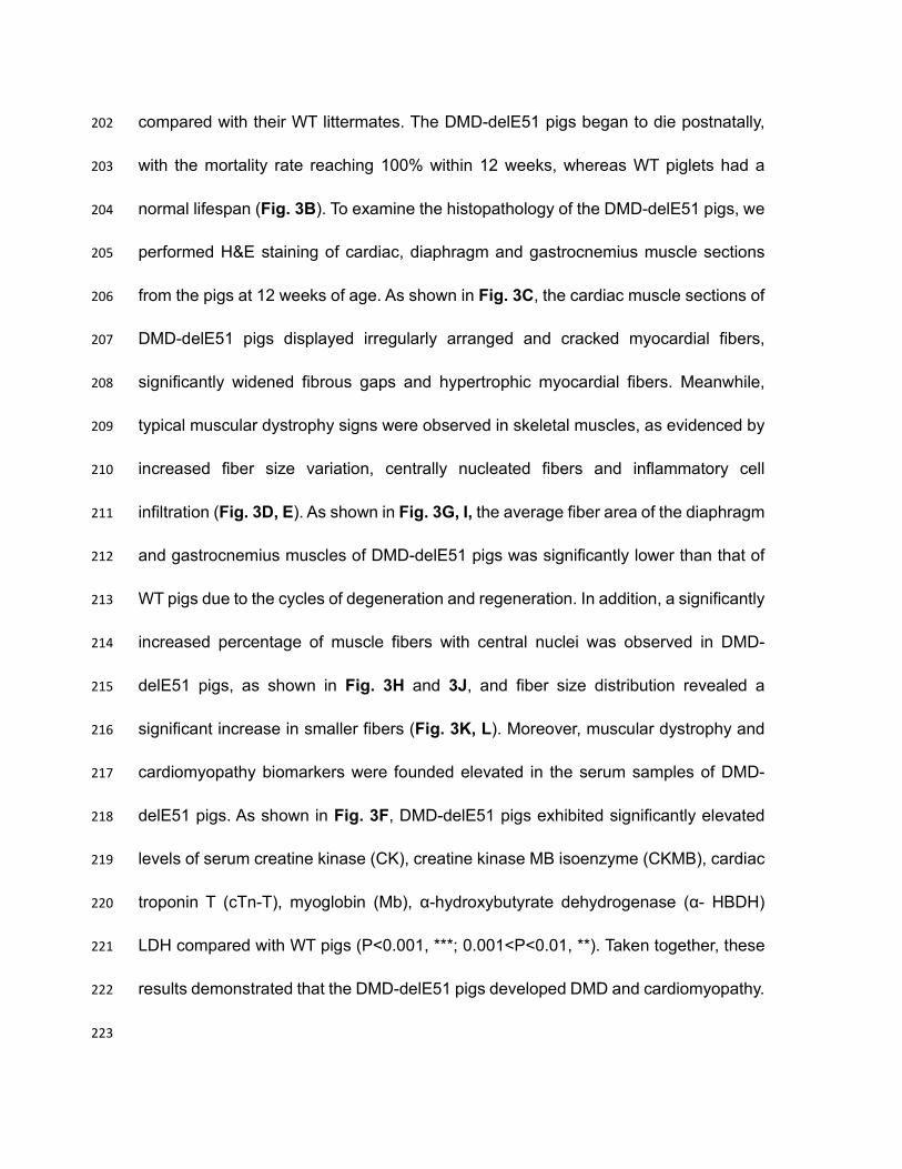
with the mortality rate reaching 100% within 12 weeks, whereas WT piglets had a 203
normal lifespan (Fig. 3B). To examine the histopathology of the DMD-delE51 pigs, we 204
performed H&E staining of cardiac, diaphragm and gastrocnemius muscle sections 205
from the pigs at 12 weeks of age. As shown in Fig. 3C, the cardiac muscle sections of 206
DMD-delE51 pigs displayed irregularly arranged and cracked myocardial fibers, 207
significantly widened fibrous gaps and hypertrophic myocardial fibers. Meanwhile, 208
typical muscular dystrophy signs were observed in skeletal muscles, as evidenced by 209
increased fiber size variation, centrally nucleated fibers and inflammatory cell 210
infiltration (Fig. 3D, E). As shown in Fig. 3G, I, the average fiber area of the diaphragm 211
and gastrocnemius muscles of DMD-delE51 pigs was significantly lower than that of 212
WT pigs due to the cycles of degeneration and regeneration. In addition, a significantly 213
increased percentage of muscle fibers with central nuclei was observed in DMD-214
delE51 pigs, as shown in Fig. 3H and 3J, and fiber size distribution revealed a 215
significant increase in smaller fibers (Fig. 3K, L). Moreover, muscular dystrophy and 216
cardiomyopathy biomarkers were founded elevated in the serum samples of DMD-217
delE51 pigs. As shown in Fig. 3F, DMD-delE51 pigs exhibited significantly elevated 218
levels of serum creatine kinase (CK), creatine kinase MB isoenzyme (CKMB), cardiac 219
troponin T (cTn-T), myoglobin (Mb), α-hydroxybutyrate dehydrogenase (α- HBDH) 220
LDH compared with WT pigs (P<0.001, ***; 0.001<P<0.01, **). Taken together, these 221
results demonstrated that the DMD-delE51 pigs developed DMD and cardiomyopathy. 222
223

Pathological alterations in the small intestine linked to malnutrition in DMD-224
delE51 pigs 225
The growth and development of DMD-delE51 pigs and WT pigs were monitored. 226
Compared with their WT littermates, DMD-delE51 pigs exhibited smaller body size 227
(Fig. 4A) and lighter body weight (Fig. 4B). Subsequent serological tests showed that 228
the levels of serum albumin (ALB) and prealbumin (PA) were significantly reduced in 229
DMD-delE51 pigs, indicating malnutrition (Fig. 4C, D). To further examine the 230
histopathology of the DMD-delE51 pigs, we performed H&E staining of small intestinal 231
tissue sections from the pigs at 12 weeks of age. As shown in Fig. 4E, 4G and Suppl. 232
Fig. 3A, compared with that of the controls, the intestinal villus height of DMD-delE51 233
pigs was significantly decreased, and the ratio of villus height to crypt depth was 234
approximately 60% reduced. Meanwhile, the thickness of the intestinal wall was 235
reduced by approximately 40% (Fig. 4F). In addition, we also examined the expression 236
of the DMD gene and dystrophin protein in the small intestine. The expression of the 237
DMD gene and dystrophin protein were significantly disrupted in the stomach and 238
small intestine (Suppl. Fig. 3B-E). 239
240
DMD-delE51 pigs suffered from gastric dysfunction 241
H&E staining of the stomach revealed that the gastric gland of DMD-delE51 pigs was 242
significantly thinner than that of WT pigs (Fig. 5A, B). Importantly, compared with WT 243
pigs, serum gastrin 17 (G-17) levels in DMD-delE51 pigs were significantly 244
downregulated at week 7 (Fig. 5C), serum pepsinogen I (PGI) levels were upregulated 245

at week 6 and reached the peak at week 8, but then sharply declined to a minimum 246
by week 12 (Fig. 5D). Serum pepsinogen II levels began to be significantly 247
upregulated at week 7 and remained higher than those in WT pigs (Fig. 5E). The ratio 248
of PGI/PGII (PGR) at week 7 was significantly lower in DMD-delE51 pigs than in WT 249
pigs (Fig. 5F). Collectively, DMD-delE51 pigs with disrupted dystrophin expression 250
exhibited morphological abnormality and functional impairment in the stomach. 251
252
Discussion 253
254
DMD is a fatal disease with multisystem involvement. Existing mouse23, rat24, 25, 255
rabbit26, dog27, pig28-31 models have been instrumental to understand the pathogenesis 256
of DMD and to develop therapeutic strategies. In this study, we established a new pig 257
model of DMD using CRISPR-genome editing and SCNT, and studied the pathology 258
in skeletal muscle, heart, stomach and small intestine. We demonstrated that the 259
DMD-delE51 pigs were born with abnormal posture, developed muscular dystrophy, 260
cardiomyopathy and gastrointestinal defects, and died within 12 weeks. In DMD 261
patients, weakness and difficulty in ambulation are first noted between 3 and 7 years 262
old8. A waddling gait is common, and patients become wheelchair-bound by the age 263
of 1232. Death usually occurs in the third decade of life due to heart or respiratory 264
failure. Our suggest that DMD-delE51 pigs have a markedly accelerated disease 265
progression as compared with that of DMD patients, similar to the other pig model of 266
DMD reported earlier28-31. 267

Individuals with DMD often have gastrointestinal or nutritional impairments33. Previous 268
research has demonstrated that the fundic gland and intestinal wall are the most 269
important parts of the digestive system34. The thickness of the fundic gland and 270
intestinal wall is closely related to the rhythmic contraction of the gastrointestinal tract 271
and mechanical digestion and reflects the rates of digestion and absorption of nutrients. 272
Moreover, the ratio of villus height to crypt depth comprehensively reflects the 273
digestive and absorption function of the small intestine35. When the ratio decreases, 274
the mucous membrane is likely to be damaged, and the digestive and absorptive 275
capacity is reduced, often accompanied by diarrhea and growth inhibition36. In this 276
study, DMD-delE51 pigs showed decreased gastric fundic gland and intestinal wall 277
thickness and shortened intestinal villus height. Our findings indicate that DMD-delE51 278
pigs have abnormal digestion and absorption in the gastrointestinal tract, leading to 279
malnutrition. Previous studies demonstrated abnormal levels of G-17, PGI, PGII and 280
PGR in serum of patients with gastritis37. Notably, the changes of G-17, PGI and PGII 281
in DMD-delE51 pigs were highly consistent with those in human patients with atrophic 282
gastritis (AG)38. In AG dominated by antral atrophy, antral mucosal atrophy can lead 283
to a decrease in the number of G cells and a decrease in the secretion of G-17, 284
resulting in a decrease in the content of G-17 in blood circulation39. Studies have 285
shown that low levels of serum PGI and PGR are biological markers of gastric body 286
atrophy, and the level of serum PGI decreases gradually with increasing severity of 287
mucosal atrophy40. Therefore, our findings suggest that DMD-delE51 pigs developed 288
gastric dysfunction, which likely contributes to the accelerated disease progression. 289

290
Previous studies have reported that the clinical manifestations of gastric dilatation and 291
intestinal pseudo-obstruction in patients with DMD are potentially related to the lack of 292
dystrophin in muscle41. We found that dystrophin was expressed in the gastrointestinal 293
tissues of WT pigs but disrupted in DMD-delE51 pigs, suggesting that the loss of 294
dystrophin protein is likely involved in the pathological alterations of the 295
gastrointestinal tract, weakening the digestion and absorption capacity of the 296
gastrointestinal tract and eventually inducing malnutrition and gastric dysfunction in 297
DMD-delE51 pigs. 298
299
The gastrointestinal dysfunction is of clinical importance and likely contributes to the 300
overall disease progression. As the life expectancy of DMD patients increases due to 301
better management of the life-threatening complications, the gastrointestinal 302
dysfunction could become more significant. It is thus necessary to consider the 303
gastrointestinal track as a therapeutic target organ for DMD patients. 304
305
Materials and Methods 306
Ethics statement 307
All animal studies were approved by the Animal Welfare and Research Ethics 308
Committee at Jilin University, and all procedures were conducted strictly in accordance 309
with the Guide for the Care and Use of Laboratory Animals. All surgeries were 310
performed under anesthesia, and every effort was made to minimize animal suffering 311

(SY201901013). 312
313
Construction of Cas9/sgRNA targeting vector 314
The CRISPR/Cas9 system was constructed as previously described42. Briefly, the 315
plasmid containing the U6-sgRNA and Cas9 expression elements was obtained from 316
Addgene (#42230). The targeting sgRNA is CTTGGACAGAACTTACCGAC. A pair of 317
complementary sgRNA oligo DNAs were synthesized, annealed into double-strand 318
DNAs, and ligated to the BbsI sites of the vector to form the intact plasmid, which was 319
confirmed by sequence analysis. 320
321
Isolation and culture of PFFs 322
The isolation and culture of PFFs were performed as previously described43. Thirty-323
three-day-old fetuses were chosen and separated from Bama miniature sows. First, 324
fetuses, without the head, tail, limb bones, and viscera, were cut into small pieces. 325
Then, these small pieces were digested and cultured in DMEM (GIBCO) 326
supplemented with 15% fetal bovine serum (FBS) at 39 °C and 5% CO2 in a humidified 327
incubator. PFFs at passage 1 were frozen in FBS containing 10% dimethyl sulfoxide 328
(DMSO). 329
330
Electrotransfection and single-cell colony selection 331
First, PFFs were thawed and cultured in 10-cm culture dishes. Then, 3x10⁶ PFFs were 332
electrotransfected with 200 μL of Opti-MEM (GIBCO) using 2-mm gap cuvettes and a 333
BTX ECM 2001 electroporator. The parameters for electrotransfection were as follows: 334
340 V, 1 ms, 3 pulses for 1 repeat. During these experiments, a total of 30 μg plasmids 335
were added to the reaction media. At 36 h after electrotransfection, the cells were 336
plated into ten 10-cm dishes at a density of 4x10³ cells per dish. Single-cell colonies 337
were picked and cultured in 24-well plates. When the plates reached 90% confluence, 338
10% of cells from each plate was lysed using 10 μL of lysis buffer (0.45% NP-40 plus 339
0.6% Proteinase K) for 60 min at 56 °C and then 10 min at 95 °C. The lysate was used 340
as a template for PCR. The forward and reverse primers were 5’-341
CAGCTAAACAGAGTAAAGAG-3’ and 5’-GATTTCCCTAGAGTCCACTT-3’, 342
respectively. The PCR conditions were 94 °C for 5 min; 94 °C for 30 s, 55 °C for 30 s, 343
and 72 °C for 1 min for 35 cycles; 72 °C for 5 min; and hold at 16 °C. The PCR products 344
were sequenced, and some PCR products were ligated into the PLB vector (Tiangen, 345
Beijing, China) and sequenced to identify the mutations. The positive cell colonies 346
were expanded and cryopreserved. 347
348
Off-target assay 349
Potential off-target sites (OTSs) were predicted by scanning the porcine genome using 350
BLAST based on the homology to the sgRNA plus protospacer adjacent motif (PAM). 351
The genomic DNA of the mutant cell clones was analyzed via PCR and DNA 352
sequencing to determine the target effects. The primer sequences used for analyzed 353
the off-target activities are listed in Suppl. Table 2. 354
355

Somatic nuclear transfer technology (SCNT) and genotyping of mutant piglets 356
Somatic cell nuclear transfer and embryo transfer were performed as previously 357
reported44. Positive colonies were screened and selected as donor cells for SCNT. 358
First, a single donor cell was microinjected into the enucleated pig oocyte. Second, 359
reconstructed embryos were electroactivated. Finally, embryos were transferred into 360
synchronized recipient pigs. After piglets were delivered, genomic DNA samples were 361
extracted from the tail tissue for genotyping. 362
363
Survival curve and bodyweight 364
The bodyweights of age- and sex-matched WT and DMD-modified pigs were 365
measured biweekly. A minimum of three individual animals of DMD-modified pigs was 366
used in all experiments. 367
368
Serum biochemical analysis 369
After piglets were born, serum samples were collected and measured by enzyme-370
linked immunosorbent assay (ELISA) following the manufacturer’s instructions, in an 371
infinite 200 PRO Microplate Reader (Tecan, Switzerland). Samples were measured in 372
triplicate, and the absorbance was monitored at 37 °C. 373
374
Neutral red dye (NRD) uptake assay 375
The cell viability was analyzed by the Neutral Red Cell Proliferation and Cytotoxicology 376
Assay Kit (Beyotime, China). The absorbance (OD) was measured at 540 nm, and the 377

value at the reference wavelength of 630 nm was subtracted. The assay was 378
performed on each cell clone in triplicate, and values were averaged from 4–6 wells 379
per plate. 380
381
Lactate dehydrogenase (LDH) assay 382
LDH activity in the medium was measured using the LDH assay kit (Beyotime, China) 383
according to the manufacturer’s instruction. The OD was measured by an infinite 200 384
PRO Microplate Reader (Tecan, Switzerland) at a wavelength of 490 nm. LDH activity 385
was calculated according to the formula provided by the instruction. 386
387
Analysis of apoptotic cells by flow cytometry 388
Annexin V-FITC antibody immunofluorescence combined with propidium iodide (PI) 389
was used to perform a fluorescent analysis of apoptosis, according to the instructions 390
of the Annexin V-FITC apoptosis detection kit (Beyotime, China). A total of 1×10⁵ cells 391
were collected and incubated with Annexin V-FITC and PI in the provided binding 392
buffer for 25 min in the dark at 4 ˚C, and analyzed by flow cytometry. 393
394
Western blotting 395
Equal amounts of proteins were separated through SDS-PAGE on a 5% separating 396
gel, and the protein bands were electrophoretically transferred to polyvinylidene 397
fluoride (PVDF) membranes. Then, the membranes were blocked for 2 h in TBST 398
buffer with 5% milk at room temperature. The membranes were subsequently 399

incubated with the primary antibodies (rabbit anti-light chain-3B (LC3B) antibody, 400
BM4827, Boster, 1:400; anti-dystrophin antibody, ab15277, Abcam, 1:400) overnight 401
at 4°C. After wash 3 times for 10 min with TBST buffer, the membranes were incubated 402
for 1 h with the secondary antibody. Finally, the protein bands were detected with the 403
ECL-Plus Western blotting reagent. 404
405
Quantitative reverse transcription PCR (RT-PCR) 406
For detection of relative mRNA expression of the DMD gene, total RNA was isolated 407
from muscle samples using TRNzol-A+ Reagent (TIANGEN, DP421) following the 408
manufacturer’s recommendations. The RNA (1 μg) was reverse-transcribed (RT) to 409
generate cDNA using a FastKing RT Kit (with gDNase) (TIANGEN, KR116) according 410
to the manufacturer’s manual. The reaction conditions were 95 °C for 5 min and 10 s; 411
60 °C for 20 s and 72 °C for 30 s for 40 cycles; and 95-55 °C for 30 s (melting curve). 412
The fluorescence intensity and amplification plots were analyzed by a BIO-RAD 413
iCycler Thermal Cycler with iQ5 Optical Module for RT-PCR (Bio-Rad, ABI 7500, iQ5). 414
The results were expressed via the comparative cycle threshold (CT) method as 415
described before, and expression levels were represented by fold changes over values 416
derived from healthy pigs. GAPDH was utilized as a reference gene. The primers used 417
in RT-PCR are listed in Suppl. Table 6. 418
419
Immunohistochemistry (IHC) 420
Dystrophin expression and location were examined in fixed sections. Muscle samples 421

were fixed in 4% paraformaldehyde (PFA), embedded in paraffin, and sectioned at 5 422
μm after harvest. IHC was performed according to standard techniques. Briefly, 423
muscle sections were deparaffinized, and antigen retrieval was performed. After slices 424
were cooled to 25 °C and washed twice with PBS, the slides were blocked with 5% 425
BSA for 15 min at 25 °C. Then, the slides were incubated with the primary anti-426
dystrophin antibody (MANDYS8, GeneTex, 1:400) overnight at 4 °C. Following 427
incubation and 3 washes with 0.05% Tween-20 in PBS, sections were incubated with 428
Alexa Fluor 488-conjugated Affinipure goat anti-mouse IgG (H+L) (SA0006-1, 429
ProteinTech, 1:100) for 1 h at 25 °C and washed again. Slides were mounted with 430
SlowFade Gold Antifade Reagent with DAPI (Invitrogen). For analysis of protein 431
abundance following IHC, 5 nonoverlapping pictures were randomly taken from each 432
section. 433
434
Hematoxylin and eosin (H&E) staining 435
Fresh tissues including heart, gastrocnemius, diaphragm, stomach and intestine, were 436
fixed in 4% PFA, embedded in paraffin, and sectioned at 5 μm. H&E staining was 437
performed with standard techniques. Briefly, sections were incubated in Mayer’s 438
hematoxylin. Then, sections were rinsed with tap water and counterstained with 1% 439
eosin. Finally, the sections were dehydrated, and coverslips were applied. Five 440
nonoverlapping pictures were randomly taken from each muscle section. 441
442
Morphometric analysis of muscle 443

Morphometric analyses were performed on H&E-stained muscle sections of DMD 444
exon 51-modified pigs and age-matched WT controls following the manufacturer’s 445
instructions, and five different regions were counted per section. The fiber size and 446
percentage of central nucleated fibers were calculated by ImagePro Plus software 447
(v6.0, Media Cybernetics, Silver Spring, MD, USA). 448
449
Statistical analysis 450
All data are expressed as the means ± standard error of the mean (SEM), and 451
Student’s t-test was used for statistical analysis. A single asterisk indicates statistical 452
significance at P<0.05. Double asterisks indicate a strong statistical significance at 453
P<0.01. Triple asterisks indicate even stronger statistical significance at P<0.001. All 454
statistical analyses were completed using GraphPad Prism 7.0 software. 455
456
457
458

References 459
1. Moser, H. Duchenne Muscular-Dystrophy - Pathogenetic Aspects and Genetic 460
Prevention. Hum Genet 66, 17-40 (1984). 461
2. Mendell, J.R. & Lloyd-Puryear, M. Report of MDA muscle disease symposium 462
on newborn screening for Duchenne muscular dystrophy. Muscle Nerve 48, 21-463
26 (2013). 464
3. Moat, S.J., Bradley, D.M., Salmon, R., Clarke, A. & Hartley, L. Newborn 465
bloodspot screening for Duchenne muscular dystrophy: 21 years experience in 466
Wales (UK). Eur J Hum Genet 21, 1049-1053 (2013). 467
4. Mercuri, E. & Muntoni, F. Muscular dystrophies. Lancet 381, 845-860 (2013). 468
5. Birnkrant, D.J. et al. The respiratory management of patients with duchenne 469
muscular dystrophy: a DMD care considerations working group specialty article. 470
Pediatr Pulmonol 45, 739-748 (2010). 471
6. Kohler, M. et al. Disability and survival in Duchenne muscular dystrophy. J 472
Neurol Neurosurg Psychiatry 80, 320-325 (2009). 473
7. Hoffman, E.P., Brown, R.H., Jr. & Kunkel, L.M. Dystrophin: the protein product 474
of the Duchenne muscular dystrophy locus. Cell 51, 919-928 (1987). 475
8. Blake, D.J., Weir, A., Newey, S.E. & Davies, K.E. Function and genetics of 476
dystrophin and dystrophin-related proteins in muscle. Physiol Rev 82, 291-329 477
(2002). 478
9. Coffey, A.J. et al. Construction of a 2.6-Mb Contig in Yeast Artificial 479
Chromosomes Spanning the Human Dystrophin Gene Using an Sts-Based 480

Approach. Genomics 12, 474-484 (1992). 481
10. Monaco, A.P., Walker, A.P., Millwood, I., Larin, Z. & Lehrach, H. A yeast artificial 482
chromosome contig containing the complete Duchenne muscular dystrophy 483
gene. Genomics 12, 465-473 (1992). 484
11. Koenig, M. et al. The molecular basis for Duchenne versus Becker muscular 485
dystrophy: correlation of severity with type of deletion. Am J Hum Genet 45, 486
498-506 (1989). 487
12. Monaco, A.P. et al. Detection of Deletions Spanning the Duchenne Muscular-488
Dystrophy Locus Using a Tightly Linked DNA Segment. Nature 316, 842-845 489
(1985). 490
13. Kole, R. & Krieg, A.M. Exon skipping therapy for Duchenne muscular dystrophy. 491
Adv Drug Deliv Rev 87, 104-107 (2015). 492
14. Birnkrant, D.J. et al. Diagnosis and management of Duchenne muscular 493
dystrophy, part 1: diagnosis, and neuromuscular, rehabilitation, endocrine, and 494
gastrointestinal and nutritional management. Lancet Neurol 17, 251-267 (2018). 495
15. Mule, F., Vannucchi, M.G., Corsani, L., Serio, R. & Faussone-Pellegrini, M.S. 496
Myogenic NOS and endogenous NO production are defective in colon from 497
dystrophic (mdx) mice. Am J Physiol-Gastr L 281, G1264-G1270 (2001). 498
16. Mule, F., Amato, A. & Serio, R. Gastric emptying, small intestinal transit and 499
fecal output in dystrophic (mdx) mice. J Physiol Sci 60, 75-79 (2010). 500
17. Feder, D. et al. Evaluation of the gastrointestinal tract in mdx mice: an 501
experimental model of Duchenne muscular dystrophy. Apmis 126, 693-699 502

(2018). 503
18. Chamberlain, J.S., Metzger, J., Reyes, M., Townsend, D. & Faulkner, J.A. 504
Dystrophin-deficient mdx mice display a reduced life span and are susceptible 505
to spontaneous rhabdomyosarcoma. Faseb J 21, 2195-2204 (2007). 506
19. Wells, D.J. Tracking progress: an update on animal models for Duchenne 507
muscular dystrophy. Disease models & mechanisms 11 (2018). 508
20. Patterson, J.K., Lei, X.G. & Miller, D.D. The pig as an experimental model for 509
elucidating the mechanisms governing dietary influence on mineral absorption. 510
Exp Biol Med 233, 651-664 (2008). 511
21. Heinritz, S.N., Mosenthin, R. & Weiss, E. Use of pigs as a potential model for 512
research into dietary modulation of the human gut microbiota. Nutr Res Rev 26, 513
191-209 (2013). 514
22. Kuzmuk, K.N. & Schook, L.B. Pigs as a Model for Biomedical Sciences. 515
Genetics of the Pig, 2nd Edition, 426-444 (2011). 516
23. Bulfield, G., Siller, W.G., Wight, P.A. & Moore, K.J. X chromosome-linked 517
muscular dystrophy (mdx) in the mouse. Proc Natl Acad Sci U S A 81, 1189-518
1192 (1984). 519
24. Larcher, T. et al. Characterization of dystrophin deficient rats: a new model for 520
Duchenne muscular dystrophy. PloS one 9, e110371 (2014). 521
25. Nakamura, K. et al. Generation of muscular dystrophy model rats with a 522
CRISPR/Cas system. Scientific reports 4, 5635 (2014). 523
26. Sui, T. et al. A novel rabbit model of Duchenne muscular dystrophy generated 524
by CRISPR/Cas9. Disease models & mechanisms 11 (2018). 525
27. Sharp, N.J. et al. An error in dystrophin mRNA processing in golden retriever 526
muscular dystrophy, an animal homologue of Duchenne muscular dystrophy. 527
Genomics 13, 115-121 (1992). 528
28. Klymiuk, N. et al. Dystrophin-deficient pigs provide new insights into the 529
hierarchy of physiological derangements of dystrophic muscle. Hum Mol Genet 530
22, 4368-4382 (2013). 531
29. Frohlich, T. et al. Progressive muscle proteome changes in a clinically relevant 532
pig model of Duchenne muscular dystrophy. Scientific reports 6 (2016). 533
30. Hollinger, K. et al. Dystrophin insufficiency causes selective muscle 534
histopathology and loss of dystrophin-glycoprotein complex assembly in pig 535
skeletal muscle. Faseb J 28, 1600-1609 (2014). 536
31. Nonneman, D.J., Brown-Brandl, T., Jones, S.A., Wiedmann, R.T. & Rohrer, G.A. 537
A defect in dystrophin causes a novel porcine stress syndrome. BMC Genomics 538
13, 233 (2012). 539
32. Kohler, M. et al. Quality of life, physical disability, and respiratory impairment in 540
Duchenne muscular dystrophy. Am J Respir Crit Care Med 172, 1032-1036 541
(2005). 542
33. Davidson, Z.E. & Truby, H. A review of nutrition in Duchenne muscular 543
dystrophy. J Hum Nutr Diet 22, 383-393 (2009). 544
34. Dibner, J.J. & Richards, J.D. The digestive system: Challenges and 545
opportunities. J Appl Poultry Res 13, 86-93 (2004). 546

35. Pluske, J.R. et al. Maintenance of villus height and crypt depth, and 547
enhancement of disaccharide digestion and monosaccharide absorption, in 548
piglets fed on cows' whole milk after weaning. Brit J Nutr 76, 409-422 (1996). 549
36. Almeida, J.A. et al. Lactose malabsorption in the elderly: Role of small intestinal 550
bacterial overgrowth. Scand J Gastroentero 43, 146-154 (2008). 551
37. Wang, T.C. et al. Synergistic interaction between hypergastrinemia and 552
Helicobacter infection in a mouse model of gastric cancer. Gastroenterology 553
118, 36-47 (2000). 554
38. Sipponen, P. et al. Serum levels of amidated gastrin-17 and pepsinogen I in 555
atrophic gastritis: An observational case-control study. Scand J Gastroentero 556
37, 785-791 (2002). 557
39. Cao, Q., Ran, Z.H. & Xiao, S.D. Screening of atrophic gastritis and gastric 558
cancer by serum pepsinogen, gastrin-17 and Helicobacter pylori 559
immunoglobulin G antibodies. J Digest Dis 8, 15-22 (2007). 560
40. Kekki, M., Samloff, I.M., Varis, K. & Ihamaki, T. Serum Pepsinogen-I and Serum 561
Gastrin in the Screening of Severe Atrophic Corpus Gastritis. Scand J 562
Gastroentero 26, 109-116 (1991). 563
41. Ricotti, V. et al. Recurrent pseudo-obstruction and sigmoid volvulus in 564
Duchenne Muscular Dystrophy: A case report. Neuromuscular Disord 22, 887-565
888 (2012). 566
42. Wang, K.K. et al. CRISPR/Cas9-mediated knockout of myostatin in Chinese 567
indigenous Erhualian pigs. Transgenic Res 26, 799-805 (2017). 568

43. Zou, X.D. et al. Preparation of a new type 2 diabetic miniature pig model via the 569
CRISPR/Cas9 system. Cell Death Dis 10 (2019). 570
44. Zhou, X.Q. et al. Generation of CRISPR/Cas9-mediated gene-targeted pigs via 571
somatic cell nuclear transfer. Cellular and Molecular Life Sciences 72, 1175-572
1184 (2015). 573
574
575
576

FIGURES 577
578
Figure 1. PFFs with DMD exon 51 deficiency showed impaired cell membrane 579
integrity and early cell apoptosis. (A) Sanger sequencing of PFFs showed different 580
mutations induced by Cas9/sgRNA electrotransfection. WT sequence is shown at the 581
top of the targeting sequence. PAM sequences are highlighted in red. (B) NRD uptake 582
assay of PFF clones carrying DMD exon 51 mutations at different cell densities. ***P< 583
0.001, **P<0.01 and *P<0.05. (C) LDH activities in culture medium at different time 584
points were measured by the LDH-kit. ***P< 0.001, **P<0.01 and *P<0.05. (D) Cell 585
apoptosis was analyzed by flow cytometry. *P<0.05. (E) Western blotting analysis of 586
autophagy in PFFs did not detect significant difference between WT and DMD-mutant 587
PFFs. 588
589
590
591
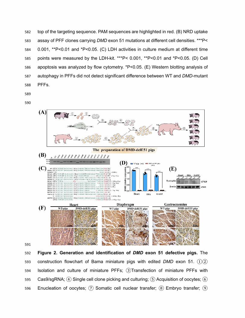
Figure 2. Generation and identification of DMD exon 51 defective pigs. The 592
construction flowchart of Bama miniature pigs with edited DMD exon 51. ①② 593
Isolation and culture of miniature PFFs; ③Transfection of miniature PFFs with 594
Cas9/sgRNA; ④ Single cell clone picking and culturing; ⑤ Acquisition of oocytes; ⑥ 595
Enucleation of oocytes; ⑦ Somatic cell nuclear transfer; ⑧ Embryo transfer; ⑨ 596

Delivery and identification of DMD-delE51 pigs. (B) PCR analysis of DMD exon 51 in 597
all piglets. (C) Mutation analysis by T-cloning and Sanger sequencing in all piglets. WT 598
sequence is shown at the top. PAM sites are highlighted in red; target sequences are 599
shown in green. (D) The relative expression of DMD mRNA in DMD-delE51 pigs and 600
the age-matched wild-type pigs. DIA, Diaphragm; GAS, Gastrocnemius; ***P< 0.001. 601
(E) Western blotting showed the disrupted expression of dystrophin in DMD-delE51 602
pigs. (F) IHC analysis of dystrophin expression in heart, diaphragm and gastrocnemius 603
muscles. Scale bars: 50 µm and 100 µm. 604
605
606

607
Figure 3. Development of muscular dystrophy and cardiomyopathy in DMD-608
delE51 pigs. (A) Piglets carrying DMD exon 51 mutations showed abnormal posture. 609
(B) Survival curves of DMD-delE51 pigs and WT-pigs. (C-E) H&E staining of heart, 610
diaphragm and gastrocnemius muscle sections from WT and DMD-delE51 pigs at the 611
age of 12 weeks. Scale bars: 50 µm. Fiber fracture (green arrows) and hypertrophic 612
fiber (black arrows) were seen in heart sections. Excessive fiber size variation (red 613
arrows), central nucleated fibers (purple arrows), fiber fracture (green arrows) and 614
inflammatory cell infiltration (blue arrows) were readily visible in muscle sections. (F) 615
Serum biochemical profiles of WT and DMD-delE51 pigs. ***P< 0.001, **P<0.01 and 616

*P<0.05. (G, I) Quantification of mean fiber area (MMA) of diaphragm and 617
gastrocnemius muscle in WT and DMD-delE51 pigs. ***P< 0.001. (H, J) Quantification 618
of centrally nucleated fiber (CNF) percentage in WT and DMD-delE51 pigs. ***P< 619
0.001. (K, L) Size distribution of diaphragm and gastrocnemius muscle in WT and 620
DMD-delE51 pigs. 621
622
623
624

625
Figure 4. DMD-delE51 pigs exhibited symptoms of malnutrition. Photograph of 626
DMD-delE51 pigs and WT control at the age of one week. (B) Body mass of DMD-627
delE51 and WT pigs from birth to 12 weeks of age. ***P< 0.001, **P<0.01. (C, D) 628
Serum ALB (C) and PA (D) levels of WT and DMD-delE51 pigs. ***P< 0.001; *P<0.05. 629
(E) H&E staining of the small intestine sections of WT and DMD-delE51 pigs. Red 630
dotted lines indicate the thickness of the intestinal wall, black arrows indicate the 631
height of the small intestine villi and blue arrows indicate the depth of the small 632

intestine crypt. Scale bars: 50 µm and 100 µm. (F) The relative thickness of small 633
intestinal wall in WT and DMD-delE51 pigs. *** p < 0.01. (G) The relative ratio of small 634
intestine villus height/crypt depth of WT and DMD-delE51 pigs. *** p < 0.01. 635
636
637
638
639

640
Figure 5. DMD-delE51 pigs suffered from atrophic gastritis. (A) H&E staining of 641
stomach sections from WT and DMD-delE51 pigs at the age of 12 weeks. The 642
thickness of gastric fundus glands (red rectangle) was reduced in DMD-delE51 pigs. 643
Scale bars: 50 µm and 100 µm. (B) Quantification of the relative thickness of gastric 644
fundus glands in WT and DMD-delE51 pigs. *** p < 0.01. (C) Measurements of serum 645
G-17 in WT and DMD-delE51 pigs. ***P< 0.001. (D) Measurement of serum PGI in 646
WT and DMD-delE51 pigs. ***P< 0.001. (E) Measurement of serum PGII in WT and 647

DMD-delE51 pigs. ***P< 0.001. (F) The ratio of PGI/PGII in WT and DMD-delE51 pigs. 648
*** p < 0.01. 649

Supplementary Information 1
Supplementary Table 1: List of putative off-target sites (PAM sequences are labeled 2
in blue. Base substitutions are shown in red.) 3
sgRNA CTTGGACAGAACTTACCGACTGG
OTS1 CCAGTACAGAACTTACAGACCGG
OTS2 CTTTGATAGGACTTACCGATTAG
OTS3 CTTCTCCAGAACTTACCGCCCAG
OTS4 CTTGTACAGAAATCACCGACCAG
OTS5 CTTGGACACATCTTACCAACAGG
OTS6 CCTGTACAGAACTTACCTATGAG
OTS7 CTTGGCCAGCATTTACCGTCAGG
OTS8 ATTTGACAGAACATACCAACAGG
4
Supplementary Table 2: List of primers for PCR amplification of off-target sites. 5
Primers Sequences (5' to 3') Amplicon (bp)
DMD- sgRNA-OTS1 AAGGAAATTGAGACTCAGAGAAGA 502
TGCTTTTCATTGGCTCTGGC
DMD- sgRNA-OTS2 TGCCAGTGTGGTTGGTTTCT 551
CCAGTCCATTCCCCCATCAC
DMD - sgRNA-OTS3 GTTTACCGCAGACCCACAGA 589
GTGCGTAGAGACCCAAACCA
DMD - sgRNA-OTS4 GGCTGGTCATGGTTAGCACT 522
CTGAACACCCTTCCTCCACC
DMD - sgRNA-OTS5 TTTGACCCCAATCCATGCGT 577
TGCTCTATGCCACTTCGCTT
DMD - sgRNA-OTS6 TGTGTCTTGGTGGGTGATGG 508
GTGTGGATGGGTGTATGCCA
DMD - sgRNA-OTS7 AGGGGCTTATGCTTGTGGTC 522
TCAGAAGCCTGCCCTTCATG
DMD - sgRNA-OTS8 GGTCCTGACCCTTTGGATGT 593
AGGCTGAATTATCTGAGTGCCA
6
7
8
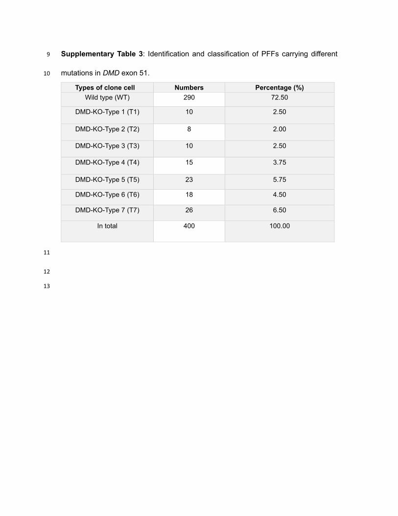
Supplementary Table 3: Identification and classification of PFFs carrying different 9
mutations in DMD exon 51. 10
Types of clone cell Numbers Percentage (%)
Wild type (WT) 290 72.50
DMD-KO-Type 1 (T1) 10 2.50
DMD-KO-Type 2 (T2) 8 2.00
DMD-KO-Type 3 (T3) 10 2.50
DMD-KO-Type 4 (T4) 15 3.75
DMD-KO-Type 5 (T5) 23 5.75
DMD-KO-Type 6 (T6) 18 4.50
DMD-KO-Type 7 (T7) 26 6.50
In total 400 100.00
11
12
13
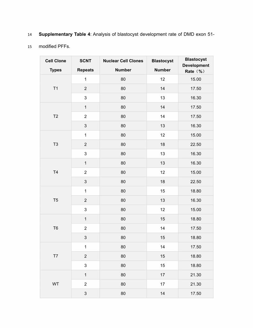
Supplementary Table 4: Analysis of blastocyst development rate of DMD exon 51-14
modified PFFs. 15
Cell Clone
Types
SCNT
Repeats
Nuclear Cell Clones
Number
Blastocyst
Number
Blastocyst
Development
Rate(%)
T1
1 80 12 15.00
2 80 14 17.50
3 80 13 16.30
T2
1 80 14 17.50
2 80 14 17.50
3 80 13 16.30
T3
1 80 12 15.00
2 80 18 22.50
3 80 13 16.30
T4
1 80 13 16.30
2 80 12 15.00
3 80 18 22.50
T5
1 80 15 18.80
2 80 13 16.30
3 80 12 15.00
T6
1 80 15 18.80
2 80 14 17.50
3 80 15 18.80
T7
1 80 14 17.50
2 80 15 18.80
3 80 15 18.80
WT
1 80 17 21.30
2 80 17 21.30
3 80 14 17.50

Supplementary Table 5: SCNT results for the generation of DMD-delE51 pigs. 16
Target
gene
Transferred
embryos
No.
recipients
No.(%)
pregnancies
No. born No. (%)
mutants
DMD
200 1 1 4 2
200 1 0 0 0
200 1 1 4 3
200 1 0 0 0
200 1 1 7 4
Total 1000 5 3 15 9
17
Supplementary Table 6: List of primers used for RT-PCR. 18
RT-DMD-F (5’-3’) CCCTGGACTGACCACTAT
RT-DMD-R (5’-3’) CTCTGTGATTTTATAACTCG
RT-GAPDH-F (5’-3’) ATCCTGGGCTACACTGAGGA
RT-GAPDH-R (5’-3’) TGTCGTACCAGGAAATGAGCT
19
20
21

22
23
Supplementary Figure 1. Sequence analysis of pig dystrophin and design of 24
CRISPR-targeting strategy. (A) Comparison of amino acid sequences of dystrophin 25
proteins among different species. (B)Schematic representation of the sgRNA targeting 26
porcine DMD exon 51. PAM is highlighted in red. (C) Sanger sequencing traces of 27
PCR amplicons from WT and electrotransfected PFFs. The cleavage sites are labeled 28
with a red arrow. (D) The analysis of off-target sites (OTS). The corresponding 29
sequencing chromatograms for the top OTS are shown. 30
31

32
Supplementary Figure 2. Analysis of PFFs with DMD mutations. The relative 33
expression of DMD mRNA in DMD-edited and WT PFFs. ***P< 0.001. (B) Western 34
blotting analysis of dystrophin expression in DMD-modified and WT PFFs. (C) NRD 35
uptake analysis of PFF cell clones carrying DMD exon 51 mutations at different cell 36
densities; ***P< 0.001, **P<0.01 and *P<0.05. (D) LDH activity in culture medium at 37

different time periods. ***P< 0.001, **P<0.01 and *P<0.05. (E, F) Cell apoptosis 38
analyzed by flow cytometry. (G) Representative images of blastocysts at 8.5 days after 39
nuclear transfer. PFFs with DMD exon 51 modified could develop normally into 40
blastocysts. (H) The analysis of blastocyst development rate of PFFs carrying DMD 41
exon 51mutations. NS, no significant. 42
43
44
Supplementary Figure 3. Dystrophin expression in porcine stomach and small 45
intestine. (A) The relative height of small intestine villus in WT and DMD-delE51 pigs. 46
*** p < 0.01. (B) The relative expression of DMD mRNA in stomach and small intestine 47
from DMD-delE51 pigs and the age-matched wild-type pigs; ***P< 0.001. (C) Western 48
blotting analysis of dystrophin in stomach and small intestine of WT and DMD-delE51 49
pigs. (D, E) IHC staining of dystrophin in stomach (D) and small intestine (E) of pigs. 50
Scale bars: 50 µm and 100 µm. 51

Figures
Figure 1
PFFs with DMD exon 51 de�ciency showed impaired cell membrane integrity and early cell apoptosis. (A)Sanger sequencing of PFFs showed different mutations induced by Cas9/sgRNA electrotransfection. WTsequence is shown at the top of the targeting sequence. PAM sequences are highlighted 582 in red. (B)

NRD uptake assay of PFF clones carrying DMD exon 51 mutations at different cell densities. ***P< 0.001,**P<0.01 and *P<0.05. (C) LDH activities in culture medium at different time points were measured by theLDH-kit. ***P< 0.001, **P<0.01 and *P<0.05. (D) Cell apoptosis was analyzed by �ow cytometry. *P<0.05.(E) Western blotting analysis of autophagy in PFFs did not detect signi�cant difference between WT andDMD-mutant PFFs.
Figure 2
Generation and identi�cation of DMD exon 51 defective pigs. The construction �owchart of Bamaminiature pigs with edited DMD exon 51. Isolation and culture of miniature PFFs; Transfection ofminiature PFFs with Cas9/sgRNA; Single cell clone picking and culturing; Acquisition of oocytes; Enucleation of oocytes; Somatic cell nuclear transfer; Embryo transfer; Delivery and identi�cation ofDMD-delE51 pigs. (B) 597 PCR analysis of DMD exon 51 in all piglets. (C) Mutation analysis by T-cloningand Sanger sequencing in all piglets. WT sequence is shown at the top. PAM sites are highlighted in red;

target sequences are shown in green. (D) The relative expression of DMD mRNA in DMD-delE51 pigs andthe age-matched wild-type pigs. DIA, Diaphragm; GAS, Gastrocnemius; ***P< 0.001. (E) Western blottingshowed the disrupted expression of dystrophin in DMD-delE51 pigs. (F) IHC analysis of dystrophinexpression in heart, diaphragm and gastrocnemius muscles. Scale bars: 50 μm and 100 μm.
Figure 3

Development of muscular dystrophy and cardiomyopathy in DMD-delE51 pigs. (A) Piglets carrying DMDexon 51 mutations showed abnormal posture. (B) Survival curves of DMD-delE51 pigs and WT-pigs. (C-E)H&E staining of heart, diaphragm and gastrocnemius muscle sections from WT and DMD-delE51 pigs atthe age of 12 weeks. Scale bars: 50 μm. Fiber fracture (green arrows) and hypertrophic �ber (blackarrows) were seen in heart sections. Excessive �ber size variation (red arrows), central nucleated �bers(purple arrows), �ber fracture (green arrows) and in�ammatory cell in�ltration (blue arrows) were readilyvisible in muscle sections. (F) Serum biochemical pro�les of WT and DMD-delE51 pigs. ***P< 0.001,**P<0.01 and *P<0.05. (G, I) Quanti�cation of 617 mean �ber area (MMA) of diaphragm andgastrocnemius muscle in WT and DMD-delE51 pigs. ***P< 0.001. (H, J) Quanti�cation of centrallynucleated �ber (CNF) percentage in WT and DMD-delE51 pigs. ***P< 0.001. (K, L) Size distribution ofdiaphragm and gastrocnemius muscle in WT and DMD-delE51 pigs.

Figure 4
DMD-delE51 pigs exhibited symptoms of malnutrition. Photograph of DMD-delE51 pigs and WT controlat the age of one week. (B) Body mass of DMD-delE51 and WT pigs from birth to 12 weeks of age. ***P<0.001, **P<0.01. (C, D) Serum ALB (C) and PA (D) levels of WT and DMD-delE51 pigs. ***P< 0.001;*P<0.05. (E) H&E staining of the small intestine sections of WT and DMD-delE51 pigs. Red dotted linesindicate the thickness of the intestinal wall, black arrows indicate the height of the small intestine villi and

blue arrows indicate the depth of the small intestine crypt. Scale bars: 50 μm and 100 μm. (F) The relative633 thickness of small intestinal wall in WT and DMD-delE51 pigs. *** p < 0.01. (G) The relative ratio ofsmall intestine villus height/crypt depth of WT and DMD-delE51 pigs. *** p < 0.01.
Figure 5
MD-delE51 pigs suffered from atrophic gastritis. (A) H&E staining of stomach sections from WT andDMD-delE51 pigs at the age of 12 weeks. The thickness of gastric fundus glands (red rectangle) was

reduced in DMD-delE51 pigs. Scale bars: 50 μm and 100 μm. (B) Quanti�cation of the relative thicknessof gastric fundus glands in WT and DMD-delE51 pigs. *** p < 0.01. (C) Measurements of serum G-17 inWT and DMD-delE51 pigs. ***P< 0.001. (D) Measurement of serum PGI in WT and DMD-delE51 pigs.***P< 0.001. (E) Measurement of serum PGII in WT and DMD-delE51 pigs. ***P< 0.001. (F) The ratio of648 PGI/PGII in WT and DMD-delE51 pigs. *** p < 0.01.