pathogenetical factors contributing to high iga levels and ... · pdf fileund liquor...
TRANSCRIPT

Department of Small Animal Medicine and Surgery
University of Veterinary Medicine Hannover
Center for Systems Neuroscience Hannover
Pathogenetical factors contributing to high IgA levels and marked
neutrophilic pleocytosis in canine Steroid-responsive Meningitis-Arteritis
Thesis
Submitted in partial fulfillment of the requirements for the degree
DOCTOR OF PHILOSOPHY (PhD)
at the Center for Systems Neuroscience Hannover
awarded by the University of Veterinary Medicine Hannover
by
Malte Schwartz
(Neumünster)
Hannover 2009

Supervisor: Prof. Dr. A. Tipold
1st referee: Prof. Dr. A. Tipold
2nd referee: Prof. Dr. W. Baumgärtner
3rd referee: PD Dr. K. Krampfl/Prof. Dr. M. Stangel
External referee: Prof. Dr. T.J. Anderson (University of Glasgow, Scotland)
Date of final exam: 12/09/2009

Preliminary results of flow cytometric measurements have already been included in:
Schwartz, M., 2007. Durchflußzytometrische Untersuchungen von Leukozyten in Blut
und Liquor cerebrospinalis bei Hunden mit steril-eitriger Meningitis-Arteriitis. Thesis.
Department of Small Animal Medicine and Surgery, University of Veterinary
Medicine, Hannover, Germany.
Further accepted or submitted publications that include the presented results:
Schwartz, M., Puff, C., Stein, V.M., Baumgärtner, W., Tipold, A., 2009,
Pathogenetical factors for excessive IgA production: Th2-dominated immune response
in canine steroid-responsive Meningitis-Arteritis. Submitted.
Schwartz, M., Carlson, R., Tipold, A., 2008, Selective CD11a upregulation on
neutrophils in the acute phase of steroid-responsive meningitis-arteritis in dogs. Vet
Immunol Immunopathol 126, 248-255.
Schwartz, M., Puff, C., Stein, V.M., Baumgärtner, W., Tipold, A., 2009, Marked
MMP-2 transcriptional up-regulation in mononuclear leukocytes invading the
subarachnoidal space in aseptic suppurative Steroid-responsive Meningitis-Arteritis in
dogs. Vet Immunol Immunopathol. Article in Press.
This work was funded by a Georg-Christoph-Lichtenberg-Scholarship provided by the
Department of Science and Culture, Lower Saxony, Germany

Index
Chapter 1 General Introduction .............................................................................................. 1
Chapter 2 Pathogenetical factors for excessive IgA production: Th2-dominated immune
response in canine steroid-responsive Meningitis-Arteritis ................................... 8
2.1 Abstract ...................................................................................................................... 9
2.2 Introduction .............................................................................................................. 11
2.3 Materials and Methods ............................................................................................. 13
2.3.1 Animals and Samples ....................................................................................... 13
2.3.2 RNA purification and reverse transcription ..................................................... 15
2.3.3 Primers ............................................................................................................. 16
2.3.4 Standard dilution series .................................................................................... 17
2.3.5 Real-time PCR.................................................................................................. 18
2.3.6 Statistical analysis ............................................................................................ 19
2.4 Results ...................................................................................................................... 20
2.5 Discussion ................................................................................................................ 28
2.6 Conclusion................................................................................................................ 32
2.7 Acknowlegements .................................................................................................... 32
2.8 Conflict of interest statement ................................................................................... 33
Chapter 3 Selective CD11a upregulation on neutrophils in the acute phase of Steroid
responsive Meningitis-Arteritis in dogs ............................................................... 34
3.1 Abstract .................................................................................................................... 35
3.2 Introduction .............................................................................................................. 37
3.3 Materials and Methods ............................................................................................. 39
3.3.1 Animals and Samples ....................................................................................... 39
3.3.2 Monoclonal antibodies (mAbs)........................................................................ 41
3.3.3 Sample processing for the ex vivo examination ............................................... 42
3.3.4 Sample processing for the in vitro study.......................................................... 43
3.3.5 Flow cytometric analysis.................................................................................. 44
3.3.6 Preliminary experiments .................................................................................. 45
3.3.7 Statistical analysis ............................................................................................ 45

3.4 Results ...................................................................................................................... 46
3.4.1 Results of the ex vivo examination................................................................... 46
3.4.2 Correlation of parameters................................................................................. 49
3.4.3 Results of the in vitro study.............................................................................. 50
3.5 Discussion ................................................................................................................ 53
3.6 Conclusion................................................................................................................ 57
3.7 Acknowledgments .................................................................................................... 57
Chapter 4 Marked MMP-2 transcriptional up-regulation in mononuclear leukocytes
invading the subarachnoidal space in aseptic suppurative Steroid-responsive
Meningitis-Arteritis in dogs ................................................................................. 58
4.1 Abstract .................................................................................................................... 59
4.2 Introduction .............................................................................................................. 60
4.3 Materials and Methods ............................................................................................. 62
4.3.1 Animals and Samples ....................................................................................... 62
4.3.2 RNA purification and reverse transcription ..................................................... 64
4.3.3 Primers ............................................................................................................. 65
4.3.4 Standard dilution series .................................................................................... 67
4.3.5 Real-time PCR.................................................................................................. 67
4.3.6 Statistical analysis ............................................................................................ 69
4.4 Results ...................................................................................................................... 69
4.5 Discussion ................................................................................................................ 77
4.6 Conclusion................................................................................................................ 83
4.7 Acknowlegements .................................................................................................... 83
4.8 Conflict of interest statement ................................................................................... 83
Chapter 5 General Discussion............................................................................................... 84
Chapter 6 Summary .............................................................................................................. 89
Chapter 7 Zusammenfassung................................................................................................ 91
Chapter 8 References ............................................................................................................ 94
Chapter 9 Acknowledgements ............................................................................................ 107

List of abbreviations
bp base pairs
°C degree celcius
CBC complete blood count
CD Cluster of Differentiation
cDNA complementary DNA
CNS central nervous system
CO2 carbon dioxide
ConA concanavalin A
COX cyclooxygenase
CSF cerebrospinal fluid
CSF WBC cerebrospinal fluid white blood cell
CT computed tomography
DNA desoxyribonucleic acid
EDTA ethylene diamine tetraacetic acid
EF elongation factor
ELISA enzyme-linked immunosorbent assay
Fig. figure
g gravity
GAPDH glyceraldehyd-3-phosphate dehydrogenase
GRO-α growth-related gene product alpha
h hour
HUVEC human umbilical vascular endothelial cell

i.e. that is
Ig immunoglobulin
IFN interferon
IL interleukin
kg kilogram
KS Kawasaki Syndrome
mAb monoclonal antibody
mg milligram
µg microgram
µl microliter
min minutes
ml milliliter
MNC mononuclear cell
mM millimolar
MMP matrix metalloproteinase
MRI magnetic resonance imaging
mRNA messenger RNA
ng nanogram
nM nanomolar
NTC no template control
OD optical density
PB peripheral blood
PBMNC peripheral blood mononuclear cell
PBPMN peripheral blood polymorphonuclear cell

PBS phosphate-buffered saline
PCR polymerase chain reaction
PMN polymorphonuclear cell
PNS peripheral nervous system
rSpear Spearman´s rank correlation coefficient
RNA ribonucleic acid
RPE R-phycoerythrine
SID once per day
SRMA Steroid-responsive Meningitis-Arteritis
Th1 T helper 1
Th2 T helper 2
Th17 T helper 17
TIMP tissue inhibitor of metalloproteinase
U enzymatic activity
WBC white blood cell

Chapter 1
1
Chapter 1: General Introduction
Canine Steroid-responsive Meningitis-Arteritis (SRMA) is known under various terms, all
reflecting certain characteristics of the disorder. These names include Beagle Pain Syndrome
(Hayes et al., 1989), Canine Pain Syndrome (Burns et al., 1991), Necrotizing Vasculitis
(Brooks, 1984), Polyarteritis (Harcourt, 1978), Canine Juvenile Polyarteritis Syndrome
(Felsburg et al., 1992), Aseptic Suppurative Meningitis (Presthus, 1991) and Corticosteroid-
responsive Meningomyelitis (Irving and Chrisman, 1990). Results of post mortem
examinations strongly suggest that these descriptions all refer to the identical disease entity of
SRMA.
Steroid-responsive Meningitis-Arteritis is one of the most frequently diagnosed inflammations
of the central nervous system (CNS) of dogs and was reported to account for 15 % of these
cases (Tipold, 1995). In this species SRMA is known to be the most common type of
meningitis (Meric, 1988). In another study SRMA was found to be the cause for fever in 11 %
of dogs that had been referred for diagnostic work-up of this clinical sign (Battersby et al.,
2006).
Juvenile to young adult dogs are most frequently affected with ages at the time of diagnosis
ranging from 2 months (Russo et al., 1983) to 9 years (Cizinauskas et al., 2000).
Individuals from all breeds and cross-breed dogs seem to be susceptible, however, breed
predispositions have been identified for Boxers (Behr and Cauzinille, 2006; Tipold and Jaggy,
1994), Bernese Mountain Dogs (Cizinauskas et al., 2000; Presthus, 1991; Tipold and Jaggy,
1994) and Beagles (Joshua and Ishmael, 1968), of which many are kept as laboratory animals

Chapter 1
2
(Brooks, 1984; Felsburg et al., 1992; Harcourt, 1978; Hayes et al., 1989; Scott-Moncrieff et
al., 1992). No sex predilection was found in any of these studies.
The natural course of SRMA is relapsing with a tendency to progressively deteriorate
(Brooks, 1984). The classical acute and a chronic protracted form of SRMA have been
described (Tipold and Jaggy, 1994). The latter may develop with inadequate treatment
resulting in relapses (Tipold, 2000).
The acute form is characterized by fever and pain upon palpation and manipulation of the
cervical region. No neurological deficits, suggesting involvement of the CNS parenchyma, are
evident on a neurological examination. The chronic form, however, may be accompanied by
deficits indicative for a spinal cord or multi-focal CNS lesion (Tipold and Jaggy, 1994).
These lesions most frequently become evident as gait abnormalities (Tipold and Jaggy, 1994)
that rarely progress to a state of plegia (Hoff and Vandevelde, 1981; Joshua and Ishmael,
1968). Additional neurological symptoms may include central vestibular signs (Behr and
Cauzinille, 2006; Tipold and Jaggy, 1994), optic nerve lesions (Meric et al., 1985; Russo et
al., 1983) and myoclonus (Tipold and Jaggy, 1994) or generalized tremor (Gandini et al.,
2003).
A hematological examination shows a neutrophilic leukocytosis in the majority of patients
(Scott-Moncrieff et al., 1992; Tipold and Jaggy, 1994). In some patients, especially in those
that are presented for a relapse (Cizinauskas et al., 2000), the complete blood count (CBC)
reveals a lymphopenia (Burns et al., 1991), in other patients, however, a lymphocytosis may
be evident (Meric et al., 1986). Further hematological abnormalities may include monocytosis
(Cizinauskas et al., 2000; Scott-Moncrieff et al., 1992), mild normocytic, normochromic
anemia (Burns et al., 1991; Harcourt, 1978; Scott-Moncrieff et al., 1992; Tipold and Jaggy,
1994) and thrombocytosis (Hayes et al., 1989). The albumin concentration is decreased in

Chapter 1
3
some dogs (Bathen-Noethen et al., 2008; Burns et al., 1991; Hayes et al., 1989; Lowrie et al.,
2008; Scott-Moncrieff et al., 1992) and globulin-/α2-globulin levels may be elevated (Behr
and Cauzinille, 2006; Brooks, 1984; Cizinauskas et al., 2000; Hayes et al., 1989; Scott-
Moncrieff et al., 1992; Tipold and Jaggy, 1994).
Findings in cerebrospinal fluid (CSF) of affected dogs depend on whether animals suffer from
the acute or chronic form of SRMA. Changes in the acute form are characterized by a
moderate to marked pleocytosis with predominance of non-degenerated neutrophils (Behr and
Cauzinille, 2006; Harcourt, 1978; Tipold and Jaggy, 1994). The content in total protein is
usually moderately to markedly elevated (Behr and Cauzinille, 2006; Tipold and Jaggy,
1994). Frank blood (Behr and Cauzinille, 2006; Irving and Chrisman, 1990) or xanthochromia
(Bathen-Noethen et al., 2008; Meric et al., 1986) may be found in CSF of some dogs
reflecting hemorrhage into the subarachnoidal space due to vasculitis-induced destruction of
pial blood vessels. In dogs that have entered the chronic stage of SRMA (Tipold and Jaggy,
1994) or have received glucocorticosteroid treatment prior to presentation (Behr and
Cauzinille, 2006; Poncelet and Balligand, 1993) a mild to moderate mononuclear to mixed
pleocytosis is usually present. In most of these cases the protein content is normal to slightly
elevated (Tipold and Jaggy, 1994).
A gross pathological examination reveals changes only in some individuals. These consist of
varying degrees of hemorrhage, ranging from petechial (Snyder et al., 1995) to massive
bleedings (Joshua and Ishmael, 1968; Vandevelde and Fankhauser, 1972). In addition,
amyloid depositions in various organs (Snyder et al., 1995), enlarged lymph nodes (Scott-
Moncrieff et al., 1992) and meningeal fibrosis resulting in obstructive hydrocephalus internus
(Gerhardt et al., 1998; Tipold and Jaggy, 1994) may be evident.

Chapter 1
4
Multi-focal mainly extra-parenchymal lesions are found in histopathological examinations
(Burns et al., 1991). These consist of a peri- and panarteritis of small to medium-sized vessels
(Brooks, 1984), characterized by progressive fibrinoid necrosis of the vessel (Meric et al.,
1986) with infiltrations of macrophages, plasma cells, lymphocytes and variable numbers of
neutrophils (Brooks, 1984; Tipold et al., 1995). These findings occur mainly within the spinal
meninges, in which concurrent meningitis is present (Tipold and Jaggy, 1994). In some dogs
identical vascular changes may also be detected in extra-mural coronary arteries,
mediastinum, thyroid gland, thymus and different segments of the gastro-intestinal and uro-
genital tract (Hayes et al., 1989; Scott-Moncrieff et al., 1992). In the chronic phase of SRMA
fibrinoid enlargement of the vessel walls may cause compression of the surrounding nervous
tissue, resulting in neurological deficits (Tipold and Jaggy, 1994; Tipold et al., 1995). These
acute and chronic changes frequently occur concurrently (Snyder et al., 1995).
Similar vascular changes, mainly within the extra-mural coronary arteries, were found in
many asymptomatic Beagles that had been included in various toxicological studies, either as
control dogs or as recipients of tested agents (Hartman, 1987, 1989; Kemi et al., 1990; Ruben
et al., 1989; Spencer and Greaves, 1987; Stejskal et al., 1982).
Diagnosis of the acute form of SRMA is straightforward in most cases whereas recognition of
the chronic form may be difficult. Identification of SRMA is based on the combination of the
following findings: pain upon cervical palpation, fever, marked neutrophilic leuko- and
pleocytosis, immediate and sustained response to glucocorticosteroid treatment, exclusion of
other disease processes that may cause similar symptoms. Concomitant elevation of
immunoglobulin (Ig) A concentration in serum and CSF supports a diagnosis of SRMA
(Cizinauskas et al., 2000; Tipold and Jaggy, 1994). Detection of increased serum levels of
Chapter 1
5
acute phase proteins is helpful in supporting the diagnosis of SRMA (Bathen-Noethen et al.,
2008; Lowrie et al., 2008).
Steroid-responsive Meningitis-Arteritis is treated with a long-term course of
glucocorticosteroids (≥ 6 months), starting with 4 mg/kg body mass once daily, decreasing to
0.5 mg/kg every second day. While in mild cases treatment with non-steroidal anti-
inflammatory drugs may be sufficient to achieve resolution of clinical signs, refractory
courses of the disease may require additional administration of cytostatic agents. Decreasing
the drug dosage should be attempted only when hematological and CSF analyses have yielded
results within reference values. Concurrent administration of protective drugs to prevent
damage of gastro-intestinal mucosal membranes is recommended (Tipold, 2000). Treatment is
usually well tolerated (Cizinauskas et al., 2000; Tipold and Jaggy, 1994) and prognosis is
favorable if appropriate therapy is initiated early in the course of SRMA (Behr and Cauzinille,
2006; Cizinauskas et al., 2000; Gandini et al., 2003; Meric et al., 1985; Poncelet and
Balligand, 1993). Prognosis is considered guarded in older individuals, in which transition
into the chronic form has occurred (Cizinauskas et al., 2000; Tipold and Jaggy, 1994).
The etiology and pathogenesis of SRMA are largely unknown. A dysregulation resulting in an
overshooting immune reaction is thought to occur, the triggering event, however, is yet
obscure. All attempts to identify an infectious agent have failed until now (Cizinauskas et al.,
2000; Harcourt, 1978; Meric et al., 1985; Poncelet and Balligand, 1993; Scott-Moncrieff et
al., 1992; Tipold and Jaggy, 1994) and no genetic factor promoting development of SRMA
could be identified to date (Hayes et al., 1989; Meric et al., 1986; Poncelet and Balligand,
1993; Ruben et al., 1989; Scott-Moncrieff et al., 1992).
Chapter 1
6
In a previous study our group demonstrated that the inflammatory response in the acute form
of SRMA is associated with high proportions of B cells among lymphocytes of the peripheral
blood and CSF. In addition, the CD4+:CD8α+ lymphocyte ratio is increased in comparison to
controls, suggesting low numbers of circulating cytotoxic T cells in SRMA (Schwartz et al.,
2008b). These findings in combination with an increased systemic and intrathecal IgA
production (Cizinauskas et al., 2000; Tipold and Jaggy, 1994) indicate that a type 2 immune
response with predominant involvement of Th2 cells occurs in SRMA.
We thus investigated mRNA expression of Th1 (interleukin (IL)-2, interferon (IFN)-γ) and
Th2 signature cytokines (IL-4, -5, -10) in peripheral blood mononuclear cells (PBMNCs) and
CSF white blood cells (CSF WBCs) by means of reverse-transcriptase real-time polymerase
chain reaction (PCR) and compared values from dogs in the acute phase of SRMA with those
under glucocorticosteroid treatment for SRMA and dogs suffering from other CNS
inflammations and neoplasias.
Another intriguing finding in the classical form of SRMA is the invasion of enormous
numbers of leukocytes, mainly neutrophils, into the subarachnoidal space (Behr and
Cauzinille, 2006; Harcourt, 1978; Tipold and Jaggy, 1994). This attraction is at least partially
due to an increased chemotactic activity for peripheral blood polymorphonuclear cells
(PBPMNs) in the CSF of affected dogs (Burgener et al., 1998). Additional factors, however,
are required to allow extravasation and migration of leukocytes into the CSF. These include
β2 integrin mediated leukocyte adhesion to and crawling along endothelial cells (Phillipson et
al., 2006) and crossing of the basement membrane as one component of the blood-CSF-
barrier (Ransohoff et al., 2003). Matrix metalloproteinases (MMPs)-2 and -9 were shown to

Chapter 1
7
be potent substances that degrade this basement membrane in inflammations of the CNS
(Mun-Bryce and Rosenberg, 1998; Paul et al., 1998).
We hypothesized that up-regulation of β2 integrins occurs on PBPMNs in the acute form of
SRMA and that this represents one mechanism that facilitates the development of marked
neutrophilic pleocytosis. We therefore quantitated CD11a, b and c expression on these cells
by immunophenotyping and subsequent flow cytometric measurement. In addition, we
suspected that in the acute phase of SRMA leukocytic up-regulation of MMP-2 and -9 allows
their migration into the CSF and that concomitant counter-regulation of their tissue inhibitors
of metalloproteinases (TIMPs)-2 and -1 may occur. To investigate this assumption mRNA
expression levels of the respective MMPs and TIMPs were determined in PBMNCs,
PBPMNs and CSF WBCs by means of reverse-transcriptase real-time PCR.
In both studies results were compared to those from individuals under glucocorticosteroid
treatment for SRMA and dogs with other inflammatory and neoplastic diseases of the CNS.

Chapter 2
8
Chapter 2: Pathogenetical factors for excessive IgA production: Th2-
dominated immune response in canine steroid-responsive Meningitis-
Arteritis
M. Schwartza,b,c*, C. Puffc, V.M. Steina, W. Baumgärtnerb,c, A. Tipolda,b
a Department of Small Animal Medicine and Surgery, School of Veterinary Medicine
Hannover, Bischofsholer Damm 15, 30173 Hannover, Germany
b Center for Systems Neuroscience, School of Veterinary Medicine Hannover, Buenteweg 17,
30559 Hannover, Germany
c Department of Pathology, School of Veterinary Medicine Hannover, Buenteweg 17, 30559
Hannover, Germany
*Corresponding author. Tel.: +49-511-856-8965; fax: +49-511-856-7686; e-mail:
Submitted to The Veterinary Journal.

Chapter 2
9
2.1 Abstract
Canine Steroid-responsive Meningitis-Arteritis (SRMA) is a systemic inflammatory disease
with a predominant manifestation within the cervical meninges, increased immunoglobulin A
(IgA) levels in serum and cerebrospinal fluid (CSF), and a shift of the B:T cell ratio towards a
higher percentage of B cells. We therefore hypothesized that the inflammatory reaction in
SRMA is associated with a Th2-dominated immune response.
Samples from dogs in the acute phase of SRMA (n = 16) and under glucocorticosteroid
treatment for SRMA (n = 16) were investigated in comparison with other inflammatory (n =
9) and neoplastic disorders (n = 10) of the central nervous system. Pellets of peripheral blood
mononuclear cells (PBMNCs) and CSF white blood cells (CSF WBCs) were studied for
interleukin (IL)-2, interferon (IFN)-γ, IL-4, IL-5 and IL-10 mRNA expression by means of
reverse-transcriptase real-time polymerase chain reaction (PCR). Values were normalized to
the geometric mean of the internal reference genes glyceraldehyd-3-phosphate dehydrogenase
(GAPDH), β-actin and elongation factor (EF)-1α.
PBMNCs of dogs in the acute phase of SRMA expressed low levels of Th1 response related
cytokines (IL-2, IFN-γ) whereas IL-4, which is indicative for a Th2-dominated immune
reaction, was up-regulated in comparison to the other diseases. Interleukin-5 and -10 levels
were similar among all groups. The IL4:IL2 ratio was similar in PBMNCs and CSF WBCs in
SRMA and IL-10 levels were increased in CSF WBCs when compared to PBMNCs.
These findings indicate that SRMA is associated with a Th2-dominated immune response
with a pronounced production of IL-4 and the distribution of Th1 and Th2 subsets is similar
in PBMNCs and CSF WBCs in this disease. The described type 2 immune reaction may be an

Chapter 2
10
important pathogenetical factor for high systemic and intrathecal IgA production in the acute
phase of SRMA and under glucocorticosteroid treatment.
Keywords: CNS immune response; Dog; humoral; IL-4; T helper 2 cells
Abbreviations: CBC = complete blood count; cDNA = complementary DNA; CSF =
cerebrospinal fluid; CSF WBCs = cerebrospinal fluid white blood cells; CNS = central
nervous system; EF = elongation factor; GAPDH = glyceraldehyd-3-phosphate
dehydrogenase; IFN = interferon; IL = interleukin; PBMNCs = peripheral blood mononuclear
cells; SRMA = Steroid-responsive Meningitis-Arteritis

Chapter 2
11
2.2 Introduction
Steroid-responsive Meningitis-Arteritis (SRMA) is a systemic inflammatory disease with a
predominant manifestation within the cervical meninges (Tipold and Jaggy, 1994). It is one of
the most frequently diagnosed inflammatory disorders of the central nervous system (CNS) in
dogs (Tipold, 1995) and accounts for the majority of canine meningitides (Meric, 1988).
Typically, juvenile to young adult dogs are affected and the natural course of SRMA is
relapsing with episodes of severe inflammation and symptom-free intervals in-between
(Tipold and Jaggy, 1994).
Laboratory hallmarks consist of a marked neutrophilic leuko- and pleocytosis, which is, in the
majority of patients, accompanied by a simultaneous elevation of immunoglobulin A (IgA)
levels in serum and cerebrospinal fluid (CSF) (Tipold and Jaggy, 1994; Tipold et al., 1995).
To date, all attempts to identify an infectious agent have failed (Cizinauskas et al., 2000;
Harcourt, 1978; Meric et al., 1985; Poncelet and Balligand, 1993; Scott-Moncrieff et al.,
1992; Tipold and Jaggy, 1994) and the disease is suspected to result from a dysregulation of
the immune system.
In a previous study we demonstrated that, in addition to increased IgA levels, a shift of the
B:T cell ratio towards a high percentage of B cells is present in the peripheral blood of dogs
with SRMA. This shift is even more pronounced in the CSF and a large proportion of B cells
is positively correlated with high IgA levels. In addition, the CD4:CD8α ratio in the
peripheral blood is increased in these dogs suggesting low numbers of circulating cytotoxic
lymphocytes (Schwartz et al., 2008b).
Since the mid 1980´s it is known that the CD4+ T helper cell population is not homogenous
regarding its cytokine profile and according to the Th1/Th2 paradigm may be classified as

Chapter 2
12
either T helper 1 (Th1) or T helper 2 (Th2) cells. T helper 1 cells are mainly characterized by
their signature cytokines interferon-γ (IFN-γ) and interleukin-2 (IL-2) whereas Th2 cells
express IL-4, -5, and -10. T helper 2 cells support the humoral arm of the immune system by
stimulation of B cells and their cytokines are crucial in the initiation of IgA production
(Briere et al., 1994; Harriman et al., 1988; Murray et al., 1987). Both subsets promote the
predominance of their own kind and suppress cells of the other sub-category. Therefore
prolonged immune reactions may result in a polarized type 1 or type 2 response with a
predominant involvement of the respective cytokines (Abbas et al., 1996; London et al., 1998;
Mosmann and Sad, 1996; O'Garra, 1998; Romagnani, 1997).
We thus hypothesized that the inflammatory response in SRMA is associated with a type 2-
skewed immune response and investigated the mRNA expression pattern of cytokines that are
indicative for either a type 1 or type 2 immune response in peripheral blood mononuclear
cells (PBMNCs) and cerebrospinal fluid white blood cells (CSF WBCs) of dogs in the acute
phase of SRMA. These values were compared to those of dogs that were under
glucocorticosteroid treatment for SRMA and dogs that suffered from other CNS inflammatory
diseases and neoplasias.
Chapter 2
13
2.3 Materials and Methods
2.3.1 Animals and Samples
Samples used for mRNA quantitation derived from dogs with SRMA and other CNS
disorders (Table 1) and included pellets consisting of PBMNCs and CSF WBCs.
Immunophenotyping of these lymphocytes had been carried out in a previous study (Schwartz
et al., 2008b).
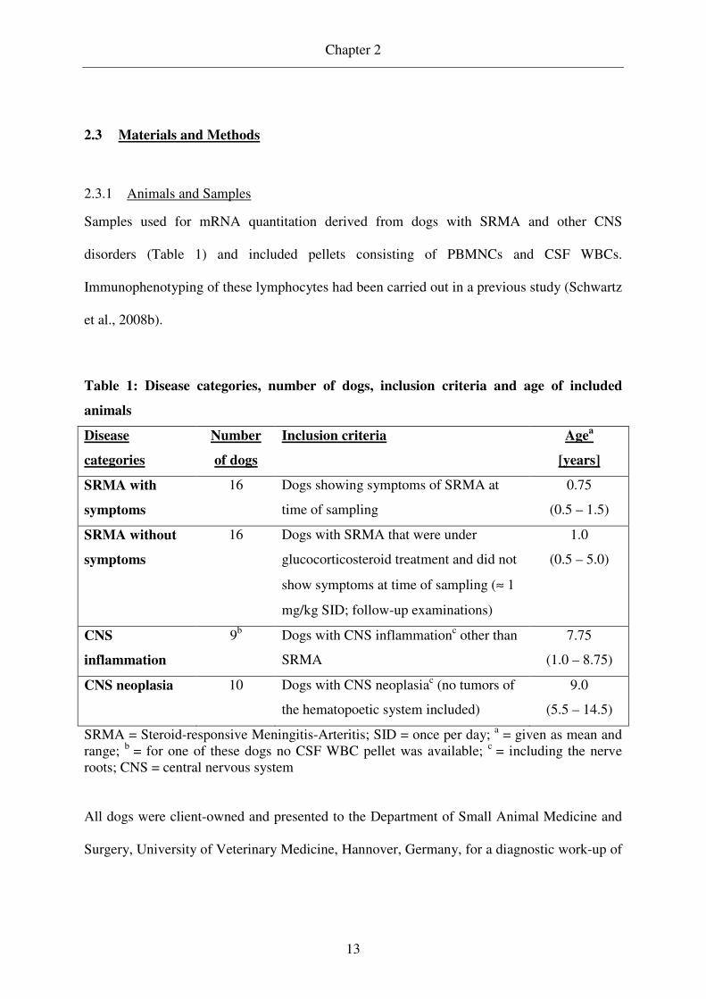
Table 1: Disease categories, number of dogs, inclusion criteria and age of included
animals
Disease
categories
Number
of dogs
Inclusion criteria Agea
[years]
SRMA with
symptoms
16 Dogs showing symptoms of SRMA at
time of sampling
0.75
(0.5 – 1.5)
SRMA without
symptoms
16 Dogs with SRMA that were under
glucocorticosteroid treatment and did not
show symptoms at time of sampling (≈ 1
mg/kg SID; follow-up examinations)
1.0
(0.5 – 5.0)
CNS
inflammation
9b Dogs with CNS inflammationc other than
SRMA
7.75
(1.0 – 8.75)
CNS neoplasia 10 Dogs with CNS neoplasiac (no tumors of
the hematopoetic system included)
9.0
(5.5 – 14.5)
SRMA = Steroid-responsive Meningitis-Arteritis; SID = once per day; a = given as mean and range; b = for one of these dogs no CSF WBC pellet was available; c = including the nerve roots; CNS = central nervous system
All dogs were client-owned and presented to the Department of Small Animal Medicine and
Surgery, University of Veterinary Medicine, Hannover, Germany, for a diagnostic work-up of

Chapter 2
14
neurological signs. Animals were ascribed to one of the following groups: “SRMA with
symptoms”, “SRMA without symptoms”, “CNS inflammation”, and “CNS neoplasia”. Each
patient was subject to a general and neurological examination, which was followed by a
complete blood count (CBC), routine blood chemistry, and urinalysis. In addition, a
suboccipital CSF tap and CSF analysis (WBC count, cytomorphological differentiation, and
total protein) were performed in every dog. Samples containing erythrocytes were accepted
only if xanthochromia, suggesting in vivo leakage of red blood cells into the subarachnoid
space due to impaired vascular integrity, was present (Jamison and Lumsden, 1988).
Immunoglobulin A (IgA) content in serum and CSF was determined in all animals using a
previously described enzyme-linked immunosorbent assay (ELISA) to support the diagnosis
of SRMA (Tipold et al., 1994). Ancillary tests performed to obtain a definitive diagnosis
included radiographic studies, ultrasonography, computed tomography (CT), magnetic
resonance imaging (MRI), as well as electrodiagnostic testing. If available, results of tests for
infectious agents and pathological examinations were considered to achieve the diagnosis. A
possible history of glucocorticosteroid administration within one week prior to presentation
(within 6 weeks if depot formulations were given) was noted for each patient.
Diagnosis of SRMA was based on the combination of the following findings: pain upon
cervical palpation, fever, marked neutrophilic leuko- and pleocytosis, immediate and
sustained response to glucocorticosteroid treatment, and exclusion of other disease processes
that may cause similar symptoms. Concomitant elevation of IgA in serum and CSF supported
the diagnosis (Tipold and Jaggy, 1994).
The groups “CNS inflammation” and “CNS neoplasia” included diagnoses of
meningoencephalitis of unknown etiology (n = 2), granulomatous meningoencephalitis (n =
1), CNS neosporosis (n = 2), acute polyradiculoneuritis (n = 2), bacterial meningomyelitis (n

Chapter 2
15
= 2), and intracranial meningioma (n = 5), pituitary macroadenoma (n = 1), malignant
blastoma of the spine/spinal cord (n = 2), and malignant nerve sheath neoplasia (n = 2).
Isolation of PBMNCs was achieved by means of density gradient centrifugation with two
subsequent washing steps (Schwartz et al., 2008b). Cerebrospinal fluid WBCs were spun
down (200 x g; 10 min; room temperature) and the supernatant was removed. Cerebrospinal
fluid WBC pellets therefore contained a mixture of leukocyte populations and were
immediately frozen and stored at - 80°C until RNA isolation was performed.
2.3.2 RNA purification and reverse transcription
Purification of total RNA from cell pellets was performed using RNeasy Mini Kit columns
(Qiagen) according to the manufacturer’s protocol for animal cells. To prevent contamination
with genomic DNA an on-column DNase digestion step with RNase-free DNase (Qiagen)
following the manufacturer’s instructions was included. The yield of RNA was subsequently
determined with a GeneQuant pro device (Biochrom Ltd.) by means of spectrophotometry at
OD260. The remaining eluate was immediately placed in liquid nitrogen and then stored at -
80°C until reverse transcription was undertaken.
Reverse transcription of RNA into complementary DNA (cDNA) was performed with
Omniscript RT (Qiagen), Random Primers (Promega), and RNaseOUTTM Recombinant
Ribonuclease Inhibitor (Invitrogen). Forty microliters of master mix containing ≤ 2 µg of total
RNA was produced according to Qiagen’s protocol for reverse transcription. Incubation for
60 min at 37°C took place in a thermocycler (PTC-200 Peltier Thermal Cycler; MJ
Research).Complementary DNA was stored at - 20°C in aliquots until its use in real-time
PCR experiments.

Chapter 2
16
2.3.3 Primers
Primers used in this study (Table 2 and 3) were either taken from the literature (as cited) or
designed with Primer3 software (Rozen and Skaletsky, 2000) or Beacon Designer version 2.1
software (Premier Biosoft International). Primers were produced by Eurofins MWG Operon.
The specificity of each PCR product was confirmed by DNA sequencing (AGOWA
genomics).
Table 2: Primers used for production of standard dilution series
Target
gene
Primer sequence in 5´- 3´ (forward primers on
top, reverse primers on bottom)
Size of
amplicon
[bp]
GenBank
accession
number
IL-2 ACCTCAACTCCTGCCACAAT 289 D30710
GCACTTCCTCCAGGTTTTTG
IFN-γa GCAAGTAATCCAGATGTATCG 283 S41201
TTATCGCCTTGCGCTGGACC
IL-4b CTGATTCCAACTCTGGTCTGC 283 AF239917
TTGCCATGCTGCTGAGGTTC
IL-5c AGGCAAACACTGAACATTTC 468 AF331919
TCTCCAAAATCTTCCACTAC
IL-10a CCTGGGTTGCCAAGCCCTGTC 212 U33843
ATGCGCTCTTCACCTGCTCC
GAPDHd AAGGTCGGAGTCAACGGATT 365 AB038240
GCAGAAGAAGCAGAGATGATG
β-actin AACACCCCAGCCATGTATGT 333 NM_001003349
CTTCTCCAGGGAGGACGAG
EF-1αe AGCCCTTGCGCCTGCCTCTC 219 X03558
CAGACACATTCTTGACATTGAAGC
bp = base pairs; IL = interleukin; IFN = interferon; GAPDH = glyceraldehyd-3-phosphate dehydrogenase; EF = elongation factor; a Markus et al. (2002); b Barnes et al. (2000); c Yang et al. (2001); d Puff et al. (2008); e von Smolinski et al. (2005)

Chapter 2
17
Table 3: Primers used for real-time PCR
Target
gene
Primer sequence in 5´- 3´(forward primers on
top, reverse primers on bottom)
Size of
amplicon
[bp]
GenBank
accession
number
IL-2 CCAACTCTCCAGGATGCTCAC 81 D30710
TCTGCTAGACATTGAAGGTGTGTA
IFN-γ AGCATGGATACCATCAAGGAAGA 104 S41201
AGATCGTTCACAGGAATTTGAATCA
IL-4 CTCCAAAGAACACAAGCGATAAGG 84 AF239917
TGTTGGAGCAGTTGTGTGTATAGA
IL-5 CCTATGTTTCTGCCTTTGCTGTAG 95 AF331919
GCCTATCAGCCAAGTTCGATGA
IL-10 GGTGGGAGCCAGCCGACACCAG 120 U33843
AAGAAGATCTTCACCCACCCGAAGG
GAPDHa GTCATCAACGGGAAGTCCATCTC 84 AB038240
AACATACTCAGCACCAGCATCAC
β-actin TCTACGAGGGGTACGCCTTG 149 NM_001003349
TTCCTTGATGTCACGCACGAT
EF-1αb AGCCCTTGCGCCTGCCTCTC 219 X03558
CAGACACATTCTTGACATTGAAGC
bp = base pairs; IL = interleukin; IFN = interferon; GAPDH = glyceraldehyd-3-phosphate dehydrogenase; EF = elongation factor; a Puff et al. (2008); b von Smolinski et al. (2005)
2.3.4 Standard dilution series
In order to allow calculation of a standard curve tenfold dilution series (102 – 108 copies) were
produced for each target gene. These dilution series also served as standards for inter-run
calibration to account for run-to-run variations. Amplicons were produced by conventional
end-point PCR (PTC-200 Peltier Thermal Cycler; MJ Research), agarose gel electrophoresis,
and DNA extraction with NucleoSpin Extract II columns (Macherey-Nagel) according to the
Chapter 2
18
manufacturer’s protocol. Spectrophotometrical determination of DNA concentration was
performed at OD260 (GeneQuant pro; Biochrom Ltd.) and copy numbers per volume were
calculated according to the following formula: copies/µl = 6 x 10²³ x DNA concentration
(ng/µl) x 10-9/[size of amplicon (base pairs) x 660].
Complementary DNA of canine PBMNC that were stimulated with concanavalin A (Con A)
(5µg/ml) for 24 h served as template for IL-2, -4, -5, and IFN-γ. Template for IL-10, GAPDH,
β-actin, and EF-1α was cDNA from DH-82 cells (Wellman et al., 1988). Reaction conditions
were as follows: initial denaturation at 94°C for 1 minute; 40 cycles of denaturation at 94°C
for 1 minute, annealing at 59°C (IL-2, IL-10, IFN-γ, GAPDH) / 60°C (β-actin, EF-1α) for 2
minutes and elongation at 72°C for 1 minute; final elongation at 72°C for 5 minutes. PCR
conditions for IL-4 and IL-5 were set according to Barnes et al. (2000) and Yang et al. (2001),
respectively. Amplification was achieved with recombinant Taq DNA Polymerase
(Invitrogen) in 1x PCR Buffer (Invitrogen) with 1.25 mM MgCl2 (Invitrogen), 0.2 mM dNTP
mixture (Applied Biosystems), and 300 nM of each primer (Table 2).
2.3.5 Real-time PCR
Real-time PCRs were run on an Mx3005P QPCR system (Stratagene) with Brilliant SYBR
Green QPCR Core Reagent Kit (Stratagene) in 8x Strip Tubes with Optical Cap (Stratagene).
Due to the number of samples, experiments for each target gene were run on separate plates
for PBMNC and CSF WBC samples. Measurements were conducted the same day using the
identical standard dilution series. Standards were kept at + 4°C in-between runs. The plates
included the standard dilution series as well as a no template control (NTC) in duplicate.
Reaction volume was 25 µl including 1 µl template per reaction.

Chapter 2
19
Reaction conditions were as follows: initial denaturation at 95°C for 10 minutes; 40 cycles
with denaturation at 95°C for 30 seconds, annealing at 59°C (IL-2, IL-5, IFN-γ) / 60°C (β-
actin, EF-1α) / 61°C (IL-10) / 64°C (IL-4, GAPDH) for 1 minute and elongation at 72°C for
30 seconds; final extension at 72°C for 1 minute; dissociation program beginning at 95°C for
1 minute followed by increasing the temperature by 0.5°C every 30 seconds from 55°C to
95°C. The master mix contained 0.025 U/µl SureStart Taq DNA Polymerase in 1x core PCR
buffer with 2.5 mM (IL-10, IFN-γ, GAPDH) / 5.0 mM (IL-2, IL-4, IL-5, β-actin, EF-1α)
MgCl2, 800 µM dNTP mix, 150 nM of each primer (Table 3), 8 % glycerol, and 4 % dimethyl
sulphoxide (DMSO) (3 % for IL-10, IFN-γ, GAPDH). Final SYBR Green concentration was
0.25x and Rox was used as a reference dye at 30 nM.
The software tool “Multiple Experiment Analysis” (MxPro QPCR Software v4.01;
Stratagene) allowed run-to-run comparison between the results for PBMNC and CSF WBC
samples.
To account for variations in sample input, extraction and reaction efficiencies, and presence
of inhibitors, copy numbers of the genes of interest were normalized against the geometric
mean of the internal reference genes GAPDH, β-actin and EF-1α: copiesnormalized (target gene)
[%] = copiesnon-normalized (target gene) x 100/ [copies (GAPDH) x copies (β-actin) x copies (EF-
1α)]^(1/3).
2.3.6 Statistical analysis
In addition to descriptive methods, the Wilcoxon rank sum test was applied for comparison of
the results deriving from the different disease categories. For these comparisons results of the
groups “CNS inflammation” and “CNS neoplasia” were combined. It was tested whether

Chapter 2
20
results of dogs with a history of glucocorticosteroid pretreatment differed significantly from
the non-pretreated ones. The Wilcoxon signed-rank test served as a tool to compare values
deriving from PBMNCs and CSF WBCs of dogs in the acute phase of SRMA. The
Spearman´s rank correlation coefficients (rSpear) were calculated to detect overall correlations
among the whole study population and correlations for “SRMA with symptoms”. The
parameters included normalized copy numbers and results of CBC, CSF, IgA level, and flow
cytometric lymphocyte subtype analysis (Schwartz et al., 2008b). Statistical significance was
set at the 5% level (p ≤ 0.05).
2.4 Results
Dissociation procedures at the end of real-time PCRs for IL-2, INF-γ, IL-4, IL-5, IL-10,
GAPDH, β-actin, and EF-1α, produced clearly defined single melting temperatures at 77,5°C,
79,1°C, 78,5°C, 79,6°C, 83,8°C, 81,1°C, 85,4°C, and 82,3°C, respectively. PCR products for
these genes were considered specific because temperatures corresponded with those of the
standard dilution series. These standards contained a single gene product that had been
sequenced and recognized as the respective gene of interest.
All reference genes could be measured in every PBMNC sample and normalization was
therefore always possible. However, copy numbers for only 1 or 2 of the reference genes
could be determined in 9 CSF WBC samples and these samples were excluded from further
analysis since an accurate normalization was not possible. These included 3 samples
belonging to the category “SRMA without symptoms”, 1 to “CNS inflammation”, and 5
samples to “CNS neoplasia”.

Chapter 2
21
Results of IL-2 mRNA expression analysis are displayed in Figure 1. After normalization
PBMNC samples of dogs in the acute phase of SRMA contained the least copy numbers
followed by dogs treated for SRMA. Although the distribution of values was fairly
homogenous within the group “SRMA with symptoms”, there was a large degree of variation
within the remaining groups.
Figure 1: Results of real-time PCR for IL-2 mRNA expression of PBMNCs (a) and CSF WBCs (b). Each
dot represents the copy number in one sample. Measured copy numbers are normalized to the geometric
mean of reference genes GAPDH, ββββ-actin, and EF-1αααα and are given as the percentage of the respective
geometric mean. Horizontal bars give median values. Asterisks indicate statistically significant differences
from the disease category “SRMA with symptoms” (** p ≤≤≤≤ 0.01). For purpose of statistical analysis
samples from “CNS inflammation” and “CNS neoplasia” were grouped as one disease category (IL =
interleukin; PBMNC = peripheral blood mononuclear cells; CSF WBC = cerebrospinal fluid white blood
cells; SRMA = Steroid-responsive Meningitis-Arteritis; GAPDH = glyceraldehyd-3-phosphate
dehydrogenase; EF = elongation factor).

Chapter 2
22
As displayed in Figure 2, IFN-γ mRNA was expressed least in PBMNC samples that derived
from individuals in the acute phase of SRMA and the level of expression was highest in
“CNS neoplasia”. Similarly to IL-2, IFN-γ expression was most homogenous in dogs with
symptoms of SRMA.
Figure 2: Results of real-time PCR for IFN-γγγγ mRNA expression of PBMNCs (a) and CSF WBCs (b). Each
dot represents the copy number in one sample. Measured copy numbers are normalized to the geometric
mean of reference genes GAPDH, ββββ-actin, and EF-1αααα and are given as the percentage of the respective
geometric mean. Horizontal bars give median values. Asterisks indicate statistically significant differences
from the disease category “SRMA with symptoms” (** p ≤≤≤≤ 0.01). For purpose of statistical analysis
samples from “CNS inflammation” and “CNS neoplasia” were grouped as one disease category. Please
note the non-uniform scaling of the 2 diagrams (IFN = interferon; PBMNC = peripheral blood
mononuclear cells; CSF WBC = cerebrospinal fluid white blood cells; SRMA = Steroid-responsive
Meningitis-Arteritis; GAPDH = glyceraldehyd-3-phosphate dehydrogenase; EF = elongation factor).
Chapter 2
23
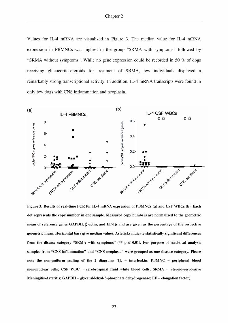
Values for IL-4 mRNA are visualized in Figure 3. The median value for IL-4 mRNA
expression in PBMNCs was highest in the group “SRMA with symptoms” followed by
“SRMA without symptoms”. While no gene expression could be recorded in 50 % of dogs
receiving glucocorticosteroids for treatment of SRMA, few individuals displayed a
remarkably strong transcriptional activity. In addition, IL-4 mRNA transcripts were found in
only few dogs with CNS inflammation and neoplasia.
Figure 3: Results of real-time PCR for IL-4 mRNA expression of PBMNCs (a) and CSF WBCs (b). Each
dot represents the copy number in one sample. Measured copy numbers are normalized to the geometric
mean of reference genes GAPDH, ββββ-actin, and EF-1αααα and are given as the percentage of the respective
geometric mean. Horizontal bars give median values. Asterisks indicate statistically significant differences
from the disease category “SRMA with symptoms” (** p ≤≤≤≤ 0.01). For purpose of statistical analysis
samples from “CNS inflammation” and “CNS neoplasia” were grouped as one disease category. Please
note the non-uniform scaling of the 2 diagrams (IL = interleukin; PBMNC = peripheral blood
mononuclear cells; CSF WBC = cerebrospinal fluid white blood cells; SRMA = Steroid-responsive
Meningitis-Arteritis; GAPDH = glyceraldehyd-3-phosphate dehydrogenase; EF = elongation factor).

Chapter 2
24
Normalized values of IL-5 mRNA transcripts are given in Figure 4. Results for PBMNCs
were comparable among the disease categories with slightly higher median values in “SRMA
without symptoms” and “CNS neoplasia”.
Figure 4: Results of real-time PCR for IL-5 mRNA expression of PBMNCs (a) and CSF WBCs (b). Each
dot represents the copy number in one sample. Measured copy numbers are normalized to the geometric
mean of reference genes GAPDH, ββββ-actin, and EF-1αααα and are given as the percentage of the respective
geometric mean. Horizontal bars give median values. Asterisks indicate statistically significant differences
from the disease category “SRMA with symptoms” (* p ≤≤≤≤ 0.05; ** p ≤≤≤≤ 0.01). For purpose of statistical
analysis samples from “CNS inflammation” and “CNS neoplasia” were grouped as one disease category.
Please note the non-uniform scaling of the 2 diagrams (IL = interleukin; PBMNC = peripheral blood
mononuclear cells; CSF WBC = cerebrospinal fluid white blood cells; SRMA = Steroid-responsive
Meningitis-Arteritis; GAPDH = glyceraldehyd-3-phosphate dehydrogenase; EF = elongation factor).

Chapter 2
25
Figure 5 shows the quantity of mRNA transcripts for IL-10. Median values in PBMNC pellets
did not differ significantly among the different disease categories, a greater degree of
dispersion, however, was noticeable in dogs with neoplastic and non-SRMA inflammatory
disorders.
Figure 5: Results of real-time PCR for IL-10 mRNA expression of PBMNCs (a) and CSF WBCs (b). Each
dot represents the copy number in one sample. Measured copy numbers are normalized to the geometric
mean of reference genes GAPDH, ββββ-actin, and EF-1αααα and are given as the percentage of the respective
geometric mean. Horizontal bars give median values. Asterisks indicate statistically significant differences
from the disease category “SRMA with symptoms” (** p ≤≤≤≤ 0.01). For purpose of statistical analysis
samples from “CNS inflammation” and “CNS neoplasia” were grouped as one disease category. Please
note the non-uniform scaling of the 2 diagrams (IL = interleukin; PBMNC = peripheral blood
mononuclear cells; CSF WBC = cerebrospinal fluid white blood cells; SRMA = Steroid-responsive
Meningitis-Arteritis; GAPDH = glyceraldehyd-3-phosphate dehydrogenase; EF = elongation factor).

Chapter 2
26
Pretreatment with glucocorticosteroids, after exclusion of “SRMA without symptoms”, was
recorded for 8 dogs (4/16 in “SRMA with symptoms”, 3/9 in “CNS inflammation”, 1/10 in
“CNS neoplasia”) and did not cause significantly differing results in CBC, CSF analysis, flow
cytometrically determined lymphocyte subsets, and mRNA expression of genes of interest,
except for a more severe pleocytosis (p < 0.05), higher CSF IgA content (p < 0.01), and
increased PBMNC IL-10 mRNA levels (p < 0.05) in pretreated dogs. Studying dogs in the
acute phase of SRMA separately, the only significant difference that could be detected were
lower IL-10 mRNA levels in CSF WBCs of pretreated individuals (p < 0.05).
The IL-4:IL-2 ratio was used as an indicator for the Th2:Th1 ratio and was highest in
PBMNCs of “SRMA with symptoms” (median: 0.77; range: 0 – 8.84). Values were
significantly higher when compared to “CNS inflammation” + “CNS neoplasia” (p ≤ 0.05)
(median: 0.03; range: 0 – 1.42), whereas differences to “SRMA without symptoms” did not
reach the level of significance (p = 0.338) (median: 0.20; range: 0 – 8.84). The Th2:Th1 ratio
in peripheral blood of dogs in the acute phase of SRMA did not differ significantly from that
in the CSF (p = 0.826) (median: 0.22; range: 0 – 1.79).
In CSF WBC pellets belonging to “SRMA with symptoms” the expression of all cytokines
could be recorded, whereas they were found only in single samples of the “CNS
inflammation” and “CNS neoplasia” group. Moreover, none were detected in the “SRMA
without symptoms” group. Normalized values for cytokine mRNA in CSF cell pellets were
below those of PBMNCs. These differences were statistically significant for IL-4, IL-5, and
IFN-γ (p ≤ 0.05). An exception were IL-10 mRNA levels, which were higher in CSF WBCs
when compared to PBMNC levels. Cerebrospinal fluid WBC pellets contained a considerable

Chapter 2
27
proportion of neutrophils that also contributed to the normalization factors, calculated from
reference gene mRNA expression levels, thus causing a certain “dilution effect”.
For the entire study population no statistically significant correlation could be identified
between cytokine mRNA expression and B:T cell or CD4:CD8α T cell ratios. Correlations
that reached the level of significance were weak (rSpear < ± 0.5) and reflect that findings
typically present in SRMA (marked neutrophilic leuko- and pleocytosis + increased IgA
levels) are associated with low mRNA expression for IL-2 and IFN-γ and strong expression
for IL-4 in PBMNCs (Table 4).
Table 4: Correlations between quantity of mRNA expression and other tested
parameters (for the entire study population)
Cytokine Cellular response Spearman´s rank
correlation
coefficient
Level of
significance
IL-2 in PBMNCs Degree of neutrophilic
leukocytosis - 0.393** p = 0.004
Degree of pleocytosis - 0.365** p = 0.008
IgA level in serum - 0.403** p = 0.002
IFN-γ in PBMNCs IgA level in CSF - 0.319* p = 0.022
IL-4 in PBMNCs Degree of neutrophilic
leukocytosis
+ 0.365** p = 0.008
IL = interleukin; PBMNCs = peripheral blood mononuclear cells; IgA = immunoglobulin A; IFN = interferon; CSF = cerebrospinal fluid

Chapter 2
28
Within “SRMA with symptoms” high expression of IFN-γ in PBMNCs was associated with a
low B:T cell ratio in the peripheral blood (rSpear = - 0.487; p ≤ 0.05) and strong IL-10
expression in CSF WBCs correlated with milder degrees of pleocytosis (rSpear = - 0.500; p ≤
0.05).
2.5 Discussion
Steroid-responsive Meningitis-Arteritis is frequently diagnosed in dogs (Battersby et al.,
2006; Meric, 1988; Tipold, 1995) and our knowledge about the pathogenesis of SRMA is still
limited. The disease is suspected to arise from a dysregulation of the immune system and the
presented data show that SRMA is associated with a type 2-skewed immune response. In a
previous study we could demonstrate that the acute phase of SRMA is associated with a
lymphocyte shift towards high proportions of B cells, in the peripheral blood and particularly
obvious in the CSF. This observation correlates positively with high IgA levels (Schwartz et
al., 2008b). Immunoglobulin A production is known to be positively influenced by IL-4, -5, -
6, and -10 (Briere et al., 1994; Murray et al., 1987; Ramsay et al., 1994), all belonging to the
classical Th2 cytokine spectrum (Abbas et al., 1996). We therefore hypothesized that T helper
cells in SRMA predominantly belong to the Th2 subset and thus investigated the cytokine
expression pattern in PBMNCs and CSF WBCs by means of reverse-transcriptase real-time
PCR. Values from dogs in the acute phase of SRMA were compared to those from dogs under
treatment and dogs with other inflammatory and neoplastic diseases of the CNS. In order to
assure that regulation of reference genes does not influence the results, data were normalized
to a set of three reference genes each belonging to a different functional class in cell

Chapter 2
29
metabolism thus rendering a simultaneous up- or down-regulation unlikely (Vandesompele et
al., 2002).
Indeed, PBMNCs of dogs with signs of SRMA expressed low levels of IL-2 and IFN-γ,
whereas IL-4 expression was higher in these samples. These findings are compatible with a
polarized type 2 immune response with a pronounced production of IL-4. In contrast, IL-5
and -10 mRNA levels were within the same range as those from dogs with other
inflammatory and neoplastic CNS disorders. In general, there was a strong overlap of values
for all cytokines among the different disease categories and variations of values were most
pronounced in “CNS inflammation” and “CNS neoplasia” reflecting the heterogeneity of
these groups.
Polarization of the immune response is a dynamic process that progresses and thus requires
time to develop. Polarization is therefore most noticeable in chronic diseases while early
states of inflammation are usually associated with mixed patterns of cytokine production
(London et al., 1998; O'Garra, 1998). This explains our findings of a mixed cytokine pattern
with a predominance of type 2 associated cytokines in dogs with SRMA. Due to the highly
painful nature of the disease dogs are usually referred to specialty facilities within hours to
few days after onset of clinical signs and this time span may be too short to develop full-
blown polarization of the immune response. In addition, it was realized that the Th1/Th2
paradigm is an oversimplification for the description of in vivo inflammations in many cases.
It is now suggested that Th1 and Th2 cells with their classical cytokine expression patterns
rather represent extreme phenotypes and that there is a large variety of cytokine expression
patterns in single CD4+ T cells. On a population level, however, the cytokine profile may
certainly be skewed in either type 1 or type 2 direction (Kelso, 1995; London et al., 1998) as
found in SRMA. Whether inflammatory reactions tend to polarize towards a type 1 or 2

Chapter 2
30
immune response depends on multiple factors. Among the promoters for type 2 immune
responses are a predominance of IL-4 in the microenvironment of naïve T helper cells and
increasing dosages of antigen (Abbas et al., 1996; London et al., 1998; Mosmann and Sad,
1996; Romagnani, 1997). The latter and the finding that repeated T helper cell stimulation
promotes type 2 immune responses make environmental antigens typical elicitors of
inflammatory reactions with high IL-4 levels (Abbas et al., 1996). Involvement of such an
environmental factor in the pathogenesis of SRMA has been suggested earlier (Tipold et al.,
1999). There is also some evidence that antigen-presentation by B cells stimulates Th2 cell
development (Abbas et al., 1996), which is of interest in light of the finding that high
percentages of B cells circulate in peripheral blood and CSF of dogs with SRMA (Schwartz et
al., 2008b). Another factor that participates in determination which route an inflammatory
reaction takes is the genetic background of the host (London et al., 1998; O'Garra, 1998).
Although no genetic factor could be identified to date, the fact that certain breeds are over-
represented in the population of diseased dogs (Behr and Cauzinille, 2006; Cizinauskas et al.,
2000; Harcourt, 1978; Scott-Moncrieff et al., 1992; Tipold and Jaggy, 1994) suggests that a
certain genetic background could facilitate the development of SRMA, possibly because of a
more pronounced tendency to develop a type 2 immune reaction.
Although IL-5 is one of the signature cytokines of Th2 cells, levels of expression were within
a comparable range among the disease categories, which can be explained by the fact that
each cytokine gene may be expressed independently (Kelso, 1995; London et al., 1998).
Interleukin-5 was ascribed an important role in the differentiation of IgA B cells into IgA-
secreting plasma cells (Harriman et al., 1988) and presence of IL-4 further boosts this
secretion (Murray et al., 1987). Messenger RNA encoding for IL-5 could be detected in each
PBMNC pellet of dogs with SRMA, both in the acute phase and under treatment, suggesting

Chapter 2
31
that IL-5, with the aid of IL-4, contributes to increased IgA levels and is most probably
responsible for persistently elevated levels in many dogs under glucocorticosteroid treatment
(Cizinauskas et al., 2000).
The expression level of the immunomodulatory cytokine IL-10 did not show considerable
differences in PBMNC samples of the different disease categories. Whereas IL-10 was
originally considered a typical Th2 cytokine, it is now clear that also lymphocytes of the Th1
subset as well as B cells and monocytes, amongst others, are capable to produce IL-10.
Regulatory T cells (TReg) are now considered a major source of IL-10 (Taylor et al., 2006).
This cytokine acts suppressive on both Th1 and Th2 cells and given the strong, presumably
overshooting, immune response in SRMA it is not surprising that no up-regulation of this
cytokine could be recognized in PBMNCs. The finding of increased IL-10 mRNA levels in
CSF WBCs when compared to PBMNCs may represent an endogenous response for CNS
protection. In addition to its immunosuppressive function, IL-10 also has the potential to
induce differentiation of B cells into IgA secreting plasma cells (Briere et al., 1994).
Comparison of mRNA expression levels in PBMNCs of dogs in the acute phase of SRMA
and after 2 months of glucocorticosteroid treatment revealed a tendency towards a
“depolarization” of the immune response with single individuals exhibiting an even more
pronounced type 2 immune response. Glucocorticosteroids are known to inhibit the
transcription of both Th1 and Th2 associated cytokines, long-term treatment, however, tends
to result in a dominance of Th2 cells (Elenkov, 2004; Liberman et al., 2009). This may
explain that IgA levels remain elevated in a number of individuals despite treatment with
clinical improvement (Cizinauskas et al., 2000).
In the present study we compared results of dogs suffering from SRMA with inflammatory
and neoplastic diseases of the CNS. It was shown that CNS neoplasias tend to be associated

Chapter 2
32
with serum cytokine levels that are shifted towards a type 2 immune response (Kumar et al.,
2006). The same applies for bacterial infections of the CNS (Raziuddin et al., 1995).
Therefore, one can assume that the overall results of the groups “CNS inflammation” and
“CNS neoplasia” are rather shifted towards a Th2-dominated than towards a Th1-dominated
immune response, indicating that SRMA is associated with an even more pronounced shift
towards an IL-4 dominated type 2 immune response.
2.6 Conclusion
Canine SRMA is associated with a lymphocyte shift towards high proportions of B cells and
increased IgA levels. The presented data on mRNA expression levels suggest that the immune
response in SRMA tends to polarize towards a type 2 immune reaction. The inflammation is
characterized by comparatively high IL-4 and low IL-2 and IFN-γ levels in PBMNCs,
whereas IL-5 and IL-10 expression was similar to those of dogs suffering from other
inflammatory and neoplastic CNS disorders. Cytokine profiles in CSF WBCs showed a
similar Th2:Th1 ratio to that in PBMNCs. We thus conclude that distribution of Th1 and Th2
subsets does not differ significantly between peripheral blood and CSF and that the type 2
immune reaction represents an important pathogenetical factor for high systemic and
intrathecal IgA production in the acute phase of SRMA and under glucocorticosteroid
treatment.
2.7 Acknowlegements
This work was supported by a Georg-Christoph-Lichtenberg-Scholarship donated by the
Department of Science and Culture of the federal state of Lower Saxony, Germany (MS), and

Chapter 2
33
a grant from the Frauchiger Foundation, Switzerland (VMS). In addition, we thank Dr. E.A.
Orlando for kindly providing us with primers for β-actin and Drs. M. and R. Kreutzer for their
excellent technical advice.
2.8 Conflict of interest statement
None of the authors of this paper has a financial or personal relationship with other people or
organizations that could inappropriately influence or bias the content of the paper.

Chapter 3
34
Chapter 3: Selective CD11a upregulation on neutrophils in the acute phase
of Steroid-responsive Meningitis-Arteritis in dogs
M. Schwartza,b*, R. Carlsona, A. Tipolda,b
a Department of Small Animal Medicine and Surgery, School of Veterinary Medicine
Hannover, Bischofsholer Damm 15, 30173 Hannover, Germany
b Center for Systems Neuroscience, School of Veterinary Medicine Hannover, Buenteweg 17,
30559 Hannover, Germany
*Corresponding author. Tel.: +49-511-856-8965; fax: +49-511-856-7686; e-mail:
Vet Immunol Immunopathol 126, 248-255.

Chapter 3
35
3.1 Abstract
Steroid-responsive Meningitis-Arteritis (SRMA) is a systemic inflammatory disease of
juvenile to young adult dogs with a relapsing course and most prominent manifestation in the
cervical meninges. The most important laboratory finding is a marked neutrophilic
pleocytosis.
Integrin (CD11a, b, c) expression on polymorphonuclear cells (PMNs) was quantified by
immunophenotyping and subsequent flow cytometric measurements. Values were determined
for peripheral blood in the acute phase of SRMA (n = 14) as well as during
glucocorticosteroid treatment (n =16). Results were compared to those from dogs with other
neurological diseases (n = 49) and healthy individuals (n = 7). Integrin expression was also
investigated on PMNs deriving from cerebrospinal fluid (CSF) of dogs in the acute phase of
SRMA (n = 14). In a second part of the study PMNs of healthy dogs were incubated with sera
of dogs in the acute phase of SRMA (n = 12). The influence on integrin expression was
studied and results were compared to those after incubation with pooled sera of dogs suffering
from idiopathic epilepsy (n = 3).
PMNs in peripheral blood of dogs in the acute phase of SRMA showed higher values of
CD11a expression when compared to dogs under treatment and to control groups, whereas
CD11b and c expression was comparable among the different groups. In the acute phase of
SRMA CD11b expression on PMNs in CSF was increased in comparison to that in peripheral
blood. Incubation with SRMA sera caused a stronger upregulation of CD11a than did pooled
epilepsy sera in 9/12 cases whereas an upregulation of CD11b and c was observed in single
cases only.

Chapter 3
36
High CD11a expression on PMNs in peripheral blood appears to be an important factor in the
pathogenesis of SRMA. This integrin is known to be essential for adhesion of PMNs within
the neutrophil recruitment cascade and therefore might mediate the enhanced invasion of
neutrophils into the subarachnoidal space eventually leading to meningitis and clinical signs.
Since sera of dogs suffering from SRMA selectively induce an upregulation of CD11a it can
be suspected that this fluid contains one or multiple factors being responsible for this.
Key words: canine, CNS immune response, compartmentalization, flow cytometry,
immunoglobulin A, integrin
Abbreviation list: CBC = complete blood cell count; CNS = central nervous system; CSF =
cerebrospinal fluid; IgA = immunoglobuline A; IL = interleukin; KS = Kawasaki Syndrome;
mAb = monoclonal antibody; PBS = phosphate buffered saline; PMN = polymorphonuclear
cell; SRMA = Steroid-responsive Meningitis-Arteritis; WBC = white blood cell
Chapter 3
37
3.2 Introduction
Steroid-responsive Meningitis-Arteritis (SRMA) is a systemic inflammatory disease with a
relapsing course. Juvenile to young adult dogs are affected and inflammation is most
prominent in the cervical meninges. In addition, extraneural signs may be detected elsewhere,
e.g., in the extramural coronary arteries (Hayes et al., 1989; Scott-Moncrieff et al., 1992).
There is a breed predisposition for Boxers, Bernese Mountain Dogs and Beagles, however,
multiple breeds and crossbred dogs have also been reported to be affected (Meric et al., 1985;
Scott-Moncrieff et al., 1992; Tipold and Jaggy, 1994). SRMA is a naturally occurring animal
model for several human vasculitides of unknown origin, including Kawasaki Syndrome (KS)
and Polyarteritis nodosa (Burns et al., 1991; Snyder et al., 1995). Furthermore, SRMA allows
studies on the general phenomenon of compartmentalization of immune responses into the
subarachnoid space.
Most prominent laboratory findings in SRMA are a marked neutrophilic leuko- and
pleocytosis as well as an increase in immunoglobuline (Ig) A levels in serum and
cerebrospinal fluid (CSF) (Tipold and Jaggy, 1994).
Neutrophilic pleocytosis is an uncommon finding in neurologic diseases of dogs. In addition
to SRMA, it is also observed in a small percentage of dogs with intracranial meningiomas
(Dickinson et al., 2006) and in dogs suffering from bacterial central nervous system (CNS)
infection. These bacterial meningoencephalomyelitides, however, are exceptionally rare in
dogs (Radaelli and Platt, 2002). Attraction of neutrophils into the CNS can, amongst others,
be mediated by interleukin-8 (IL-8 = CXCL8) and growth-related gene product alpha (GRO-
α = CXCL1), which both are significantly upregulated in CSF of human patients with
bacterial meningitis (Spanaus et al., 1997). And in fact, Burgener et al. (1998) could show
Chapter 3
38
that also in CSF of dogs with SRMA, chemotactic activity for neutrophils is increased and
that this is, at least in part, caused by IL-8 or an IL-8-like factor. Since high chemotactic
activity persists during successful glucocorticosteroid treatment with clearance of pleocytosis,
Burgener and colleagues suspected that a decrease in endothelial adhesion might be
responsible for the lack of neutrophil invasion into the CSF. This endothelial adhesion, which
is followed by crawling of the migrating white blood cells (WBCs) along the vessel wall, is
crucial for the extravasation of WBCs. Adhesion is mediated by CD11a whereas CD11b is
responsible for crawling (Phillipson et al., 2006). Linked to CD18 these leukocytic surface
molecules and CD11c form a heterodimer and are referred to as β2 integrins (Abbas and
Lichtman, 2003). Blockage of CD18 in rabbits with experimentally induced bacterial
meningitis could prevent the development of pleocytosis, confirming the importance of these
molecules in the pathogenesis of meningitis (Tuomanen et al., 1989).
Since neutrophil migration into CSF is a key feature of SRMA and CD11a, b and c might
play a crucial role in this event, the purpose of the study was to characterize the pattern of
integrin expression in blood and CSF of dogs with SRMA and to compare these findings to
those of various control groups. Furthermore we investigated if sera of SRMA patients
contain substances, which have the ability to induce changes in β2 integrin expression on non-
activated neutrophils in vitro.

Chapter 3
39
3.3 Materials and Methods
3.3.1 Animals and Samples
Blood samples to determine in vivo integrin expression on polymorphonuclear cells (PMNs)
in peripheral blood, derived from 71 dogs of various breeds and both sexes. All dogs were
presented in the Department of Small Animal Medicine and Surgery, University of Veterinary
Medicine, Hannover, Germany between July 2006 and March 2008 because of neurological
disorders. The categorization into one of the groups “SRMA with symptoms”, “SRMA
without symptoms”, “Idiopathic epilepsy”, “Spinal cord traumata”, “Inflammatory diseases”,
“Neoplastic diseases” and “Miscellaneous” (Table 1) was based on results of general and
neurological examinations and laboratory testing (blood, CSF, urine) under consideration of
electrodiagnostic studies (electromyography, nerve conduction velocity studies,
electroencephalography) and imaging findings (X-ray, computed tomography, magnetic
resonance imaging). In those cases where results of a pathological examination were
available, these contributed to the diagnoses. Diagnosis of SRMA was based on the presence
of the following findings: fever, cervical pain, neutrophilic leukocytosis, neutrophilic
pleocytosis, increased IgA levels in serum and CSF, immediate and sustained response to
glucocorticosteroid treatment and the absence of other causes for these signs (Tipold and
Jaggy, 1994). Possible glucocorticosteroid treatment within seven days prior to sampling (in
case of depot formulations within six weeks prior to sampling) was noted for all dogs. The
integrin expression was also determined in seven clinically healthy Beagle dogs summarized
as “Healthy controls”. Health status in these animals was determined by general examination
and complete blood cell count (CBC). In dogs with symptoms of SRMA (n = 14) integrin
expression was also investigated on PMNs in CSF.

Chapter 3
40
Table 1: Disease categories (age given as mean and range [years])
Disease
categories
Number
of dogs
Inclusion criteria Age
SRMA with
symptoms
14a Dogs showing symptoms of SRMA at
time of sampling
1.0
(0.5 – 1.5)
SRMA without
symptoms
16 Dogs with SRMA that were under
glucocorticosteroid treatment and did not
show symptoms at time of sampling
(control examinations)b
1.0
(0.5 – 5.0)
Idiopathic
epilepsy
6 Dogs with idiopathic epilepsy 3.75
(1.75 – 8.5)
Spinal cord
traumata
6 Dogs with intervertebral disk
protrusion/herniation, vertebral
subluxation
8.25
(6.0 – 11.25)
Inflammatory
diseases
12 Dogs with inflammatory diseases of the
CNS or PNS
7.5
(1.0 – 9.75)
Neoplastic
diseases
10 Dogs with CNS neoplasia 9.0
(4.25 – 14.5)
Miscellaneous 15 Dogs with neurological symptoms due to
other diseases
6.0
(1.0 – 10.5)
Healthy controls 7 Healthy dogs 5.0
(4.75 – 6.5)
SRMA = Steroid-responsive Meningitis-Arteritis; a = eight individuals are also included in “SRMA without symptoms” at a later date (follow up study); b = one dog did not receive medication and was in a subclinical phase of SRMA; CNS = central nervous system; PNS = peripheral nervous system

Chapter 3
41
For flow cytometric quantification of granulocytic CD11a, b and c expression in peripheral
blood samples 10 ml blood was collected from either the cephalic or saphenal vein in tubes
containing ethylene diamine tetraacetic acid (EDTA). Mean WBC count of dogs with SRMA
was 27 500 WBCs/µl (range: 16 200 – 48 000/µl). CSF samples were taken from the
cerebellomedullary cistern in general anesthesia with dogs being in lateral recumbency. CSF
analysis was performed immediately after sample collection and consisted of cell count,
cytomorphological differentiation and total protein content determination. Samples containing
erythrocytes were only included in this study if CSF was xanthochrome and blood
contamination was therefore considered to be due to arteritis (Jamison and Lumsden, 1988).
Mean WBC count in CSF of dogs with SRMA was 1 670 WBCs/µl (range: 18 – 8 534/µl).
IgA levels in serum and CSF were determined at time of sampling using an enzyme linked
immunosorbent assay (ELISA) system established by Tipold et al. (1994).
Whether an integrin upregulation could be induced by incubation of non-activated
granulocytes with serum of dogs with SRMA was tested in a second part of the study. Three
healthy large breed dogs served as PMN donors. To ensure that neutrophils of these dogs
were in a physiological activation state a general examination and CBC were carried out in
each dog. Twenty-five milliliter blood of each donor dog was collected in EDTA tubes as
described above.
3.3.2 Monoclonal antibodies (mAbs)
Monoclonal antibodies (mAb) of murine origin and directed against canine surface antigens,
and their dilutions used in this study are listed in Table 2. Primary antibodies belonged to the
IgG1 isotype and were purchased from Serotec (Duesseldorf, Germany). An F(ab´)2-fragment

Chapter 3
42
specific RPE-labeled goat-anti-mouse IgG antibody (Dianova, Hamburg, Germany) served as
secondary antibody (1:200 dilution).
Table 2: Monoclonal antibodies
Target antigen Clone Dilution
CD11a CA11.4D3 1:300
CD11b CA16.3E10 1:6
CD11c CA11.6A1 1:6
IgG1 negative control W3/25 1:6
3.3.3 Sample processing for the ex vivo examination
The method of indirect membrane immunofluorescence with subsequent flow cytometric
measurement was elected for determination of integrin expression.
Sample processing for the first part of the study, addressing the in vivo CD11a, b and c
expression on peripheral blood PMNs, was initiated immediately after venipuncture. PMNs
were obtained by means of density gradient centrifugation as described by Danilenko et al.
(1992) with the exception that phosphate buffered saline (PBS) was used instead of 6 %
dextran for dilution of blood and no autologous plasma was added to PBS for washing
isolated PMNs. After the separation cell viability was determined by trypan blue exclusion
and PMNs were counted using a hemocytometer. Cell suspension was then adjusted to 5 x 10⁵
vital PMNs/50 µl by adding CellWASH solution (BD Biosciences, Erembodegem, Belgium).
To prevent unspecific Fc-receptor binding of mAbs the cell suspension was incubated with
heat-inactivated (56°C; 30 min) pooled goat serum for 10 min (1:11 dilution) (Schwartz,
2007). Negative controls consisted of an isotype-matched primary antibody and incubation
with the secondary antibody only. For each mAb 50 µl of cell suspension was used. A 30-min

Chapter 3
43
incubation period for the primary antibodies was followed by two washing steps at 200 x g
for 6 min using CellWASH solution. An incubation period with the secondary antibody (30
min) and subsequent washing steps identical to the before mentioned ones followed.
Immediately after the second washing step cell suspensions were diluted with BD
FACSFlowTM solution (BD Biosciences, Erembodegem, Belgium) in a ratio of 2:5 and
integrin expression was flow cytometrically quantified using a FACSCaliburTM device with
BD CellQuestTM Pro Version 5.2.1 software (Becton Dickinson, Heidelberg, Germany).
Preparation of PMNs from CSF was initiated immediately after tapping. CSF was centrifuged
at 200 x g for 10 min and subsequently the supernatant was removed. The absolute number of
leukocytes was calculated from CSF cell count and cell concentration was then adjusted to 5 x
105 leukocytes/50 µl by adding CellWASH solution. Blocking of Fc-receptors, washing steps
and flow cytometric measurements were equal to those used for peripheral blood PMNs,
duration of incubation with antibodies, however, was reduced to 15 min to minimize cell loss.
In one dog only CD11b staining was performed.
3.3.4 Sample processing for the in vitro study
In the second part of the study changes in integrin expression on non-activated PMNs
following incubation with sera of dogs with SRMA in comparison to dogs with idiopathic
epilepsy were investigated. Three experiments were performed. In each experiment
granulocytes of one donor dog were incubated with sera of four dogs with SRMA and with
pooled sera from three epileptic dogs, which all had a normal general examination and
laboratory results (incl. IgA levels) within reference range. Incubation with PBS served as an
additional negative control. The method for isolation of PMNs was identical to that described

Chapter 3
44
above (2.3. Sample processing for the ex vivo examination). Twenty microliter of serum was
added to 100 µl of cell suspension containing 3 x 10⁵ PMNs (adjusted with PBS) and a 25-
min incubation period (37°C; 5 % CO2) followed. All samples were processed as duplicates.
After this period PMNs were washed three times at 200 x g for 6 min using CellWASH.
Immunostaining was performed as described in the first part of the study (2.3. Sample
processing for the ex vivo examination) with the exception that pooled goat serum was not
used as a blocking agent to prevent interference of different sera types. Negative controls as
used in the first part of the study helped to rule out unspecific mAb binding. All sera were
stored at – 20°C prior to usage.
3.3.5 Flow cytometric analysis
Ten thousand events per sample were flow cytometrically registered. Setting of the region
containing granulocytes was based on their size and complexity characteristics as illustrated
in Figure 1.
Figure 1: Result of flow cytometric
measurement of polymorphonuclear
cells (PMNs) from peripheral blood
displayed as dot plot. PMNs were
isolated by means of density gradient
centrifugation. Setting of PMN
containing region was based on size
and complexity characteristics of cells.
Note the absence of any lymphocytic
contamination.

Chapter 3
45
Mean fluorescence values were used for statistical analysis. In the second part of the study the
arithmetic mean was calculated from values deriving from duplicate samples. For comparison
of these values among the three experiments, fluorescence following incubation with pooled
“Idiopathic epilepsy” sera (FIE) was given a value of 100. Values resulting from incubation
with PBS and with sera of dogs with SRMA (FA) were put in relation to this given value
resulting in a relative value (FR). The following formula was used for this purpose: FR = FA x
100/FIE.
3.3.6 Preliminary experiments
Preliminary experiments were performed to determine PMN viability at the time of flow
cytometric measurement. Staining with To-Pro-3 (Molecular ProbesTM, Leiden, The
Netherlands) resulted in > 99 % vital cells within the PMN region.
3.3.7 Statistical analysis
In addition to descriptive methods the Wilcoxon rank sum test was applied for comparison of
the results deriving from the groups “SRMA with symptoms” and “SRMA without
symptoms”. Results from these two groups were also compared to those of the remaining
disease categories. The Wilcoxon signed-rank test served as a tool to compare values deriving
from blood and CSF of the same individual. The Spearman´s rank correlation coefficients
were calculated to detect correlations of parameters within the two groups of SRMA. These
parameters included neutrophil count in peripheral blood and CSF, IgA levels in serum and

Chapter 3
46
CSF and values of in vivo integrin expression. Statistical significance was set at the 5 % level
(P < 0.05).
3.4 Results
Three out of 14 dogs with symptoms of SRMA received a glucocorticosteroid treatment prior
to sampling. In the group of animals with SRMA not showing any symptoms all but one dog
where under glucocorticosteroid therapy with daily doses ranging from 0.8 – 1.0 mg/kg
prednisolone. This one dog was in an asymptomatic phase of the disease at the time of
sampling.
3.4.1 Results of the ex vivo examination
The results of the first part of the study addressing the quantification of integrin expression on
PMNs in peripheral blood are illustrated as Box-and-Whisker Plots in Figure 2 a-c. The
median value for fluorescence intensity after staining of CD11a was highest in the group of
dogs suffering from symptoms of SRMA. Values within this group were significantly higher
(P < 0.05) compared to the remaining disease categories with the exception of “Spinal cord
traumata” and “Healthy controls”. Distribution of values showed a wide range within “SRMA
with symptoms” with some individuals expressing as little CD11a as dogs in the other groups.
However, only five dogs not displaying clinical signs of SRMA at time of sampling (i.e. < 7
%) showed higher CD11a expression than the median value of “SRMA with symptoms”.
There was no more than one of these individuals per group. Determination of CD11b
expression revealed a comparatively wide range of results throughout all groups except for
“Healthy controls”. CD11b expression in dogs with signs of SRMA and under therapy was

Chapter 3
47
significantly lower (P < 0.05) than that of healthy Beagle dogs, which was highest among all
groups. Also median expression of CD11c was highest in “Healthy controls”. CD11c
expression in dogs displaying symptoms of SRMA was significantly higher (P < 0.05) than
that of dogs with CNS neoplasia
(a)
(b)
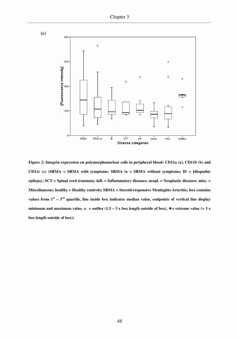
Chapter 3
48
Figure 2: Integrin expression on polymorphonuclear cells in peripheral blood: CD11a (a), CD11b (b) and
CD11c (c) (SRMA = SRMA with symptoms; SRMA tx = SRMA without symptoms; IE = Idiopathic
epilepsy; SCT = Spinal cord traumata; infl. = Inflammatory diseases; neopl. = Neoplastic diseases; misc. =
Miscellaneous; healthy = Healthy controls; SRMA = Steroid-responsive Meningitis-Arteritis; box contains
values from 1st – 3rd quartile, line inside box indicates median value, endpoints of vertical line display
minimum and maximum value, o = outlier (1.5 – 3 x box length outside of box), ����= extreme value (> 3 x
box length outside of box))
(c)
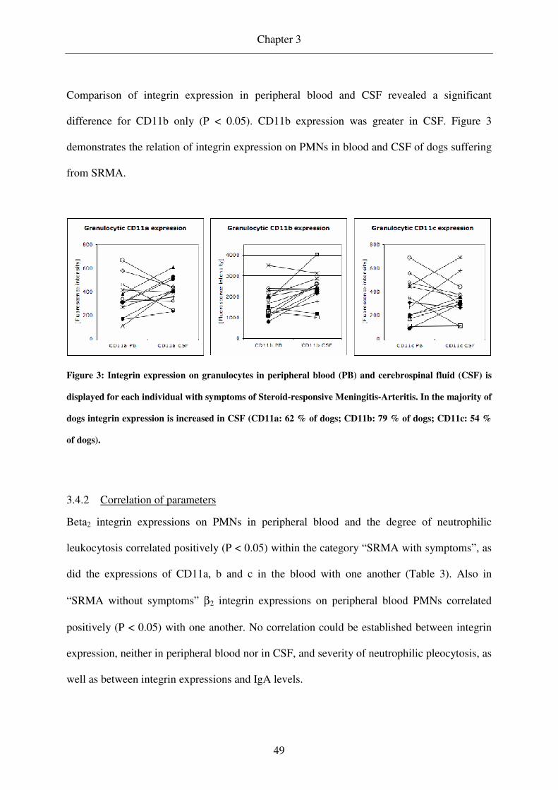
Chapter 3
49
Comparison of integrin expression in peripheral blood and CSF revealed a significant
difference for CD11b only (P < 0.05). CD11b expression was greater in CSF. Figure 3
demonstrates the relation of integrin expression on PMNs in blood and CSF of dogs suffering
from SRMA.
Figure 3: Integrin expression on granulocytes in peripheral blood (PB) and cerebrospinal fluid (CSF) is
displayed for each individual with symptoms of Steroid-responsive Meningitis-Arteritis. In the majority of
dogs integrin expression is increased in CSF (CD11a: 62 % of dogs; CD11b: 79 % of dogs; CD11c: 54 %
of dogs).
3.4.2 Correlation of parameters
Beta2 integrin expressions on PMNs in peripheral blood and the degree of neutrophilic
leukocytosis correlated positively (P < 0.05) within the category “SRMA with symptoms”, as
did the expressions of CD11a, b and c in the blood with one another (Table 3). Also in
“SRMA without symptoms” β2 integrin expressions on peripheral blood PMNs correlated
positively (P < 0.05) with one another. No correlation could be established between integrin
expression, neither in peripheral blood nor in CSF, and severity of neutrophilic pleocytosis, as
well as between integrin expressions and IgA levels.

Chapter 3
50
Table 3: Significant correlations for the group “SRMA with symptoms” (P < 0.05)
Parameter Parameter Spearman´s rank
correlation coefficient
CD11a in PB Number of neutrophils in PB + 0.824**
CD11b in PB Number of neutrophils in PB + 0.662**
CD11c in PB Number of neutrophils in PB + 0.688**
CD11a in PB CD11b in PB + 0.727**
CD11a in PB CD11c in PB + 0.727**
CD11b in PB CD11c in PB + 0.657
SRMA = Steroid-responsive Meningitis-Arteritis; PB = peripheral blood; ** = P < 0.01
3.4.3 Results of the in vitro study
In the second part of the study changes in integrin expression following incubation with sera
of dogs with symptoms of SRMA were investigated. Figure 4 summarizes the results of the
three single experiments. In general, incubation of neutrophils with sera of either group
(SRMA and epilepsy) led to higher integrin expressions in comparison to incubation with
PBS. Nine out of 12 sera from dogs with SRMA (75 %) induced a stronger CD11a
upregulation in comparison to incubation with epilepsy sera. The histogram plot in Figure 5
shows an example for this comparatively strong upregulation of CD11a. A PMN
subpopulation expressing particularly large amounts of CD11a is visible (marked with arrow).
The remaining three dogs, whose sera resulted in FR < 100, showed either very low or very
high in vivo CD11a expression. In contrast to this finding, a stronger upregulation of CD11b
and c was detected in only two and one out of 12 tested SRMA sera, respectively, when
compared to pooled sera from epileptic dogs. All other sera caused lower CD11b and c
expressions in comparison to epilepsy sera.
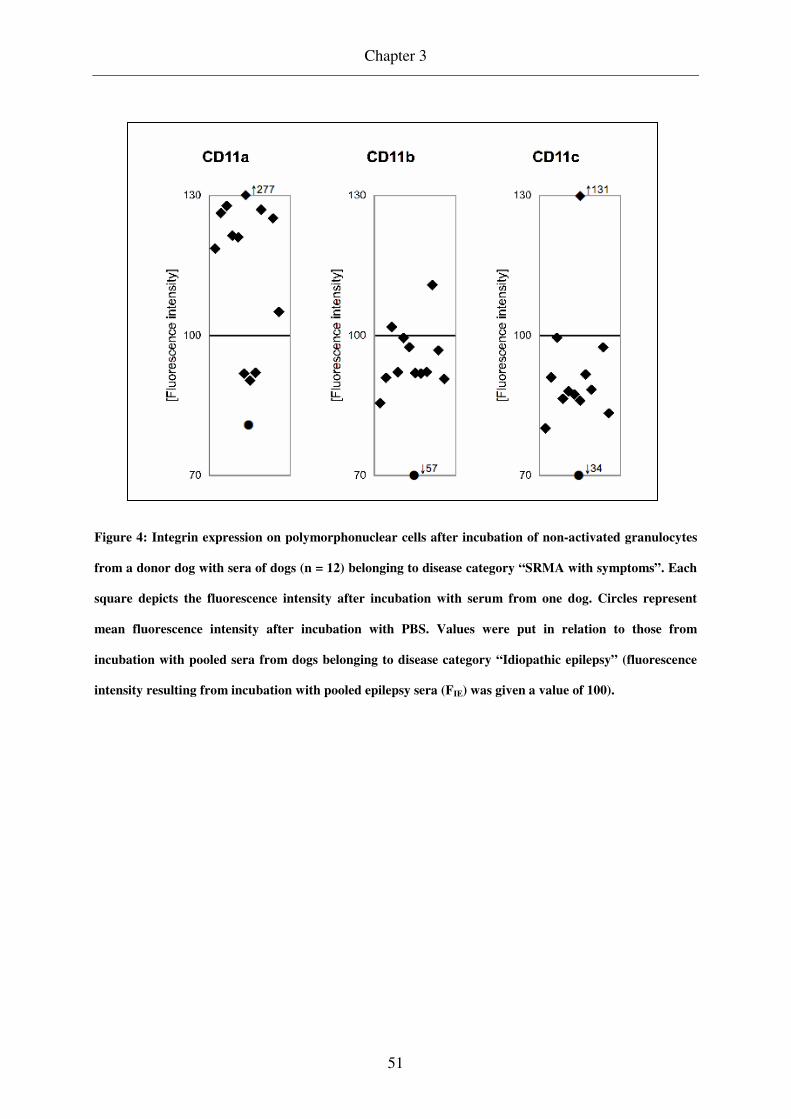
Chapter 3
51
Figure 4: Integrin expression on polymorphonuclear cells after incubation of non-activated granulocytes
from a donor dog with sera of dogs (n = 12) belonging to disease category “SRMA with symptoms”. Each
square depicts the fluorescence intensity after incubation with serum from one dog. Circles represent
mean fluorescence intensity after incubation with PBS. Values were put in relation to those from
incubation with pooled sera from dogs belonging to disease category “Idiopathic epilepsy” (fluorescence
intensity resulting from incubation with pooled epilepsy sera (FIE) was given a value of 100).

Chapter 3
52
Figure 5: Histogram plot displaying CD11a expression after incubation with sera (negative controls for
immunostaining procedure included (dashed arrow)). Grey graphs result from incubation with pooled
serum from dogs belonging to disease category “Idiopathic epilepsy”, bold black graphs from incubation
with serum from a dog belonging to “SRMA with symptoms”. Graphs show a comparatively strong
upregulation of CD11a after incubation with SRMA serum and a separation of a cell subpopulation with a
particularly marked upregulation (solid arrow) (x-axis displays fluorescence intensity).

Chapter 3
53
3.5 Discussion
SRMA is a naturally occurring animal model for various systemic vasculitides of unknown
origin in humans. Especially Kawasaki Syndrome (KS) and SRMA bear many similarities
(Burns et al., 1991). In both diseases an involvement of the meninges occurs (in ≈ 25 % of KS
patients) and in each of the disorders increased IgA levels are detected without their
deposition being responsible for initiating vascular lesions (Rowley and Shulman, 1998;
Tipold et al., 1995). As an animal model SRMA is of a great value due to its comparatively
high incidence. As one of the most frequently occurring canine CNS inflammations, it
accounts for approximately 15 % of these cases (Tipold, 1995). Since the systemic disease
SRMA shows a strong tendency to manifest in the cervical meninges, findings in this animal
model might as well be applied to other medical conditions, in which a compartmentalization
of the immune response occurs. Extensive studies on this canine disease will help to elucidate
pathogenetical principles of the development of immune mediated meningitides and immune
response compartmentalization in general. This in long-term might give rise to the
development of new treatment strategies for both, humans and dogs. The aim of this study
was to quantify integrin expression on PMNs in SRMA, as one striking feature of the disease
is the migration of enormous numbers of neutrophils into CSF (Tipold and Jaggy, 1994) and
integrins are crucial for this extravasation (Tuomanen et al., 1989). It was shown that this
attraction of neutrophils in SRMA is, at least partially, due to the presence of IL-8 or an IL-8-
like factor in the CSF (Burgener et al., 1998).
Our study showed that neutrophils of dogs with symptoms of SRMA exhibit stronger CD11a
expression than those of the remaining disease categories, whereas expression of CD11b and
c was comparable among all groups. The wide range of integrin expression is likely to reflect

Chapter 3
54
the situation that patients with SRMA are presented in different periods of the symptomatic
phase. In some dogs, a compensatory endogenous anti-inflammatory response might already
have been initiated, as it was shown to occur in canines with multiple organ dysfunction
syndrome secondary to sepsis (Weiss et al., 2004). In the cited study examinations were
performed for the β2 integrin CD11b/CD18 only. It seems reasonable, however, to assume
that such an anti-inflammatory reaction also affects CD11a/CD18 and CD11c/CD18. Our
finding that the expression of investigated integrins correlated positively with one another
supports this assumption. The natural course of SRMA, with its clinically silent periods in
between periods with fever and cervical pain, might be attributable to such compensatory
mechanisms. A reduced migration due to decreased CD11a-mediated endothelial adhesion
would explain that no or only few neutrophils are present in the subarachnoid space in these
asymptomatic periods (Phillipson et al., 2006; Tuomanen et al., 1989) while chemotactic
activity of the CSF remains high (Burgener et al., 1998). Since glucocorticosteroids are
known to reduce β2 integrin expression on activated human neutrophils in vitro, assumedly
via induction of downregulating proteins (Filep et al., 1997), and > 90 % of our patients with
SRMA without symptoms were under prednisolone therapy, it would be extremely interesting
in this regard to monitor integrin expressions in the natural course of disease with its waxing
and waning pattern. Due to the highly painful nature of SRMA, however, ethical
considerations prohibit such an approach. The expression of all integrins in peripheral blood
correlated positively with the severity of neutrophilic leukocytosis in the state of
inflammation. Thus, a strong systemic immune response was accompanied by high β2 integrin
expression. This was most obvious for CD11a and led to the suspicion that this particular
surface molecule plays a major role in the pathogenesis of the suppurative meningitis as the

Chapter 3
55
cardinal manifestation of SRMA. This molecule was shown to be essential for neutrophil
adhesion to endothelial cells, which in turn is a crucial step within the leukocyte recruitment
cascade (Phillipson et al., 2006). The selective upregulation of CD11a is in accordance with
results of Sahin et al. (1996) who investigated neutrophil adhesion properties in Behçet´s
disease, a human vasculitis with recurrent episodes of remission and possible affection of the
CNS (Hirohata and Kikuchi, 2003). As in SRMA elevated IgA levels are detected in serum
(Sunakawa and Ohshio, 1989) and, if the CNS is affected, in CSF (Jongen et al., 1992). Their
in vitro experiments led to the insight that patients´ sera cause an upregulation of CD11a on
neutrophils deriving from healthy controls whereas CD11b expression is unaffected. This
coincides with increased adhesive properties. Furthermore, blockage with anti-CD11a mAbs
causes a more pronounced inhibition of neutrophil adhesion to cultured human umbilical
vascular endothelial cells (HUVECs) than does blockage with anti-CD11b mAbs.
The second part of our experiment supports our in vivo findings in peripheral blood. Initially
non-activated neutrophils expressed CD11a to a greater extent after incubation with 75 % of
SRMA patients´ sera when compared to a serum pool of dogs with idiopathic epilepsy. In
contrast to this, CD11b and c expression was higher in only 17 % and 8 %, respectively,
meaning that the vast majority of sera caused a decreased CD11b and c upregulation in
comparison to the epilepsy pool. The three dogs that had relative fluorescence values < 100,
i.e. their sera induced less CD11a than did sera form epileptic dogs, exhibited either
extremely high or low in vivo CD11a expressions. This discrepancy of high CD11a
expression in vivo and poor stimulatory properties in vitro could be explained with an anti-
inflammatory response that was already initiated in vivo, without having affected integrin
levels on neutrophils yet.

Chapter 3
56
We suppose that sera of diseased dogs contain one or multiple factors that exhibit a selective
boosting effect on CD11a whereas CD11b and c expressions remain unaffected or are even
depressed. One can suspect that high CD11a expression in SRMA reflects high expression of
the functional CD11a/CD18 molecule. In the past, a considerable redundancy between the
function of β2 integrins was suspected. A recent study using an in vivo approach with CD11a
and CD11b knock-out mice, however, could demonstrate fundamentally different roles within
the neutrophil recruitment cascade. While initial adhesion is mediated by CD11a, subsequent
crawling along endothelium was shown to be largely dependent on CD11b (Phillipson et al.,
2006). High CD11a expression in SRMA therefore is most likely associated with increased
neutrophil adhesion properties to endothelial cells. This eventually would facilitate neutrophil
migration to CSF. The depression of the factor causing the CD11a upregulation in SRMA and
changes on endothelial cells in the natural course of disease as well as under treatment might
be the cause for the absence of neutrophils in CSF of dogs in subclinical periods despite the
persistence of increased chemotactic activity in CSF (Burgener et al., 1998).
Comparison of integrin expression on neutrophils in peripheral blood and CSF led to variable
results. In a marked number of dogs (39 %) lower CD11a expression on cells isolated from
CSF was measured. Since it is known that CD11a is an important molecule for neutrophils´
crossing of the blood-CSF barrier, it is conceivable, that in these patients a previously present
upregulation of CD11a had already been reversed, either by a distinct milieu of stimulating
factors in CSF or due to changes that occur in perishing neutrophils. It is known from ex vivo
examinations that especially neutrophils have only a short survival time in CSF (Steele et al.,
1986).
Taking into account the results of Weiss et al. (2004) who showed that in dogs with non-
septic and septic inflammatory diseases an increase in CD11b and decrease in CD18 on

Chapter 3
57
peripheral blood neutrophils occurs, integrin expression pattern in SRMA, with its selective
increase in CD11a, seems to differ from that in other inflammatory conditions.
3.6 Conclusion
Our results demonstrate a disproportionally strong upregulation of CD11a on neutrophils in
peripheral blood of dogs with SRMA, whereas CD11b and c expressions are comparable to
those in control groups. Since incubation of sera from dogs in a clinically apparent phase of
SRMA with initially non-activated neutrophils results in a comparatively strong CD11a
expression, whereas CD11b and c expressions remain lower than after incubation with control
sera, we hypothesize that SRMA serum contains one or multiple factors that promote this
selective increase of CD11a. CD11a is highly suspicious to account for the massive invasion
of neutrophils into the CSF and therefore might play a major role in the pathogenesis of the
disease.
3.7 Acknowledgments
This work was supported by a grant of the Swiss Society for Bernese Mountain Dogs and a
Georg-Christoph-Lichtenberg-Scholarship donated by the Department of Science and Culture
of the federal state of Lower Saxony, Germany.

Chapter 4
58
Chapter 4: Marked MMP-2 transcriptional up-regulation in mononuclear
leukocytes invading the subarachnoidal space in aseptic suppurative
Steroid-responsive Meningitis-Arteritis in dogs
M. Schwartza,b,c*, C. Puffc, V.M. Steina, W. Baumgärtnerb,c, A. Tipolda,b
a Department of Small Animal Medicine and Surgery, School of Veterinary Medicine
Hannover, Bischofsholer Damm 15, 30173 Hannover, Germany
b Center for Systems Neuroscience, School of Veterinary Medicine Hannover, Buenteweg 17,
30559 Hannover, Germany
c Department of Pathology, School of Veterinary Medicine Hannover, Buenteweg 17, 30559
Hannover, Germany
*Corresponding author. Tel.: +49-511-856-8965; fax: +49-511-856-7686; e-mail:
Vet Immunol Immunopathol. Article in Press.

Chapter 4
59
4.1 Abstract
Canine Steroid-responsive Meningitis-Arteritis (SRMA) is a suitable animal model for studies
on the development of neutrophilic pleocytosis in aseptic meningitis.
Samples of dogs in the acute phase of SRMA (n = 16) were examined for gene expression of
matrix metalloproteinases (MMP) -2 and -9 and tissue inhibitors of metalloproteinases
(TIMP) -1 and -2. Results were compared to those of dogs under glucocorticosteroid
treatment for SRMA (n = 16) and dogs with other inflammatory and neoplastic diseases of the
central nervous system (CNS) (n = 19). Samples included mononuclear (PBMNCs) and
polymorphonuclear cells (PBPMNs) of peripheral blood and cerebrospinal fluid white blood
cells (CSF WBCs).
In the acute phase of SRMA CSF WBCs showed mRNA expression for MMP-2 and -9 and
TIMP-1 and -2, highlighting a contribution of these cells to the overall content of MMPs and
TIMPs in CSF. MMP-2 mRNA levels in CSF WBCs were significantly up-regulated in
comparison to PBMNC expression levels, suggesting that MMP-2 is relevant for PBMNC
invasion into the subarachnoidal space and that the expression is influenced by migratory
activity through the blood-CSF-barrier.
Keywords: Canine, Blood-brain-barrier, Leukocyte extravasation, Matrix metalloproteinases
Abbreviations: CBC = complete blood count; cDNA = complementary DNA; CSF =
cerebrospinal fluid; CSF WBCs = cerebrospinal fluid white blood cells; CNS = central
nervous system; EF = elongation factor; GAPDH = Glyceraldehyd-3-phosphate
dehydrogenase; MMP = matrix metalloproteinase; PBMNCs = peripheral blood mononuclear
cells; PBPMNs = peripheral blood polymorphonuclear cells; SRMA = Steroid-responsive
Meningitis-Arteritis; TIMP = tissue inhibitors of metalloproteinases
Chapter 4
60
4.2 Introduction
Aseptic meningitis with predominance of polymorphonuclear cells (PMNs) is diagnosed in
the human (Amir et al., 1991; Negrini et al., 2000) and in the canine patient (Tipold, 1995).
However, understanding of principles that lead to the development of these suppurative
meningitides is still incomplete.
Since standards for diagnostic work-up and treatment of companion animals, in particular the
dog, are similar to that in human patients, investigation of analogous veterinary diseases
offers a great opportunity to increase the knowledge about pathomechanisms in both species.
While bacterially induced inflammation of the meninges is rare in dogs (Radaelli and Platt,
2002), by far the most frequently occurring canine disease associated with a
polymorphonuclear pleocytosis is Steroid-responsive Meningitis-Arteritis (SRMA) (Meric,
1988; Tipold, 1995). This meningitis is associated with inflammation of small to medium-
sized arteries, predominantely within the spinal meninges (Brooks, 1984; Scott-Moncrieff et
al., 1992; Tipold and Jaggy, 1994). To date every attempt to identify a causative infectious
agent has failed and the inflammation is believed to result from a dysregulation of the
immune system (Cizinauskas et al., 2000; Harcourt, 1978; Meric et al., 1986; Meric et al.,
1985; Poncelet and Balligand, 1993; Scott-Moncrieff et al., 1992; Tipold and Jaggy, 1994).
Steroid-responsive Meningitis-Arteritis is associated with cerebrospinal fluid (CSF)
neutrophil counts of several thousands per microliter and thus accounts for an ideal animal
model to study neutrophil invasion into the central nervous system (CNS), especially since
follow-up examinations with repeated CSF taps are part of the routine treatment protocol.
Entry of cells and molecules from the peripheral blood directly into the subarachnoidal space
is limited and controlled by the special architecture of the choroid plexus and the pial vessels

Chapter 4
61
(Allt and Lawrenson, 1997; Ransohoff et al., 2003). Opening of this protective shield with its
tight junctions and underlying basement membrane is required to allow extravasation of
leukocytes into the CSF.
Matrix metalloproteinases (MMPs) are a family of zinc- and calcium-dependent
endopeptidases that are involved in remodelling of the extracellular matrix and also function
as activators of latent forms of MMPs and cytokines. These enzymes may thus exhibit both,
beneficial and detrimental effects. To counteract the potentially harmful actions, the MMP
production and activation of their proforms is tightly regulated (Candelario-Jalil et al., 2009;
Yong et al., 2001). Important in this respect are the tissue inhibitors of metalloproteinases
(TIMPs) of which TIMP-1 forms a specific complex with proMMP-9 and TIMP-2 with
proMMP-2 (Brew et al., 2000).
Experimental studies have shown that MMP-2 (gelatinase A) and MMP-9 (gelatinase B) are
potent substances to interfere with the integrity of the barrier between blood circulation and
CNS mainly by degradation of type IV collagen, the main component of the basement
membrane (Mun-Bryce et al., 2002; Mun-Bryce and Rosenberg, 1998; Paul et al., 1998;
Rosenberg et al., 1992; Timpl, 1989). These enzymes have also been implicated in the
development of systemic vasculitis (Gavin et al., 2003). We therefore hypothesized that
leukocytes migrating from peripheral blood into the cerebrospinal fluid might increase their
transcriptional activity for MMPs as one important mechanism to allow crossing-over from
the vascular bed into the subarchnoidal space in SRMA. and that counter-regulation of their
respective tissue inhibitors may take place.
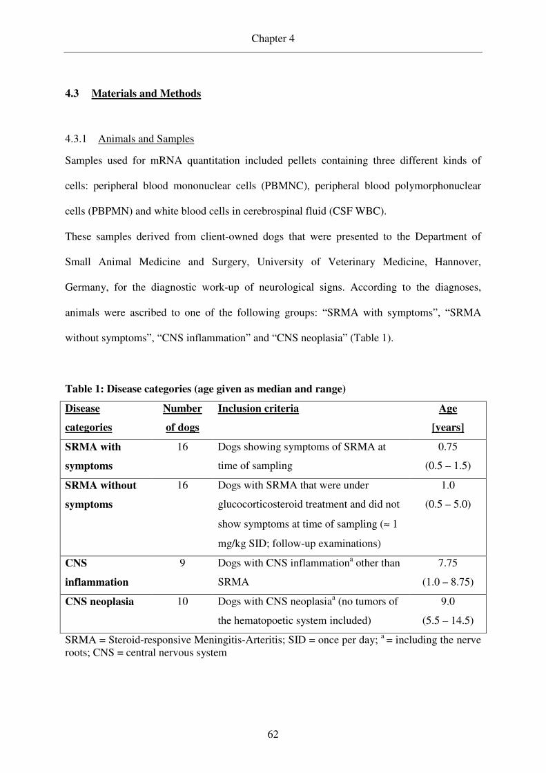
Chapter 4
62
4.3 Materials and Methods
4.3.1 Animals and Samples
Samples used for mRNA quantitation included pellets containing three different kinds of
cells: peripheral blood mononuclear cells (PBMNC), peripheral blood polymorphonuclear
cells (PBPMN) and white blood cells in cerebrospinal fluid (CSF WBC).
These samples derived from client-owned dogs that were presented to the Department of
Small Animal Medicine and Surgery, University of Veterinary Medicine, Hannover,
Germany, for the diagnostic work-up of neurological signs. According to the diagnoses,
animals were ascribed to one of the following groups: “SRMA with symptoms”, “SRMA
without symptoms”, “CNS inflammation” and “CNS neoplasia” (Table 1).
Table 1: Disease categories (age given as median and range)
Disease
categories
Number
of dogs
Inclusion criteria Age
[years]
SRMA with
symptoms
16 Dogs showing symptoms of SRMA at
time of sampling
0.75
(0.5 – 1.5)
SRMA without
symptoms
16 Dogs with SRMA that were under
glucocorticosteroid treatment and did not
show symptoms at time of sampling (≈ 1
mg/kg SID; follow-up examinations)
1.0
(0.5 – 5.0)
CNS
inflammation
9 Dogs with CNS inflammationa other than
SRMA
7.75
(1.0 – 8.75)
CNS neoplasia 10 Dogs with CNS neoplasiaa (no tumors of
the hematopoetic system included)
9.0
(5.5 – 14.5)
SRMA = Steroid-responsive Meningitis-Arteritis; SID = once per day; a = including the nerve roots; CNS = central nervous system

Chapter 4
63
Each patient was subject to a general and neurological examination, which was followed by a
complete blood count (CBC), routine blood chemistry and urinalysis. In addition, a
suboccipital CSF tap and CSF analysis (WBC count, cytomorphological differentiation and
total protein) was performed in every dog. Samples containing erythrocytes were included
only if xanthochromia, suggesting in vivo leakage of red blood cells into the subarachnoid
space, was present (Jamison and Lumsden, 1988). Immunoglobulin A (IgA) content in serum
and CSF was determined in all animals using a previously described enzyme-linked
immunosorbent assay (ELISA) system to support diagnosis of SRMA (Tipold et al., 1994).
Ancillary tests performed to obtain a definitive diagnosis included radiographic studies,
ultrasonography, computed tomography (CT), magnetic resonance imaging (MRI) as well as
electrodiagnostic testing. If available, results of tests for infectious agents and of pathological
examinations contributed to the diagnoses. A history of glucocorticosteroid administration
within one week prior to presentation (within 6 weeks if depot formulations were given) was
recorded for each patient.
Diagnosis of SRMA was based on the combination of the following findings: pain upon
cervical palpation, fever, marked neutrophilic leuko- and pleocytosis, immediate and
sustained response to glucocorticosteroid treatment, exclusion of other disease processes that
may cause similar symptoms. Concomitant elevation of IgA levels in serum and CSF
supported this diagnosis (Tipold and Jaggy, 1994).
Separation of WBC populations deriving from peripheral blood was achieved by means of
density gradient centrifugation with two subsequent washing steps (Schwartz et al., 2008a;
Schwartz et al., 2008b). Cerebrospinal fluid WBCs were spun down (200 x g; 10 min; room
temperature) and supernatant was removed, therefore pellets contained a mixture of WBC

Chapter 4
64
populations. Cell pellets were immediately frozen and stored at - 80°C until RNA isolation
was undertaken.
In a few cases not all of the three kinds of cell pellets were available for investigation of
mRNA expression. Two of the 16 dogs in “SRMA with symptoms” were lacking a PBPMN
pellet, in 1/9 dogs in “CNS inflammation” a PBMNC pellet was available only and in 2/10
dogs in “CNS neoplasia” a PBPMN pellet was missing.
4.3.2 RNA purification and reverse transcription
Purification of total RNA from the cell pellets was performed using RNeasy Mini Kit
columns (Qiagen) according to the manufacturer’s protocol for animal cells. To prevent
contamination with genomic DNA an on-column DNase digestion step with RNase-free
DNase (Qiagen) following the manufacturer’s instructions was included. The yield of RNA
was subsequently determined with a GeneQuant pro device (Biochrom Ltd.) by means of
spectrophotometry at OD260. The remaining eluate was immediately placed in liquid nitrogen
and then stored at - 80°C until reverse transcription.
Reverse transcription of RNA into complementary DNA (cDNA) was performed with
Omniscript RT (Qiagen), Random Primers (Promega) and RNaseOUTTM Recombinant
Ribonuclease Inhibitor (Invitrogen). Forty microliters of master mix containing ≤ 2 µg of total
RNA was produced according to Qiagen’s protocol for reverse transcription. Incubation for
60 min at 37°C took place in a thermocycler (PTC-200 Peltier Thermal Cycler; MJ Research).
Complementary DNA was stored at - 20°C in aliquots until its use in real-time PCR
experiments.

Chapter 4
65
4.3.3 Primers
Primers used in this study (Table 2 and 3) were either taken from the literature (as cited) or
designed with Primer3 software (Rozen and Skaletsky, 2000) or Beacon Designer version 2.1
software (Premier Biosoft International). The probe for MMP-9 was designed with the latter
program. Primers and probe were purchased from Eurofins MWG Operon. The specificity of
each PCR product was confirmed by DNA sequencing (AGOWA genomics).
Table 2: Primers for production of standard dilution series
Target
gene
Primer sequence in 5´- 3´ (forward primers on
top, reverse primers on bottom)
Size of
amplicon
[bp]
GenBank
accession
number
MMP-2a CCAAGAACTTCCGTCTGTCC 606 AF177217
AGCTATGACCACTGCCTTGC
MMP-9a GAGGTTCGACGTGAAGGCGCAGAT 200 J05070
AGGTCACGTAGCCCACTTCGTCCAC
TIMP-1b GCGTTATGAGATCAAGATGAC 345 AF077817
CTGGTCCGTCCACAAGCA
TIMP-2c,a GCAGAYGTAGTGATCAGRGR 315 AF095638
GTTYAGGCTCTTCTTCTGGG
GAPDHa AAGGTCGGAGTCAACGGATT 365 AB038240
GCAGAAGAAGCAGAGATGATG
β-Actin AACACCCCAGCCATGTATGT 333 NM_001003349
CTTCTCCAGGGAGGACGAG
EF-1αd AGCCCTTGCGCCTGCCTCTC 219 X03558
CAGACACATTCTTGACATTGAAGC
bp = base pairs; TIMP = tissue inhibitor of matrix metalloproteinases; MMP = matrix metalloproteinase; GAPDH = Glyceraldehyd-3-phosphate dehydrogenase; EF = elongation factor; a Puff et al. (2008); b Barnes et al. (2000); c Clegg et al. (1999); d von Smolinski et al. (2005)

Chapter 4
66
Table 3: Primers for real-time PCR
Target
gene
Primer sequence in 5´- 3´(forward primers on
top, reverse primers on bottom)
Size of
amplicon
[bp]
GenBank
accession
number
MMP-2a GGAGATCTTCTTCTTCAAGGACCG 89 AF177217
AGAATGTGGCTACCAGCAGGG
MMP-9a CATGACATCTTCCAGTACCAAG 85 AB006421
GGTTCACCTCATTCCGAGAA
Probe:
FAM-CTACTTCTGCCAGGACCGCTTCTACT-
TAMRA
TIMP-1a ACGGACACTTGCAGATCAAC 94 AF077817
GCAGCATAGGTCTTGGTGAA
TIMP-2a CCATCAAGCGGATTCAGT 89 AF095638
GGAAGGAGCCGTGTAGATAA
GAPDHa GTCATCAACGGGAAGTCCATCTC 84 AB038240
AACATACTCAGCACCAGCATCAC
β-Actin TCTACGAGGGGTACGCCTTG 149 NM_001003349
TTCCTTGATGTCACGCACGAT
EF-1αb AGCCCTTGCGCCTGCCTCTC 219 X03558
CAGACACATTCTTGACATTGAAGC
bp = base pairs; TIMP = tissue inhibitor of matrix metalloproteinases; MMP = matrix metalloproteinase; GAPDH = Glyceraldehyd-3-phosphate dehydrogenase; EF = elongation factor; a Puff et al. (2008); b von Smolinski et al. (2005)
Chapter 4
67
4.3.4 Standard dilution series
In order to allow calculation of standard curves tenfold dilution series (102 – 108 copies) were
produced for each target gene. These dilution series also served as standards for inter-run
calibration to account for run-to-run variation. Amplicons were produced by conventional
end-point PCR (PTC-200 Peltier Thermal Cycler; MJ Research), agarose gel electrophoresis
and DNA extraction with NucleoSpin Extract II columns (Macherey-Nagel) according to the
manufacturer’s protocol. Spectrophotometrical determination of DNA concentration was
performed at OD260 (GeneQuant pro; Biochrom Ltd.) and copy numbers per volume were
calculated according to the following formula: copies/µl = 6 x 10²³ x DNA concentration
(ng/µl) x 10-9/[size of amplicon (base pairs) x 660].
Template was cDNA from DH-82 cells (Wellman et al., 1988). Reaction conditions were as
follows: initial denaturation at 94°C for 1 minute; 40 cycles of denaturation at 94°C for 1
minute, annealing at 59°C (TIMP-1, TIMP-2, MMP-2, MMP-9, GAPDH) / 60°C (β-Actin,
EF-1α) for 2 minutes and elongation at 72°C for 1 minute; final elongation at 72°C for 5
minutes. Amplification was achieved with recombinant Taq DNA Polymerase (Invitrogen) in
1x PCR Buffer (Invitrogen) with 1.25 mM MgCl2 (Invitrogen), 0.2 mM dNTP mixture
(Applied Biosystems) and 300 nM of each primer (Table 2).
4.3.5 Real-time PCR
Real-time PCRs were run on an Mx3005P QPCR system (Stratagene) with Brilliant SYBR
Green QPCR Core Reagent Kit (Stratagene) in 8x Strip Tubes with Optical Cap (Stratagene).
Due to the number of samples, experiments for each target gene were run on separate plates
for PBMNC, PBPMN and CSF WBC samples. Measurements were conducted the same day

Chapter 4
68
using the identical standard dilution series. Standards were kept at + 4°C in-between runs.
The plates included the standard dilution series as well as a no template control (NTC) in
duplicate. Reaction volume was 25 µl including 1 µl template per reaction.
Reaction conditions were as follows: initial denaturation at 95°C for 10 minutes; 40 cycles
with denaturation at 95°C for 30 seconds, annealing at 57°C (TIMP-1, TIMP-2) / 60°C (β-
Actin, EF-1α) / 64°C (MMP-2, GAPDH) for 1 minute and elongation at 72°C for 30 seconds;
final extension at 72°C for 1 minute; dissociation program beginning at 95°C for 1 minute
followed by increasing temperature by 0.5°C every 30 seconds from 55°C to 95°C. The
master mix contained 0.025 U/µl SureStart Taq DNA Polymerase in 1x core PCR buffer with
2.5 mM (MMP-2, GAPDH) / 4.0 mM (TIMP-1, TIMP-2) / 5.0 mM (β-Actin, EF-1α) MgCl2,
800 µM dNTP mix, 150 nM of each primer (Table 3), 8 % glycerol and 4 % dimethyl
sulphoxide (DMSO) (3 % for MMP-2, GAPDH). Final SYBR Green concentration was 0.25x
and Rox was used as a reference dye at 30 nM.
The constitution of the master mix for MMP-9 mRNA quantification differed because a
sequence specific probe was used (Table 3). It contained 0.025 U/µl SureStart Taq DNA
Polymerase in 1 x core PCR buffer with 5.0 mM MgCl2, 800 µM dNTP mix, 300 nM of each
primer, 200 nM of TaqMan probe and 76 nM Rox. The thermal profile was: initial
denaturation at 95°C for 10 minutes; 40 cycles with 95°C for 15 seconds and 60°C for 1
minute.
The software tool “Multiple Experiment Analysis” (MxPro QPCR Software v4.01;
Stratagene) allowed run-to-run comparison between the results for PBMNC, PBPMN and
CSF WBC samples.

Chapter 4
69
To account for variations in sample input, extraction and reaction efficiencies and presence of
inhibitors, copy numbers of the genes of interest were normalized against the geometric mean
of the internal reference genes GAPDH, β-Actin and EF-1α: copiesnormalized (target gene) [%]
= copiesnon-normalized (target gene) x 100/ [copies (GAPDH) x copies (β-Actin) x copies (EF-
1α)]^(1/3).
4.3.6 Statistical analysis
In addition to descriptive methods the Wilcoxon rank sum test was applied for comparison of
the results deriving from the different disease categories. For these comparisons results of the
groups “CNS inflammation” and “CNS neoplasia” were combined. It was also tested whether
results for dogs with a history of glucocorticosteroid pretreatment differed significantly from
those of the non-pretreated ones. The Wilcoxon signed-rank test served as a tool to compare
values deriving from PB and CSF of dogs in the acute phase of SRMA. The Spearman´s rank
correlation coefficients were calculated to detect correlations for “SRMA with symptoms”.
The parameters included normalized copy numbers and results of CBC and CSF analysis.
Statistical significance was set at the 5% level (p ≤ 0.05).
4.4 Results
Dissociation procedures at the end of real-time PCRs for MMP-2, TIMP-1, TIMP-2, GAPDH,
β-Actin and EF-1α produced clearly defined single melting temperatures at 82,9°C, 84,4°C,
76,9°C, 81,1°C, 85,4°C and 82,3°C respectively. PCR products for these genes were
considered specific because temperatures corresponded with those of the standard dilution

Chapter 4
70
series. These standards contained a single gene product only that had been sequenced and
recognized as the respective gene of interest.
Copy numbers for the three reference genes could be measured in every PBMNC and
PBPMN sample and normalization was therefore always possible. In 9 CSF WBC samples,
however, copy numbers for only 1 or 2 of the reference genes could be determined and these
samples were excluded from further analysis since accurate normalization was not possible.
In these cases a mild degree of pleocytosis or a normal CSF cell count reduced the size of the
cell pellets, containing considerably less leukocytes than those of the other dogs.
Pretreatment with glucocorticosteroids, after exclusion of “SRMA without symptoms”, was
recorded for 8 dogs (4/16 in “SRMA with symptoms”, 3/9 in “CNS inflammation”, 1/10 in
“CNS neoplasia”) and did not cause significantly differing results neither in CBC and CSF
analysis nor in mRNA expression, except for a more severe pleocytosis and higher CSF IgA
levels in pretreated dogs (p < 0.05 and p < 0.01, respectively). Considering dogs in the acute
phase of SRMA only, no differences were detected in pretreated and not pretreated
individuals. Pretreated patients were therefore not excluded from further analysis.
Results of MMP-2 mRNA expression analysis are displayed in Figure 1. In most dogs no
mRNA expression could be recorded in PBMNC samples. Exceptions were 1/16 dogs from
the group “SRMA with symptoms” and 3/16 dogs from “SRMA without symptoms”. No or
only slight expression was detected in PBPMN pellets. In contrast, 15/16 CSF WBCs samples
from dogs in the acute phase of SRMA showed clear evidence for mRNA expression.
Normalized copy numbers were significantly higher than those in samples from PBMNCs and
PBPMNs (p ≤ 0.001). The 4 positive CSF samples not belonging to dogs in the acute phase of
SRMA all derived from dogs that also had marked pleocytoses with considerable

Chapter 4
71
participation of neutrophils (50 – 89 %). Two of these dogs suffered from bacterial
meningomyelitis, in the other 2 individuals an intracranial meningioma was diagnosed.
Figure 1: Results of real-time PCR for MMP-2 mRNA expression of PBMNCs (a), PBPMNs (b) and CSF
WBCs (c) in all disease categories and PBMNCs + CSF WBCs in the acute phase of SRMA (d). Each dot
represents the copy number in one sample. Measured copy numbers are normalized to the geometric
mean of reference genes GAPDH, ββββ-Actin and EF-1αααα and are given as the percentage of the respective

Chapter 4
72
geometric mean. Horizontal bars give median values. Asterisks indicate statistically significant differences
from the disease category “SRMA with symptoms” (** p ≤≤≤≤ 0.01). For statistical analysis samples from
“CNS inflammation” and “CNS neoplasia” were grouped as one disease category. Please note the non-
uniform scaling of the diagrams (MMP = matrix metalloproteinase; PBMNCs = peripheral blood
mononuclear cells; PBPMNs = peripheral blood polymorphonuclear cells; CSF WBCs = cerebrospinal
fluid white blood cells; SRMA = Steroid-responsive Meningitis-Arteritis; GAPDH = Glyceraldehyd-3-
phosphate dehydrogenase; EF = elongation factor)
Messenger RNA expression for MMP-9 is visualized in Figure 2. Expression in PBMNCs
was lowest in “SRMA with symptoms” and the overall expression in this cell type was
markedly below that of PBPMNs (p ≤ 0.001). In these, values of dogs in acute stage of
SRMA were comparable to those of dogs suffering from other CNS inflammations than
SRMA (p = 0.815) and showed higher MMP-9 mRNA content than individuals from “SRMA
without symptoms” (p ≤ 0.05) and “CNS neoplasia” (p = 0.07). In 15/16 CSF WBC pellets
from dogs with clinical signs due to SRMA mRNA expression of this gene could be noted.
The remaining positive CSF samples derived from the two dogs with meningiomas that also
showed significant mRNA expression for MMP-2. Levels of MMP-9 mRNA expression in
“SRMA with symptoms” were lowest in PBMNCs, intemediate in CSF WBCs and highest in
PBPMNs (p ≤ 0.01).
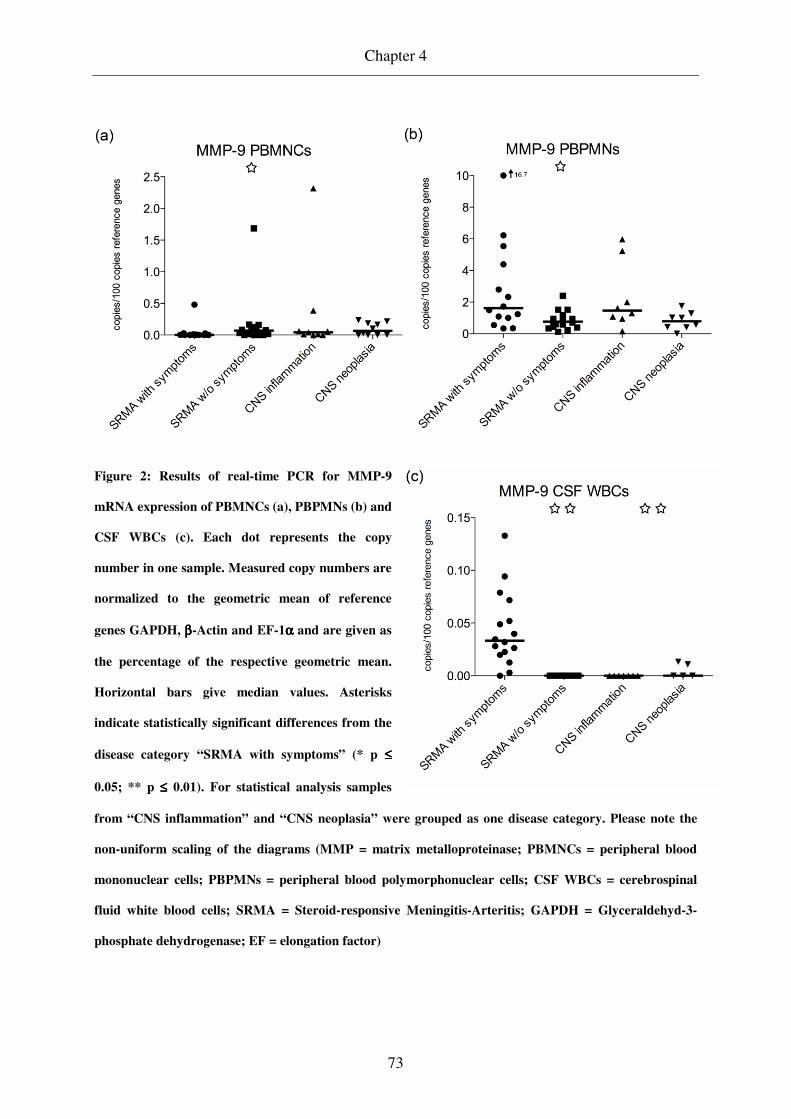
Chapter 4
73
Figure 2: Results of real-time PCR for MMP-9
mRNA expression of PBMNCs (a), PBPMNs (b) and
CSF WBCs (c). Each dot represents the copy
number in one sample. Measured copy numbers are
normalized to the geometric mean of reference
genes GAPDH, ββββ-Actin and EF-1αααα and are given as
the percentage of the respective geometric mean.
Horizontal bars give median values. Asterisks
indicate statistically significant differences from the
disease category “SRMA with symptoms” (* p ≤≤≤≤
0.05; ** p ≤≤≤≤ 0.01). For statistical analysis samples
from “CNS inflammation” and “CNS neoplasia” were grouped as one disease category. Please note the
non-uniform scaling of the diagrams (MMP = matrix metalloproteinase; PBMNCs = peripheral blood
mononuclear cells; PBPMNs = peripheral blood polymorphonuclear cells; CSF WBCs = cerebrospinal
fluid white blood cells; SRMA = Steroid-responsive Meningitis-Arteritis; GAPDH = Glyceraldehyd-3-
phosphate dehydrogenase; EF = elongation factor)

Chapter 4
74
Figure 3 displays the quantity of TIMP-1 mRNA expression after normalization. Peripheral
blood MNCs and CSF WBCs of dogs in the acute phase of SRMA showed the highest
expression among the disease categories whereas copy numbers of PBPMN samples are
similar within the 4 groups. Messenger RNA expression in PBMNCs and CSF WBCs was
within the same magnitude while that of PBPMNs was markedly lower. The difference of
CSF WBCs and PBPMNs in the acute phase of SRMA was statistically significant (p ≤ 0.01).
Normalized values for TIMP-2 mRNA expression are given in Figure 4. Overall, samples
from “SRMA with symptoms” showed the lowest level of expression. An exception were
those deriving from CSF. In the acute phase of SRMA the amount of mRNA encoding for
TIMP-2 per cell was lowest in PBMNCs, intermediate in CSF WBCs and highest in
PBPMNs. These differences were statistically significant (p ≤ 0.01).

Chapter 4
75
Figure 3: Results of real-time PCR for TIMP-1
mRNA expression of PBMNCs (a), PBPMNs (b) and
CSF WBCs (c). Each dot represents the copy
number in one sample. Measured copy numbers are
normalized to the geometric mean of reference genes
GAPDH, ββββ-Actin and EF-1αααα and are given as the
percentage of the respective geometric mean.
Horizontal bars give median values. Asterisks
indicate statistically significant differences from the
disease category “SRMA with symptoms” (* p ≤≤≤≤
0.05; ** p ≤≤≤≤ 0.01). For statistical analysis samples
from “CNS inflammation” and “CNS neoplasia” were grouped as one disease category. Please note the
non-uniform scaling of the diagrams (MMP = matrix metalloproteinase; PBMNCs = peripheral blood
mononuclear cells; PBPMNs = peripheral blood polymorphonuclear cells; CSF WBCs = cerebrospinal
fluid white blood cells; SRMA = Steroid-responsive Meningitis-Arteritis; GAPDH = Glyceraldehyd-3-
phosphate dehydrogenase; EF = elongation factor)

Chapter 4
76
Figure 4: Results of real-time PCR for TIMP-2
mRNA expression of PBMNCs (a), PBPMNs (b) and
CSF WBCs (c). Each dot represents the copy
number in one sample. Measured copy numbers are
normalized to the geometric mean of reference
genes GAPDH, ββββ-Actin and EF-1αααα and are given as
the percentage of the respective geometric mean.
Horizontal bars give median values. Asterisks
indicate statistically significant differences from the
disease category “SRMA with symptoms” (* p ≤≤≤≤
0.05; ** p ≤≤≤≤ 0.01). For statistical analysis samples
from “CNS inflammation” and “CNS neoplasia” were grouped as one disease category. Please note the
non-uniform scaling of the diagrams (MMP = matrix metalloproteinase; PBMNCs = peripheral blood
mononuclear cells; PBPMNs = peripheral blood polymorphonuclear cells; CSF WBCs = cerebrospinal
fluid white blood cells; SRMA = Steroid-responsive Meningitis-Arteritis; GAPDH = Glyceraldehyd-3-
phosphate dehydrogenase; EF = elongation factor)

Chapter 4
77
No statistically significant differences were present for any of the genes between samples of
dogs that were under treatment for SRMA and those that derived from individuals with other
CNS inflammatory diseases and CNS neoplasias.
Statistically significant correlations and non-significant trends are given in Table 4.
Table 4: Significant correlations and non-significant trends between quantity of mRNA
expression and other tested parameters (for the group “SRMA with symptoms”)
Parameter Parameter Spearman´s rank
correlation
coefficient
Level of
significance
MMP-2 in CSF WBCs Percentage of lympho-
cytes in CSF WBCs + 0.600* p = 0.018
TIMP-1 in PBMNCs TIMP-2 in PBMNCs + 0.744** p = 0.001
Degree of pleocytosis - 0.591* p = 0.016
TIMP-1 in PBPMNs TIMP-1 in CSF WBCs + 0.556* p = 0.039
Degree of neutrophilia
in peripheral blood - 0.481 p = 0.081
TIMP-1 in CSF WBCs TIMP-2 in CSF WBCs - 0.632** p = 0.009
TIMP-2 in PBMNCs Degree of pleocytosis - 0.612* p = 0.012
TIMP-2 in CSF WBCs Degree of pleocytosis - 0.471 p = 0.066
SRMA = Steroid-responsive Meningitis-Arteritis; MMP = matrix metalloproteinase; CSF WBCs = white blood cells in cerebrospinal fluid; TIMP = tissue inhibitor of metalloproteinases; PBMNCs = peripheral blood mononuclear cells; PBPMNs = peripheral blood polymorphonuclear cells
4.5 Discussion
Meningitis with predominance of polymorphonuclear cells (PMNs) occurs in the human and
canine patient. While, for a long time, these suppurative meningitides have been considered
indicative for a bacterial etiology in human medicine, it is now recognized that a considerable
Chapter 4
78
proportion of aseptic meningitis cases show a preponderance of neutrophils (Amir et al.,
1991; Negrini et al., 2000) and that these cytomorphological characteristics may persist
beyond 24 hours of initial symptoms (Negrini et al., 2000). The disease SRMA may provide a
suitable model to increase the understanding of general pathogenetical principles that lead to
the development of suppurative meningitis. A particular advantage of this naturally occurring
canine model, in comparison to the established experimental rodent and rabbit models, is the
possibility to perform repeated CSF taps with yield of large volumes.
Steroid-responsive Meningitis-Arteritis accounts for the majority of meningitis cases in dogs
and is among the most frequently diagnosed CNS inflammations in this species (Meric, 1988;
Tipold, 1995). It is the most likely cause for marked polymorphonuclear pleocytosis (Tipold,
1995), which is additionally seen in a small percentage of dogs suffering from intracranial
meningiomas (Dickinson et al., 2006) and in the rare cases of bacterial CNS infection
(Radaelli and Platt, 2002).
To allow development of pleocytosis leukocytes need to pass the tightly controlled barrier
between vasculature and CNS. This may take place at different sites: 1. from parenchymal
vessels into the perivascular space of brain and spinal cord tissue, 2. at the choroid plexus into
the ventricles 3. from blood vessels within the meninges into the subarchnoidal space
(Ransohoff et al., 2003). Since perivascular leukocytic infiltrates are lacking within CNS gray
and white matter in SRMA (Brooks, 1984; Hayes et al., 1989; Scott-Moncrieff et al., 1992;
Tipold et al., 1995), it seems rather unlikely that leukocyte trafficking takes place at this level.
A number of leukocytes may certainly enter the CSF via the choroid plexus, the most
plausible localization for the bulk of migration, however, are pial blood vessels, especially
since their integrity is impaired by concurrent arteritis. And in fact, xanthochromia or non-
iatrogenic hemorrhage is regularly seen in CSF samples (Behr and Cauzinille, 2006; Irving
Chapter 4
79
and Chrisman, 1990; Meric et al., 1986), supporting the idea that this site of the blood-CSF-
barrier is the one that is easiest to cross. In addition, leakage of albumin into the CSF of
diseased dogs was demonstrated, which also indicates loss of integrity of the blood-CSF-
barrier (Behr and Cauzinille, 2006).
Matrixmetalloproteinases play an important role in neuroinflammation. They contribute to its
perpetuation by conversion of pro-forms of inflammatory molecules into mature forms and
allow leukocyte recruitment into the CNS by permeabilizing the protective shield between
blood stream and CSF (Candelario-Jalil et al., 2009; Yong et al., 2001). The action of these
enzymes, with their potential to cause severe damage to surrounding substrates, is tightly
regulated. Steps of regulation include the transcriptional and translational levels as well as the
requirement for activation and finally the possibility of their subsequent inactivation
(Rosenberg, 2002; Yong et al., 2001). Production of MMPs within the CNS occurs in a
multitude of residential and migratory cells. These include endothelial cells, pericytes,
astrocytes, oligodendrocytes, microglia, neurons and invading leukocytes (Rosenberg, 2002;
Yong et al., 2001).
MMP-2 and -9 were shown to mediate opening of the blood-brain-barrier (Mun-Bryce and
Rosenberg, 1998; Rosenberg et al., 1992), which made them good candidates to be
investigated in our canine meningitis model. In contrast to most previous studies that have
focused on the enzymatic activity in CSF (Bergman et al., 2002; Kolb et al., 1998; Leppert et
al., 2000; Levine et al., 2006; Paul et al., 1998; Turba et al., 2007) or transcriptional activity
in CNS parenchyma (Kieseier et al., 1999; Leib et al., 2000), we investigated the contribution
of invading leukocytes themselves to this phenomenon. In addition, mRNA expression of
tissue inhibitors of metalloproteinases, TIMP-2 and TIMP-1, that interact predominantly with
MMP-2 and MMP-9 (Brew et al., 2000) was quantitated.

Chapter 4
80
Albeit our measurements of mRNA encoding for MMP-2 in peripheral blood cells of dogs
suffering from SRMA revealed only few samples with mRNA content above the detection
limit, more than 90 % of CSF WBC specimens from the group “SRMA with symptoms”
contained detectable amounts of MMP-2 mRNA with levels exceeding those found in
PBMNCs. Also the CSF WBC sample of the dog, that had detectable mRNA in PBMNCs in
the acute stage of SRMA, showed a > 25-fold increase of transcriptional activity for MMP-2
in comparison to the value in PBMNCs. The remaining 4 CSF samples, in which MMP-2
mRNA could be detected, all derived from dogs whose CNS pathology was also characterized
by a predominantly polymorphonuclear pleocytosis. It was shown that MMP-2 is not
produced by neutrophils (Opdenakker et al., 2001), therefore it is highly suspicious that the
mononuclear leukocyte population accounts for the MMP-2 mRNA content in CSF samples.
Detectable amounts of MMP-2 mRNA in some PBPMN pellets may arise from non-
neutrophil PBPMNs. Since differential cell counts revealed predominance of
polymorphonuclear cells in all CSF samples and these cells contribute to the quantity of
reference genes, the actual increase in mRNA expression will even be stronger than indicated
by our data.
Presence of MMP-2 in CSF of healthy individuals was confirmed by multiple studies and
these levels remained unchanged in subjects with meningitis characterized by predominance
of polymorphonuclear cells (Azeh et al., 1998; Kieseier et al., 1999; Leib et al., 2000; Paul et
al., 1998). Messenger RNA expression was also unchanged in CSF WBCs in experimental
pneumococcal meningitis in rats (Leib et al., 2001). In contrast to our experimental set-up,
however, comparison was drawn to CSF WBC samples at the time of infection and not to
expression levels in PBMNCs. In addition, copy numbers were normalized to the single
reference gene GAPDH only and up-regulation might have remained undetected if it

Chapter 4
81
coincided with that of GAPDH mRNA. Multiple studies have demonstrated that there is no
universally applicable reference gene (Vandesompele et al., 2002) and in vitro GAPDH
expression was shown to be increased by up to 70-fold in stimulated human lymphocytes
(Bas et al., 2004). Vandesompele et al. (2002) therefore recommend normalization of reverse-
transcriptase real-time PCR data to the geometric mean of at least 3 reference genes. It is
conceivable that all PBMNCs that migrate into the subarachnoidal space will up-regulate their
MMP-2 mRNA expression to facilitate crossing of the basement membrane, but that the
amount of MMP-2 production is not sufficient to change the overall content of MMP-2 in
CSF to be detectable by zymography or by ELISA. This assumption is supported by the
finding that, conversely to the situation in the CSF, human T lymphocytes constitutively
express MMP-9 and MMP-2 only upon stimulation. This MMP-2 expression is dose-
dependant and activation is accompanied by increased migratory activity through a basement
membrane model (Leppert et al., 1995). The fact that in our study MMP-2 mRNA expression
levels in CSF WBCs were positively correlated with the proportion of lymphocytes in CSF
supports the assumption that these cells largely account for the increase in mRNA encoding
for MMP-2. Thus, SRMA seems to be a unique naturally occurring animal model to study
MMP mRNA up-regulation in leukocytes that exhibit migratory activity through the blood-
CSF-barrier.
Expression of TIMP-2 was present in each CSF WBC sample of dogs in the acute phase of
SRMA. The role of TIMP-2 in the regulation of MMP-2 activity is not clear-cut. It was
shown that TIMP-2 can either enhance activation of proMMP-2 or inhibit MMP-2 activity in
a dose-dependent manner (Brew et al., 2000; Rosenberg et al., 1992). This inhibitor might
therefore modulate the migration behavior of leukocytes into the CSF of dogs with SRMA.

Chapter 4
82
We were able to show that leukocytes that have invaded the subarachnoidal space in the
course of SRMA, exhibited transcriptional activity for MMP-9. This might be attributable to
all participating cell populations as PBMNCs and PBPMNs both produce mRNA encoding
for MMP-9. Given the fact that MMP-9 activity in CSF is a constant finding in bacterial
meningitis (Azeh et al., 1998; Kieseier et al., 1999; Leib et al., 2000; Leppert et al., 2000;
Lindberg et al., 2006; Paul et al., 1998) it seems plausible that leukocytes and their MMP-9
production also contribute to the pathogenesis of SRMA, amongst others by altering the
integrity of the blood-CSF-barrier (Mun-Bryce and Rosenberg, 1998). Other studies have
suggested that, beside leukocytes, also parenchymal cells up-regulate their production of
MMP-9 and thus contribute to its activity in CSF (Leppert et al., 2000; Lindberg et al., 2006;
Paul et al., 1998).
In addition, leukocytes that had entered the subarachnoidal space in the acute stage of SRMA
also revealed transcriptional activity for TIMP-1, which inhibits MMP-9 activity by formation
of a strong bond (Rosenberg, 2002). Detection of TIMP-1 in the CSF is a consistent finding in
healthy controls and concentration is increased in meningitis patients (Kolb et al., 1998;
Leppert et al., 2000; Paul et al., 1998). Our results support the idea that increase of TIMP-1 in
CNS inflammation is at least partially due to invading leukocytes.
Steroid-responsive Meningitis-Arteritis is treated with long-term courses of
glucocorticosteroids. One of the multiple modes of action by which glucocorticosteroids
cause suppression of inflammation is the reduction of transcriptional activity for MMP-2 and
-9 which was shown to follow drug application in the human (Aljada et al., 2001; Cundall et
al., 2003). Our results with no detectable MMP-2 expression in PBMNCs of the majority of
dogs under treatment and decreased MMP-9 expression in PBPMNs after initiation of
treatment are in line with these findings.

Chapter 4
83
4.6 Conclusion
Our data clearly show that transcriptional activity for MMP-2 increases markedly in
mononuclear cells that have invaded the subarachnoidal space. This suggests that, although
overall levels of concentration and activity are unchanged in meningitis, MMP-2 plays its role
in migration of PBMNCs through the blood-CSF-barrier. In addition, we could demonstrate
that leukocytes that have migrated into the CSF in meningitis express mRNA encoding for
MMP-9 and TIMP-1 and -2. It is therefore highly likely that these cells themselves contribute
to MMP and TIMP content in CSF.
4.7 Acknowlegements
This work was supported by a Georg-Christoph-Lichtenberg-Scholarship donated by the
Department of Science and Culture of the federal state of Lower Saxony, Germany (MS), and
a grant from the Frauchiger Foundation, Switzerland (VMS). In addition, we would like to
thank Dr. Enzo A. Orlando for kindly providing us with primers for β-Actin.
4.8 Conflict of interest statement
None of the authors of this paper has a financial or personal relationship with other people or
organizations that could inappropriately influence or bias the content of the paper.

Chapter 5
84
Chapter 5: General Discussion
Steroid-responsive Meningitis-Arteritis is a systemic inflammatory disease in dogs that
predominantly manifests within the cervical meninges. It accounts for one of the most
frequently diagnosed inflammatory disorders involving the CNS (Tipold, 1995) and is
suspected to arise from a dysregulation of the immune system. Hallmarks of the disease are
increased systemic and intrathecal IgA concentrations (Cizinauskas et al., 2000; Tipold and
Jaggy, 1994) and a marked neutrophilic pleocytosis in the acute stage (Behr and Cauzinille,
2006; Harcourt, 1978; Tipold and Jaggy, 1994).
Canine SRMA is a recognized animal model for systemic vasculitides in humans (Burns et
al., 1991; Felsburg et al., 1992; Snyder et al., 1995) and improvement in our knowledge of the
involved pathogenetical principles will help both, the canine and human patient, and may
eventually allow the development of more specific treatment strategies for these disorders.
In a previous study we demonstrated that, in addition to increased systemic and intrathecal
IgA concentrations (Cizinauskas et al., 2000; Tipold and Jaggy, 1994), SRMA is associated
with high proportions of circulating B cells and a high CD4+:CD8α+ lymphocyte ratio in
peripheral blood (Schwartz et al., 2008b). In the first part of our study we therefore
investigated whether, according to our hypothesis, the inflammation in the acute phase of
SRMA is associated with a type 2 immune response with predominance of Th2 cells.
Our results reveal that the immune reaction in the acute form of SRMA is indeed
characterized by strong IL-4 and weak IL-2 and IFN-γ expression with IL-5 and -10

Chapter 5
85
expression levels being comparable to control groups. This finding is consistent with an
incomplete type 2 skewed immune reaction (Abbas et al., 1996; London et al., 1998;
Mosmann and Sad, 1996; O'Garra, 1998; Romagnani, 1997). The situation that affected dogs
are usually presented soon after recognition of the first clinical signs may account for the fact
that, in the most part, only tendencies could be detected. Complete immune response
polarization, either towards Th1 or Th2 dominance, necessitates time to develop (London et
al., 1998; O'Garra, 1998), thus explaining a lack of statistically significant differences for
most cytokines. The production of IL-4 and -5 in both, PBMNCs and CSF WBCs, is highly
likely to contribute to elevated IgA concentrations in SRMA since IL-5 was shown to induce
B cell differentiation into IgA-secreting plasma cells (Harriman et al., 1988) and IL-4 to
further enhance IgA secretion (Murray et al., 1987). In affected dogs increased IL-10 levels in
CSF WBCs in comparison to values in PBMNCs may represent an endogenous counter-
regulatory reaction that is initiated to protect the CNS.
At the time of the first recheck following initiation of glucocorticosteroid treatment, the
cytokine profile reveals a “depolarization” of the immune response in most patients, with
some individuals exhibiting an ongoing polarization towards Th2 dominance. This
progressive polarization under the influence of glucocorticosteroids was described earlier
(Elenkov, 2004) and may well explain why IgA levels remain elevated throughout treatment
in some individuals (Cizinauskas et al., 2000).
Parts two and three of the presented study focused on mechanisms that facilitate the
development of the marked neutrophilic pleocytosis in the acute stage of SRMA.

Chapter 5
86
The β2 integrin expression on PBPMNs was quantitated since these mediate the interaction
between white blood cells and endothelial cells allowing the subsequent leukocyte
extravasation (Tuomanen et al., 1989). Our results demonstrate that a selective up-regulation
of CD11a occurs and that incubation of non-activated PBPMNs with sera of affected dogs
also induces this selective up-regulation. CD11a is a surface molecule that is crucial for the
neutrophil adhesion to the endothelium, which in turn is essential for the following cell
migration towards extravascular sites of inflammation (Phillipson et al., 2006). Low CD11a
expression levels under successful glucocorticosteroid treatment in SRMA may be one
explanation why no or only few numbers of neutrophils are present in the CSF of treated dogs
despite a persistently increased chemotactic activity on PBPMNs in CSF (Burgener et al.,
1998). Glucocorticosteroids were shown to exhibit this down-regulatory effect on activated
human neutrophils in vitro (Filep et al., 1997). The finding that sera of affected dogs induce
such a selective CD11a up-regulation on non-activated PBPMNs in vitro suggests the
presence of one or multiple hitherto unidentified factors that have stimulating properties on
CD11a expression, leaving CD11b and c levels unaffected or even suppressing their
expression.
We additionally investigated MMP-2 and -9 plus TIMP-1 and -2 expression in leukocytes
deriving from peripheral blood and CSF. Matrix metalloproteinases may promote leukocyte
invasion into the subarachnoidal space by opening of the blood-CSF-barrier (Mun-Bryce and
Rosenberg, 1998; Rosenberg et al., 1992) and endogenous counter-regulation by TIMP
production may take place to control potentially harmful effects of MMPs (Brew et al., 2000).
Cerebrospinal fluid WBCs causing pleocytosis in the acute form of SRMA exhibit
transcriptional activity for the studied MMPs suggesting their contributory role for leukocyte

Chapter 5
87
entry into the subarachnoidal compartment. While previous studies on MMP content in the
CSF mainly focused on demonstrating the sole presence of these gelatinases by zymography
or ELISA (Bergman et al., 2002; Kolb et al., 1998; Leppert et al., 2000; Levine et al., 2006;
Paul et al., 1998; Turba et al., 2007) our data strongly suggest that invading leukocytes
represent one source of this MMP-2 and -9 content. Also TIMP expression is present in all
CSF WBC samples of dogs in the acute stage of SRMA, which indicates that some degree of
counter-regulation occurs in these cells. Expression of MMP-9 in PBPMNs of dogs in the
acute phase of SRMA is markedly higher than in individuals under treatment. This down-
regulation under therapy may represent, in addition to CD11a suppression, another
mechanism by which glucocorticosteroids prevent neutrophil migration across the blood-
CSF-barrier (Cundall et al., 2003) despite persistent neutrophil-attracting properties of the
CSF (Burgener et al., 1998). Our results also demonstrate that migration of the mononuclear
fraction of circulating leukocytes into the CNS is associated with marked MMP-2 up-
regulation in diseased dogs.
Further studies should investigate the existance of Th17 lymphocytes in the canine species
and, if existant, their role in the pathogenesis of SRMA. Strikingly many of the properties
ascribed to this lymphocyte subtype are also characteristic features of the disease.
In addition, the determination of the actual MMP activity by zymography will further
elucidate the enzymes’ role in leukocyte migration into the CNS of dogs with SRMA.
In conclusion, we were able to show that the inflammatory response in the acute form of
SRMA is associated with a type 2 skewed immune reaction. This dominance of Th2 cells may
explain previous findings of elevated systemic and intrathecal IgA levels and a high B:T cell
Chapter 5
88
ratio in peripheral blood and CSF. Also the increase in CD4+:CD8α+ lymphocyte ratio in
peripheral blood, suggesting reduced numbers of circulating cytotoxic T cells, is in line with a
Th2-dominated immune reaction.
In addition, the presented data demonstrate that a selective up-regulation of CD11a occurs in
the acute form of SRMA and that sera of affected dogs are capable to induce this selective
augmentation on non-activated PBPMNs in vitro. These results most likely reflect increased
adhesion properties of neutrophils to the endothelium in vivo and are likely to represent one
mechanism that is involved in the development of the marked neutrophilic pleocytosis
associated with the acute stage of SRMA. Matrix metalloproteinases-2 and -9, which are
produced by leukocytes that migrate into the subarachnoidal space, seem to represent other
factors that contribute to the pathogenesis of pleocytosis by permitting leukocyte migration
across the blood-CSF-barrier.

Chapter 6
89
Chapter 6: Summary
Pathogenetical factors contributing to high IgA levels and marked neutrophilic
pleocytosis in canine Steroid-responsive Meningitis-Arteritis
Malte Schwartz
Canine Steroid-responsive Meningitis-Arteritis (SRMA) is a systemic inflammatory disease
with predominant manifestation within the cervical meninges. Laboratory characteristics
include simultaneous elevation of immunoglobulin A (IgA) concentrations in serum and
cerebrospinal fluid (CSF) and marked neutrophilic pleocytosis. The first part of the presented
study addressed the hypothesis that increased IgA levels are due to a Th2-dominated immune
response. In a second and third part factors potentially facilitating neutrophil migration into
the CSF were investigated. These included leukocytic matrix metalloproteinase (MMP)
expression and β2 integrin expression on peripheral blood polymorphonuclear cells
(PBPMNs). Whereas MMPs have the potential to induce blood-CSF-barrier permeabilization,
β2 integrins are responsible for neutrophil-endothelium-interaction, which represents a crucial
event for subsequent extravasation.
Throughout the study values deriving from dogs in the acute phase of SRMA were compared
to those of dogs with SRMA under glucocorticosteroid treatment and individuals suffering
from various neurological disorders. To detect Th2-dominance, pellets containing peripheral
blood mononuclear cells (PBMNCs) and CSF white blood cells (CSF WBCs) were examined
for their cytokine mRNA expression. Cytokines that were investigated included Th1-
associated interleukin (IL)-2 and interferon (IFN)-γ as well as Th2 signature cytokines IL-4, -
5, and -10. All results were normalized to the expression of 3 reference genes. A similar
Chapter 6
90
approach allowed quantitation of MMP-2 and -9 expression in PBMNCs, PBPMNs and CSF
WBCs. In addition, mRNA expression for tissue inhibitors of metalloproteinases (TIMP)-1
and -2 was investigated. Beta2 integrin expression (CD11a, CD11b, CD11c) on PBPMNs was
quantitated by means of indirect immunophenotyping and subsequent flow cytomentry.
The immune response occurring in SRMA is characterized by high levels of IL-4 and low IL-
2 and IFN-γ expression, whereas IL-5 and -10 levels are similar among all disease categories.
These results are consistent with a Th2 skewed immune reaction in the acute phase of SRMA,
which may well explain previous findings of high IgA and B cell levels. In addition, increased
IL-10 levels are found in CSF WBCs of affected dogs. This may reflect a counter-regulatory
reaction that prevents involvement and damage of the central nervous system (CNS)
parenchyma. Cerebrospinal fluid WBCs produce both, MMP-2 and -9, suggesting their
contribution to leukocyte migration into the subarachnoidal space by disruption of the blood-
CSF-barrier. These cells additionally produce TIMP-1 and -2, which may represent another
mechanism that prevents destruction of the adjacent CNS parenchyma. Investigation of the β2
integrin expression showed that selective CD11a expression occurs in SRMA, whereas
CD11b and CD11c expression remains unchanged. The surface molecule CD11a is necessary
for leukocyte adhesion to endothelial cells and up-regulation is likely to display another
mechanism that contributes to high CSF neutrophil counts in the acute phase of SRMA.
In conclusion, our study reveals that SRMA is associated with a Th2-dominated immune
response and that facilitation of leukocyte migration into the subarachnoidal space is mediated
by MMP-2 and -9 production as well as CD11a up-regulation.
Chapter 7
91
Chapter 7: Zusammenfassung
Pathogenetische Merkmale zur Entstehung hoher IgA-Spiegel und einer neutrophilen
Pleozytose bei der steril-eitrigen Meningitis-Arteriitis des Hundes
Malte Schwartz
Die steril-eitrige Meningitis-Arteriitis (SRMA) des Hundes ist eine systemische Erkrankung
mit vornehmlicher Manifestation der Entzündung im Bereich der zervikalen Meningen.
Charakteristische Laborparameter sind erhöhte Immunglobulin A (IgA)-Spiegel in Serum und
Liquor cerebrospinalis (CSF) sowie eine hochgradige neutrophile Pleozytose. Im ersten Teil
dieser Studie wurde die Hypothese, daß erhöhte IgA-Konzentrationen auf eine Th2-
dominierte Immunantwort zurückzuführen sind, bearbeitet. In einem zweiten und dritten Teil
wurden Faktoren zur Entstehung der neutrophilen Pleozytose untersucht. In diesem
Zusammenhang wurden leukozytäre Matrixmetalloproteinase (MMP)-Expression sowie
Beta2-Integrinexpression auf polymorphkernigen Zellen des peripheren Blutes (PBPMNs)
untersucht. Während MMPs in der Lage sind, die Blut-Liquor-Schranke zu permeabilisieren,
spielen Beta2-Integrine bei der Extravasation von Leukozyten eine entscheidende Rolle,
indem sie den Leukozyten-Endothel-Kontakt vermitteln.
Die Quantifizierung der Beta2-Integrine (CD11a, b und c) auf polymorphkernigen Zellen des
peripheren Blutes (PBPMNs) erfolgte mittels indirekter Immunphänotypisierung und
anschießender durchflußzytometrischer Auswertung. Die Untersuchung der Zytokine sowie
der MMPs und deren Inhibitoren (TIMP-1 und -2) wurde mittels mRNA-Quantifizierung in
mononukleären Zellen des peripheren Blutes (PBMNCs) sowie in Leukozyten aus dem
Liquor cerebrospinalis (CSF WBCs) durchgeführt. Die Expression von MMPs und TIMPs

Chapter 7
92
wurde zudem in PBPMNs bestimmt. Interleukin (IL)-2 und Interferon (IFN)-γ wurden zum
Nachweis einer Th1-Immunantwort untersucht, während die Zytokine IL-4, -5 und -10 der
Identifikation einer Th2-Antwort dienten. Sämtliche Resultate wurden mithilfe der
Expression von drei Referenzgenen normalisiert. Resultate von Hunden in der akuten Phase
der SRMA wurden mit denen von Tieren verglichen, die sich zur Zeit der Probenentnahme
wegen einer SRMA-Erkrankung unter Glukokortikosteroidtherapie befanden, sowie mit
Ergebnissen von Hunden mit verschiedenen anderen neurologischen Erkrankungen.
Die bei SRMA ablaufende Immunreaktion ist durch eine hohe IL-4- und niedrige IL-2- und
IFN-γ-Expression gekennzeichnet, wohingegen IL-5- und IL-10-Spiegel mit denen der
übrigen Krankheitsgruppen vergleichbar sind. Diese Ergebnisse sprechen dafür, daß bei
SRMA eine Verschiebung des Th1:Th2-Verhältnisses in Richtung einer verstärkten
Beteiligung von IL-4-produzierenden Lymphozyten zu einer verstärkten IgA-Synthese und
einem hohen Anteil zirkulierender und intrathekaler B-Lymphozyten führt. Eine gesteigerte
IL-10-Expression von in den Subarachnoidalraum eingewanderten Leukozyten stellt einen
möglichen Mechanismus dar, der eine Beteiligung und Schädigung des Neuroparenchyms im
Zuge einer SRMA verhindert. Die CSF WBCs von Hunden in der akuten Phase der SRMA
produzieren sowohl MMP-2- als auch -9-mRNA, was eine Beteiligung dieser Gelatinasen an
der Störung der Blut-Liquor-Schranke sowie am Übertritt der Zellen in den
Subarachnoidalraum nahelegt. Zusätzlich produzieren sie TIMP-1 und -2, die eine zusätzliche
Schädigung des Neuroparenchyms bei SRMA verhindern können. Die PBPMNs der Hunde in
der akuten Phase der SRMA zeigen eine selektive Aufregulation von CD11a. Diese konnte
zudem durch Inkubation mit Sera von Hunden in der akuten Phase der SRMA induziert
werden. Da CD11a die Adhäsion von Leukozyten an das Endothel vermittelt, erscheint es

Chapter 7
93
wahrscheinlich, daß dessen Aufregulation bei SRMA zu der verstärkten Einwanderung der
Neutrophilen in den Liquorraum beiträgt.
Zusammenfassend konnte in der vorliegenden Studie gezeigt werden, daß SRMA mit einer
Th2-dominierten Immunreaktion einhergeht. Zudem wurden mit der CD11a-Aufregulation
und MMP-Produktion zwei Mechanismen identifiziert, die das Einwandern von Neutrophilen
in den Liquorraum begünstigen.

Chapter 8
94
Chapter 8: References
Abbas, A.K., Lichtman, A.H., 2003, Cellular and Molecular Immunology. Saunders,
Philadelphia, PA, USA, pp. 119-121.
Abbas, A.K., Murphy, K.M., Sher, A., 1996, Functional diversity of helper T lymphocytes.
Nature 383, 787-793.
Aljada, A., Ghanim, H., Mohanty, P., Hofmeyer, D., Tripathy, D., Dandona, P., 2001,
Hydrocortisone suppresses intranuclear activator-protein-1 (AP-1) binding activity in
mononuclear cells and plasma matrix metalloproteinase 2 and 9 (MMP-2 and MMP-
9). J Clin Endocrinol Metab 86, 5988-5991.
Allt, G., Lawrenson, J.G., 1997, Is the pial microvessel a good model for blood-brain barrier
studies? Brain Res Brain Res Rev 24, 67-76.
Amir, J., Harel, L., Frydman, M., Handsher, R., Varsano, I., 1991, Shift of cerebrospinal
polymorphonuclear cell percentage in the early stage of aseptic meningitis. J Pediatr
119, 938-941.
Azeh, I., Mader, M., Smirnov, A., Beuche, W., Nau, R., Weber, F., 1998, Experimental
pneumococcal meningitis in rabbits: the increase of matrix metalloproteinase-9 in
cerebrospinal fluid correlates with leucocyte invasion. Neurosci Lett 256, 127-130.
Barnes, A., Bee, A., Bell, S., Gilmore, W., Mee, A., Morris, R., Carter, S.D., 2000,
Immunological and inflammatory characterisation of three canine cell lines: K1, K6
and DH82. Vet Immunol Immunopathol 75, 9-25.
Bas, A., Forsberg, G., Hammarstrom, S., Hammarstrom, M.L., 2004, Utility of the
housekeeping genes 18S rRNA, beta-actin and glyceraldehyde-3-phosphate-

Chapter 8
95
dehydrogenase for normalization in real-time quantitative reverse transcriptase-
polymerase chain reaction analysis of gene expression in human T lymphocytes.
Scand J Immunol 59, 566-573.
Bathen-Noethen, A., Carlson, R., Menzel, D., Mischke, R., Tipold, A., 2008, Concentrations
of acute-phase proteins in dogs with steroid responsive meningitis-arteritis. J Vet
Intern Med 22, 1149-1156.
Battersby, I.A., Murphy, K.F., Tasker, S., Papasouliotis, K., 2006, Retrospective study of
fever in dogs: laboratory testing, diagnoses and influence of prior treatment. J Small
Anim Pract 47, 370-376.
Behr, S., Cauzinille, L., 2006, Aseptic suppurative meningitis in juvenile boxer dogs:
retrospective study of 12 cases. J Am Anim Hosp Assoc 42, 277-282.
Bergman, R.L., Inzana, K.D., Inzana, T.J., 2002, Characterization of matrix
metalloproteinase-2 and -9 in cerebrospinal fluid of clinically normal dogs. Am J Vet
Res 63, 1359-1362.
Brew, K., Dinakarpandian, D., Nagase, H., 2000, Tissue inhibitors of metalloproteinases:
evolution, structure and function. Biochim Biophys Acta 1477, 267-283.
Briere, F., Bridon, J.M., Chevet, D., Souillet, G., Bienvenu, F., Guret, C., Martinez-Valdez,
H., Banchereau, J., 1994, Interleukin 10 induces B lymphocytes from IgA-deficient
patients to secrete IgA. J Clin Invest 94, 97-104.
Brooks, P.N., 1984, Necrotizing vasculitis in a group of Beagles. Laboratory Animals 18,
285-290.
Burgener, I., Van Ham, L., Jaggy, A., Vandevelde, M., Tipold, A., 1998, Chemotactic activity
and IL-8 levels in the cerebrospinal fluid in canine steroid responsive meningitis-
arteriitis. J Neuroimmunol 89, 182-190.

Chapter 8
96
Burns, J.C., Felsburg, P.J., Wilson, H., Rosen, F.S., Glickman, L.T., 1991, Canine pain
syndrome is a model for the study of Kawasaki disease. Perspect Biol Med 35, 68-73.
Candelario-Jalil, E., Yang, Y., Rosenberg, G.A., 2009, Diverse roles of matrix
metalloproteinases and tissue inhibitors of metalloproteinases in neuroinflammation
and cerebral ischemia. Neuroscience 158, 983-994.
Cizinauskas, S., Jaggy, A., Tipold, A., 2000, Long-term treatment of dogs with steroid-
responsive meningitis-arteritis: clinical, laboratory and therapeutic results. J Small
Anim Pract 41, 295-301.
Clegg, P.D., Radford, A.D., Carter, S.D., 1999, Molecular studies of matrix
metalloproteinases and tissue inhibitors of metalloproteinases in equine joints. Vet J
157, 336-338.
Cundall, M., Sun, Y., Miranda, C., Trudeau, J.B., Barnes, S., Wenzel, S.E., 2003, Neutrophil-
derived matrix metalloproteinase-9 is increased in severe asthma and poorly inhibited
by glucocorticoids. J Allergy Clin Immunol 112, 1064-1071.
Danilenko, D.M., Moore, P.F., Rossitto, P.V., 1992, Canine leukocyte cell adhesion
molecules (LeuCAMs): characterization of the CD11/CD18 family. Tissue Antigens
40, 13-21.
Dickinson, P.J., Sturges, B.K., Kass, P.H., LeCouteur, R.A., 2006, Characteristics of cisternal
cerebrospinal fluid associated with intracranial meningiomas in dogs: 56 cases (1985-
2004). J Am Vet Med Assoc 228, 564-567.
Elenkov, I.J., 2004, Glucocorticoids and the Th1/Th2 balance. Ann N Y Acad Sci 1024, 138-
146.
Felsburg, P.J., HogenEsch, H., Somberg, R.L., Snyder, P.W., Glickman, L.T., 1992,
Immunologic abnormalities in canine juvenile polyarteritis syndrome: a naturally

Chapter 8
97
occurring animal model of Kawasaki disease. Clin Immunol Immunopathol 65, 110-
118.
Filep, J.G., Delalandre, A., Payette, Y., Foldes-Filep, E., 1997, Glucocorticoid receptor
regulates expression of L-selectin and CD11/CD18 on human neutrophils. Circulation
96, 295-301.
Gandini, G., Brini, E., Bellotti, D., Cipone, M., 2003, Clinical and clinicopathologic findings
in three dogs with steroid-responsive meningitis-arteritis (SRMA). Vet Res Commun
27 Suppl 1, 763-765.
Gavin, P.J., Crawford, S.E., Shulman, S.T., Garcia, F.L., Rowley, A.H., 2003, Systemic
arterial expression of matrix metalloproteinases 2 and 9 in acute Kawasaki disease.
Arterioscler Thromb Vasc Biol 23, 576-581.
Gerhardt, A., Risse, R., Meyer-Lindenberg, A., 1998, Nekrotisierende Vaskulitis der
Leptomeninx cerebralis und spinalis bei einem Berner Sennenhund. Dtsch Tierarztl
Wochenschr 105, 139-141.
Harcourt, R.A., 1978, Polyarteritis in a colony of beagles. Vet Rec 102, 519-522.
Harriman, G.R., Kunimoto, D.Y., Elliott, J.F., Paetkau, V., Strober, W., 1988, The role of IL-
5 in IgA B cell differentiation. J Immunol 140, 3033-3039.
Hartman, H.A., 1987, Idiopathic extramural coronary arteritis in beagle and mongrel dogs.
Vet Pathol 24, 537-544.
Hartman, H.A., 1989, Spontaneous extramural coronary arteritis in dogs. Toxicol Pathol 17,
138-144.
Hayes, T.J., Roberts, G.K., Halliwell, W.H., 1989, An idiopathic febrile necrotizing arteritis
syndrome in the dog: beagle pain syndrome. Toxicol Pathol 17, 129-137.
Hirohata, S., Kikuchi, H., 2003, Behcet's disease. Arthritis Res Ther 5, 139-146.

Chapter 8
98
Hoff, E.J., Vandevelde, M., 1981, Necrotizing vasculitis in the central nervous systems of two
dogs. Vet Pathol 18, 219-223.
Irving, G., Chrisman, C., 1990, Long-term outcome of five cases of corticosteroid-responsive
meningomyelitis. Journal of the American Animal Hospital Association 26, 324-328.
Jamison, E.M., Lumsden, J.H., 1988, Cerebrospinal fluid analysis in the dog: methodology
and interpretation. Semin Vet Med Surg (Small Anim) 3, 122-132.
Jongen, P.J., Daelmans, H.E., Bruneel, B., den Hartog, M.R., 1992, Humoral and cellular
immunologic study of cerebrospinal fluid in a patient with Behcet encephalitis. Arch
Neurol 49, 1075-1078.
Joshua, J., Ishmael, J., 1968, Pain syndrome associated with spinal hemorrhage in the dog.
Vet Rec 83, 165-169.
Kelso, A., 1995, Th1 and Th2 subsets: paradigms lost? Immunol Today 16, 374-379.
Kemi, M., Usui, T., Narama, I., Takahashi, R., 1990, Histopathology of spontaneous
panarteritis in beagle dogs. Nippon Juigaku Zasshi 52, 55-61.
Kieseier, B.C., Paul, R., Koedel, U., Seifert, T., Clements, J.M., Gearing, A.J., Pfister, H.W.,
Hartung, H.P., 1999, Differential expression of matrix metalloproteinases in bacterial
meningitis. Brain 122 (Pt 8), 1579-1587.
Kolb, S.A., Lahrtz, F., Paul, R., Leppert, D., Nadal, D., Pfister, H.W., Fontana, A., 1998,
Matrix metalloproteinases and tissue inhibitors of metalloproteinases in viral
meningitis: upregulation of MMP-9 and TIMP-1 in cerebrospinal fluid. J
Neuroimmunol 84, 143-150.
Kumar, R., Kamdar, D., Madden, L., Hills, C., Crooks, D., O'Brien, D., Greenman, J., 2006,
Th1/Th2 cytokine imbalance in meningioma, anaplastic astrocytoma and glioblastoma
multiforme patients. Oncol Rep 15, 1513-1516.

Chapter 8
99
Leib, S.L., Clements, J.M., Lindberg, R.L., Heimgartner, C., Loeffler, J.M., Pfister, L.A.,
Tauber, M.G., Leppert, D., 2001, Inhibition of matrix metalloproteinases and tumour
necrosis factor alpha converting enzyme as adjuvant therapy in pneumococcal
meningitis. Brain 124, 1734-1742.
Leib, S.L., Leppert, D., Clements, J., Tauber, M.G., 2000, Matrix metalloproteinases
contribute to brain damage in experimental pneumococcal meningitis. Infect Immun
68, 615-620.
Leppert, D., Leib, S.L., Grygar, C., Miller, K.M., Schaad, U.B., Hollander, G.A., 2000,
Matrix metalloproteinase (MMP)-8 and MMP-9 in cerebrospinal fluid during bacterial
meningitis: association with blood-brain barrier damage and neurological sequelae.
Clin Infect Dis 31, 80-84.
Leppert, D., Waubant, E., Galardy, R., Bunnett, N.W., Hauser, S.L., 1995, T cell gelatinases
mediate basement membrane transmigration in vitro. J Immunol 154, 4379-4389.
Levine, J.M., Ruaux, C.G., Bergman, R.L., Coates, J.R., Steiner, J.M., Williams, D.A., 2006,
Matrix metalloproteinase-9 activity in the cerebrospinal fluid and serum of dogs with
acute spinal cord trauma from intervertebral disk disease. Am J Vet Res 67, 283-287.
Liberman, A.C., Druker, J., Garcia, F.A., Holsboer, F., Arzt, E., 2009, Intracellular molecular
signaling. Basis for specificity to glucocorticoid anti-inflammatory actions. Ann N Y
Acad Sci 1153, 6-13.
Lindberg, R.L., Sorsa, T., Tervahartiala, T., Hoffmann, F., Mellanen, L., Kappos, L., Schaad,
U.B., Leib, S.L., Leppert, D., 2006, Gelatinase B [matrix metalloproteinase (MMP)-9]
and collagenases (MMP-8/-13) are upregulated in cerebrospinal fluid during aseptic
and bacterial meningitis in children. Neuropathol Appl Neurobiol 32, 304-317.

Chapter 8
100
London, C.A., Abbas, A.K., Kelso, A., 1998, Helper T cell subsets: heterogeneity, functions
and development. Vet Immunol Immunopathol 63, 37-44.
Lowrie, M., Penderis, J., Eckersall, P.D., McLaughlin, M., Mellor, D., Anderson, T.J., 2008,
The role of acute phase proteins in diagnosis and management of steroid-responsive
meningitis arteritis in dogs. Vet J in press, doi:10.1016/j.tvjl.2008.05.001.
Markus, S., Failing, K., Baumgärtner, W., 2002, Increased expression of pro-inflammatory
cytokines and lack of up-regulation of anti-inflammatory cytokines in early distemper
CNS lesions. J Neuroimmunol 125, 30-41.
Meric, S.M., 1988, Canine meningitis. A changing emphasis. J Vet Intern Med 2, 26-35.
Meric, S.M., Child, G., Higgins, R.J., 1986, Necrotizing vasculitis of the spinal
pachyleptomeningeal arteries in three Bernese mountain dog littermates. Journal of the
American Animal Hospital Association 21, 459-465.
Meric, S.M., Perman, V., Hardy, R.M., 1985, Corticosteroid-responsive meningitis in ten
dogs. Journal of the American Animal Hospital Association 21, 677-684.
Mosmann, T.R., Sad, S., 1996, The expanding universe of T-cell subsets: Th1, Th2 and more.
Immunol Today 17, 138-146.
Mun-Bryce, S., Lukes, A., Wallace, J., Lukes-Marx, M., Rosenberg, G.A., 2002, Stromelysin-
1 and gelatinase A are upregulated before TNF-alpha in LPS-stimulated
neuroinflammation. Brain Res 933, 42-49.
Mun-Bryce, S., Rosenberg, G.A., 1998, Gelatinase B modulates selective opening of the
blood-brain barrier during inflammation. Am J Physiol 274, R1203-1211.
Murray, P.D., McKenzie, D.T., Swain, S.L., Kagnoff, M.F., 1987, Interleukin 5 and
interleukin 4 produced by Peyer's patch T cells selectively enhance immunoglobulin A
expression. J Immunol 139, 2669-2674.

Chapter 8
101
Negrini, B., Kelleher, K.J., Wald, E.R., 2000, Cerebrospinal fluid findings in aseptic versus
bacterial meningitis. Pediatrics 105, 316-319.
O'Garra, A., 1998, Cytokines induce the development of functionally heterogeneous T helper
cell subsets. Immunity 8, 275-283.
Opdenakker, G., Van den Steen, P.E., Dubois, B., Nelissen, I., Van Coillie, E., Masure, S.,
Proost, P., Van Damme, J., 2001, Gelatinase B functions as regulator and effector in
leukocyte biology. J Leukoc Biol 69, 851-859.
Paul, R., Lorenzl, S., Koedel, U., Sporer, B., Vogel, U., Frosch, M., Pfister, H.W., 1998,
Matrix metalloproteinases contribute to the blood-brain barrier disruption during
bacterial meningitis. Ann Neurol 44, 592-600.
Phillipson, M., Heit, B., Colarusso, P., Liu, L., Ballantyne, C.M., Kubes, P., 2006,
Intraluminal crawling of neutrophils to emigration sites: a molecularly distinct process
from adhesion in the recruitment cascade. J Exp Med 203, 2569-2575.
Poncelet, L., Balligand, M., 1993, Steroid responsive meningitis in three boxer dogs. Vet Rec
132, 361-362.
Presthus, J., 1991, Aseptic suppurative meningitis in Bernese mountain dogs. The European
Journal of Companion Animal Practice 1, 24-28.
Puff, C., Krudewig, C., Imbschweiler, I., Baumgärtner, W., Alldinger, S., 2008, Influence of
persistent canine distemper virus infection on expression of RECK, matrix-
metalloproteinases and their inhibitors in a canine macrophage/monocytic tumour cell
line (DH82). Vet J in press, doi:10.1016/j.tvjl.2008.03.026.
Radaelli, S.T., Platt, S.R., 2002, Bacterial meningoencephalomyelitis in dogs: a retrospective
study of 23 cases (1990-1999). J Vet Intern Med 16, 159-163.
Chapter 8
102
Ramsay, A.J., Husband, A.J., Ramshaw, I.A., Bao, S., Matthaei, K.I., Koehler, G., Kopf, M.,
1994, The role of interleukin-6 in mucosal IgA antibody responses in vivo. Science
264, 561-563.
Ransohoff, R.M., Kivisakk, P., Kidd, G., 2003, Three or more routes for leukocyte migration
into the central nervous system. Nat Rev Immunol 3, 569-581.
Raziuddin, S., el-Awad, M.E., Telmesani, A.W., Bilal, N.E., al-Janadi, M., 1995, CD4+ Th2
cell response cytokine production in bacterial meningitis. J Clin Immunol 15, 338-
348.
Romagnani, S., 1997, The Th1/Th2 paradigm. Immunol Today 18, 263-266.
Rosenberg, G.A., 2002, Matrix metalloproteinases in neuroinflammation. Glia 39, 279-291.
Rosenberg, G.A., Kornfeld, M., Estrada, E., Kelley, R.O., Liotta, L.A., Stetler-Stevenson,
W.G., 1992, TIMP-2 reduces proteolytic opening of blood-brain barrier by type IV
collagenase. Brain Res 576, 203-207.
Rowley, A.H., Shulman, S.T., 1998, Kawasaki syndrome. Clin Microbiol Rev 11, 405-414.
Rozen, S., Skaletsky, H., 2000, Primer3 on the WWW for general users and for biologist
programmers. Methods Mol Biol 132, 365-386.
Ruben, Z., Deslex, P., Nash, G., Redmond, N.I., Poncet, M., Dodd, D.C., 1989, Spontaneous
disseminated panarteritis in laboratory beagle dogs in a toxicity study: a possible
genetic predilection. Toxicol Pathol 17, 145-152.
Russo, E.A., Lees, G.E., Hall, C.L., 1983, Corticosteroid-responsive aseptic suppurative
meningitis in three dogs. Southwestern Veterinarian 35, 197-201.
Sahin, S., Akoglu, T., Direskeneli, H., Sen, L.S., Lawrence, R., 1996, Neutrophil adhesion to
endothelial cells and factors affecting adhesion in patients with Behcet's disease. Ann
Rheum Dis 55, 128-133.
Chapter 8
103
Schwartz, M., 2007. Durchflußzytometrische Untersuchungen von Leukozyten in Blut und
Liquor cerebrospinalis bei Hunden mit steril-eitriger Meningitis-Arteriitis. Thesis.
Department of Small Animal Medicine and Surgery, University of Veterinary
Medicine, Hannover, Germany.
Schwartz, M., Carlson, R., Tipold, A., 2008a, Selective CD11a upregulation on neutrophils in
the acute phase of steroid-responsive meningitis-arteritis in dogs. Vet Immunol
Immunopathol 126, 248-255.
Schwartz, M., Moore, P.F., Tipold, A., 2008b, Disproportionally strong increase of B cells in
inflammatory cerebrospinal fluid of dogs with Steroid-responsive Meningitis-Arteritis.
Vet Immunol Immunopathol 125, 274-283.
Scott-Moncrieff, J.C., Snyder, P.W., Glickman, L.T., Davis, E.L., Felsburg, P.J., 1992,
Systemic necrotizing vasculitis in nine young beagles. J Am Vet Med Assoc 201,
1553-1558.
Snyder, P.W., Kazacos, E.A., Scott-Moncrieff, J.C., HogenEsch, H., Carlton, W.W.,
Glickman, L.T., Felsburg, P.J., 1995, Pathologic features of naturally occurring
juvenile polyarteritis in beagle dogs. Vet Pathol 32, 337-345.
Spanaus, K.S., Nadal, D., Pfister, H.W., Seebach, J., Widmer, U., Frei, K., Gloor, S., Fontana,
A., 1997, C-X-C and C-C chemokines are expressed in the cerebrospinal fluid in
bacterial meningitis and mediate chemotactic activity on peripheral blood-derived
polymorphonuclear and mononuclear cells in vitro. J Immunol 158, 1956-1964.
Spencer, A., Greaves, P., 1987, Periarteritis in a beagle colony. J Comp Pathol 97, 121-128.
Steele, R.W., Marmer, D.J., O'Brien, M.D., Tyson, S.T., Steele, C.R., 1986, Leukocyte
survival in cerebrospinal fluid. J Clin Microbiol 23, 965-966.

Chapter 8
104
Stejskal, V., Havu, N., Malmfors, T., 1982, Necrotizing vasculitis as an immunological
complication in toxicity study. Arch Toxicol Suppl 5, 283-286.
Sunakawa, M., Ohshio, G., 1989, Serum secretory IgA levels in patients with Behcet disease.
Metab Pediatr Syst Ophthalmol 12, 110-112.
Taylor, A., Verhagen, J., Blaser, K., Akdis, M., Akdis, C.A., 2006, Mechanisms of immune
suppression by interleukin-10 and transforming growth factor-beta: the role of T
regulatory cells. Immunology 117, 433-442.
Timpl, R., 1989, Structure and biological activity of basement membrane proteins. Eur J
Biochem 180, 487-502.
Tipold, A., 1995, Diagnosis of inflammatory and infectious diseases of the central nervous
system in dogs: a retrospective study. J Vet Intern Med 9, 304-314.
Tipold, A., 2000, Steroid-Responsive Meningitis-Arteritis in Dogs, in: Kirk´s Current
Veterinary Therapie XIII - Small Animal Practice. pp. 978-981.
Tipold, A., Jaggy, A., 1994, Steroid responsive meningitis-arteritis in dogs: Long-term study
of 32 cases. J Small Anim Pract 35, 311-316.
Tipold, A., Moore, P., Zurbriggen, A., Vandevelde, M., 1999, Lymphocyte subset distribution
in steroid responsive meningitis-arteriitis in comparison to different canine
encephalitides. Zentralbl Veterinarmed A 46, 75-85.
Tipold, A., Pfister, H., Zurbriggen, A., Vandevelde, M., 1994, Intrathecal synthesis of major
immunoglobulin classes in inflammatory diseases of the canine CNS. Vet Immunol
Immunopathol 42, 149-159.
Tipold, A., Vandevelde, M., Zurbriggen, A., 1995, Neuroimmunological studies in steroid-
responsive meningitis-arteritis in dogs. Res Vet Sci 58, 103-108.

Chapter 8
105
Tuomanen, E.I., Saukkonen, K., Sande, S., Cioffe, C., Wright, S.D., 1989, Reduction of
inflammation, tissue damage, and mortality in bacterial meningitis in rabbits treated
with monoclonal antibodies against adhesion-promoting receptors of leukocytes. J
Exp Med 170, 959-969.
Turba, M.E., Forni, M., Gandini, G., Gentilini, F., 2007, Recruited leukocytes and local
synthesis account for increased matrix metalloproteinase-9 activity in cerebrospinal
fluid of dogs with central nervous system neoplasm. J Neurooncol 81, 123-129.
Vandesompele, J., De Preter, K., Pattyn, F., Poppe, B., Van Roy, N., De Paepe, A., Speleman,
F., 2002, Accurate normalization of real-time quantitative RT-PCR data by geometric
averaging of multiple internal control genes. Genome Biol 3, RESEARCH0034.
Vandevelde, M., Fankhauser, R., 1972, Zur Pathologie der Rückenmarksblutungen beim
Hund. Schweiz Arch Tierheilkd 114, 463-475.
von Smolinski, D., Leverkoehne, I., von Samson-Himmelstjerna, G., Gruber, A.D., 2005,
Impact of formalin-fixation and paraffin-embedding on the ratio between mRNA copy
numbers of differently expressed genes. Histochem Cell Biol 124, 177-188.
Weiss, D.J., Welle, M., Mortiz, A., Walcheck, B., 2004, Evaluation of leukocyte cell surface
markers in dogs with septic and nonseptic inflammatory diseases. Am J Vet Res 65,
59-63.
Wellman, M.L., Krakowka, S., Jacobs, R.M., Kociba, G.J., 1988, A macrophage-monocyte
cell line from a dog with malignant histiocytosis. In Vitro Cell Dev Biol 24, 223-229.
Yang, S., Sellins, K.S., Weber, E., McCall, C., 2001, Canine interleukin-5: molecular
characterization of the gene and expression of biologically active recombinant protein.
J Interferon Cytokine Res 21, 361-367.

Chapter 8
106
Yong, V.W., Power, C., Forsyth, P., Edwards, D.R., 2001, Metalloproteinases in biology and
pathology of the nervous system. Nat Rev Neurosci 2, 502-511.

Chapter 9
107
Chapter 9: Acknowledgements
I especially thank my main supervisor Prof. Dr. Andrea Tipold for her fantastic and dedicated
support throughout the course of my PhD project. She was a wonderful mentor in every
aspect of academic life including research and clinical neurology. For all this I owe her a
great debt of gratitude.
I also acknowledge my co-supervisors Prof. Dr. Wolfgang Baumgärtner and PD Dr. Klaus
Krampfl who, together with my main supervisor, made an excellent team to guide me through
my PhD-study.
Also my colleagues from the Department of Small Animal Medicine and Surgery (and in
particular those from my lab and the neurology service) as well as the colleagues from the
Department of Pathology were great in supporting me with my studies. Thanks a lot!
I thank the members of the Coordination office of the Center for Systems Neuroscience for
their marvelous support.
Finally, I thank Dr. Jonas Güse for helping me out with formatting the thesis. No idea what I
would have done without your help!!

DECLARATION
I herewith declare that I autonomously carried out the PhD-thesis entitled
Pathogenetical factors contributing to high IgA levels and marked
neutrophilic pleocytosis in canine Steroid-responsive Meningitis-Arteritis.
I did not receive any assistance in return for payment by consulting agencies or any other
person. No one received any kind of payment for direct or indirect assistance in correlation to
the content of the submitted thesis.
I conducted the project at the following institutions:
Department of Small Animal Medicine and Surgery, University of Veterinary Medicine
Hannover, Hannover, Germany.
Department of Pathology, University of Veterinary Medicine Hannover, Hannover,
Germany.
The thesis has not been submitted elsewhere for an exam, as thesis or for evaluation in a
similar context.
I hereby affirm the above statements to be complete and true to the best of my knowledge.
___________________ Malte Schwartz