patho-genetics of clostridium chauvoei - boris.unibe.ch patho-genetics of clostridium...
TRANSCRIPT
+ MODEL
Research in Microbiology xx (2014) 1e9www.elsevier.com/locate/resmic
Patho-genetics of Clostridium chauvoei
Joachim Frey a,*, Laurent Falquet b
a Institute of Veterinary Bacteriology, University of Bern, L€anggasstrasse 122, 3001 Bern, Switzerlandb Biochemistry Unit, Dept. of Biology, University of Fribourg and Swiss Institute of Bioinformatics, Fribourg, Switzerland
Received 30 June 2014; accepted 26 October 2014
Abstract
The genomic sequence of Clostridium chauvoei, the etiological agent of blackleg, a severe disease of ruminants with high mortality specifiedby a myonecrosis reveals a chromosome of 2.8 million base-pairs and a cryptic plasmid of 5.5 kilo base-pairs. The chromosome contains themain pathways like glycolysis/gluconeogenesis, sugar metabolism, purine and pyrimidine metabolisms, but the notable absence of genes of thecitric acid cycle and deficient or partially deficient amino acid metabolism for Histidine, Tyrosine, Phenylalanine, and Tryptophan. Theseessential amino acids might be acquired from host tissue damage caused by various toxins and by protein metabolism that includes 57 genes forpeptidases, and several ABC transporters for amino acids import.© 2014 The Authors. Published by Elsevier Masson SAS on behalf of Institut Pasteur. This is an open access article under the CC BY-NC-NDlicense (http://creativecommons.org/licenses/by-nc-nd/3.0/).
Keywords: Blackleg; Virulence; Toxins; Metabolic pathway; Hostepathogen interactions; Mode of replication
1. Introduction
Clostridium chauvoei is a highly pathogenic, histotoxic,anaerobic, endospore forming Gram-positive bacteriumcausing blackleg, a severe disease of cattle, sheep and otherdomestic animals. Blackleg is globally spread among rumi-nants specified primarily as a myonecrosis with high mortalitycausing significant losses in livestock production[15,18,22,29]. C. chauvoei is one of the most pathogenicClostridium species. Although C. chauvoei is mainly consid-ered to be specific to ruminants, rare fatal cases of fulminanthuman gas gangrene and neutropenic enterocolitis caused byC. chauvoei have been reported and it is assumed that preva-lence of C. chauvoei causing disease in humans may be higherthan currently diagnosed [38,26]. Infection of ruminants by C.chauvoei is caused by exposure of the animals to the pathogenpresent in form of spores in the soil of “poisoned” pastures.However, C. chauvoei has been detected in manure which alsorepresents a source of infection and can lead to contamination
* Corresponding author. Tel.: þ41 31 631 2414; fax: þ41 31 631 2634.
E-mail address: [email protected] (J. Frey).
Please cite this article in press as: Frey J, Falquet L, Patho-genetics of Clostrid
j.resmic.2014.10.013
http://dx.doi.org/10.1016/j.resmic.2014.10.013
0923-2508/© 2014 The Authors. Published by Elsevier Masson SAS on behalf of I
(http://creativecommons.org/licenses/by-nc-nd/3.0/).
of pastures [2]. The infection by C. chauvoei leads to myo-necrosis, oedemic lesions and fever, followed by lameness anddeath [18,34]. Pathology of blackleg is mostly found inmuscular tissue of infected animals from where the pathogengenerally is isolated. Blackleg in cattle and sheep is controlledworldwide by commercial vaccines that consist of whole,inactivated bacteria and chemically toxoided culture super-natants [35]. Furthermore, outer membrane proteins andflagella have been proposed as immunogens against C. chau-voei infections [5,6,24,25,31,32]. The molecular mechanismsof pathogenicity of C. chauvoei in particular the spreading ofthis pathogen from the digestive tract where it is taken up tothe muscle tissue where lesions are most abundant and wherethe pathogen is found at high amounts assumingly due toreplication, is largely unknown.
Current knowledge reveals that toxins, DNAse, hyaluroni-dase, neuraminidase and flagella seem to make a majorcontribution to pathogenicity of C. chauvoei[5,13,17,24,25,30,32,33,36]. Among the postulated toxins ofC. chauvoei, including an oxygen stable haemolysin, an oxy-gen labile haemolysin and 27 kDa not further specified hae-molytic protein. “Clostridium chauvoei toxin A” (CctA) a
ium chauvoei, Research in Microbiology (2014), http://dx.doi.org/10.1016/
nstitut Pasteur. This is an open access article under the CC BY-NC-ND license

2 J. Frey, L. Falquet / Research in Microbiology xx (2014) 1e9
32 kDa protein b-barrel pore forming toxin belonging to theleucocidin superfamily of bacterial toxins has recently beenreported and was analysed in detail both genetically and bio-chemically [13]. This leucocidin CctA represents the mainhaemolytic and cytotoxic activity of C. chauvoei. Recombi-nant polyhistidine tailed CctA produced in Escherichia coliand purified after solubilisation in 6 M guanidine hydrochlo-ride, by Ni2þ chelate affinity chromatography, is stronglyhaemolytic [13] and does not lose haemolytic activity afterexposure to atmospheric oxygen [13]. Furthermore, guineapigs vaccinated with inactivated recombinant CctA were pro-tected against challenge with virulent C. chauvoei, thusshowing its central role in virulence and its potential in im-mune protection. Besides CctA, the genetic basis for the sia-lidase NanA has been determined [36]. Besides these twospecific virulence genes and genes encoding flagellar antigens,little is known on the genetics of C. chauvoei. Very recently,we have determined the genome sequence of a virulent strainof C. chauvoei (JF4335) isolated from a cattle that succumbedof blackleg in 2004 [11]. This is currently the only compre-hensive genomic dataset of C. chauvoei (GenBank/EMBLaccession number CBML0000000000.1). The current studyrepresents a comprehensive genomic analysis of C. chauvoeiaiming in enhancing knowledge about virulence and adapta-tion of this clostridium to infect via ingestion and inducemyonecrosis in muscle tissue.
2. Results and discussion
Fig. 1. Agarose gel analysis of total genomic DNA extracted from C. chauvoei
JF4335 revealing the presence of a medium copy number plasmid marked by
2.1. C. chauvoei general genome statistics overview andcomparative genomics
arrow.
Table 1
General genomic features predicted for C. chauvoei JF4335.
General feature Number or % of the genome
Genome
Number of ORFs 2630
Chromosome
Size (bp) 208250126G þ C content 28.17
Number of ORFs 20628CDS numbers 20567Assigned function 10935Ribosomal RNA operons 5e10
Transfer RNA 50
Bacteriophage or phage-like 2 1
Plasmid 1
Plasmid size (bp) 50566Plasmid G þ C content 25.28
The genome of C. chauvoei strain JF4335 is estimated toconsist of a single circular chromosome of 2.8 million base-pairs (Mbp). The currently available draft genome sequence iscomposed of 12 contigs covering a genome size of 208250126base-pairs (bp) [11]. Moreover it contains a cryptic plasmid of5.5 kilo base-pairs (kbp), which is present as medium copiesper chromosome equivalent as estimated from total genomicDNA extraction (Fig. 1). The genome of the most frequentlyencountered pathogenic and also commensal Clostridiumspecies, Clostridium perfringens, varies between 2.9 and3.4 Mbp and contains various plasmids, that of Clostridiumbotulinum between 3.2 and 4.2 MBp and that of the meta-bolically diverse species Clostridium acetobutylicum is4.1 Mbp. The relatively small genome of C. chauvoei ascompared to other Clostridium sp. reflects its restricted envi-ronment, the bovine, caprine and ovine host, to which it isadapted and where the bacterium is able to replicate. The draftgenome of C. chauvoei JF4335 contains 2630 predicted openreading frames (ORFs) of which 1935 protein sequences couldbe assigned and 632 ORFs represent hypothetical proteins thatcould not be assigned (Table 1). Of the assigned proteins most(259) are devoted to carbohydrate biosynthesis as revealed byRAST analysis (http://rast.nmpdr.org/) [28], showing theimportance of carbohydrates for this pathogen both in thecapsulated vegetative form during infection and in the
Please cite this article in press as: Frey J, Falquet L, Patho-genetics of Clostrid
j.resmic.2014.10.013
sporulated form during rest and persistence in the environment(Fig. 2). Next frequent, 158 genes are involved in biosynthesisof cofactors, vitamins and prosthetic groups, 138 represent cellwall and capsule formation genes, 69 are directly involved indormancy and sporulation, 65 are devoted to motility andchemotaxis, 60 represent stress protein genes, 44 are involvedin antibiotic and metal resistance and 13 encode confirmed or
ium chauvoei, Research in Microbiology (2014), http://dx.doi.org/10.1016/

Fig. 2. Representation assigned proteins of the genome sequence of C. chauvoei arranged by the various functional groups.
3J. Frey, L. Falquet / Research in Microbiology xx (2014) 1e9
primary virulence factors including 4 hemolysins/leucocidins(Table 2).
The putative origin of replication oriC consists of a DnaAbinding domain including the nona-nucleotide consensussequence TTATCCACA, followed by CDS comprising thereplication initiation protein DnaA, DNA polymerase III beta-clamp DnaN, RecF GyrB and GyrA. The locus of the origin ofreplication was further supported by GC skew analysiscalculated in 1000 bp windows across the genome. The scaf-fold containing the DnaA gene (NZ_CBML010000008.1 GI:546551543) displays a characteristic peak near the position ofthis gene, adding more weight to the prediction (Fig. 3) (http://gcat.davidson.edu/DGPB/gc_skew/gc_skew.html).
The genome of C. chauvoei JF4335 reveals six genesencoding the RNA polymerase s70 factor, and each a gene foralternative sigma factors s54, s24 and sH. Furthermore there isa gene encoding an anti-sigma F factor and a gene for an anti-sigma F factor antagonist, which both are supposedly involvedin the regulation of sporulation [10].
Analysis of the genome for the presence of prophages usingthe PHAST server [39] lead to the discovery of an intactprophage (26.2 kbp) similar to the C. perfringens bacterio-phage fCP51 described recently [14] and having a similargene organisation as bacteriophage f3626 [40] showing inparticular genes for small and large subunit terminase, portalprotein, prohead protease, phage integrase, tail tape measureprotein and major tail protein (Fig. 4).
Please cite this article in press as: Frey J, Falquet L, Patho-genetics of Clostrid
j.resmic.2014.10.013
The genome of C. chauvoei is very distant compared toother known clostridia genomes even to its closest availableneighbour C. perfringens str.13. Comparing them usingMAUVE [8] we confirmed the very low conservation as onlyfew small co-linear blocks were identified (Fig. 5).
2.2. Metabolic genes
Comparing metabolic pathway with RAST, we found thatthe full presence of the main pathways like glycolysis/gluconeogenesis (Fig. 6), sugar metabolism, purine and py-rimidine metabolisms, as well as many amino acid metabo-lisms. Here we report the notable absence of genes of thecitric acid cycle confirming similar discoveries in otheranaerobic Clostridium species [1]. However, bypass genes likeSi-citrate synthase or Re-citrate synthase [23] could not beidentified indicating that C. chauvoei must use another bypassto the citric acid cycle.
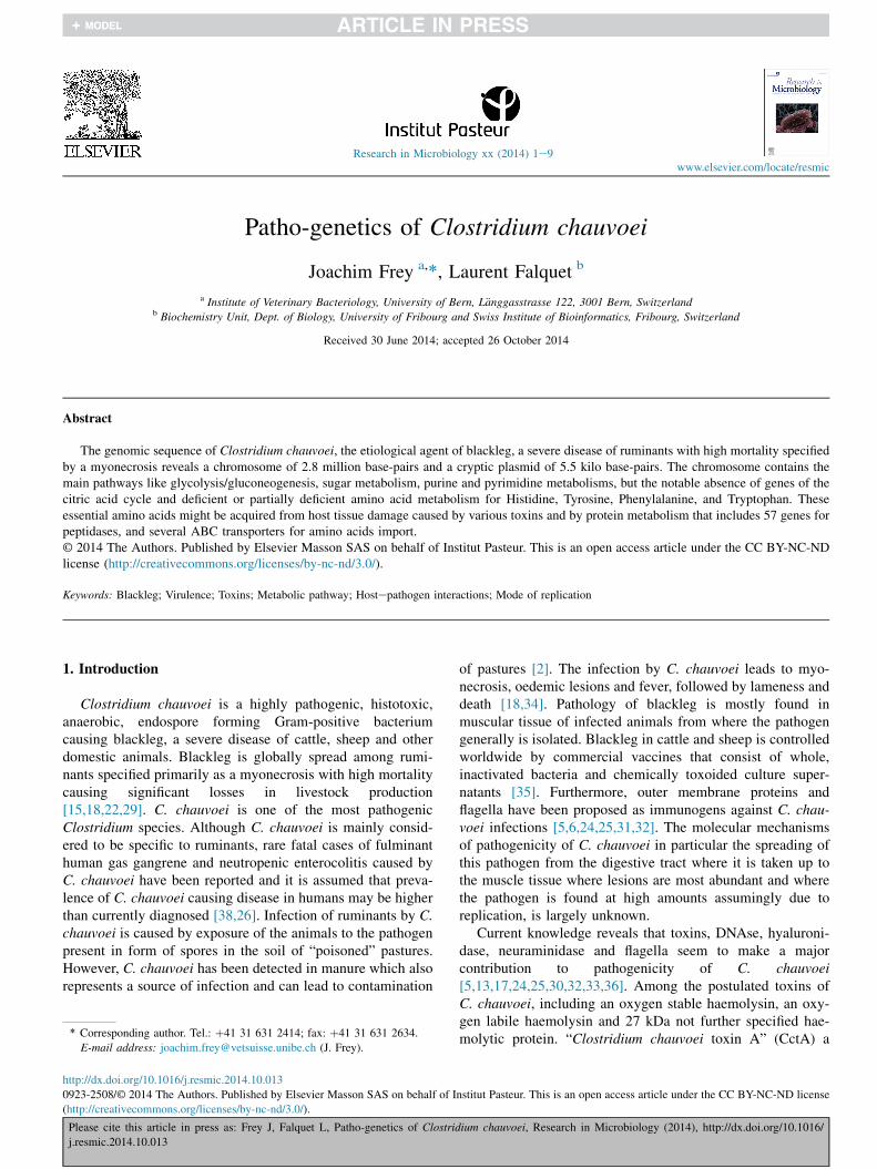
Other pathways are deficient or partially deficient, mainlyin the amino acid metabolism like Histidine, Tyrosine,Phenylalanine, and Tryptophan. These amino acids areessential and therefore it explains why RAST analysis revealedthe presence of 136 functions involved in protein metabolismwhereof 57 genes, mostly encoding for peptidases, play a rolein protein degradation (Table 2). In addition several ABCtransporters responsible for amino acids import are alsoidentified. This reflects the protein rich environment, the
ium chauvoei, Research in Microbiology (2014), http://dx.doi.org/10.1016/
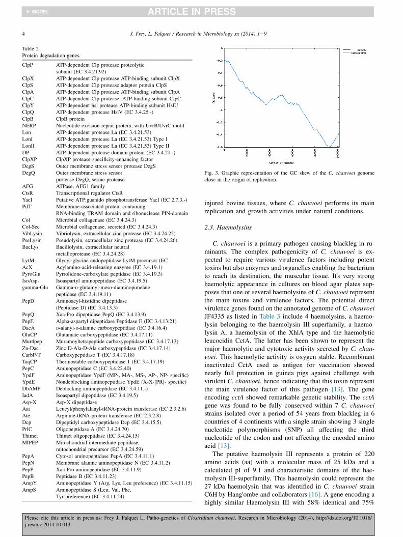
Table 2
Protein degradation genes.
ClpP ATP-dependent Clp protease proteolytic
subunit (EC 3.4.21.92)
ClpX ATP-dependent Clp protease ATP-binding subunit ClpX
ClpS ATP-dependent Clp protease adaptor protein ClpS
ClpA ATP-dependent Clp protease ATP-binding subunit ClpA
ClpC ATP-dependent Clp protease, ATP-binding subunit ClpC
ClpY ATP-dependent hsl protease ATP-binding subunit HslU
ClpQ ATP-dependent protease HslV (EC 3.4.25.-)
ClpB ClpB protein
NERP Nucleotide excision repair protein, with UvrB/UvrC motif
Lon ATP-dependent protease La (EC 3.4.21.53)
LonI ATP-dependent protease La (EC 3.4.21.53) Type I
LonII ATP-dependent protease La (EC 3.4.21.53) Type II
DP ATP-dependent protease domain protein (EC 3.4.21.-)
ClpXP ClpXP protease specificity-enhancing factor
DegS Outer membrane stress sensor protease DegS
DegQ Outer membrane stress sensor
protease DegQ, serine protease
AFG ATPase, AFG1 family
CtsR Transcriptional regulator CtsR
YacI Putative ATP:guanido phosphotransferase YacI (EC 2.7.3.-)
PilT Membrane-associated protein containing
RNA-binding TRAM domain and ribonuclease PIN-domain
Col Microbial collagenase (EC 3.4.24.3)
Col-Sec Microbial collagenase, secreted (EC 3.4.24.3)
VibLysin Vibriolysin, extracellular zinc protease (EC 3.4.24.25)
PseLysin Pseudolysin, extracellular zinc protease (EC 3.4.24.26)
BacLys Bacillolysin, extracellular neutral
metalloprotease (EC 3.4.24.28)
LytM Glycyl-glycine endopeptidase LytM precursor (EC
AcX Acylamino-acid-releasing enzyme (EC 3.4.19.1)
PyroGlu Pyrrolidone-carboxylate peptidase (EC 3.4.19.3)
IsoAsp- Isoaspartyl aminopeptidase (EC 3.4.19.5)
gamma-Glu Gamma-D-glutamyl-meso-diaminopimelate
peptidase (EC 3.4.19.11)
PepD Aminoacyl-histidine dipeptidase
(Peptidase D) (EC 3.4.13.3)
PepQ Xaa-Pro dipeptidase PepQ (EC 3.4.13.9)
PepE Alpha-aspartyl dipeptidase Peptidase E (EC 3.4.13.21)
DacA D-alanyl-D-alanine carboxypeptidase (EC 3.4.16.4)
GluCP Glutamate carboxypeptidase (EC 3.4.17.11)
Mur4pep Muramoyltetrapeptide carboxypeptidase (EC 3.4.17.13)
Zn-Dac Zinc D-Ala-D-Ala carboxypeptidase (EC 3.4.17.14)
CarbP-T Carboxypeptidase T (EC 3.4.17.18)
TaqCP Thermostable carboxypeptidase 1 (EC 3.4.17.19)
PepC Aminopeptidase C (EC 3.4.22.40)
YpdF Aminopeptidase YpdF (MP-, MA-, MS-, AP-, NP- specific)
YpdE Nondeblocking aminopeptidase YpdE (X-X-[PR]- specific)
DbAMP Deblocking aminopeptidase (EC 3.4.11.-)
IadA Isoaspartyl dipeptidase (EC 3.4.19.5)
Asp-X Asp-X dipeptidase
Aat Leucyl/phenylalanyl-tRNA-protein transferase (EC 2.3.2.6)
Ate Arginine-tRNA-protein transferase (EC 2.3.2.8)
Dcp Dipeptidyl carboxypeptidase Dcp (EC 3.4.15.5)
PrlC Oligopeptidase A (EC 3.4.24.70)
Thimet Thimet oligopeptidase (EC 3.4.24.15)
MIPEP Mitochondrial intermediate peptidase,
mitochondrial precursor (EC 3.4.24.59)
PepA Cytosol aminopeptidase PepA (EC 3.4.11.1)
PepN Membrane alanine aminopeptidase N (EC 3.4.11.2)
PepP Xaa-Pro aminopeptidase (EC 3.4.11.9)
PepB Peptidase B (EC 3.4.11.23)
AmpY Aminopeptidase Y (Arg, Lys, Leu preference) (EC 3.4.11.15)
AmpS Aminopeptidase S (Leu, Val, Phe,
Tyr preference) (EC 3.4.11.24)
Fig. 3. Graphic representation of the GC skew of the C. chauvoei genome
close in the origin of replication.
4 J. Frey, L. Falquet / Research in Microbiology xx (2014) 1e9
Please cite this article in press as: Frey J, Falquet L, Patho-genetics of Clostrid
j.resmic.2014.10.013
injured bovine tissues, where C. chauvoei performs its mainreplication and growth activities under natural conditions.
2.3. Haemolysins
C. chauvoei is a primary pathogen causing blackleg in ru-minants. The complex pathogenicity of C. chauvoei is ex-pected to require various virulence factors including potenttoxins but also enzymes and organelles enabling the bacteriumto reach its destination, the muscular tissue. It's very stronghaemolytic appearance in cultures on blood agar plates sup-poses that one or several haemolysins of C. chauvoei representthe main toxins and virulence factors. The potential directvirulence genes found on the annotated genome of C. chauvoeiJF4335 as listed in Table 3 include 4 haemolysins, a haemo-lysin belonging to the haemolysin III-superfamily, a haemo-lysin A, a haemolysin of the XhlA type and the haemolyticleucocidin CctA. The latter has been shown to represent themajor haemolytic and cytotoxic activity secreted by C. chau-voei. This haemolytic activity is oxygen stable. Recombinantinactivated CctA used as antigen for vaccination showednearly full protection in guinea pigs against challenge withvirulent C. chauvoei, hence indicating that this toxin representthe main virulence factor of this pathogen [13]. The geneencoding cctA showed remarkable genetic stability. The cctAgene was found to be fully conserved within 7 C. chauvoeistrains isolated over a period of 54 years from blackleg in 6countries of 4 continents with a single strain showing 3 singlenucleotide polymorphisms (SNP) all affecting the thirdnucleotide of the codon and not affecting the encoded aminoacid [13].
The putative haemolysin III represents a protein of 220amino acids (aa) with a molecular mass of 25 kDa and acalculated pI of 9.1 and characteristic domains of the hae-molysin III-superfamily. This haemolysin could represent the27 kDa haemolysin that was identified in C. chauvoei strainC6H by Hang'ombe and collaborators [16]. A gene encoding ahighly similar Haemolysin III with 58% identical and 75%
ium chauvoei, Research in Microbiology (2014), http://dx.doi.org/10.1016/
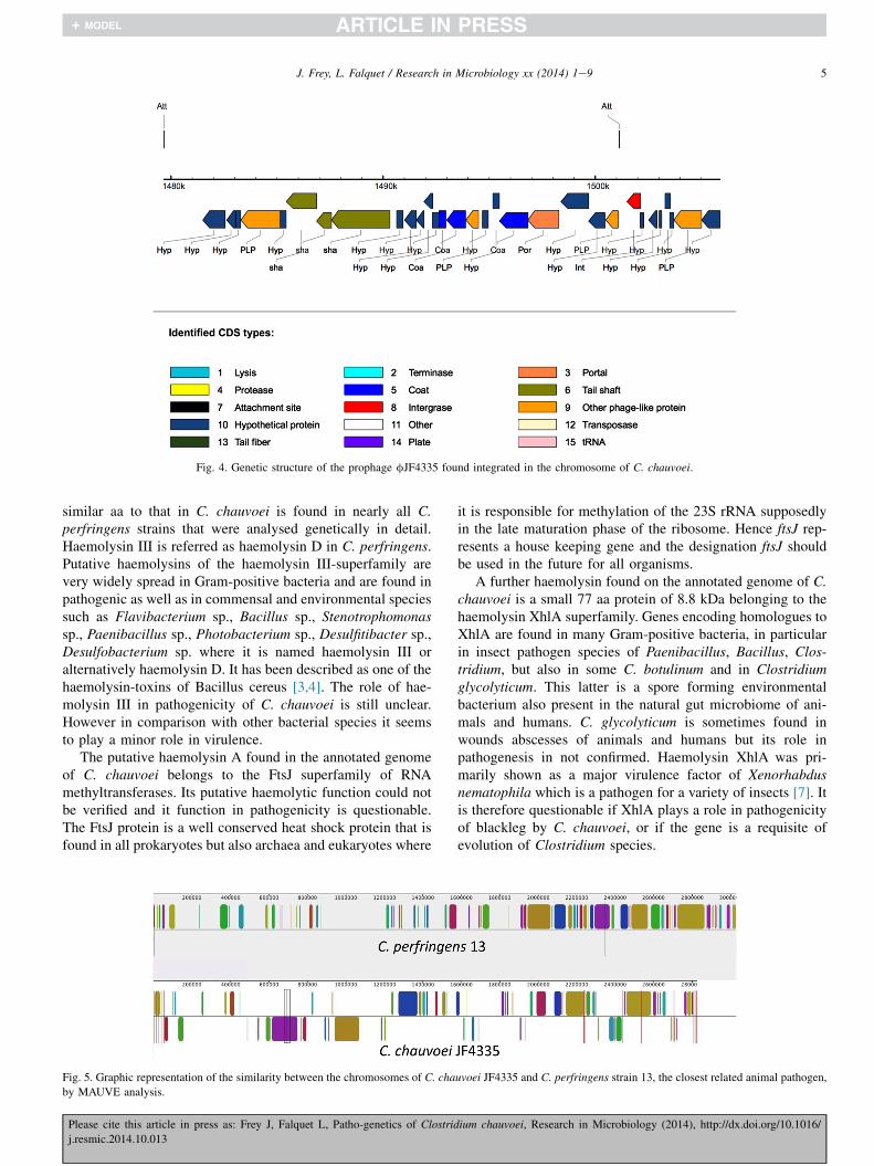
Fig. 4. Genetic structure of the prophage fJF4335 found integrated in the chromosome of C. chauvoei.
5J. Frey, L. Falquet / Research in Microbiology xx (2014) 1e9
similar aa to that in C. chauvoei is found in nearly all C.perfringens strains that were analysed genetically in detail.Haemolysin III is referred as haemolysin D in C. perfringens.Putative haemolysins of the haemolysin III-superfamily arevery widely spread in Gram-positive bacteria and are found inpathogenic as well as in commensal and environmental speciessuch as Flavibacterium sp., Bacillus sp., Stenotrophomonassp., Paenibacillus sp., Photobacterium sp., Desulfitibacter sp.,Desulfobacterium sp. where it is named haemolysin III oralternatively haemolysin D. It has been described as one of thehaemolysin-toxins of Bacillus cereus [3,4]. The role of hae-molysin III in pathogenicity of C. chauvoei is still unclear.However in comparison with other bacterial species it seemsto play a minor role in virulence.
The putative haemolysin A found in the annotated genomeof C. chauvoei belongs to the FtsJ superfamily of RNAmethyltransferases. Its putative haemolytic function could notbe verified and it function in pathogenicity is questionable.The FtsJ protein is a well conserved heat shock protein that isfound in all prokaryotes but also archaea and eukaryotes where
Fig. 5. Graphic representation of the similarity between the chromosomes of C. cha
by MAUVE analysis.
Please cite this article in press as: Frey J, Falquet L, Patho-genetics of Clostrid
j.resmic.2014.10.013
it is responsible for methylation of the 23S rRNA supposedlyin the late maturation phase of the ribosome. Hence ftsJ rep-resents a house keeping gene and the designation ftsJ shouldbe used in the future for all organisms.
A further haemolysin found on the annotated genome of C.chauvoei is a small 77 aa protein of 8.8 kDa belonging to thehaemolysin XhlA superfamily. Genes encoding homologues toXhlA are found in many Gram-positive bacteria, in particularin insect pathogen species of Paenibacillus, Bacillus, Clos-tridium, but also in some C. botulinum and in Clostridiumglycolyticum. This latter is a spore forming environmentalbacterium also present in the natural gut microbiome of ani-mals and humans. C. glycolyticum is sometimes found inwounds abscesses of animals and humans but its role inpathogenesis in not confirmed. Haemolysin XhlA was pri-marily shown as a major virulence factor of Xenorhabdusnematophila which is a pathogen for a variety of insects [7]. Itis therefore questionable if XhlA plays a role in pathogenicityof blackleg by C. chauvoei, or if the gene is a requisite ofevolution of Clostridium species.
uvoei JF4335 and C. perfringens strain 13, the closest related animal pathogen,
ium chauvoei, Research in Microbiology (2014), http://dx.doi.org/10.1016/

Fig. 6. Glycolysis/glycogenesis pathway. The blue boxed functions are annotated in the genome of C. perfringens. (For interpretation of the references to colour in
this figure legend, the reader is referred to the web version of this article.)
6 J. Frey, L. Falquet / Research in Microbiology xx (2014) 1e9
Please cite this article in press as: Frey J, Falquet L, Patho-genetics of Clostridium chauvoei, Research in Microbiology (2014), http://dx.doi.org/10.1016/
j.resmic.2014.10.013

Table 3
Primary virulence genes of Clostridium chauvoei JF4335.
Virulence factor Gene
designation
Protein nr Comment
Panton Valentine
leucocidin CctA
cctA 1863 Activity confirmed
Haemolysin III 276 Also named
haemolysin D
Haemolysin XhlA 799
(Haemolysin A)a ftsJ 1036 RNA
methyltransferase
Sialidase nanA 1464 Activity confirmed
Hyaluronidase H nagH 2049 Activity confirmed
Hyaluronidase J nagJ 1919
Patatine phospholipase 181
Patatine phospholipase 1777
Collagen binding protein 1915
Collagen binding protein 1916
Internalin A inlA1 2016
Internalin A inlA2 2017
a Correct annotation is FtsJ RNA methyltransferase.
7J. Frey, L. Falquet / Research in Microbiology xx (2014) 1e9
2.4. Non-haemolysin virulence factors
Besides haemolysin- and putative haemolysin genes, C.chauvoei contains a gene, nanA, encoding a sialidase whoseactivity has been confirmed previously by expressing it in E.coli. Sialidase activity could be obtained from a recombinantprotein expressed from a cloning vector containing the C.chauvoei nanA gene [36]. Sialidases or neuraminidaseshydrolyse the a 2e3, a 2e6, a 2e8 glycosilic linkages ofterminal sialic residues in oligosaccharides, glycosaccarides,glycoproteins and glycolipids and are known as virulencefactor by affecting the intracellular matrix and by undercuttingpathways in the host's immune regulation. Sialidase is sup-posed to underpin the rapid spread of C. chauvoei in the tissueof the infected host and has previously been suggested to playan important role in blackleg [33]. The nanA gene andsecreted sialidase activity was found in all 7 C. chauvoeistrains mentioned above. However genetic differences of thenanA gene was found among the various C. chauvoei strains,in contrast to the leucocidin gene cctA [36].
Hyaluronidase is further enzyme affecting the intracellularmatrix of the host. The genome of C. chauvoei reveals twodifferent hyaluronidase genes, nagH and nagJ (Table 3) ofwhich the activity of nagH has been confirmed in our labo-ratory (Frey and Wuthrich unpublished). Hyaluronidase,similar to sialidase is thought to be involved in loosening theintracellular matrix and tissue connections, thus favouring thespread of C. chauvoei in the infected host.
The C. chauvoei genome furthermore reveals two genesencoding for potential patatine phospholipases (PPARs).PPARs belong to the ligand inducible nuclear receptors andtranscription factors in the steroid superfamily [20]. PPARg issuggested to be beneficial in hyper-inflammatory diseases,such as sepsis, by repressing expression of inflammatory genes(inducible nitric oxide synthase, TNFa, or IL-1b) [37].However, it can also provoke apoptosis which in the hyper-
Please cite this article in press as: Frey J, Falquet L, Patho-genetics of Clostrid
j.resmic.2014.10.013
inflammatory phase of sepsis might be helpful by reducingthe number of immune cells that are involved in secreting highamounts of pro-inflammatory mediators [37]. PPARs areactivated by ligands such as fatty acids, eicosanoids and oxi-dised fatty acids [9,12,21,27].
Furthermore, two genes encoding potential collagen bind-ing proteins as well as two genes encoding homologues tointernalin A, which are supposed to be involved in virulencewere found on the chromosome of C. chauvoei.
Although flagella are often not directly involved in viru-lence, they might contribute to the infectious process by theirpotential to provide the bacterium mobility and hence to reachthe target tissue where the bacterium causes tissue damage anddisease. Furthermore flagellar antigens have been studied indetail in the view of their potential as valuable candidate an-tigens for vaccines [24,25,30e32]. Twenty flagellar biosyn-thesis genes were found on the coromosome of C. chauvoei infour clusters, probably arranged in operons: fliDMNFGIJLPSQ, flg KBCDG, flb D, the gene encoding theflagellar protein 3 and the genes for flagellar hook biosynthesisand hook length control proteins.
2.5. Antibiotic resistance
All above named strains of Clostridium chauvoei are sen-sitive to all antibiotics tested that are currently used againstclostridial infections (Frey unpublished results). The minimalinhibitory concentrations for strain JF4335 as determined inMueller Hinton II Broth using custom Sensititre™ suscepti-bility plates (Trek Diagnostics Systems, East-Grinstead, En-gland, and MCS Diagnostics BV, JL Swalmen, TheNetherlands) according to the CLSI guidelines [19] for C.chauvoei strain were: Cephalotin 2 mg/ml, Clindamycin0.5 mg/ml, Enrofloxacin 0.25 mg/ml, Erythromycin 0.25 mg/ml,Penicillin <0.012 mg/ml, Vancomycin 1 mg/ml, Tetracyclin1 mg/ml and Chloramphenicol <4 mg/ml. This reflects the factthat blackleg is not treated with antibiotics due to its virulentappearance leading to rapid death of the animal. It is thereforesurprising that C. chauvoei strain JF4335 harbours each a genepotential for penicillin resistance, a b-lactamase (EC 3.5.2.6),as well as an elongator Factor EF G type tetracycline resis-tance gene potentially involved in protection of ribosomesfrom tetracycline and catalysis and release of tetracycline(tetM, tetO analogue) and a vancomycin B-type resistanceprotein gene vanW, but no further genes potentially involved invancomycin resistance. Furthermore there are 9 genesencoding potential Multi antimicrobial extrusion proteins(Naþ/drug antiporters). However, these genes seem either notto be expressed in C. chauvoei JF4335, or are expressed asnon-functional proteins or most probably have functions otherthan export of antibiotics.
In conclusion, the genomic sequence of a virulent strain ofC. chauvoei gives insight in its particular lifestyle which im-plies the replication in damaged host tissue during infectionand the various virulence genes that allow the pathogen toreach the host tissue and prepare it for its replication. Potentialantibiotic resistance genes are present on the chromosome of
ium chauvoei, Research in Microbiology (2014), http://dx.doi.org/10.1016/

8 J. Frey, L. Falquet / Research in Microbiology xx (2014) 1e9
C. chauvoei, although this pathogen is sensitive to the corre-sponding antibiotics reflecting the fact that infected animalsare not treated with antibiotics.
Conflict of interest
The authors declare no conflict of interest.
Acknowledgements
This work was supported by the Research Foundation 3R,Reduction, Refinement and Replacement of Animal Experi-mentation grant nr. 136-13 to JF.
References
[1] Amador-Noguez D, Feng XJ, Fan J, Roquet N, Rabitz H, Rabinowitz JD.
Systems-level metabolic flux profiling elucidates a complete, bifurcated
tricarboxylic acid cycle in Clostridium acetobutylicum. J Bacteriol
2010;192:4452e61.[2] Bagge E, Lewerin SS, Johansson KE. Detection and identification by
PCR of Clostridium chauvoei in clinical isolates, bovine faeces and
substrates from biogas plant. Acta Vet Scand 2009;51:8.
[3] Baida GE, Kuzmin NP. Cloning and primary structure of a new hemo-
lysin gene from Bacillus cereus. Biochim Biophys Acta
1995;1264:151e4.
[4] Baida GE, Kuzmin NP. Mechanism of action of hemolysin III from
Bacillus cereus. BBA-Biomembranes 1996;1284:122e4.
[5] Chandler HM, Gulasekharam J. The protective antigen of a highly
immunogenic strain of Clostridium chauvoei including an evaluation
of its flagella as a protective antigen. J Gen Microbiol 1974;84:
128e34.
[6] Claus KD, Macheak ME. Preparation of a Clostridium chauvoei antigen
and determination of protective immunity by plate agglutination test. Am
J Vet Res 1972;33:1045e52.[7] Cowles KN, Goodrich-Blair H. Expression and activity of a Xenorhabdus
nematophila haemolysin required for full virulence towards Manduca
sexta insects. Cell Microbiol 2005;7:209e19.[8] Darling AE, Mau B, Perna NT. progressiveMauve: multiple genome
alignment with gene gain, loss and rearrangement. PLoS One
2010;5:e11147.
[9] Devchand PR, Keller H, Peters JM, Vazquez M, Gonzalez FJ, Wahli W.
The PPAR alpha-leukotriene B-4 pathway to inflammation control. Na-
ture 1996;384:39e43.
[10] Durre P, Hollergschwandner C. Initiation of endospore formation in
Clostridium acetobutylicum. Anaerobe 2004;10:69e74.[11] Falquet L, Calderon-Copete SP, Frey J. Draft genome sequence of the
virulent Clostridium chauvoei reference strain JF4335. Genome Announc
2013;1.
[12] Forman BM, Chen J, Evans RM. Hypolipidemic drugs, polyunsaturated
fatty acids, and eicosanoids are ligands for peroxisome proliferator-
activated receptors alpha and delta. Proc Natl Acad Sci U S A
1997;94:4312e7.[13] Frey J, Johansson A, Burki S, Vilei EM, Redhead K. Cytotoxin CctA, a
major virulence factor of Clostridium chauvoei conferring protective
immunity against myonecrosis. Vaccine 2012;30:5500e5.
[14] Gervasi T, Curto RL, Narbad A, Mayer MJ. Complete genome sequence
of PhiCP51, a temperate bacteriophage of Clostridium perfringens. Arch
Virol 2013;158:2015e7.
[15] Groseth PK, Ersdal C, Bjelland AM, Stokstad M. Large outbreak of
blackleg in housed cattle. Vet Rec 2011;169:339.
[16] Hang'ombe BM, Isogai E, Lungu J, Mubita C, Nambota A, Kirisawa R,
et al. Detection and characterization of Clostridium species in soil of
Zambia. Comp Immunol Microbiol Infect Dis 2000;23:277e84.
Please cite this article in press as: Frey J, Falquet L, Patho-genetics of Clostrid
j.resmic.2014.10.013
[17] Hang'ombe BM, Kohda T, Mukamoto M, Kozaki S. Purification and
sensitivity of Clostridium chauvoei hemolysin to various erythrocytes.
Comp Immunol Microbiol Infect Dis 2006;29:263e8.
[18] Hatheway CL. Toxigenic clostridia. Clin Microbiol Rev 1990;3:66e98.
[19] Institute CaLS. Methods for antimicrobial susceptibility testing of
anaerobic bacteria. 7th ed., vol. 27. Wayne, PA: Clinical and Laboratory
Standards Institute; 2007. no. 2. Approved standard M11-A7.
[20] Jeninga EH, Gurnell M, Kalkhoven E. Functional implications of genetic
variation in human PPAR gamma. Trends Endocrinol Metab
2009;20:380e7.
[21] Kliewer SA, Lenhard JM, Willson TM, Patel I, Morris DC, Lehmann JM.
A prostaglandin J(2) metabolite binds peroxisome proliferator-activated
receptor-gamma and promotes adipocyte differentiation. Cell
1995;83:813e9.
[22] Lange M, Neubauer H, Seyboldt C. Development and validation of a
multiplex real-time PCR for detection of Clostridium chauvoei and
Clostridium septicum. Mol Cell Probes 2010;24:204e10.
[23] Li F, Hagemeier CH, Seedorf H, Gottschalk G, Thauer RK. Re-citrate
synthase from Clostridium kluyveri is phylogenetically related to
homocitrate synthase and isopropylmalate synthase rather than to Si-
citrate synthase. J Bacteriol 2007;189:4299e304.
[24] Mattar MA, Corti~nas TI, de Guzm�an AM. Immunogenic protein varia-
tions of Clostridium chauvoei cellular antigens associated with the cul-
ture growth phase. FEMS Immunol Med Microbiol 2002;33:9e14.
[25] Mattar MA, Corti~nas TI, Stefanini AM. Extracellular proteins of Clos-
tridium chauvoei are protective in a mouse model. Acta Vet Hung
2007;55:159e70.[26] Nagano N, Isomine S, Kato H, Sasaki Y, Takahashi M, Sakaida K, et al.
Human fulminant gas gangrene caused by Clostridium chauvoei. J Clin
Microbiol 2008;46:1545e7.[27] Nagy L, Tontonoz P, Alvarez JGA, Chen HW, Evans RM. Oxidized LDL
regulates macrophage gene expression through ligand activation of PPAR
gamma. Cell 1998;93:229e40.
[28] Overbeek R, Olson R, Pusch GD, Olsen GJ, Davis JJ, Disz T, et al. The
SEED and the rapid annotation of microbial genomes using subsystems
technology (RAST). Nucleic Acids Res 2014;42:D206e14.
[29] Smith LD, Williams BL. Clostridium chauvoei. In: Thomas CC, editor.
The pathogenic anaerobia bacteria, Springfield, Illinois, USA; 1984.
p. 164e79.
[30] Tamura Y, Kijima-Tanaka M, Aoki A, Ogikubo Y, Takahashi T.
Reversible expression of motility and flagella in Clostridium chauvoei
and their relationship to virulence. Microbiology 1995;141(Pt
3):605e10.
[31] Tamura Y, Tanaka M. Opsonic activity of anti-flagellar serum against
Clostridium chauvoei by mouse polymorphonuclear leucocytes. Vet
Microbiol 1987;14:81e6.
[32] Tanaka M, Hirayama N, Tamura Y. Production, characterization, and
protective effect of monoclonal antibodies to Clostridium chauvoei
flagella. Infect Immun 1987;55:1779e83.[33] Useh NM, Ajanusi JO, Esievo KA, Nok AJ. Characterization of a sia-
lidase (neuraminidase) isolated from Clostridium chauvoei (Jakari
strain). Cell Biochem Funct 2006;24:347e52.
[34] Useh NM, Nok AJ, Esievo KA. Pathogenesis and pathology of blackleg
in ruminants: the role of toxins and neuraminidase. A short review. Vet Q
2003;25:155e9.
[35] Uzal FA. Evidence-based medicine concerning efficacy of vaccination
against Clostridium chauvoei infection in cattle. Vet Clin North Am Food
Anim Pract 2012;28:71e7.
[36] Vilei EM, Johansson A, Schlatter Y, Redhead K, Frey J. Genetic and
functional characterization of the NanA sialidase from Clostridium
chauvoei. Vet Res 2011;42:2.
[37] von Knethen A, Soller M, Brune B. Peroxisome proliferator-activated
receptor gamma (PPAR gamma) and sepsis. Arch Immunol Ther Exp
2007;55:19e25.[38] Weatherhead JE, Tweardy DJ. Lethal human neutropenic entercolitis
caused by Clostridium chauvoei in the United States: tip of the iceberg? J
Infect 2012;64:225e7.
ium chauvoei, Research in Microbiology (2014), http://dx.doi.org/10.1016/
9J. Frey, L. Falquet / Research in Microbiology xx (2014) 1e9
[39] Zhou X, Cai S, Hong A, You Q, Yu P, Sheng N, et al. Microfluidic
PicoArray synthesis of oligodeoxynucleotides and simultaneous
assembling of multiple DNA sequences. Nucleic Acids Res 2004;32:
5409e17.
Please cite this article in press as: Frey J, Falquet L, Patho-genetics of Clostrid
j.resmic.2014.10.013
[40] Zimmer M, Scherer S, Loessner MJ. Genomic analysis of Clos-
tridium perfringens bacteriophage phi3626, which integrates into
guaA and possibly affects sporulation. J Bacteriol 2002;184:
4359e68.
ium chauvoei, Research in Microbiology (2014), http://dx.doi.org/10.1016/