patellar transskeletal traction for the treatment of...
TRANSCRIPT
Case ReportPatellar Transskeletal Traction for the Treatment of ChronicPatellar Pseudoarthrosis
José Leonardo Rocha de Faria ,1 Dieno Mol Souza Portella,2 Victor Elias Titonelli,1
Naasson Trindade Cavanellas,1 Rodrigo Pires e Albuquerque,1 Eduardo Branco de Sousa ,1
and João Maurício Barretto1
1Knee Surgery Center, National Institute of Traumatology and Orthopedics of Brazil, 500 Brazil Ave.,Rio de Janeiro 20940-070, Brazil2National Institute of Traumatology and Orthopedics of Brazil, 500 Brazil Ave., Rio de Janeiro 20940-070, Brazil
Correspondence should be addressed to José Leonardo Rocha de Faria; [email protected]
Received 11 August 2018; Revised 4 December 2018; Accepted 16 December 2018; Published 22 January 2019
Academic Editor: Dimitrios S. Karataglis
Copyright © 2019 José Leonardo Rocha de Faria et al. This is an open access article distributed under the Creative CommonsAttribution License, which permits unrestricted use, distribution, and reproduction in any medium, provided the original workis properly cited.
Patellar fractures, which constitute approximately 1% of bone lesions, may lead to severe impairment of the extensormechanism. When conservative or surgical treatment fails, the patella may develop pseudoarthrosis. Neglect or delayedtreatment of this type of injury may lead to significant diastasis between the patellar fragments. There is no consensusregarding the best treatment for such cases. This study is aimed at describing a rare case of patellar pseudoarthrosis in apatient who underwent two-step surgical treatment comprising transskeletal patellar traction followed by osteosynthesiswith a tension band. A 17-year-old male patient presented with a left patellar fracture that resulted from a fall from astanding height 8 years ago. He did not undergo any type of surgical treatment during that time, but the fracture wasimmobilized for only 2 weeks. The two-step surgical treatment with transskeletal patellar traction and patellarosteosynthesis was performed and provided satisfactory functional clinical results in this patient. This two-step surgicaltreatment can be performed in cases similar to ours with satisfactory results.
1. Introduction
Patellar pseudoarthrosis, an uncommon entity in devel-oped countries, is not as rare in underdeveloped or devel-oping countries such as India, Brazil, and China [1]. Theincidence of pseudoarthrosis or delay in patellar consolida-tion ranges from 1.6% to 12.5% [2, 3]. Delayed treatmentand difficulty in accessing health services because of finan-cial and/or geographic reasons are some of the reasons forthe relatively high incidence of this pathology in theseaforementioned countries [1].
Neglect or delayed treatment may lead to significantdiastasis among the bone fragments of pseudoarthrosis.The gap between these fragments develops more fre-quently in transverse fractures. In this type of injury, the
constant contraction of the quadriceps performed overtime causes the proximal fragment of the fracture to riseup [1, 4, 5].
The treatment of patellar pseudoarthrosis with signifi-cant diastasis between bone fragments is always challeng-ing, and few studies on this topic have been reported inthe literature. Garg et al. [1] described a surgical techniquefor reducing diastasis between bone fragments of pseu-doarthrosis. They first performed transskeletal patellartraction in patients with a distance greater than 3 cmbetween the fragments, followed by osteosynthesis of thepatella with a tension band, and they obtained adequatefunctional results.
This study is aimed at describing a rare case ofchronic patellar pseudoarthrosis with 9 cm diastasis
HindawiCase Reports in OrthopedicsVolume 2019, Article ID 5915701, 6 pageshttps://doi.org/10.1155/2019/5915701

between the bone fragments in a patient who underwenttwo-step surgical treatment based on evidence reportedby Garg et al. [1].
2. Case Presentation
A 17-year-old male patient presented with a left patellarfracture that resulted from a fall from a standing height8 years ago. He did not undergo any type of surgical treat-ment during that time, but the fracture was immobilizedwith crural-crustal plaster, albeit for only 2 weeks. At thepresent consultation, the patient presented with an activerange of motion (ROM) of 70° to 120° and passive ROMof -5° to 120° (Figures 1(a) and 1(b)).
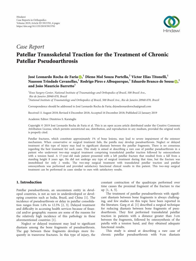
In the first phase, transskeletal patellar traction wasperformed, and a Steinmann pin with a 3.5mm thick cen-tral thread (Figures 2(a)–2(c)) was inserted transverselyinto the proximal pole. Transskeletal patellar pin assemblyis very easy to perform with the patient under sedationand local anesthesia.
The traction device placed on the patella had an initialweight of 3 kg, which was increased daily by 0.5 kg. Serialradiological images were obtained to quantify the decreasein the distance between the two poles of the area of pseu-doarthrosis (Figure 3). On day 11, diastasis between thefragments, which was 9 cm preoperatively, was reducedto 1.2 cm with the knee in full extension (Figure 4).
Then osteosynthesis was performed with a tensionband. We removed the traction device and the tractionpin from the proximal pole of the patella, with thepatient under spinal anesthesia with femoral nerve block.We performed median longitudinal surgical access andplane dissection and identified bone fragments of thepatella. We passed two 2.0mm thick Kirschner wires,longitudinally and parallelly, into the two fragments.We attempted to reduce the fragments with two Back-haus clamps (Figure 5), but the contact between thefragments was not possible.
We performed cerclage wiring with a 1.2mm thick cerclagewire followed by a figure-of-eight tension band. This tech-nique considerably reduced the distance between the pseu-doarthrosis foci, but the contact was insufficient (Figure 6).
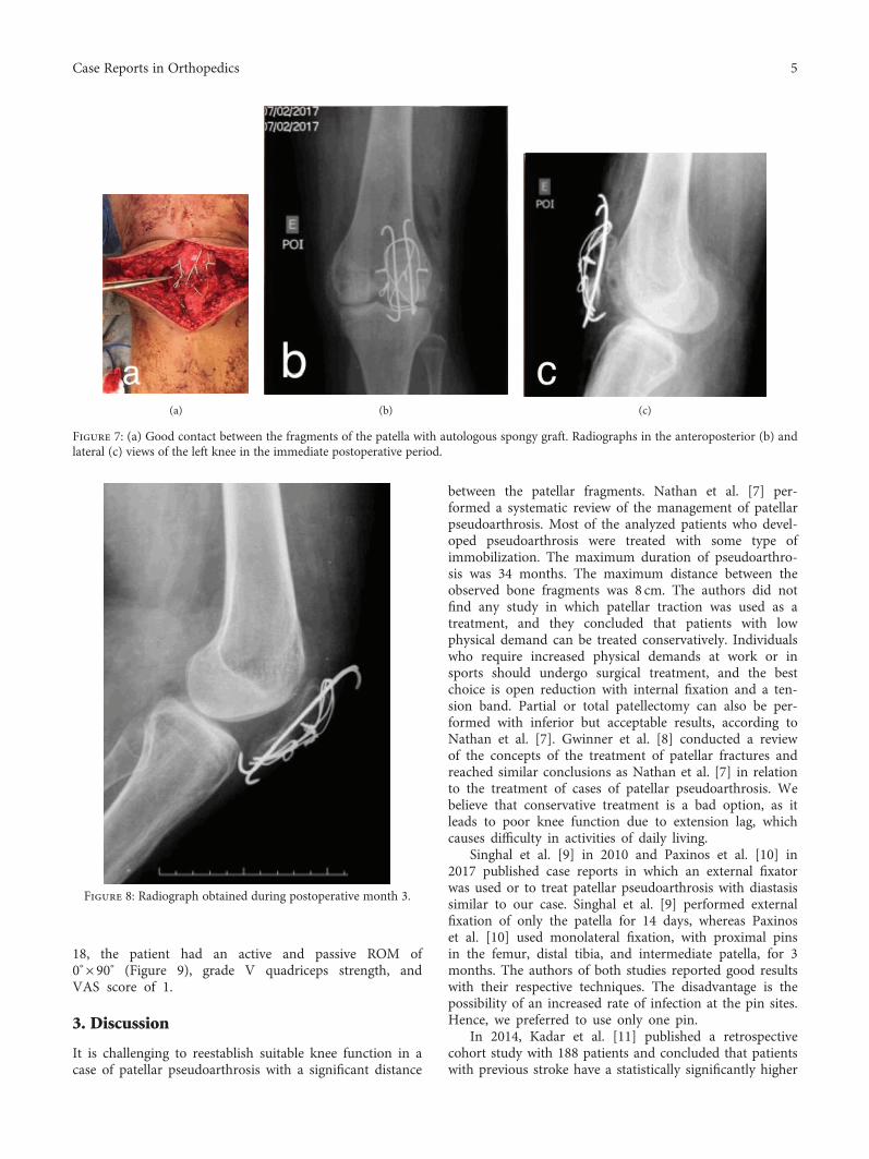
Next, we proceeded with an insertion of a 1.2mm circu-lar cerclage bonding wire, and finally, we achieved good con-tact between the bone fragments of the pseudoarthrosis.After thoroughly cleaning the site with 0.9% physiologicalsaline, we focused on the pseudoarthrosis, an autologousspongy osseous graft from the proximal tibia, and removedit through a small osseous hole made in the anteromedialtibia (Figures 7(a)–7(c)).
The patient was reassessed during the postoperativeperiod for bone consolidation (assessed by radiography),pain (visual analog scale (VAS), 0-10), quadricepsstrength (motor force rating) [6], and ROM of the limb(using a goniometer).
(a) (b)
Figure 1: (a) Preoperative radiograph. (b) Preoperative clinical examination.
2 Case Reports in Orthopedics
(a) (b)
(c)
Figure 2: The traction pin is positioned (a, b), and traction is applied (c).
Figure 3: Serial radiological images.
3Case Reports in Orthopedics

The patient was not immobilized and allowed fullweight bearing with crutches, but knee flexion wasrestricted for 8 weeks. During week 8, physiotherapy
focused on increasing the patient’s ROM. During thisperiod, the patient had a passive arch of 0° × 10°, gradeIII quadriceps strength, and a VAS score of 4, and radi-ography showed signs of bone consolidation. Duringpostoperative month 3, a passive ROM of 0° × 25°, activeROM of 10° × 25°, grade IV quadriceps strength, VASscore of 4, and consolidated patella on radiography(Figure 8) were observed. During postoperative month
Beginning of transpatellartraction-3 kg
Gap
bet
wee
n th
e fra
gmen
ts of
the p
atel
la(c
m)
Days
Increase of 0.5 kg/day
osteosynthesis
10
8
6
4
2
00 1 4 6 11
Figure 4: Measurements of the approximate distance between the bone fragments of the patella during transpatellar traction based onradiological images of the knee in full extension.
Figure 5: Contact between the fragments is not possible.
Figure 6: Cerclage wiring with the figure-of-eight tension band andtwo Backhaus clamps are used in attempt to reduce the distancebetween the pseudoarthrosis foci.
4 Case Reports in Orthopedics
18, the patient had an active and passive ROM of0° × 90° (Figure 9), grade V quadriceps strength, andVAS score of 1.
3. Discussion
It is challenging to reestablish suitable knee function in acase of patellar pseudoarthrosis with a significant distance
between the patellar fragments. Nathan et al. [7] per-formed a systematic review of the management of patellarpseudoarthrosis. Most of the analyzed patients who devel-oped pseudoarthrosis were treated with some type ofimmobilization. The maximum duration of pseudoarthro-sis was 34 months. The maximum distance between theobserved bone fragments was 8 cm. The authors did notfind any study in which patellar traction was used as atreatment, and they concluded that patients with lowphysical demand can be treated conservatively. Individualswho require increased physical demands at work or insports should undergo surgical treatment, and the bestchoice is open reduction with internal fixation and a ten-sion band. Partial or total patellectomy can also be per-formed with inferior but acceptable results, according toNathan et al. [7]. Gwinner et al. [8] conducted a reviewof the concepts of the treatment of patellar fractures andreached similar conclusions as Nathan et al. [7] in relationto the treatment of cases of patellar pseudoarthrosis. Webelieve that conservative treatment is a bad option, as itleads to poor knee function due to extension lag, whichcauses difficulty in activities of daily living.
Singhal et al. [9] in 2010 and Paxinos et al. [10] in2017 published case reports in which an external fixatorwas used or to treat patellar pseudoarthrosis with diastasissimilar to our case. Singhal et al. [9] performed externalfixation of only the patella for 14 days, whereas Paxinoset al. [10] used monolateral fixation, with proximal pinsin the femur, distal tibia, and intermediate patella, for 3months. The authors of both studies reported good resultswith their respective techniques. The disadvantage is thepossibility of an increased rate of infection at the pin sites.Hence, we preferred to use only one pin.
In 2014, Kadar et al. [11] published a retrospectivecohort study with 188 patients and concluded that patientswith previous stroke have a statistically significantly higher
(a) (b) (c)
Figure 7: (a) Good contact between the fragments of the patella with autologous spongy graft. Radiographs in the anteroposterior (b) andlateral (c) views of the left knee in the immediate postoperative period.
Figure 8: Radiograph obtained during postoperative month 3.
5Case Reports in Orthopedics

chance of developing patellar pseudoarthrosis and infection.In a meta-analysis performed in the Hospital for Special Sur-gery in New York on the rate of reoperation, pseudoar-throsis, and infection after patellar osteosynthesis, Dyet al. [3] concluded that age, sex, or the surgical tech-nique has no statistically significant influence on therates mentioned above.
In Garg et al. study [1], 35 patients with patellar pseu-doarthrosis with diastasis greater than 3 cm between bonefragments were treated and divided into three groups.The first group comprised 10 patients who underwentsingle-stage surgical treatment, which involved V-Y quad-riceps stretching and tension band osteosynthesis. The sec-ond group comprised 15 patients who underwenttwo-stage surgery, which involved patellar transskeletaltraction (mean, 8 days) and osteosynthesis with a tensionband after the space between the patellar fragments wasreduced by approximately 1 cm. The third group com-prised 10 patients who underwent two-stage surgery,which involved transskeletal patellar traction and partialdistal pole patellectomy or total patellectomy. In all aspectsevaluated, group 2 showed superior results. Thus, theauthors concluded that the treatment of choice for casesof patellar pseudoarthrosis with diastasis is osteosynthesiswith a tension band preceded by transskeletal traction ofthe patella. We achieved good results with transskeletalpatellar traction and patellar osteosynthesis in our patient,and the method appears to be effective in the treatment ofold neglected patellar fractures.
Two-step surgical treatment with transskeletal patellartraction and patellar osteosynthesis can be performed incases similar to ours with satisfactory results.
Consent
Written consent was obtained from the patient for thepublication of this report and accompanying images.
Conflicts of Interest
The authors declare that there is no conflict of interestregarding the publication of this paper.
References
[1] P. Garg, K. Satyakam, A. Garg, S. Sahoo, D. Biswas, andS. Mitra, “Patellar nonunions: comparison of various surgicalmethods of treatment,” Indian Journal of Orthopaedics,vol. 46, no. 3, pp. 304–311, 2012.
[2] J. F. Klassen and R. T. Trousdale, “Treatment of delayed andnonunion of the patella,” Journal of Orthopaedic Trauma,vol. 11, no. 3, pp. 188–194, 1997.
[3] C. J. Dy, M. T. M. Little, M. B. Berkes et al., “Meta-analysis ofre-operation, nonunion, and infection after open reductionand internal fixation of patella fractures,” Journal of Traumaand Acute Care Surgery, vol. 73, no. 4, pp. 928–932, 2012.
[4] O. Elattar, S. H. Coleman, R. F. Warren, and S. R. Rozbruch,“Neglected patellar tendon rupture with massive proximalpatellar migration treated with patellar transport and stagedallograft reconstruction: a report of 2 cases,” Orthopaedic Jour-nal of Sports Medicine, vol. 4, no. 11, 2016.
[5] Z. U. Isiklar, K. E. Varner, R. W. Lindsey, J. R. Bocell, andD. M. Lintner, “Late reconstruction of patellar ligament rup-tures using Ilizarov external fixation,” Clinical Orthopaedicsand Related Research, vol. 322, pp. 174–178, 1996.
[6] R. D. De Leffert, “Brachial plexus,” in Green’s Operative HandSurgery, Chapter 38, pp. 3453-3454, Elsevier, 1988.
[7] S. T. Nathan, B. E. Fisher, C. S. Roberts, and P. V. Giannoudis,“The management of nonunion and delayed union of patellafractures: a systematic review of the literature,” InternationalOrthopaedics, vol. 35, no. 6, pp. 791–795, 2011.
[8] C. Gwinner, S. Märdian, P. Schwabe, K.-D. Schaser, B. D. Kra-pohl, and T. M. Jung, “Current concepts review: fractures ofthe patella,” GMS Interdisciplinary Plastic and ReconstructiveSurgery DGPW, vol. 5, 2016.
[9] V. Singhal, D. Mittal, H. Lal, and R. Khare, “Gap non-union ofpatella: a treatment dilemma,” Pb Journal of Orthopaedics,vol. 12, no. 1, pp. 8–11, 2010.
[10] O. Paxinos, A. Karamitros, P. Douras, and N. Kouris,“Neglected patella nonunion successfully treated after 8 yearsby quadriceps distractive lengthening with a spanning unilat-eral external fixation system,” Knee Surgery, Sports Traumatol-ogy, Arthroscopy, vol. 26, no. 9, pp. 2784–2787, 2018.
[11] A. Kadar, H. Sherman, Y. Glazer, E. Katz, and E. L. Steinberg,“Predictors for nonunion, reoperation and infection after sur-gical fixation of patellar fracture,” Journal of Orthopaedic Sci-ence, vol. 20, no. 1, pp. 168–173, 2015.
Figure 9: Clinical examination at 18 months postoperatively.
6 Case Reports in Orthopedics

Stem Cells International
Hindawiwww.hindawi.com Volume 2018
Hindawiwww.hindawi.com Volume 2018
MEDIATORSINFLAMMATION
of
EndocrinologyInternational Journal of
Hindawiwww.hindawi.com Volume 2018
Hindawiwww.hindawi.com Volume 2018
Disease Markers
Hindawiwww.hindawi.com Volume 2018
BioMed Research International
OncologyJournal of
Hindawiwww.hindawi.com Volume 2013
Hindawiwww.hindawi.com Volume 2018
Oxidative Medicine and Cellular Longevity
Hindawiwww.hindawi.com Volume 2018
PPAR Research
Hindawi Publishing Corporation http://www.hindawi.com Volume 2013Hindawiwww.hindawi.com
The Scientific World Journal
Volume 2018
Immunology ResearchHindawiwww.hindawi.com Volume 2018
Journal of
ObesityJournal of
Hindawiwww.hindawi.com Volume 2018
Hindawiwww.hindawi.com Volume 2018
Computational and Mathematical Methods in Medicine
Hindawiwww.hindawi.com Volume 2018
Behavioural Neurology
OphthalmologyJournal of
Hindawiwww.hindawi.com Volume 2018
Diabetes ResearchJournal of
Hindawiwww.hindawi.com Volume 2018
Hindawiwww.hindawi.com Volume 2018
Research and TreatmentAIDS
Hindawiwww.hindawi.com Volume 2018
Gastroenterology Research and Practice
Hindawiwww.hindawi.com Volume 2018
Parkinson’s Disease
Evidence-Based Complementary andAlternative Medicine
Volume 2018Hindawiwww.hindawi.com
Submit your manuscripts atwww.hindawi.com