partitioning of body fluids and … · turtles were kept overnight in dry but covered boxes to...
TRANSCRIPT

J. exp. Biol. 116, 237-250 (1985) 2 3 7Printed in Great Britain © The Company of Biologists Limited 1985
PARTITIONING OF BODY FLUIDS AND CARDIOVASCULARRESPONSES TO CIRCULATORY HYPOVOLAEMIA IN THE
TURTLE, PSEUDEMYS SCRIPTA ELEGANS
BY ALLAN W. SMITS* AND MARK M. KOZUBOWSKI
Department of Physiology and Cell Biology, University of Kansas, Lawrence,KS 66045, U.SA.
Accepted 8 November 1984
SUMMARY
Investigations were conducted (1) to measure the steady state com-partmentation of body fluids and (2) to assess the efficacy of blood volume andpressure maintenance during haemorrhage-induced hypovolaemia in thepond turtle, Pseudemys scripta elegans. The pre-haemorrhage blood volume,as determined by tracer dilution of slCr-labelled erythrocytes, averaged6-89 ±0-33% of the body mass, and was part of comparatively largeextracellular (40-2 ± 0-70 %) and total body fluid volumes (75-25 ± 1-48 %).Turtles exhibited progressive reductions in systemic arterial pressurethroughout a cumulative haemorrhage of —48% of their original bloodvolume, despite dramatic increases in heart rate and comparatively largemagnitudes of transcapillary fluid transfer from interstitial to intravascularspaces. Arterial blood pressure returned to pre-haemorrhage values 2h afterexperimental haemorrhage ceased, concomitant with the restoration of theoriginal blood volume. Our results support arguments made in previousstudies that the resistance to fluid movement between vascular and extra-vascular locations in reptiles is comparatively low. Furthermore, thehaemodynamic responses of turtles to experimental hypovolaemia suggestthat barostasis through adjustments in vascular tone is less effective than thatobserved in other reptiles.
INTRODUCTION
Vertebrate animals typically maintain a relatively constant blood volume andpressure through regulatory mechanisms that are both numerous and inter-related(see review by Little, 1981), resulting in a circulatory homeostasis upon which manybodily functions depend. The efficacy of these regulatory mechanisms has beenlargely demonstrated in experiments that induce changes in circulation volume(through haemorrhage or transfusion) in mammalian models; comparatively little isknown concerning similar regulatory mechanisms operating in the cardiovascularsystems of other vertebrate taxa.
* Present address: Department of Zoology, University of Massachusetts, Amheret, MA 01003, U.S.A.Key words: Fluid volumes, haemorrhage, turtle, blood.

238 A. W. SMTTS AND M. M. KOZUBOWSKI
A unique tolerance to circulatory hypovolaemia has been demonstrated in snakes inwhich cardiovascular and respiratory homeostasis are maintained despite haemor-rhages exceeding 80-100% of the snake's initial blood volume (Lillywhite & Smith,1981; Lillywhite, Ackerman & Palacios, 1983). The primary mechanisms thatcounteract hypovolaemia in these reptiles are a rapid and extensive mobilization ofinterstitial fluid to the intravascular space (Lillywhite & Smith, 1981; Lillywhite &Pough, 1983; Smits & Lillywhite, 1985) and reflexive adjustments of cardiac per-formance and vascular tone (Lillywhite & Seymour, 1978; Lillywhite & Smith,1981). The volume of interstitial fluid is also proportionately greater in snakes than inmammals, and essentially all the fluid absorbed into the vascular space duringhaemorrhage comes from the interstitium (Smits & Lillywhite, 1985).
Turtles represent an evolutionarily distinct taxon of reptiles and have served asmodel animals in cardiovascular research for decades. However, comparatively fewdata exist that describe the compartmentation of body fluids between the vascular andnon-vascular locations in these reptiles, or the capacity for fluid movement betweenthese locations to maintain circulatory homeostasis.
In this paper we report how the intracellular and extracellular body fluid ispartitioned within the pond turtle, Pseudemys scripta. Further, the capacity for bloodvolume regulation in these reptiles has been tested by employing tracer dilutiontechniques to measure the extent and source of transcapillary fluid transfer duringhypovolaemia induced by haemorrhage.
MATERIALS AND METHODS
Animals and surgical preparations
Pond turtles {Pseudemys scripta elegans) were obtained through a commercialsupplier (Lemberger Associates, Wisconsin) and were kept in fresh water (24-28°C)on a diet of vegetables, fish and meat. The 14 adult turtles (body mass,X ± S.D. = 1466 ± 173 g) used in this study were in apparent good health and normalstate of hydration prior to experimentation.
Surgical anaesthesia was induced by lowering the turtles' body temperature in anice bath for 1 h; cold narcosis was maintained during surgery by covering the animalswith chipped ice. A hole (4 cm diameter) was drilled through the plastron to allowaccess to the major blood vessels and the heart. The left subclavian artery wasocclusively cannulated (PE-90, i.d. = 0-860 mm, o.d. = 1-270 mm) and the catheterwas advanced to the most proximal portion of the artery. The catheter passed throughthe body cavity -via a stab wound through the loose skin between the neck and leftforelimb. The patency of the catheter was maintained by occasional flushing withsmall volumes (<0-2ml) of heparinized (100 units ml"1) turtle Ringer's solution(Rogers, 1938).
Tracers and labelling procedures
The compartmentation of body fluid in each turtle prior to haemorrhage wasassessed by tracer dilution analysis. Deuterium oxide (D2O), slCr-labelled erythrocytes
Cardiovascular responses to haemorrhage in the turtle 239
and thiocyanate ion (SCN~) were used to measure total body water (TBW), wholeblood volume (BV) and extracellular fluid volume (ECFV), respectively.
The D2O (approx. 2-5 ml kg"1 body mass) was injected intraperitoneally at 16.00 hthe day prior to the experiment. Turtles were kept overnight in dry but covered boxesto reduce both cutaneous D2O exchange and evaporative water loss. A blood sampletaken immediately preceding experimental haemorrhage was vacuum distilled toremove organic components, and the D2O concentration of this body fluid wasdetermined by comparison with D2O standards (1-0—5-0ml D2OP1) using infraredspectrometry (Zweens, Frankena, Reicher & Zijlstra, 1980).
Turtle BV was measured by the dilution of autologous, 51Cr-labelled erythrocytesinfused into the arterial catheter approximately 3 h prior to experimental haemor-rhage. Details of the labelling procedure are described by Lillywhite & Smits (1985).Briefly, 1 ml of each turtle's erythrocytes was incubated with 10 /iCi of isotopic sodiumchromate at room temperature for 1 h. Following incubation the erythrocytes wererepeatedly washed with a plasma-saline solution to eliminate the unbound isotope.The erythrocytes were then diluted in normal saline to the volume originallywithdrawn from the turtle, and a known volume and specific activity of the labelledblood was infused into the arterial catheter. The blood volume was obtained bydividing the total activity infused (counts per minute) by the specific activity of theturtle blood (c.p.m. ml"1) sampled 3 h following tracer infusion. Radioactivity in allblood samples drawn for BV determination was assayed by counting each sample for5 min (approx. 1 % sigma) on a Packard Auto-Gamma 800C scintillation counter.The specific gravity of whole blood (1*05 gml"1) was used for conversions of mass tovolume (Thorson, 1968).
Dilution 'of the thiocyanate ion was used to estimate the ECFV of turtles. NaSCNwas infused into the arterial catheter (0-025 g NaSCN kg"1 body mass) 4h prior toexperimental haemorrhage. Concentrations of the thiocyanate ion in turtle plasmawere measured colourimetrically (Bradshaw & Shoemaker, 1967) using a Zeiss PMQII spectrophotometer. The pre-haemorrhage ECFV was calculated from con-centrations of the thiocyanate ion in four plasma samples (0-2 ml) collected at 15-minintervals in the hour preceding haemorrhage. The exact quantity of each of the threetracers injected was determined gravimetrically.
Tracer equilibration times were determined on one turtle (1290g), in which thetracer concentrations were measured in the turtle's blood sampled at intervals up to 3 h(51Cr) and 6 h (NaSCN) following tracer infusion (Fig. 1). These data confirmed thatadequate time was given (3 h for 51Cr; 4 h for NaSCN) for these tracers to equilibratewith their respective fluid spaces. Deuterium oxide was found to equilibrate with theTBW of turtles in 5-6 h at 25 °C; thus, adequate equilibration time was given for thistracer by administering it the day previous to the TBW determination.
Haemorrhage and fluid sampling
Immediately following measurements of steady-state fluid volumes, turtles weresubjected to a graded haemorrhage to assess the extent of inter-compartment fluidtransfer in response to hypovolemia. Turtles were conscious and at room temperature

240 A. W. SMTTS AND M. M. KOZUBOWSKI
(22-26°C) during haemorrhage, and rested in a horizontal, ventral up position.Arterial blood pressures and heart rates were measured from the subclavian arterialcatheter using a Narco P1000A Linear Core pressure transducer coupled to a DMP-4A physiograph (Narco Biosystems, Inc.). Haemorrhage was begun only after arterialblood pressure and heart rate had appeared to stabilize.
Graded haemorrhage was accomplished by withdrawing 4% of the turtle'sestimated BV at 10-min intervals until a total of 48 % of the pre-haemorrhage BV hadbeen removed. Initial blood volumes were assumed to equal 7 % of the turtle's bodymass based on previous BV measurements in the same species (Hutton, 1961; A. W.Smits & M. M. Kozubowski, unpublished observations). Each interval blood samplewas withdrawn from the subclavian arterial catheter into a clean syringe andpartitioned approximately equally for the separate analyses of whole blood (51Cr)and plasma (NaSCN). Samples of whole blood were delivered directly from thewithdrawal syringe into pre-weighed counting tubes. These tubes were reweighed,sealed with Parafilm, and counted the following day for gamma radioactivity. Plasmafractions from each withdrawal were obtained by immediate centrifugation (5 min inIEC Clinical centrifuge), and were pipetted into sealable plastic vials for later analysisof NaSCN concentration. The total osmolality of this plasma was also determinedusing vapour pressure osmometry (Wescor, Model 5100C). Haematocrits weremeasured on every blood sample by spinning blood-filled microcapillary tubes for5 min in an IEC MB microhaematocrit centrifuge (13 000 g). Arterial blood pressuresand heart rates were measured continuously between consecutive withdrawals fromthe subclavian arterial catheter; mean pressures and heart rates reported were thosemeasured during the last minute of each 10-min sampling interval. Analysis of tracer
1 4 J
Bo.u"Z 2 -
r4-0
- 3 - 0 ?_B
a
on
- 2 - 0 £
L- 1-0
Tune (h)
Fig. 1. Time-dependent changes in the activity of 51Cr in whole blood (closed circles) and thio-cyanate concentration in plasma (open circles) measured in a 1290 g female Pseudemys followingintravascular infusion of these tracers at time zero.

Cardiovascular responses to haemorrhage in the turtle 241
concentrations in 1-ml blood samples taken 0-5, 1-0 and 2-0h after haemorrhageceased (—48 %) were used to assess body fluid distribution during post-haemorrhagerecovery.
The volume and source of fluid mobilized into the vascular space were alsodetermined by tracer dilution analysis, based on the assumption that the dilution of aparticular tracer within a fluid space was proportional to the amount of unlabelledfluid entering that space. Decreases in the specific activity (c.p.m. ml~ ) of the Crwithin blood samples withdrawn during haemorrhage were used to calculate thevolume of extravascular (unlabelled) fluid transferred into the vascular space. Becausethe calculation of BV by 51Cr-labelled erythrocytes requires a knowledge of the totalradioactivity infused (see above), all calculations of BV subsequent to the initial BVmeasurement were corrected to account for slCr activity withdrawn from the turtles.The concentration of the thiocyanate ion was used to determine the source of fluidentering the vascular space during haemorrhage. Because both the plasma and theinterstitial fluid contained the same thiocyanate concentration at equilibrium, thethiocyanate concentration was assumed to remain unchanged if the source of ab-sorbed fluid was solely from the interstitium. Reductions in the plasma thiocyanateconcentration during haemorrhage were considered to indicate movement of intra-cellular (unlabelled) fluid into the extracellular space. The thiocyanate tracer is not apermanent marker within the ECF, but is normally eliminated from the extracellularspace of reptiles at a very slow rate (A. W. Smits, unpublished observations). Thetime constant of NaSCN elimination was sufficiently large (Fig. 1) to support theassumption that natural decay of this tracer during experimental haemorrhage (2 h)was negligible.
In addition to these experiments where a discrete volume of blood (48 % B V) wasremoved, five turtles were subjected to an extended haemorrhage. Four percent ofthe pre-haemorrhage BV was removed every lOmin until arterial pressure could nolonger be regulated, or until blood could no longer be withdrawn through the arterialcatheter. All procedures (surgery, tracers, haemorrhage rate, fluid sampling andanalyses) except the extent of haemorrhage were the same as described above.Albumin and globulin fractions of protein within the blood plasma (sampledthroughout haemorrhage) were assayed using Sigma Chemical Diagnostic Kits Nos.630 and 560, respectively. Following the experiment turtles were held overnight indry, plastic containers after their plastrons had been sealed with a wooden plug andmedical grade silicone rubber (Elastomer Silastic, Dow Corning).
RESULTS
Distribution of body fluid
The compartmentation of body fluid measured within turtles by tracer dilutionanalysis is schematically illustrated in Fig. 2. The whole blood volume (PV+RCV)averaged 6-89 ± 0-33 % of the body mass (X ± S.E.), and ranged from 5-74 to 8-15 %.

242 A. W. SMTTS AND M. M. KOZUBOWSKI
Physiological responses to haemorrhage
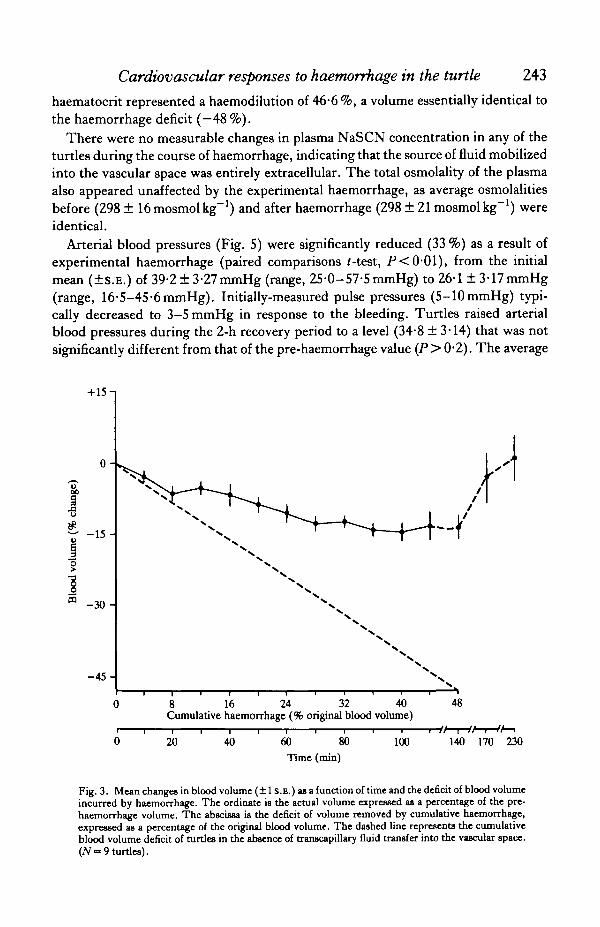
The maximum net decrease in circulating blood volume (BV) demonstrated byturtles experiencing a graded removal of whole blood averaged only 14*4% of theirpre-haemorrhage B V (Fig. 3), despite a cumulative withdrawal of 48 %. The dramaticdeparture of the lines in Fig. 3 depicting changes in BV (solid circles) and thepredicted BV changes in the absence of autotransfusion (dashed line) indicates thatturtles mobilized substantial quantities of fluid from extravascular to vascularlocations. The rate at which this fluid was transferred into the vascular spaceaccelerated as haemorrhage progressed, until the later stages of the experiment (—40to —48 % BV deficit) when rates of autotransfusion equalled the rates of experimentalhaemorrhage (Fig. 3). Restoration of the BV to pre-haemorrhage levels wasaccomplished within 1—2h after bleeding was discontinued.
Transcapillary fluid transfer determined by Cr dilution (Fig. 3) was consistentwith the degree of haemodilution observed in the haematocrit (Fig. 4). Haematocritsdecreased significantly (paired comparisons /-test; P<0-001) from (X±S.E.)22-1 + 1-89% to 13-6+1-41% during the course of haemorrhage, representing amean reduction of 38-5%. Following 2h of post-haemorrhage recovery, thehaematocrit decreased further to 11-8+1-22%. Thus, the cumulative decrease in
TBW
75-25
(1-48)
ICFV
35-08
ECFV
40-17
(0-70)
RCV 1-52
1
IFV
34-80
PV 5-37
Fig. 2. Compartmentation of body fluids measured in nine adult Pseudemys tcripta elegans (bodymass = 1475 ± 95 g, X ± S.D.), expressed as percentages of the body mass (numbers in parenthesesrepresent the S.E.M.). Red cell volumes (RCV) and plasma volumes (PV) were calculated from thewhole blood volume and the haematocrit. Intracellular fluid volumes (ICFV) and interstitial fluidvolumes (IFV) were calculated by subtraction of the extracellular fluid volume (ECFV) from the totalbody water (TBW) and subtraction of the PV from the ECFV, respectively.
Cardiovascular responses to haemorrhage in the turtle 243
haematocrit represented a haemodilution of 46-6 %, a volume essentially identical tothe haemorrhage deficit (—48%).
There were no measurable changes in plasma NaSCN concentration in any of theturtles during the course of haemorrhage, indicating that the source of fluid mobilizedinto the vascular space was entirely extracellular. The total osmolality of the plasmaalso appeared unaffected by the experimental haemorrhage, as average osmolalitiesbefore (298 ± ^mosmolkg"1) and after haemorrhage (298 ±21 mosmolkg"1) wereidentical.
Arterial blood pressures (Fig. 5) were significantly reduced (33 %) as a result ofexperimental haemorrhage (paired comparisons f-test, P<0-01), from the initialmean (±S.E.) of 39-2 +3-27mmHg (range, 25-0-57-5 mmHg) to 26-1 ± 3-17mmHg(range, 16-5-45-6mmHg). Initially-measured pulse pressures (5-10mmHg) typi-cally decreased to 3-5 mmHg in response to the bleeding. Turtles raised arterialblood pressures during the 2-h recovery period to a level (34-8 ± 3-14) that was notsignificantly different from that of the pre-haemorrhage value (P> 0-2). The average
m
+ 15 n
0 -
- 1 5 -
-30 -
- 4 5 -
0
0
8 16 24 32 40 48Cumulative haemorrhage (% original blood volume)
20 40 60 81Time (min)
100 140 170 230
Fig. 3. Mean changes in blood volume (±1 S.E.) as a function of time and the deficit of blood volumeincurred by haemorrhage. The ordinate ia the actual volume expressed as a percentage of the pre-haemorrhage volume. The abscissa is the deficit of volume removed by cumulative haemorrhage,expressed as a percentage of the original blood volume. The dashed line represents the cumulativeblood volume deficit of turtles in the absence of transcapillary fluid transfer into the vascular space.(7V~ 9 turtles).

244 A. W. SMTTS AND M. M. KOZUBOWSKI
heart rate of turtles (Fig. 5) at the cessation of haemorrhage (41-9 ± 2-19 beats min"1)was 120% higher than the initially-measured heart rate (191 ± 343 beats min"1).Although heart rate slowed significantly (P<0-02) during the recovery period to36-1 ± 2-49 beats min"1, this rate was still significantly greater (86-5 %, P< 0-001)than that measured just prior to haemorrhage.
Extended haemorrhage
The changes in B V measured during the initial 48 % haemorrhage deficit in turtlessubjected to an extended haemorrhage (Fig. 6) were similar in pattern and magnitude(—10-6 ± 3-5 %; t-test following arcsin transformation, P > 0-35) to those observed inturtles in the previous experiment (Fig. 2). Despite continued haemorrhage, BV inthese turtles increased dramatically during the later stages of the experiment to levelsthat exceeded pre-haemorrhage values by 30% (Fig. 6). The greatest increments inBV occurred following the removal of 80 % of the initial BV and concomitant with anaccelerated decline in arterial blood pressure. The maximum heart rate (42-3 ± 107
24
22-
20
c 18
16
14
12
10
0 8 16 24 32 40 48Cumulative haemorrhage (% original blood volume)
0 10 30 50 70 90Time (min)
110 150 240
Fig. 4. Mean changes in turtle haematocrits (±1 s.E.) plotted against time and the deficit of bloodvolume incurred by haemorrhage. The abscissa is the same as described in Fig. 3 (N = 9 turtles).

Cardiovascular responses to haemorrhage in the turtle 245
beats min~') was observed at an 80 % BV deficit and declined from this point until thecessation of haemorrhage.
Concentrations of albumin (16-9 ± 0-51 gdl"1) and globulin (28-5 ± l-VSgdl"1)within the plasma decreased gradually throughout extended haemorrhage to values
50
40
o.a
30
20
40
Ts 35
1o
eSu
30
20
15
Haemorrhage • •4—Recovery-^
0 8 16 24 32 40 48
Cumulative haemorrhage (% original blood volume)
0 10 30 50 70 90
Time (min)
110 150 240
Fig. 5. Average changes in arterial blood pressure (BPa) and heart rate in nine turtles (±1 S.E.) inresponse to hypovolaemia induced by haemorrhage. The abscissa is the same as described in Fig. 3.

246 A. W. SMTTS AND M. M. KOZUBOWSKI
that were 33 % (11-3 ± O ^ g d P 1 ) and 38% (17-6 ± l ^ g d r 1 ) of the pre-haemor-rhage levels, respectively. All turtles survived the extended haemorrhage to thefollowing day without access to water, and several were kept alive for 1—2 months inthe laboratory in plastic tanks containing fresh water.
DISCUSSION
Body fluid compartmentation
The mean BV of turtles at steady state (6-89% body mass) is similar to the BVassumed (7%) for the subsequent haemorrhage protocol, and is consistent with BVvalues reported for Pseudemys scripta and Chrysemys picta (combined; 8-1%)measured by the dilution of slCr-labelled erythrocytes (Wilson, Hansard & Cole,
50
OH
03
>PQ
10
~ 50I
1
10
32
T3OB
0
30
-20 I -
Globulin
28 56 84
Cumulative haemorrhage (% original blood volume)
112
Fig. 6. Changes in arterial blood pressure (BPa), heart rate (HR), fractional protein content, and thepercentage change from original blood volume (BV) measured in five turtles (X ± s.E.) in response toan extended haemorrhage where greater than 100 % of the pre-haemorrhage BV was removed.

Cardiovascular responses to haemorrhage in the turtle 247
1960). The small discrepancy in the BV of Pseudemys scripta reported here and thatmeasured by Wilson et al. (1960) or Hutton (1961; 8-75 % body mass) presumablyreflects differences in body sizes of the experimental animals, as plasma volumeappears to be inversely related to body mass in Pseudemys (Hutton, 1961).
Apparently no marker substance used in the dilution analysis of the ECFV isuniversally acceptable (Law, 1982), thus, only studies utilizing similar techniquesand tracers may be compared. The ECFV (thiocyanate space) of Pseudemys scripta(Fig. 2) exceeds the thiocyanate space measured in rats (33 % body mass; Huang &Bondurant, 1956) and is intermediate in volume compared to the thiocyanate spacesof other reptiles (Coulson & Hernandez, 1953; Minnich, 1982; Smits & Lillywhite,1985).
Total body water in Pseudemys (75-3% body mass; Fig. 2) is greater than thatmeasured in other freshwater or marine turtles (64-0-72-9%; Thorson, 1968), but isvery similar to values of TBW in other species of reptiles (review by Minnich, 1982).
Experimental haemorrhage
Decreases in systemic arterial pressure induced by haemorrhagic hypovolaemia inhigher vertebrates initiate a baroreceptor-controlled stimulation of the sympatheticnervous system that primarily increases peripheral resistance and venous returnthrough vasoconstriction, and secondarily improves cardiac output (Guyton, 1981;p. 333). Compensation for hypovolaemia via fluid resorption is typically considered amore passive process resulting from a reduction in capillary hydrostatic pressure(Djojosugito, Folkow & Kovach, 1968) and from the osmotic gradients caused by ahyperglycaemic hyperosmolality of the plasma (Ware, Norberg & Nylander, 1980),although activation of /3-adrenoreceptors (Hillman, Gustafsson & Lundvall, 1982)appears to contribute significantly to fluid resorption.
The regulation of systemic blood pressure in Pseudemys in response to hypo-volaemia appears poor despite dramatic increases in heart rate (Figs 5, 6). Systemicpressure seems largely dependent on the BV deficit (Figs 3, 5). The dependence of
Table 1. Absolute and proportional changes in heart rate, blood pressure and bloodvolume in three species of reptiles subjected to a graded haemorrhage of 32 % of their
initial blood volumes
Elaphe obsoleta* Cmtalus viridis* Pseudemys scripta
Heart rate 25->38 (+56%) 19->32(+67%) 19->37(+92%)(beats min"1)
Arterial blood 49->44(-l l%) 36—• 35 (-3%) 39-»27(-30%)pressure (mmHg)
% Change in —136% -14-4% -12-4%blood volume
Source of Extracellular Extracellular Extracellularabsorbed fluid
• Data from Smits & Lillywhite (1985).
248 A. W. SMTTS AND M. M. KOZUBOWSH
pressure regulation on BV is perhaps best exemplified by the return of arterialblood pressure to pre-haemorrhage values during the recovery period, concomitantwith a complete restoration of pre-haemorrhage BV (Figs 3, 5). This recovery ofblood pressure indicates that turtles can withstand a gradual removal of at least 48 % oftheir resting BV without entering severe and irreversible circulatory shock.
The experimental rate of haemorrhage used to demonstrate the tolerance ofterrestrial snakes to hypovolaemia (Lillywhite & Smith, 1981; Smits & Lillywhite,1985) was duplicated in the present study, thus the haemodynamic responses andchanges in B V during hypovolaemic stress in turtles and snakes may be quantitativelycompared (Table 1). Turtles and snakes are similar in their capacity to mobilizesubstantial volumes of extracellular fluid from extravascular to vascular locationsduring hypovolaemia; this supports the inference that the resistance to fluidmovement across the capillary wall in reptiles is comparatively low (Lillywhite &Smits, 1984; Smits & Lillywhite, 1985). Unlike mammals, turtles and snakes do notdemonstrate an increase in plasma osmolality during haemorrhage. Thus, the transferof fluid between extravascular and vascular locations does not appear to be facilitatedby a hyperglycaemic hyperosmolality in these reptiles. The observed stability ofextracellular osmolality also indicates that turtles do not resorb significant volumes ofdilute urine (< lOOmmolkg"1) during acute hypovolaemia.
Turtles do not maintain systemic arterial pressure as effectively as snakes inresponse to hypovolaemia, despite a proportionately greater increment in heart rate(Table 1). Baroreceptors located at the base of the pulmonary artery in Pseudemyshave been characterized by recording afferent vagal nerve activity synchronous withthe heart rate (Faraci, Shirer, Orr &Trank, 1983), but the primary effector(s) of thesemechanoreceptors remain in question. The haemodynamic responses of Pseudemysto hypovolaemia in the present study (Figs 5, 6) and to drug-induced changes in bloodpressure (Millard & Moalli, 1980) suggest that the heart, rather than the peripheralvasculature, may be the primary effector of barostasis.
Because measurements of B V in turtles are based on changes in the concentration ofslCr-labelled erythrocytes, a rapid change in haematocrit influenced by contraction ofthe spleen during haemorrhage may invalidate B V estimates. However, experimentalturtles did not demonstrate transient increases in haematocrit during any stage of thehaemorrhage (Fig. 4), thus we feel that the contribution of the spleen to erythrocyterecruitment and changes in BV during the experiment is negligible.
Turtles enduring an extended haemorrhage over-compensate for their initial BVdeficits and become increasingly hypervolaemic (Fig. 6). The restoration of BV inthese turtles does not augment the regulation of systemic blood pressure orsignificantly reduce the heart rate, in contrast to turtles bled 48 % of their initial BV(Fig. 5). This dramatic shift of fluid into the vascular space appears more related tochanges in the hydrostatic than osmotic pressures of the Starling equilibrium, as theproportional decreases in protein fractions that contribute to plasma oncotic pressureare constant throughout the experiment (Fig. 6). We have also measured dramaticincreases in the concentrations of plasma epinephrine and norepinephrine at timesconcurrent with the accelerated vascular resorption (—84 % to —96 % deficit; A. W.

Cardiovascular responses to haemorrhage in the turtle 249
Smits, M. M. Kozubowski & K. J. Renner, in preparation), and suggest that thisdramatic change in circulating BV may be related to the humoral effects of thesecatecholamines.
In summary, the capacity of aquatic turtles to counteract hypovolaemic stressthrough fluid recruitment (resorption) into the vasculature is exceptional and equals(if not exceeds) rates of autotransfusion measured in terrestrial snakes (Smits &Lillywhite, 1985). As in snakes, the ECFV is comparatively large and is the primarysource of fluid that is apparently mobilized across a low resistance between theinterstitial and intravascular spaces. Although turtles tolerate and survive substantiallosses of their blood, their haemodynamic responses to experimental hypovolaemiasuggest that barostasis through reflexogenic adjustments in vascular tone in thesereptiles is comparatively weak.
This study was supported by a Grant-in-Aid research award by the Sigma XiResearch Society to the authors, and by the U.S. Public Health Service, NIH GrantHL 24640 to H. B. Lillywhite. We thank H. W. Shirer and H. B. Lillywhite for theuse of their laboratories, and Fran Williams for typing the manuscript.
R E F E R E N C E S
BRADSHAW, S.D. & SHOEMAKER, V. H. (1967). Aspects of the water and electrolyte changes in a field populationof Amphibolurus lizards. Comp. Biochem. Physiol. 20, 855-865.
COULSON, R. A. & HERNANDEZ, T. (1953). Glucose studies in Crocodilia. Endocrinology 53, 311-320.Djojosuorro, A. M., FOLKOW, B. &KOVACH, A. G. B. (1968). The mechanism behind the rapid blood volume
restoration after hemorrhage in birds. Acta physiol. scand. 74, 114-122.FARAO, F. M., SHIRER, H. W., ORR, J. A. & TRANK, J. W. (1982). Circulatory mechanoreceptors in the pond
turtle, Pseudemys scripta. Am.J. Physiol. 242, R216-R219.GUYTON, A. C. (1981). Textbook of Medical Physiology. Philadelphia: W. B. Saunders.HILLMAN, J., GUSTAFSSON, D. & LUNDVALL, J. (1982). /S2-Adrenergic control of the plasma volume in
hemorrhage. Acta physiol. scand. 116, 175-180.HUANG, K.-C. tc BONDURANT, J. H. (1956). Simultaneous estimation of plasma volume, red cell volume and
thiocyanate space in unanesthetized normal and splenectomized rats. Am.J. Physiol. 185, 441—445.HUTTON, K. E. (1961). Blood volume, corpuscular constants, and shell weight in turtles. Am. J. Physiol. 200,
1004-1006.LAW, R. O. (1982). Techniques and application of extracellular space determination in mammalian tissues.
Experientia 38, 411-421.LILLYWHITE, H. B., ACKERMAN, R. A. & PALACIOS, L. (1983). Cardiorespiratory responses of snakes to
experimental hemorrhage. J. comp. Physiol. 152, 59-65.LILLYWHITE, H. B. & POUGH, F. H. (1983). Control of arterial pressure in aquatic sea snakes. Am.J. Physiol.
244, R66-R73.LILLYWHITE, H. B. & SEYMOUR, R. S. (1978). Regulation of arterial pressure in Australian tiger snakes. J.exp.
Biol. 75, 65-79.LILLYWHITE, H. B. & SMITH, L. H. (1981). Haemodynamic responses to haemorrhage in the snake, Elaphe
obsoleta obsoleta.J. exp. Biol. 94, 275-283.LILLYWHITE, H. B. & SMITS, A. W. (1984). Lability of blood volume in snakes and its relation to activity and
hypertension. J. exp. Biol. 110, 267-274.LITTLE, R. C. (1981). Physiology of the Heart and Circulation. Second Edition. Chicago: Year Book Medical
Publishers, Inc.MTLLARD, R. W. & M O A U J , R. (1980). Baroreflex sensitivity in an amphibian, Rana catesbeiana, and a
reptilian, Pseudemys scripta elegans.J. exp. Zool. 213, 283—288.MINNICH, J. E. (1982). The use of water. inBiologyoftheReptilia, (eds C. Gans& F. H. Pough). New York:
Academic Press.[ROGERS, C. G. (1938). Textbook of Comparative Physiology. New York: McGraw-Hill.

250 A. W. SMTTS AND M. M. KOZUBOWSKI
SMTTS, A. W. & LlLLYWHITE, H. B. (1985). Maintenance of blood volume in snakes: transcapillary shifts ofextravascular fluids during acute hemorrhage.,7. comp. Physiol. 155, 305—310.
THORSON, T. B. (1968). Body fluid partitioning in Reptilia. Copeia 1968, 592-602.WAKE, J., NORBERG, K.-A. & NYLANDER, G. (1980). Osmolar developments and altered thoracic duct lymph
flow in hypovolaemic shock. Europ. Surg. Res. 12, Suppl. 1, 48.WILSON, B., HANSARD, S. L. & COLE, B. T. (1960). Total blood volume of the turtle and the frog. Louisiana
Acad. Sci. 23, 45-52.ZWEENS, J., FRANKENA, H., REICHER, A. & ZIJLSTRA, W. G. (1980). Infrared-spectrometric determination of
D2O in biological fluids. Pflugers Arch. ges. Physiol. 385, 71-77.