part ii, muscle: mechanisms of contraction and neural ...part ii, muscle: mechanisms of contraction...
TRANSCRIPT
1
Part II, Muscle: Mechanisms of Contraction and Neural Control, Chapter 12 Outline of class notes for Physiology Objectives: After studying part II of this chapter you should be able to:
1. Discuss how contractile force is regulated 2. Describe the difference between a twitch, wave summation, tetany, and treppe in muscle
contraction. 3. Describe the difference between isometric and isotonic contractions. 4. Discuss the length tension relationship of skeletal muscle. 5. Describe the energy requirements of skeletal muscle and the creatine phosphate system. 6. What is meant by oxygen dept? 7. Describe the differences between skeletal muscle fiber types. 8. Explain the process of muscle fatigue. 9. Discuss the causes of muscle hypertrophy and muscle atrophy. 10. Discuss the characteristics of smooth and cardiac muscles and emphasize their differences in
excitation-contraction coupling. 11. Discuss the Clinical Applications from the study guide and assigned Applications to Health.
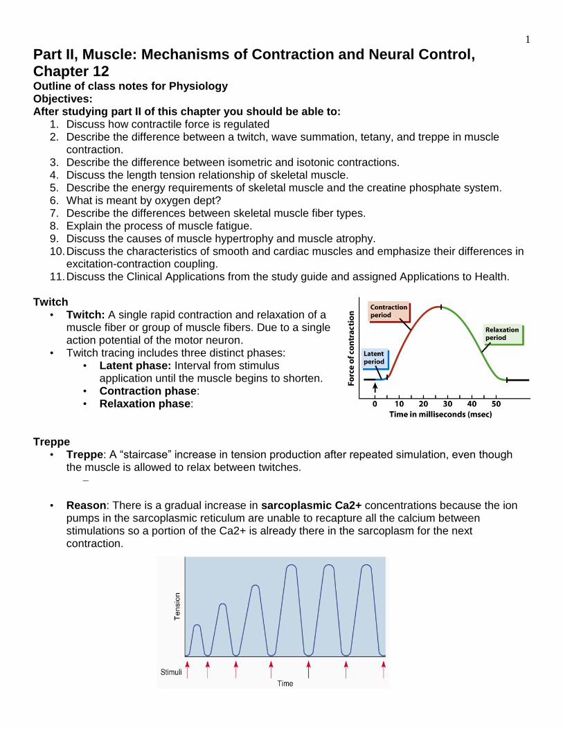
Twitch
• Twitch: A single rapid contraction and relaxation of a muscle fiber or group of muscle fibers. Due to a single action potential of the motor neuron.
• Twitch tracing includes three distinct phases: • Latent phase: Interval from stimulus
application until the muscle begins to shorten. • Contraction phase: • Relaxation phase:
Treppe
• Treppe: A “staircase” increase in tension production after repeated simulation, even though the muscle is allowed to relax between twitches.
–
• Reason: There is a gradual increase in sarcoplasmic Ca2+ concentrations because the ion pumps in the sarcoplasmic reticulum are unable to recapture all the calcium between stimulations so a portion of the Ca2+ is already there in the sarcoplasm for the next contraction.
2
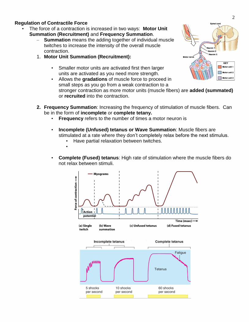
Regulation of Contractile Force • The force of a contraction is increased in two ways: Motor Unit
Summation (Recruitment) and Frequency Summation. – Summation means the adding together of individual muscle
twitches to increase the intensity of the overall muscle contraction.
1. Motor Unit Summation (Recruitment):
• Smaller motor units are activated first then larger units are activated as you need more strength.
• Allows the gradations of muscle force to proceed in small steps as you go from a weak contraction to a stronger contraction as more motor units (muscle fibers) are added (summated) or recruited into the contraction.
2. Frequency Summation: Increasing the frequency of stimulation of muscle fibers. Can
be in the form of incomplete or complete tetany. • Frequency refers to the number of times a motor neuron is
• Incomplete (Unfused) tetanus or Wave Summation: Muscle fibers are
stimulated at a rate where they don’t completely relax before the next stimulus. • Have partial relaxation between twitches. •
• Complete (Fused) tetanus: High rate of stimulation where the muscle fibers do not relax between stimuli.
3
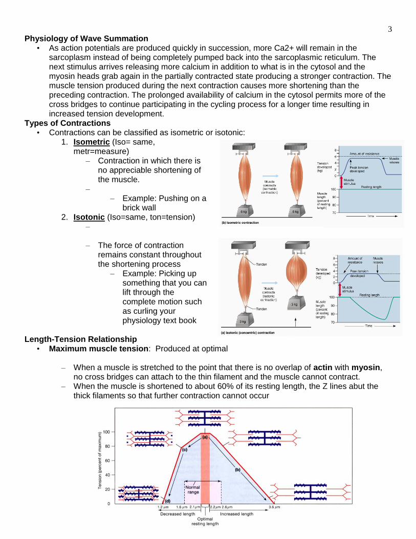
Physiology of Wave Summation • As action potentials are produced quickly in succession, more Ca2+ will remain in the
sarcoplasm instead of being completely pumped back into the sarcoplasmic reticulum. The next stimulus arrives releasing more calcium in addition to what is in the cytosol and the myosin heads grab again in the partially contracted state producing a stronger contraction. The muscle tension produced during the next contraction causes more shortening than the preceding contraction. The prolonged availability of calcium in the cytosol permits more of the cross bridges to continue participating in the cycling process for a longer time resulting in increased tension development.
Types of Contractions • Contractions can be classified as isometric or isotonic:
1. Isometric (Iso= same, metr=measure)
– Contraction in which there is no appreciable shortening of the muscle.
– – Example: Pushing on a
brick wall 2. Isotonic (Iso=same, ton=tension)
– – The force of contraction
remains constant throughout the shortening process
– Example: Picking up something that you can lift through the complete motion such as curling your physiology text book
Length-Tension Relationship
• Maximum muscle tension: Produced at optimal
– When a muscle is stretched to the point that there is no overlap of actin with myosin, no cross bridges can attach to the thin filament and the muscle cannot contract.
– When the muscle is shortened to about 60% of its resting length, the Z lines abut the thick filaments so that further contraction cannot occur
4
Muscle Tone • Muscle Tone:
– Due to spinal reflexes that alternate activity of the motor units within particular muscle resulting in a slightly contracted state.
– Their contractions do not produce enough tension to cause movement, but they do tense and firm the muscle.
– The identity of the motor units involved changes constantly.
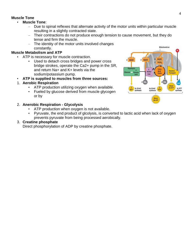
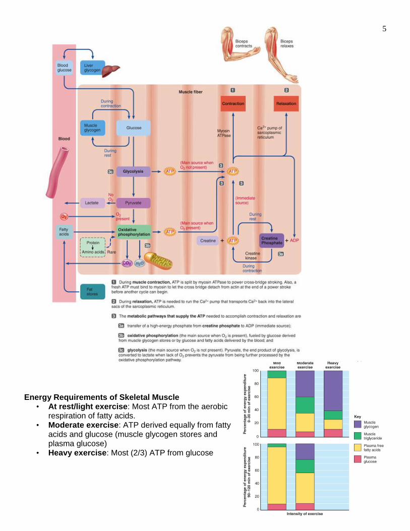
Muscle Metabolism and ATP • ATP is necessary for muscle contraction.
• Used to detach cross bridges and power cross bridge strokes, operate the Ca2+ pump in the SR, and return Na+ and K+ levels via the sodium/potassium pump.
• ATP is supplied to muscles from three sources: 1. Aerobic Respiration
• ATP production utilizing oxygen when available. • Fueled by glucose derived from muscle glycogen
or by
2. Anerobic Respiration - Glycolysis • ATP production when oxygen is not available. • Pyruvate, the end product of glcolysis, is converted to lactic acid when lack of oxygen
prevents pyruvate from being processed aerobically. 3. Creatine phosphate
Direct phosphorylation of ADP by creatine phosphate.
5
Energy Requirements of Skeletal Muscle
• At rest/light exercise: Most ATP from the aerobic respiration of fatty acids.
• Moderate exercise: ATP derived equally from fatty acids and glucose (muscle glycogen stores and plasma glucose)
• Heavy exercise: Most (2/3) ATP from glucose
6
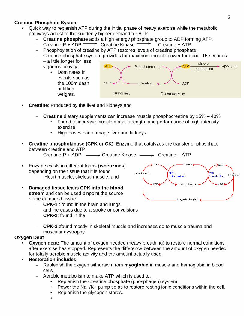
Creatine Phosphate System • Quick way to replenish ATP during the initial phase of heavy exercise while the metabolic
pathways adjust to the suddenly higher demand for ATP. – Creatine phosphate adds a high energy phosphate group to ADP forming ATP. – Creatine-P + ADP Creatine Kinase Creatine + ATP – Phosphoylation of creatine by ATP restores levels of creatine phosphate. – Creatine phosphate system provides for maximum muscle power for about 15 seconds
– a little longer for less vigorous activity.
• Dominates in events such as the 100m dash or lifting weights.
• Creatine: Produced by the liver and kidneys and
– Creatine dietary supplements can increase muscle phosphocreatine by 15% – 40% • Found to increase muscle mass, strength, and performance of high-intensity
exercise. • High doses can damage liver and kidneys.
• Creatine phosphokinase (CPK or CK): Enzyme that catalyzes the transfer of phosphate
between creatine and ATP. Creatine-P + ADP Creatine Kinase Creatine + ATP
• Enzyme exists in different forms (isoenzmes)
depending on the tissue that it is found – Heart muscle, skeletal muscle, and
• Damaged tissue leaks CPK into the blood
stream and can be used pinpoint the source of the damaged tissue.
– CPK-1 : found in the brain and lungs and increases due to a stroke or convulsions
– CPK-2: found in the
– CPK-3 :found mostly in skeletal muscle and increases do to muscle trauma and muscular dystrophy
Oxygen Debt • Oxygen dept: The amount of oxygen needed (heavy breathing) to restore normal conditions
after exercise has stopped. Represents the difference between the amount of oxygen needed for totally aerobic muscle activity and the amount actually used.
• Restoration includes: – Replenish the oxygen withdrawn from myoglobin in muscle and hemoglobin in blood
cells. – Aerobic metabolism to make ATP which is used to:
• Replenish the Creatine phosphate (phosphagen) system • Power the Na+/K+ pump so as to restore resting ionic conditions within the cell. • Replenish the glycogen stores. •
7
Muscle Fiber Types • Every muscle in the body is composed of a mixture of slow and fast muscle fibers, with other
fiber types gradated between these two extremes. The types of muscle fibers is based on their contraction speed and major pathways for forming ATP.
– Speed of Contraction: Slow and fast fibers are based on how fast their myosin ATPases split ATP resulting in filaments sliding over each other
•
– Major pathways for forming ATP: Oxidiative fibers rely mostly on aerobic pathways; glycolytic fibers rely more on anaerobic glycolysis.
Slow Fibers (Type I or Red or Slow Twitch)
• Slow fibers: Specialized for endurance; high fatigue resistance –
• Contract slowly because its myosin ATPases splits ATP at a slow rate. • Depends on oxygen for aerobic pathways to make ATP • Abundant capillaries, mitochondria, and myoglobin content to support
increased blood flow and high levels of oxidative metabolism. – Myoglobin: Pigment in muscle specialized for oxygen storage until
needed and gives the muscle cell a reddish color thus called red fibers.
• Example: soleus muscle contains more slow fibers than fast fibers.
Fast Fibers (Type II or White or Fast Twitch)
• Fast twitch: Split ATP at a rapid rate and thus have a fast contraction velocity after stimulation, but fatigue rapidly.
• Contain densely packed myofibrils, large glycogen reserves, and relatively few mitochondria and myoglobin.
• Anaerobic metabolism – Less extensive blood supply and fewer mitochondria because oxidative metabolism is of secondary importance, most ATP production is without oxygen.
• Does not contain a lot of myoglobin and appear white, thus called white fibers. • Best suited for short term, power activities such as
• Example: Most arm muscles have more fast fibers than slow fibers.
White vs Dark Meat
• Chicken’s breast is composed mainly of fast-twitch fibers giving the characteristic white meat appearance.
– Relatively little blood supply and less myoglobin than dark meat – Muscles adapted to contract rapidly for
• Chicken’s leg muscles are composed of slow-twitch fibers - meat is dark.
– Muscles adapted for endurance – running – Darker appearance is partly due to a richer blood supply and presence of myoglobin. – Myoglobin is an oxygen-carrying pigment of muscle tissue – it stores oxygen
temporarily until the muscle needs it.
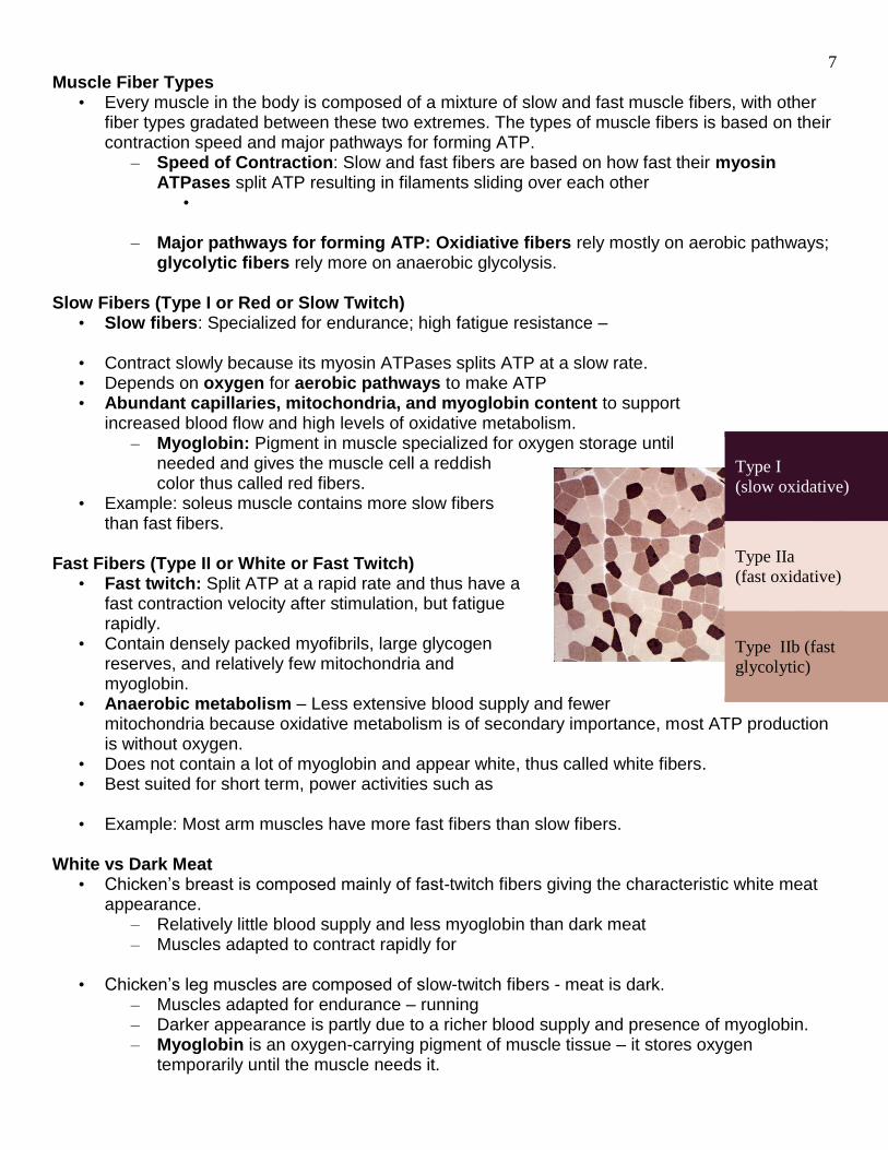
Type I
(slow oxidative)
Type IIa
(fast oxidative)
Type IIb (fast
glycolytic)
8
Muscle Fatigue • Muscle fatigue: The inability of the muscle to contract forcefully after long periods of activity.
Results from: – Accumulation of waste products (lactic acid) – Depletion of ATP, glycogen, and/or creatine phosphate – Reduced release of Ca2+ ions from sarcoplasmic reticulum. – Insufficient oxygen to make ATP – Failure of nerve impulses to release enough acetylcholine. – Depletion of intracellular
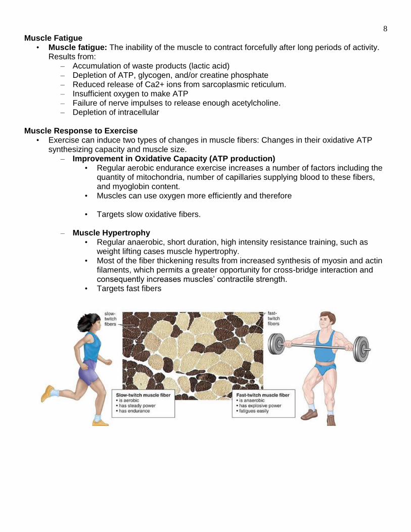
Muscle Response to Exercise
• Exercise can induce two types of changes in muscle fibers: Changes in their oxidative ATP synthesizing capacity and muscle size.
– Improvement in Oxidative Capacity (ATP production) • Regular aerobic endurance exercise increases a number of factors including the
quantity of mitochondria, number of capillaries supplying blood to these fibers, and myoglobin content.
• Muscles can use oxygen more efficiently and therefore
• Targets slow oxidative fibers.
– Muscle Hypertrophy • Regular anaerobic, short duration, high intensity resistance training, such as
weight lifting cases muscle hypertrophy. • Most of the fiber thickening results from increased synthesis of myosin and actin
filaments, which permits a greater opportunity for cross-bridge interaction and consequently increases muscles’ contractile strength.
• Targets fast fibers
9
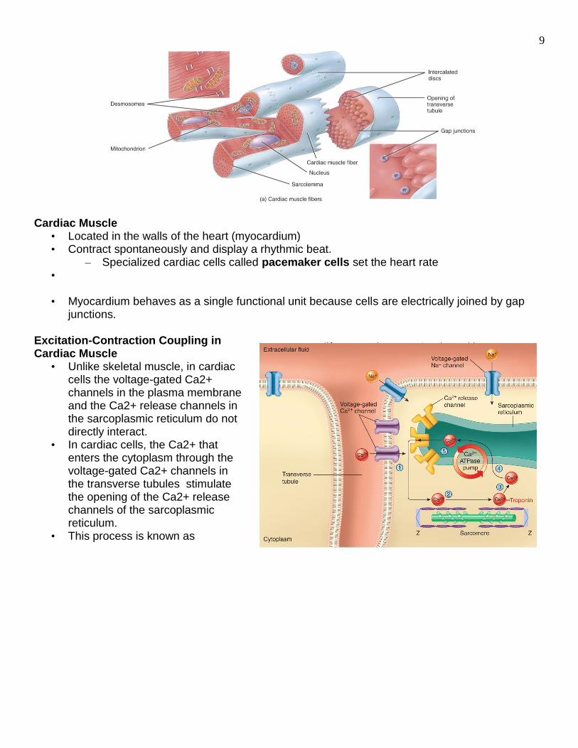
Cardiac Muscle
• Located in the walls of the heart (myocardium) • Contract spontaneously and display a rhythmic beat.
– Specialized cardiac cells called pacemaker cells set the heart rate •
• Myocardium behaves as a single functional unit because cells are electrically joined by gap
junctions. Excitation-Contraction Coupling in Cardiac Muscle
• Unlike skeletal muscle, in cardiac cells the voltage-gated Ca2+ channels in the plasma membrane and the Ca2+ release channels in the sarcoplasmic reticulum do not directly interact.
• In cardiac cells, the Ca2+ that enters the cytoplasm through the voltage-gated Ca2+ channels in the transverse tubules stimulate the opening of the Ca2+ release channels of the sarcoplasmic reticulum.
• This process is known as
10
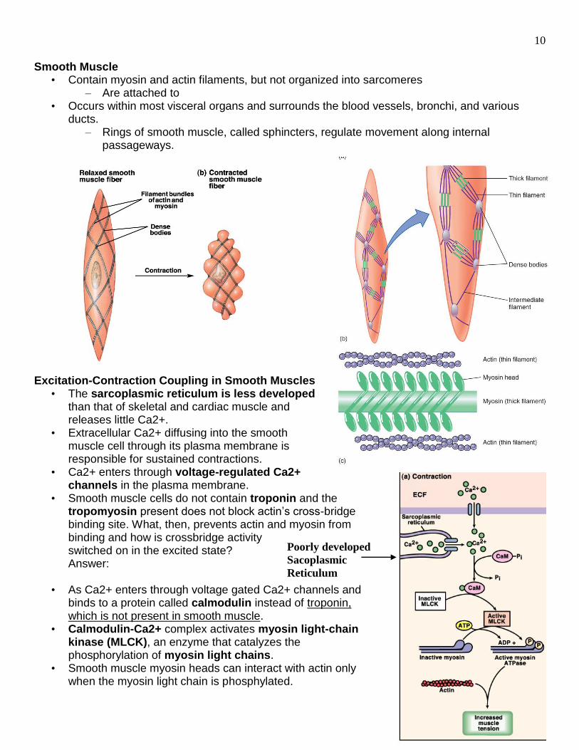
Smooth Muscle
• Contain myosin and actin filaments, but not organized into sarcomeres – Are attached to
• Occurs within most visceral organs and surrounds the blood vessels, bronchi, and various ducts.
– Rings of smooth muscle, called sphincters, regulate movement along internal passageways.
Excitation-Contraction Coupling in Smooth Muscles
• The sarcoplasmic reticulum is less developed than that of skeletal and cardiac muscle and releases little Ca2+.
• Extracellular Ca2+ diffusing into the smooth muscle cell through its plasma membrane is responsible for sustained contractions.
• Ca2+ enters through voltage-regulated Ca2+ channels in the plasma membrane.
• Smooth muscle cells do not contain troponin and the tropomyosin present does not block actin’s cross-bridge binding site. What, then, prevents actin and myosin from binding and how is crossbridge activity switched on in the excited state? Answer:
• As Ca2+ enters through voltage gated Ca2+ channels and
binds to a protein called calmodulin instead of troponin, which is not present in smooth muscle.
• Calmodulin-Ca2+ complex activates myosin light-chain kinase (MLCK), an enzyme that catalyzes the phosphorylation of myosin light chains.
• Smooth muscle myosin heads can interact with actin only when the myosin light chain is phosphylated.
Poorly developed
Sacoplasmic
Reticulum
11
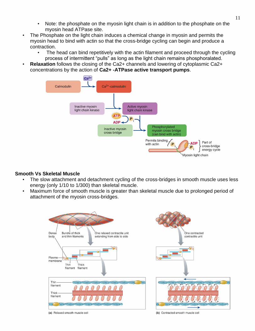
• Note: the phosphate on the myosin light chain is in addition to the phosphate on the myosin head ATPase site.
• The Phosphate on the light chain induces a chemical change in myosin and permits the myosin head to bind with actin so that the cross-bridge cycling can begin and produce a contraction.
• The head can bind repetitively with the actin filament and proceed through the cycling process of intermittent “pulls” as long as the light chain remains phosphoralated.
• Relaxation follows the closing of the Ca2+ channels and lowering of cytoplasmic Ca2+ concentrations by the action of Ca2+ -ATPase active transport pumps.
Smooth Vs Skeletal Muscle
• The slow attachment and detachment cycling of the cross-bridges in smooth muscle uses less energy (only 1/10 to 1/300) than skeletal muscle.
• Maximum force of smooth muscle is greater than skeletal muscle due to prolonged period of attachment of the myosin cross-bridges.