parotidectomy
TRANSCRIPT

PAROTIDECTOMY
PRESENTER: DR PRASHANTH L
MODERATOR: DR R M LALITHA

CONTENTS
1. INTRODUCTION
2. SURGICAL ANATOMY
3. TYPES OF PAROTIDECTOMY
4. PREOPERATIVE EVALUATION
5. SUPERFICIAL PAROTIDECTOMY
6. TOTAL PAROTIDECTOMY
7. EXTENDED TOTAL PAROTIDECTOMY
8. COMPLICATIONS
9. REFERENCES
INTRODUCTION
A parotidectomy is the surgical excision (removal) of
the parotid gland, the major and largest of the salivary
glands.
The procedure is most typically performed due to benign
or malignant tumors.
The majority of parotid gland tumors are benign,
however 20% of parotid tumors are found to be
malignant.

Rule of 80’s:
-80% of parotid tumors are benign
-80% of parotid tumors are pleomorphic adenomas
-80% of salivary gland pleomorphic adenomas
occur in the parotid
-80% of parotid pleomorphic adenomas occur in the
superficial lobe
-80% of untreated pleomorphic adenomas remain
benign

SURGICAL ANATOMY
Parotid gland
The paired parotid glands
are the largest of the major
salivary glands
weigh, on average, 15–30
g.
Preauricular region
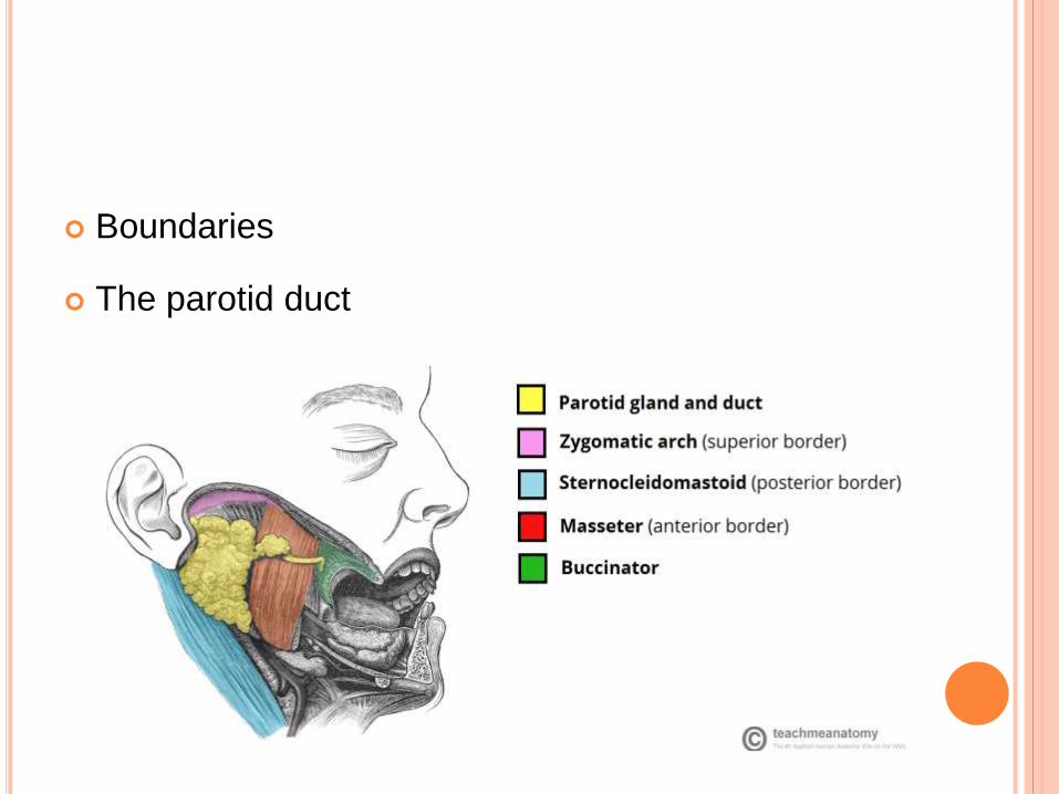
Boundaries
The parotid duct

Parotid gland is divided
by the facial nerve into
i. a superficial lobe
ii. a deep lobe

An accessory parotid gland

Superficial Muscular Aponeurotic
System (SMAS)
SMAS is a fibrous network that
invests the facial muscles, and
connects them with the dermis.
Platysma inferiorly;
Zygomatic arch superiorly
Facial nerve courses deep to the
SMAS and the platysma.
Parotid fascia
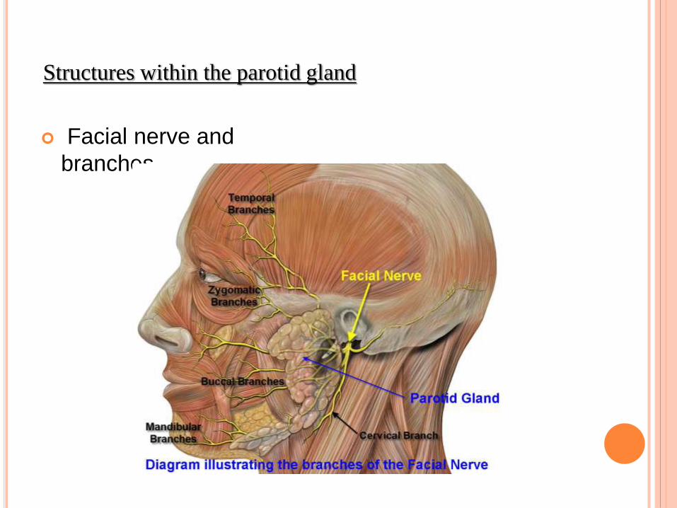
Facial nerve and
branches
Structures within the parotid gland

External carotid artery and its branches
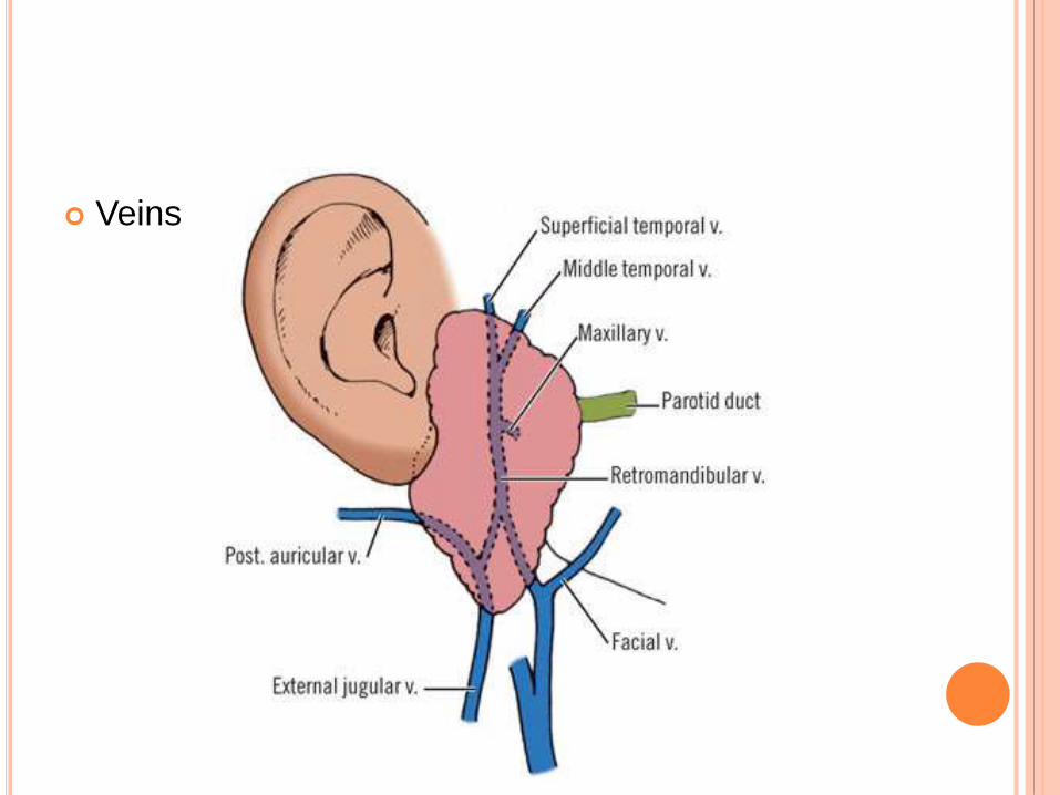
Veins

Lymphatics:
Superficial nodes drains
auricle, anterior part of
scalp, upper part of face
Deeper nodes receives
lymph from external
acoustic meatus, middle
ear, auditory tube, nose,
palate and deep parts of
cheek.
Cervical lymphnodes

RELEVANT SURGICAL RELATIONS
Posterior: Cartilage of external auditory meatus;
tympanic bone, mastoid process,
sternocleidomastoid muscle
Deep: Styloid process, stylomandibular tunnel,
parapharyngeal space, posterior belly of digastric,
sternocleidomastoid muscle
Superior: Zygomatic arch, temporomandibular joint

TYPES OF PAROTIDECTOMY
Partial parotidectomy: Resection of parotid
pathology with a margin of normal parotid tissue.
This is the standard operation for benign pathology
and low grade malignancies
Superficial parotidectomy: Resection of the entire
superficial lobe of parotid and is generally used for
metastases to parotid lymph nodes e.g. from skin
cancers, and for high grade malignant parotid
tumors.

Total parotidectomy: This involves resection of the
entire parotid gland, usually with preservation of the
facial nerve
Extended Total Parotidectomy: Removal of the
superficial and deep parotid gland also may be
extended to involve adjacent structures.

PREOPERATIVE EVALUATION
A thorough history is obtained prior to consideration
for surgery.
Symptoms of sensory loss, trismus and facial
weakness are worrisome for local tumor invasion by
a malignant neoplasm.
The past medical history should include information
regarding any prior cutaneous lesions or
malignancies.

In addition, the patient should be queried about any
prior radiation exposure to the head and neck
including dental radiographs.
Smoking is associated with Warthin’s tumor and,
therefore, should be investigated.
This tumor can also occur bilaterally, thus any
history of a prior parotid tumor should be elicited.
Cranial nerve function should be examined and
facial nerve function should be evaluated carefully.
Facial nerve paralysis is usually an indication of
nerve invasion by a malignant tumor.
Fixation to the overlying skin, limited mobility of the
mass, and associated cervical lymphadenopathy
are other signs suggestive of malignancy.
FINE-NEEDLE ASPIRATION BIOPSY (FNAB)
It is an accurate and useful investigation for the
diagnosis of a parotid mass.
FNAB allows for improved patient selection for
surgery since it can identify conditions such as
reactive lymph nodes or cysts that might mimic
parotid neoplasms clinically.
The information gained by FNAB is useful for
patient counseling, surgical timing and planning,
and guiding the direction of preoperative
consultation
RADIOLOGY
Radiological investigation is not routinely required
with parotid tumors.
It is recommended for surgical planning with tumors
that are large, fixed, and are associated with facial
nerve involvement, trismus, and parapharyngeal
space involvement.
MRI is a valuable investigation with recurrence of
pleomorphic adenoma as it is often multifocal.
PREOPERATIVE CONSENT
Scar
Anesthesia in the greater auricular distribution
Facial nerve weakness
Facial contour
Prominence of auricle
Frey’s syndrome (gustatory sweating)

PREOPERATIVE CONSENT
Scar
Anesthesia in the greater auricular distribution
Facial nerve weakness
Facial contour
Prominence of auricle
Frey’s syndrome (gustatory sweating)

SUPERFICIAL PAROTIDECTOMY
Superficial lobe parotidectomy describes removal of all
or a portion of the parotid gland superficial to the facial
nerve.
The most common indications are:
1. Benign or low grade tumor of the superficial lobe of the
parotid gland
2. metastases to parotid lymph nodes from adjacent sites
of skin cancer or melanoma, or from cancer of the
external auditory meatus.

3. Access to the deep lobe of the gland or other
structures deep to the facial nerve.
4. Chronic inflammation of parotid gland, resistant to
conservative treatment.
ANAESTHESIA
General anaesthesia
Short-acting muscle relaxation for intubation only,
so that facial nerve may be stimulated and/or
monitored
No perioperative antibiotics unless specifically
indicated
Hyperextend the head, and turn to opposite side

Infiltrate with vasoconstrictor along planned skin
incision,
Keep corner of eye and mouth exposed so as to be
able to see facial movement when facial nerve
mechanically or electrically stimulated.

TECHNIQUE
A modified Blair incision
An alternative incision is
a modified face-lift
incision.
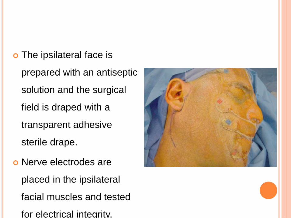
The ipsilateral face is
prepared with an antiseptic
solution and the surgical
field is draped with a
transparent adhesive
sterile drape.
Nerve electrodes are
placed in the ipsilateral
facial muscles and tested
for electrical integrity.

The skin incision is
made through the
subcutaneous tissues
and platysma muscle.
Greater auricular
nerve.
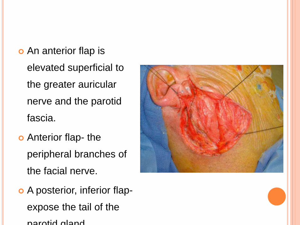
An anterior flap is
elevated superficial to
the greater auricular
nerve and the parotid
fascia.
Anterior flap- the
peripheral branches of
the facial nerve.
A posterior, inferior flap-
expose the tail of the
parotid gland.

The tail of the parotid gland is dissected
off of the sternocleidomastoid muscle
by dissecting deep to the posterior
branch of the greater auricular nerve.
Next, the posterior belly of the digastric
muscle is exposed with further
elevation of the tail of the parotid gland
The posterior belly of the digastric
muscle serves as a landmark for the
facial nerve.
During elevation of the tail of the
parotid, the integrity of the posterior
facial vein also is preserved if possible.
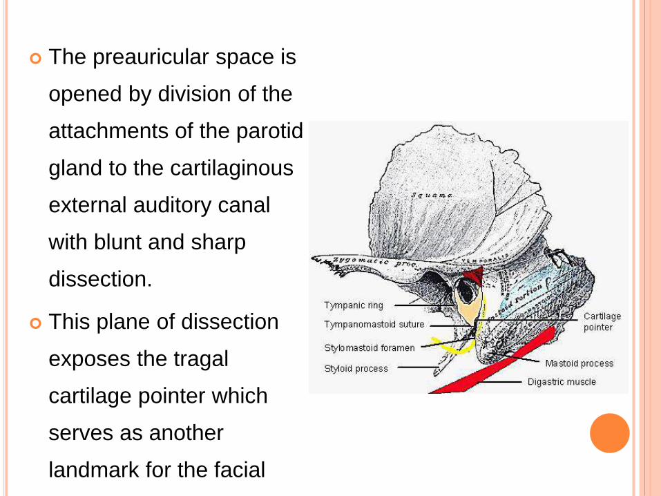
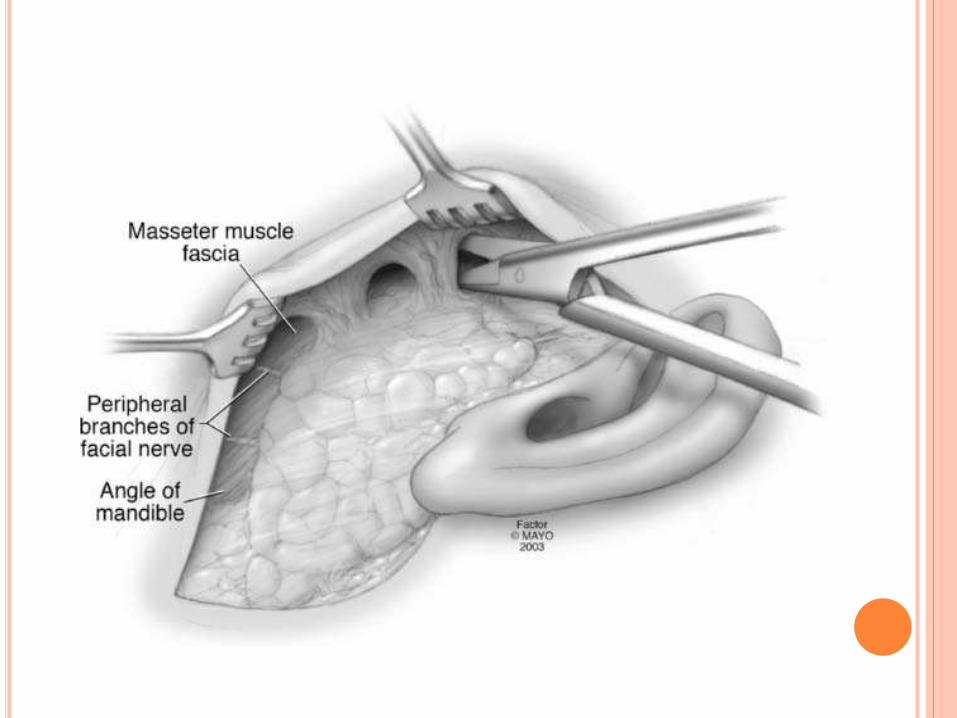
The preauricular space is
opened by division of the
attachments of the parotid
gland to the cartilaginous
external auditory canal
with blunt and sharp
dissection.
This plane of dissection
exposes the tragal
cartilage pointer which
serves as another
landmark for the facial
nerve.
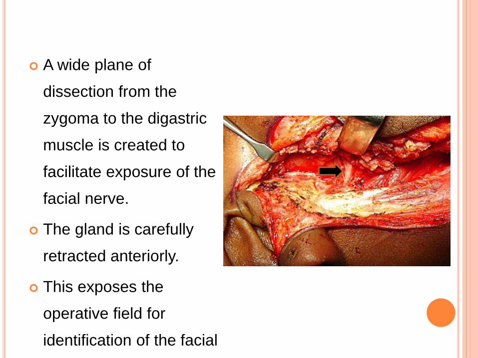
A wide plane of
dissection from the
zygoma to the digastric
muscle is created to
facilitate exposure of the
facial nerve.
The gland is carefully
retracted anteriorly.
This exposes the
operative field for
identification of the facial
nerve.

The facial nerve is
identified using
anatomic landmarks:
1. Posterior belly of the
digastric muscle
2. Mastoid tip
3. Tragal cartilage pointer
4. Tympanomastoid
suture.

If the proximal segment
of the facial nerve is
obscured, retrograde
dissection of one or
more of the peripheral
facial nerve branches
may be necessary to
identify the main trunk.

When necessary, the
facial nerve can be
identified in the
mastoid bone by
mastoidectomy and
followed peripherally.

Once the facial nerve is
identified, the parotid
gland superficial to the
facial nerve is divided
carefully, preserving the
integrity of the nerve.
The exact location of the
facial nerve should
always be determined
prior to division of the
gland tissue.

The facial nerve is
followed peripherally,
the desired portion of
the gland is dissected
from facial nerve
branches and the
specimen removed.

The facial nerve is preserved except in cases when
confirmed malignancy is found invading the nerve.
In instances of facial nerve invasion by carcinoma,
facial nerve resection is performed.
Proximal and distal margins of the resected nerve are
examined histologically by frozen section to ensure
clear surgical margins.

If the tumor involves the stylomastoid foramen,
mastoidectomy is performed to identify the proximal
facial nerve in the fallopian canal to achieve a clear
margin.
Immediate nerve reconstruction by a nerve
interposition graft is usually indicated if facial nerve
resection is performed.
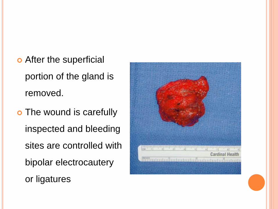
After the superficial
portion of the gland is
removed.
The wound is carefully
inspected and bleeding
sites are controlled with
bipolar electrocautery
or ligatures
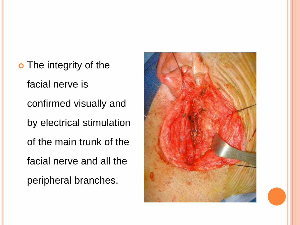
The integrity of the
facial nerve is
confirmed visually and
by electrical stimulation
of the main trunk of the
facial nerve and all the
peripheral branches.
A neck dissection is performed for clinically positive
nodes.
For the clinically negative neck, the first echelon
nodes are inspected.
Enlarged or suspicious nodes are examined and a
neck dissection is performed if metastatic disease
is confirmed by frozen section.

The wound is irrigated,
realigned, and closed in
layers over a closed-
suction drain.
The drain is usually
removed on the first
postoperative day and
the skin sutures are
removed within one
week.
Adjuvant radiation therapy is recommended for
select malignancies including
i. metastatic cutaneous squamous cell carcinoma
ii. high-grade and advanced parotid malignancies

TOTAL PAROTIDECTOMY
Total parotidectomy is the total removal of the
superficial and deep parotid gland.
The operation may involve sparing or sacrifice of
the facial nerve branches or trunk depending on
tumor extent to the nerve.

INDICATIONS:
1. Metastasis to a superficial parotid node from a
primary parotid tumor or an extraparotid
malignancy
2. Parotid malignancy that indicates metastasis by
involvement of cervical lymph nodes
3. High-grade parotid malignancy with a high risk of
metastasis.

4. Primary parotid malignancies originating in the
deep lobe and for primary malignancies that
extend outside the parotid gland.
5. Multifocal tumors, such as oncocytomas, to
ensure complete removal
EXTENDED TOTAL PAROTIDECTOMY
Removal of the superficial and deep parotid gland
also may be extended to involve adjacent
structures such as the overlying skin, the underlying
mandible, the temporal bone and external auditory
canal, or the deep musculature of the
parapharyngeal space.
These extensions are dictated by tumor growth and
behavior.

SURGICAL TECHNIQUE:
1. Preparation
2. Incisions and flap elevation
3. Deeper dissection
4. Facial nerve mobilisation

5. Removal of superficial gland
6. Deep parotidectomy
7. Total Parotidectomy with Facial Nerve Sacrifice
8. Resection of Adjacent Structures and
Reconstruction
PREPARATION
The operation is performed with the patient under
general endotracheal anesthesia.
Endotracheal tube is positioned and taped to the
oral commissure and cheek opposite to the lesion.
The patient is placed in a 45° reverse-
trendelenburg position or lounge-chair position with
the head higher than the heart.
The head is turned to the opposite side of the
lesion, and the neck is extended by placement of a
rolled sheet under the shoulders.
The patient is prepared by sterile scrub and draped
so that the ear, lateral corner of the ipsilateral eye,
ipsilateral oral commissure, and entire ipsilateral
neck are visible in the field.

If facial nerve monitoring is to be used, the nerve
monitor is placed in the orbicularis oris and
orbicularis oculi muscles to ensure upper and lower
division monitoring.
The surgeon stands on the side of the patient
ipsilateral to the gland to be dissected, the assistant
stands at the head and opposite the surgeon, and
the scrub technician stands on the side of the
surgeon.

INCISIONS AND FLAP ELEVATION
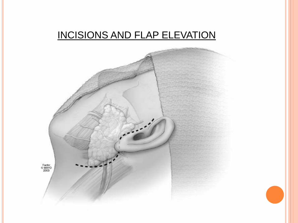
INCISIONS AND FLAP ELEVATION

INCISIONS AND FLAP ELEVATION

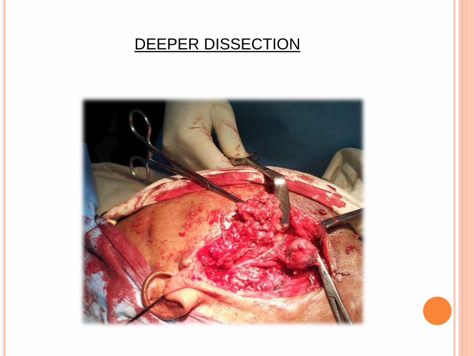
DEEPER DISSECTION


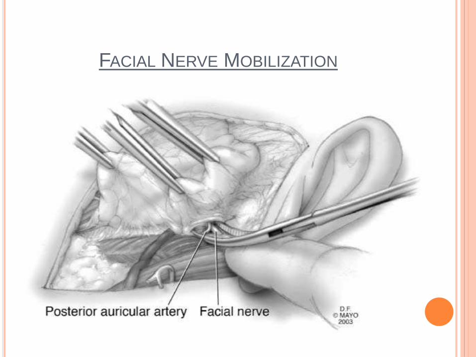
FACIAL NERVE MOBILIZATION


A small curved clamp is oriented perpendicular to
the anticipated direction of the facial trunk to
elevate tissues layer by layer.
Scissors are never used for dissection down to the
nerve, and no tissue is cut in this area until the
nerve is seen.
Blunt dissection proceeds posterior to anterior until
the surgeon identifies the nerve as a white cord 2–3
mm wide.


REMOVAL OF THE SUPERFICIAL GLAND
The gland is separated at its edge, the temporal or
marginal branches being followed to the periphery.
The thickest fascia is encountered
posterosuperiorly; this must be divided sharply or
the surgeon will make tunnels into the gland along
the nerve.
Posteriorly- branches of the superficial temporal
vein may be encountered.

Vessels directly adjacent to the nerve branches should
not be cauterized until the superficial lobe is completely
mobilized.
After following a nerve branch to its peripheral
emergence from the parotid gland, the surgeon returns
to a proximal position along that nerve and searches
for another branch to follow.
Dissection progresses from posterior to anterior and
either superiorly or inferiorly until the superficial gland
has been completely separated from the facial nerve
and the deep parotid gland.

At this point, the surgeon should have a clear
impression of the relationship of the tumor to the
facial nerve, superficial gland, deep gland, and
surrounding structures.
It may be necessary to dissect along the tumor
capsule to separate it from the deep gland and
facial nerve.
Careful retraction and meticulous dissection can
prevent rupture of the tumor capsule, which is often
pivotal in the prevention of recurrence.
The gland is now left attached to only the parotid
duct.
The surgeon inspects this area to ensure that no
buccal branches are adherent to the duct.
The duct is divided and ligated, and the specimen is
sent for examination by the pathologist.
The wound should now be irrigated and the field
inspected for bleeding vessels, which are ligated.

DEEP PAROTIDECTOMY


The gland is completely freed from attachment to any
adjacent structures and sent for frozen-section
pathologic examination.
Small vessels around the deep gland adjacent to the
mastoid and trunk can be cauterized using the bipolar
forceps.
The wound is irrigated, and meticulous hemostasis is
achieved.
If necessary, the incision can be extended for neck
dissection at this time.

At the conclusion of the operation, a suction drain is
placed in the wound through a separate stab
incision in the postauricular skin and sewn into
place.
The wound is closed with interrupted absorbable
sutures
Dressing or antibiotic ointment can be applied.
Patient is awakened and extubated.

TOTAL PAROTIDECTOMY
WITH FACIAL NERVE SACRIFICE
If facial nerve function is normal preoperatively,
even in patients with malignancy, then the nerve
can be preserved with careful dissection of the
tumor off the nerve sheath.
If the nerve is paretic or fully paralyzed
preoperatively, then it is involved with tumor and is
normally resected during tumor resection.

Nerve that is clearly invaded by high-grade
malignant tumor should be resected with the
specimen to negative proximal and distal margins.
This may necessitate sacrificing peripheral
branches, divisions, or even the main trunk of the
facial nerve.
Intraoperatively, a nerve that is infiltrated with tumor
will appear swollen and usually darker than the
normal glistening white appearance of normal facial
nerve.

After negative proximal and distal facial nerve
margins are obtained, the nerve is reconstructed
with primary neurorraphy or grafting.
Mastoidectomy and nerve mobilization may be
necessary to attain proper length of the facial nerve
for tension-free anastomosis.

Appropriate grafts include:
i. ipsilateral greater auricular nerve if it is not
involved with tumor
ii. ipsilateral sural nerve graft.
Peripheral branches can be grafted
i. proximal facial nerve
ii. ipsilateral hypoglossal nerve

RESECTION OF ADJACENT STRUCTURES
AND RECONSTRUCTION
The operation may be extended to involve resection
of adjacent structures that are involved with tumor.
It may include
i. lateral or subtotal temporal bone resection,
ii. partial mandibular resection,
iii. resection of the overlying skin,
iv. resection of portions or all of the auditory canal,
and
v. resection of surrounding musculature.
Options for reconstruction include
i. primary closure,
ii. dermal fat grafting,
iii. muscle transposition with loco regional flaps of
the sternocleidomastoid or pectoralis muscles,
iv. micro vascular cutaneous, musculocutaneous,
and innervated muscular flaps.
Again, the reconstruction will be guided by the
functional and aesthetic goals of the surgeon and
patient.

COMPLICATIONS
1. Hematoma
2. Infection
3. Facial nerve palsy
4. Salivary fistula
5. Gustatory sweating/ Frey’s syndrome
6. Cosmetic deformity

Inadequate hemostasis before
closure.
Suction drain reduces
possibility of postoperative
hematoma.
Treatment:
i. Evacuation of hematoma
ii. Control of bleeding points
iii. Reinsertion of suction drain
and closure.
HEMATOMA

Infection is rare
Some tumors presents with obstructive symptoms if
infected.
Prophylactic antibiotics are given if operating on an
infected gland.
INFECTION

Temporary or permanent
Partial or total
Neuropraxia- due to
stretching of the nerve.
If the nerve is intact at the
end of procedure-
recovery within few weeks.
FACIAL NERVE PALSY

If the palsy is severe and recovery is prolonged-
transcutaneous nerve stimulation of facial muscles.
Problems with eye closure-
i. protective glasses or tape the eyelid to prevent
exposure keratitis.
ii. Temporary tarsorrhaphy or paralysis of eyelid
elevator with botulinum toxin to allow closure of
upper eyelid.

When palsy is due to partial or total loss of facial
nerve:
i. reconstruction
ii. rehabilitation of face

Presents after suture
removal at the suture
line and posterior to
ear lobule.
Pressure dressing.
Drains
Anticholinergic drugs-
to reduce salivary
secretion
SALIVARY FISTULA


Auriculotemporal
syndrome.
60% of all
parotidectomy cases.
Discomfort, localized
facial sweating and
flushing during
mastication.
FREY’S SYNDROME

Due to parasympathetic
and sympathetic
secretomotor stimuli
misdirected to
cholinergic receptors of
sweat glands during
healing after parotid
surgery.

The iodine test administered
by applying an alcohol–
iodine–oil solution (3 g
iodine, 20 mL castor oil, and
200 mL absolute alcohol)
described by Laage-Hellman
The solution was applied on
the lateral portion of the face
that had been surgically
treated and the upper region
of the neck.

The solution was allowed
to dry and was covered
lightly with starch
powder.
The patients received
lemon candy for a
gustatory stimuli for 10
minutes.
Discoloration of the
starch iodine mixture
was interpreted as a

There is no effective treatment, but various options
are described:
i. Injection of Botulinum Toxin
ii. Surgical transection of the nerve fibers
iii. Application of an ointment containing
an anticholinergic drug such as scopolamine
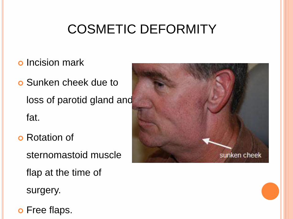
Incision mark
Sunken cheek due to
loss of parotid gland and
fat.
Rotation of
sternomastoid muscle
flap at the time of
surgery.
Free flaps.
COSMETIC DEFORMITY

REFERENCES
1. Salivary Gland Disorders: Eugene N. Myers, Robert
L. Ferris; Springer.
2. Parotidectomy : Johan Fagan : Open Access Atlas
Of Otolaryngology, Head & Neck Operative Surgery
3. Maxillofacial Surgery: Second Edition; Volume 1:
Peter Wardbooth.
4. Operative Maxillofacial Surgery; John D Langdon
and Mohan F Patel.
5. Internet

Thank You