parasitology practice 1- nematode
DESCRIPTION
PracticalTRANSCRIPT
ASCARIASIS&
Ascaris lumbricoides
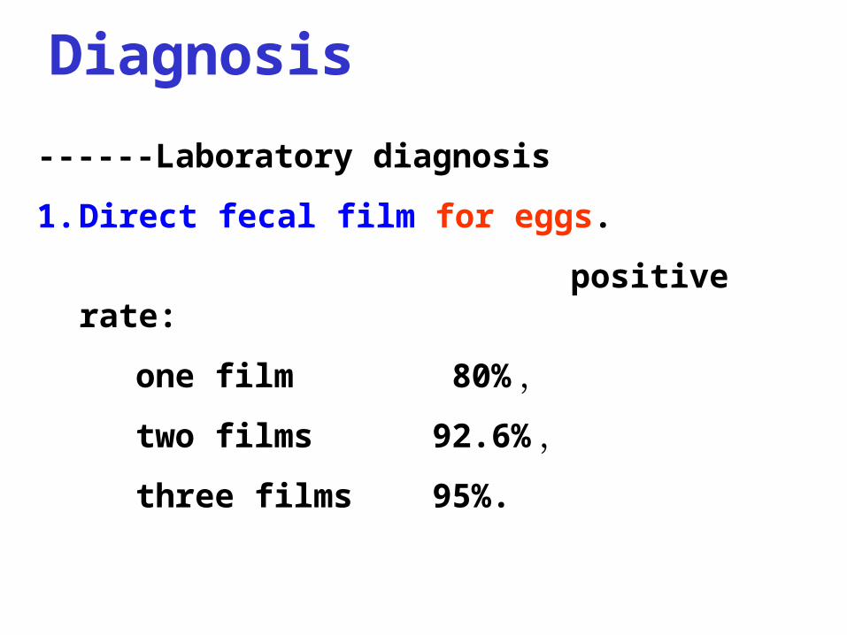
------Laboratory diagnosis
1. Direct fecal film for eggs.
positive rate:
one film 80% ,
two films 92.6% ,
three films 95%.
Diagnosis

------Laboratory diagnosis
2. Concentration techniques for eggs :
natural sediment,
flotation of saturated saline solution.
Diagnosis
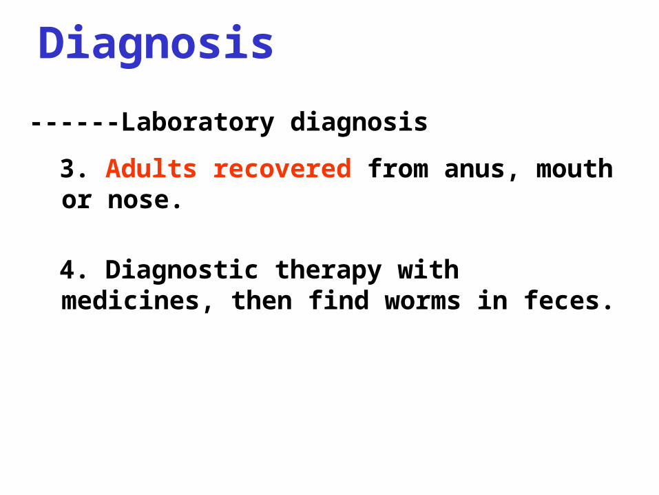
------Laboratory diagnosis
3. Adults recovered from anus, mouth or nose.
4. Diagnostic therapy with medicines, then find worms in feces.
Diagnosis
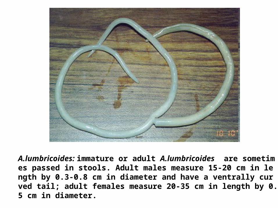
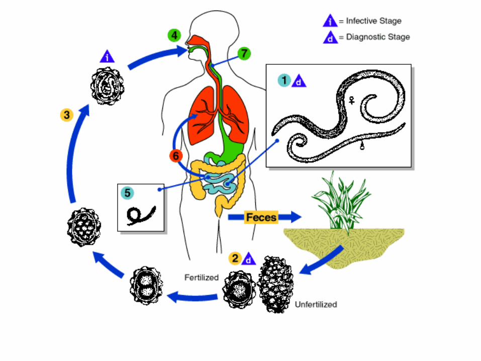
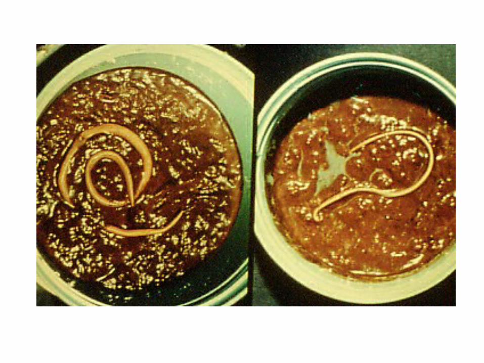
A.lumbricoides: immature or adult A.lumbricoides are sometimes passed in stools. Adult males measure 15-20 cm in length by 0.3-0.8 cm in diameter and have a ventrally curved tail; adult females measure 20-35 cm in length by 0.5 cm in diameter.
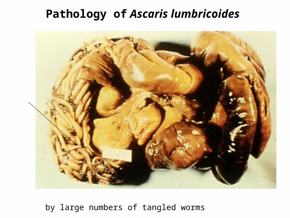
Pathology of Ascaris lumbricoides
by large numbers of tangled worms
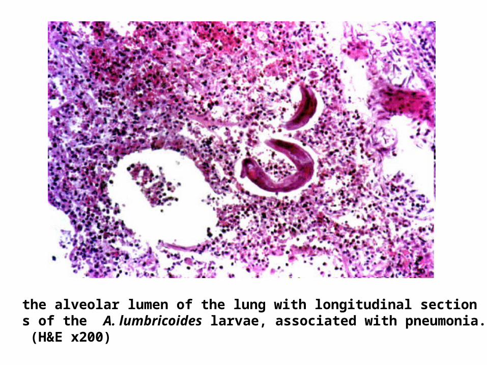
the alveolar lumen of the lung with longitudinal sections of the A. lumbricoides larvae, associated with pneumonia. (H&E x200)
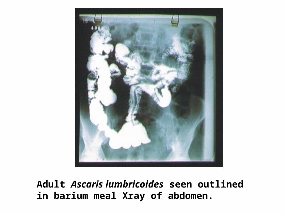
Adult Ascaris lumbricoides seen outlined in barium meal Xray of abdomen.
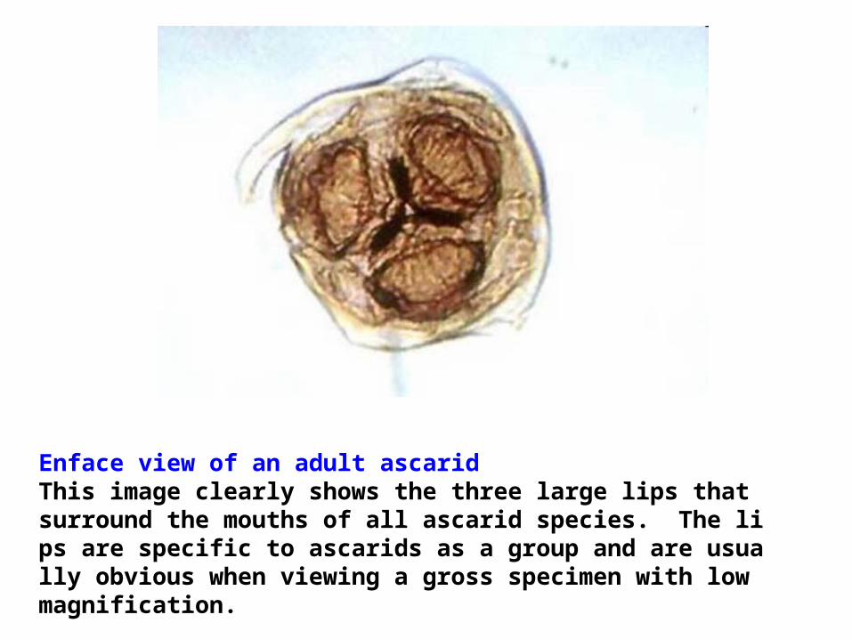
Enface view of an adult ascarid This image clearly shows the three large lips that surround the mouths of all ascarid species. The lips are specific to ascarids as a group and are usually obvious when viewing a gross specimen with low magnification.
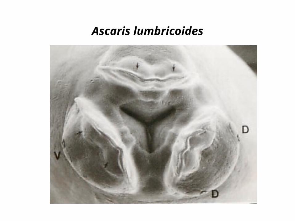
Ascaris lumbricoides
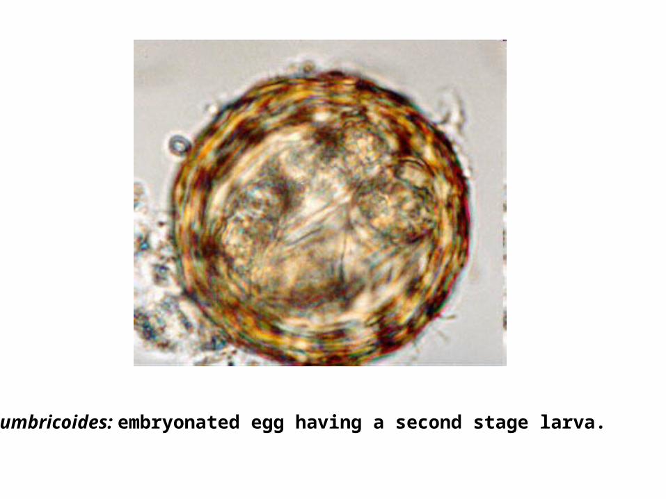
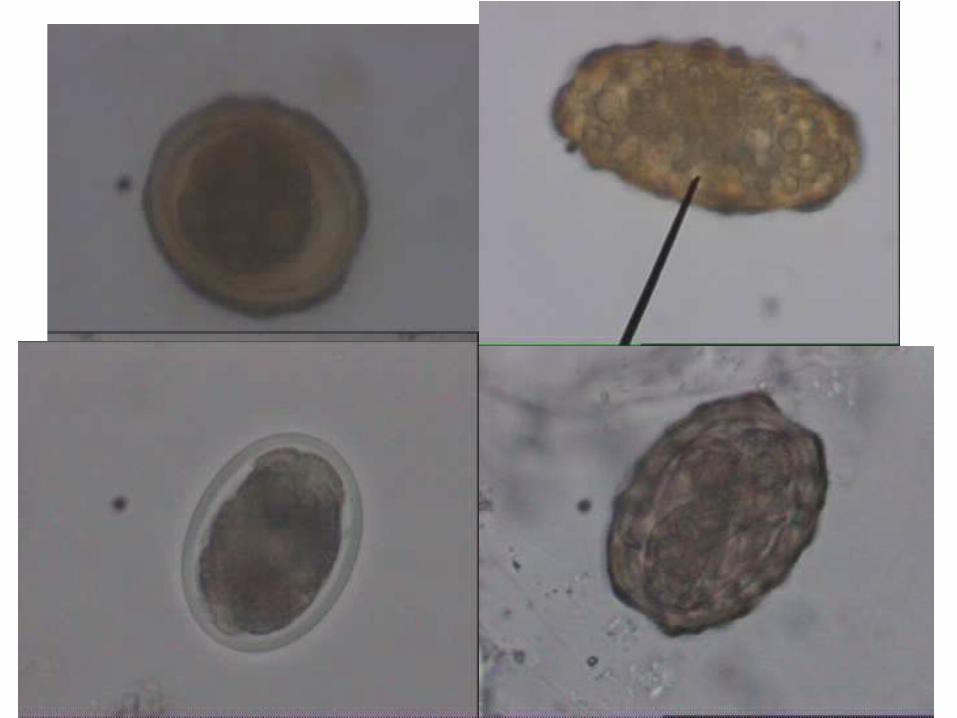
A.lumbricoides: embryonated egg having a second stage larva.
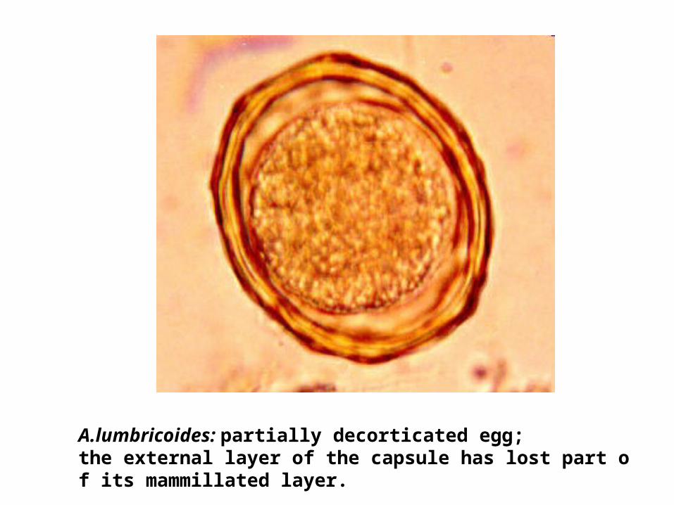
A.lumbricoides: partially decorticated egg; the external layer of the capsule has lost part of its mammillated layer.
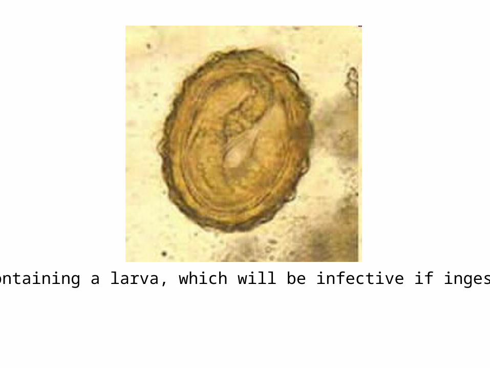
Egg containing a larva, which will be infective if ingested.
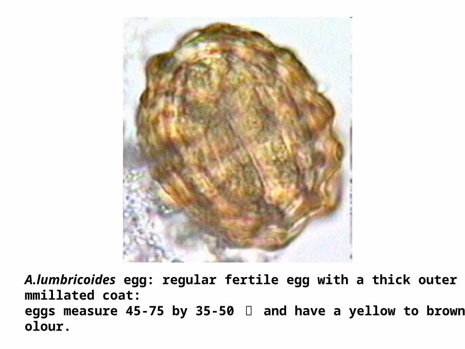
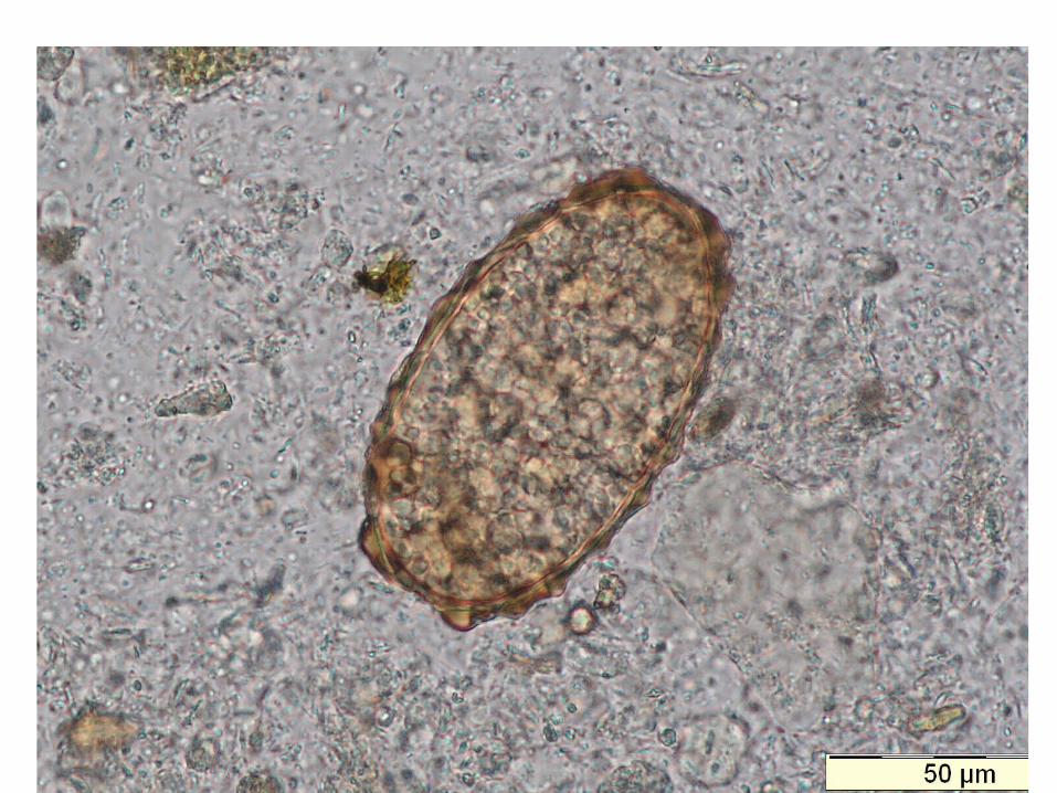
A.lumbricoides egg: regular fertile egg with a thick outer mammillated coat: eggs measure 45-75 by 35-50 祄 and have a yellow to brown colour.
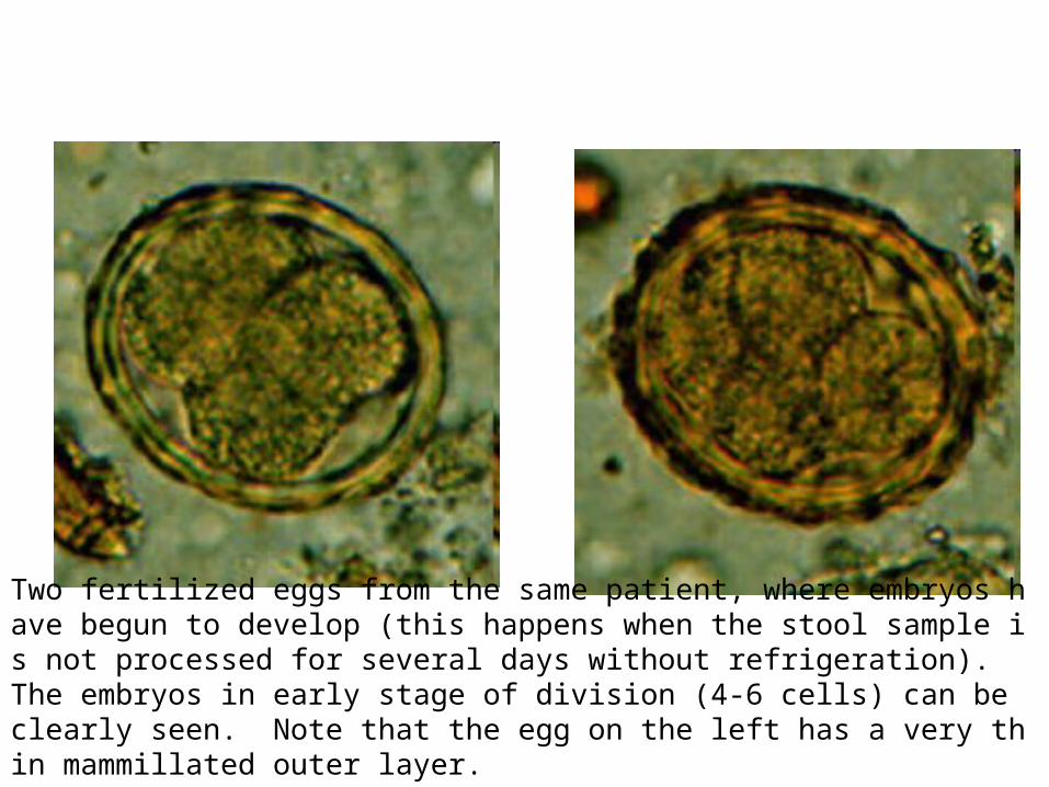
Two fertilized eggs from the same patient, where embryos have begun to develop (this happens when the stool sample is not processed for several days without refrigeration). The embryos in early stage of division (4-6 cells) can be clearly seen. Note that the egg on the left has a very thin mammillated outer layer.
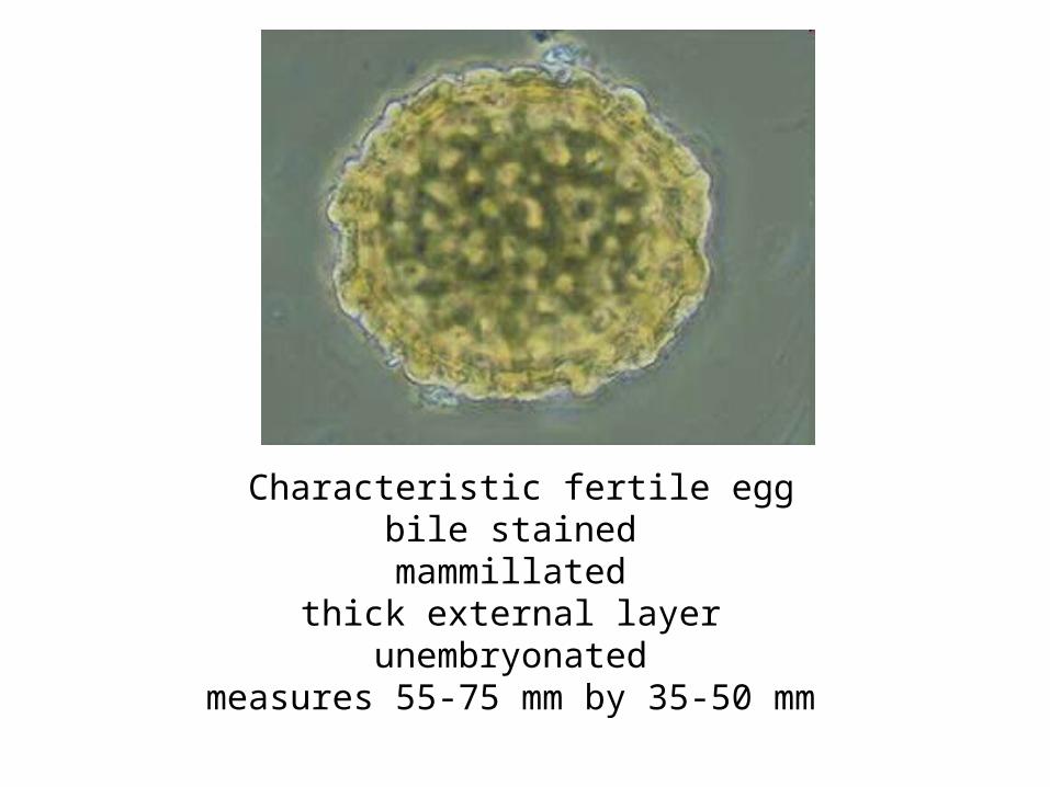
Characteristic fertile eggbile stained
mammillated thick external layer
unembryonated measures 55-75 mm by 35-50 mm
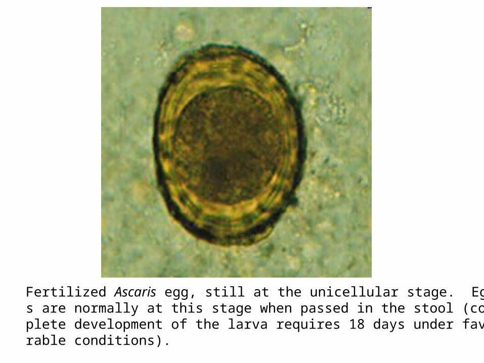
Fertilized Ascaris egg, still at the unicellular stage. Eggs are normally at this stage when passed in the stool (complete development of the larva requires 18 days under favorable conditions).
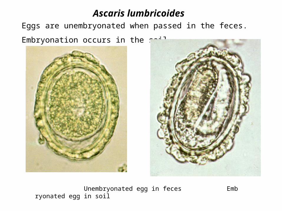
Ascaris lumbricoidesEggs are unembryonated when passed in the feces.
Embryonation occurs in the soil.
Unembryonated egg in feces Embryonated egg in soil
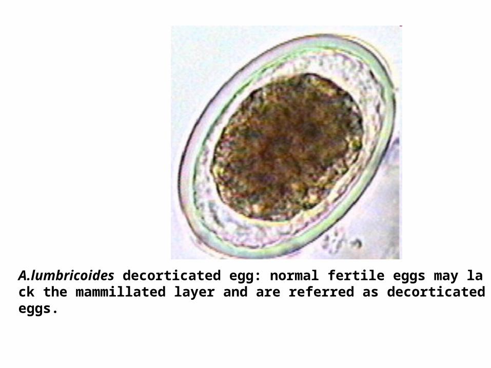
A.lumbricoides decorticated egg: normal fertile eggs may lack the mammillated layer and are referred as decorticated eggs.
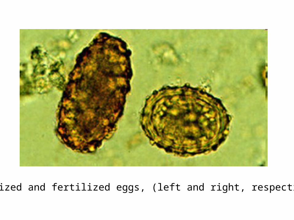
Unfertilized and fertilized eggs, (left and right, respectively).
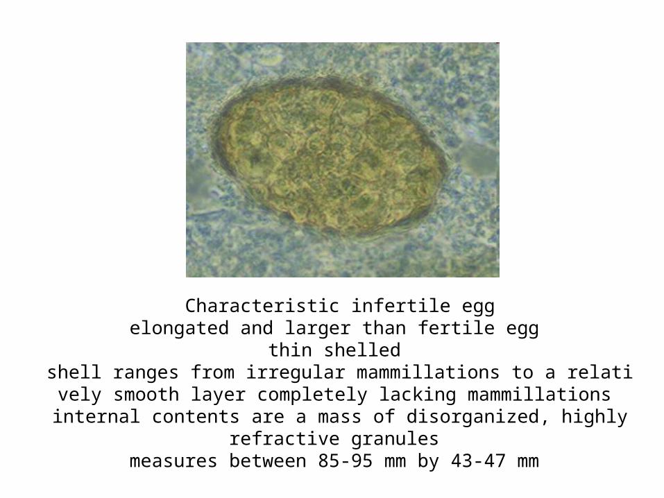
Characteristic infertile eggelongated and larger than fertile egg
thin shelled shell ranges from irregular mammillations to a relatively smooth layer complet
ely lacking mammillations internal contents are a mass of disorganized, highly refractive granules
measures between 85-95 mm by 43-47 mm
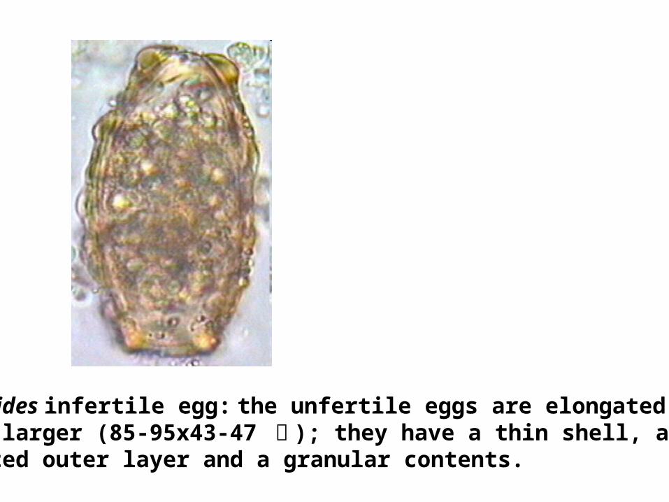
A.lumbricoides infertile egg: the unfertile eggs are elongated and much larger (85-95x43-47 祄 ); they have a thin shell, an irregular, mammillated outer layer and a granular contents.
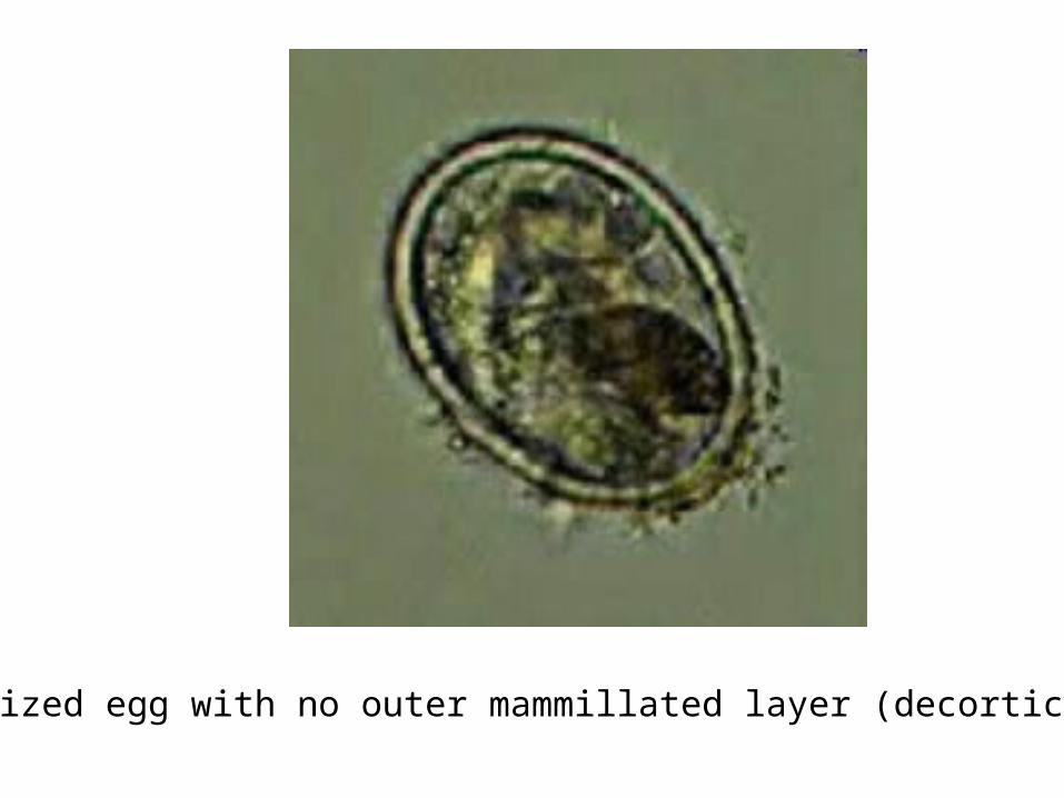
Unfertilized egg with no outer mammillated layer (decorticated).
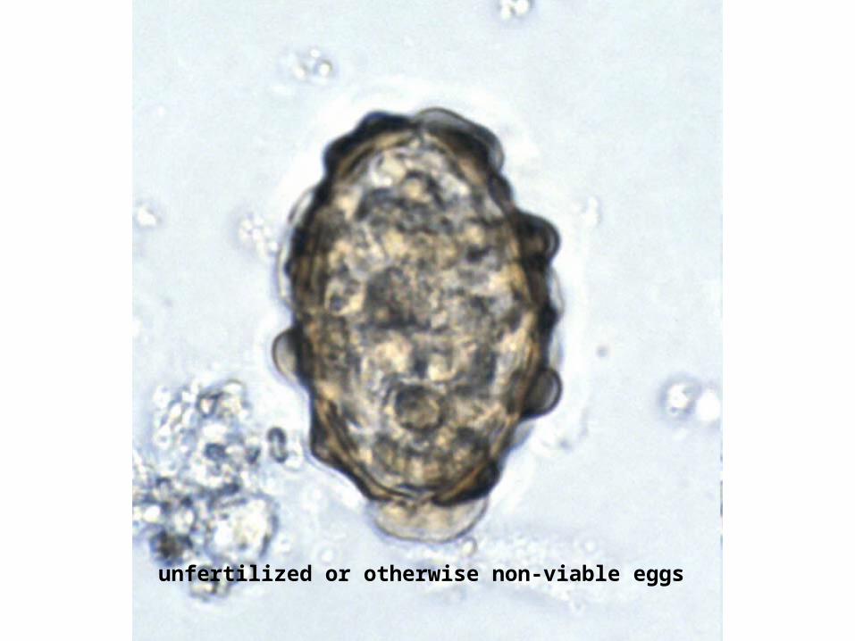
unfertilized or otherwise non-viable eggs
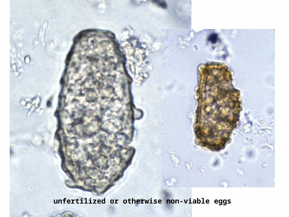
unfertilized or otherwise non-viable eggs
TRICHURIASIS&
Trichuris trichiura(whipworm)
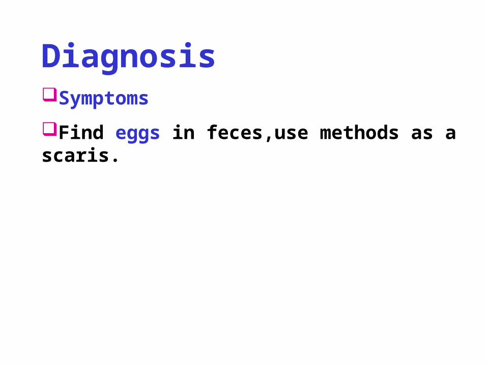
DiagnosisSymptoms
Find eggs in feces,use methods as ascaris.
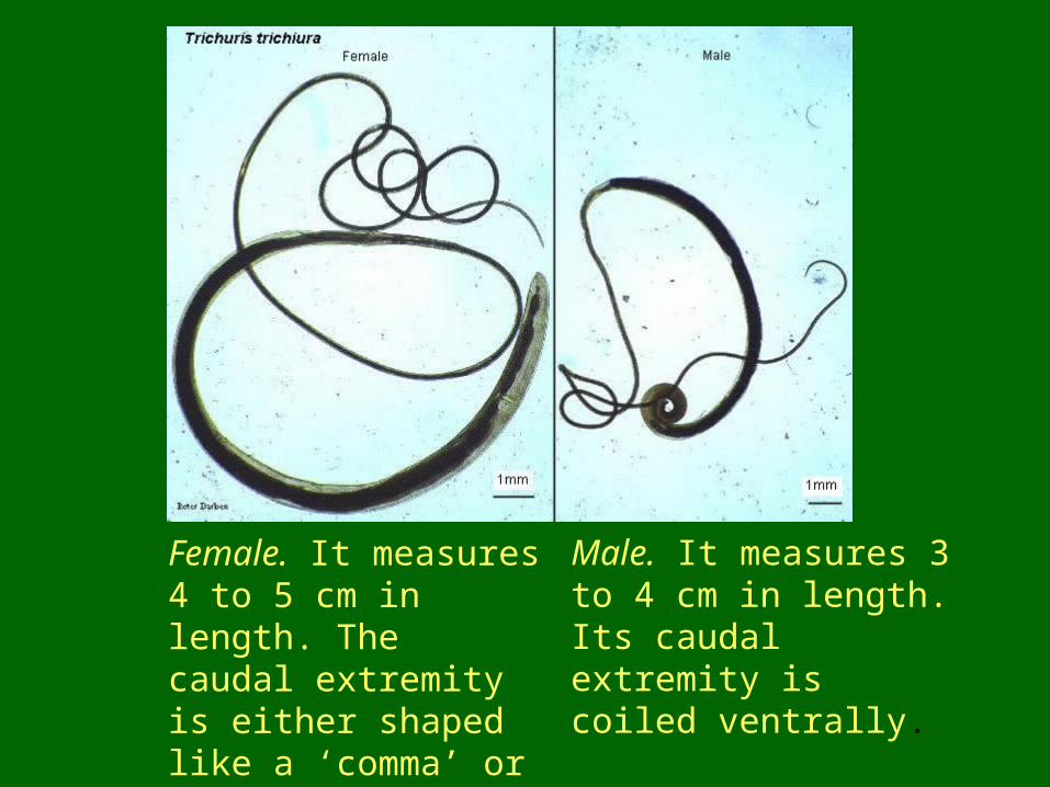
Female. It measures 4 to 5 cm in length. The caudal extremity is either shaped like a ‘comma’ or arc. The worm is oviparous.
Male. It measures 3 to 4 cm in length. Its caudal extremity is coiled ventrally.
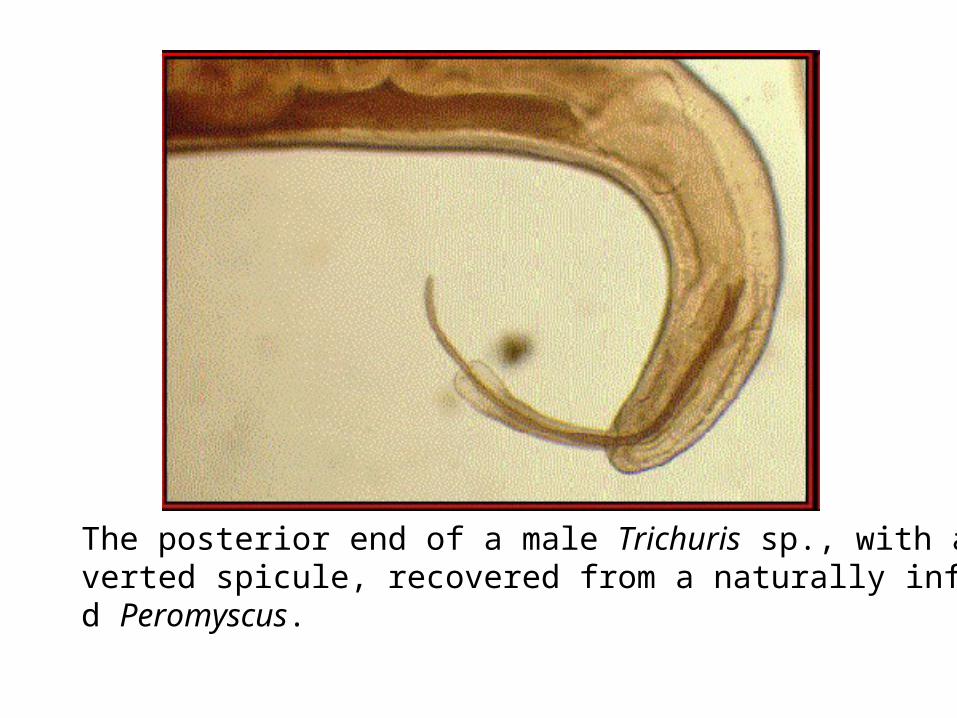
The posterior end of a male Trichuris sp., with an everted spicule, recovered from a naturally infected Peromyscus.
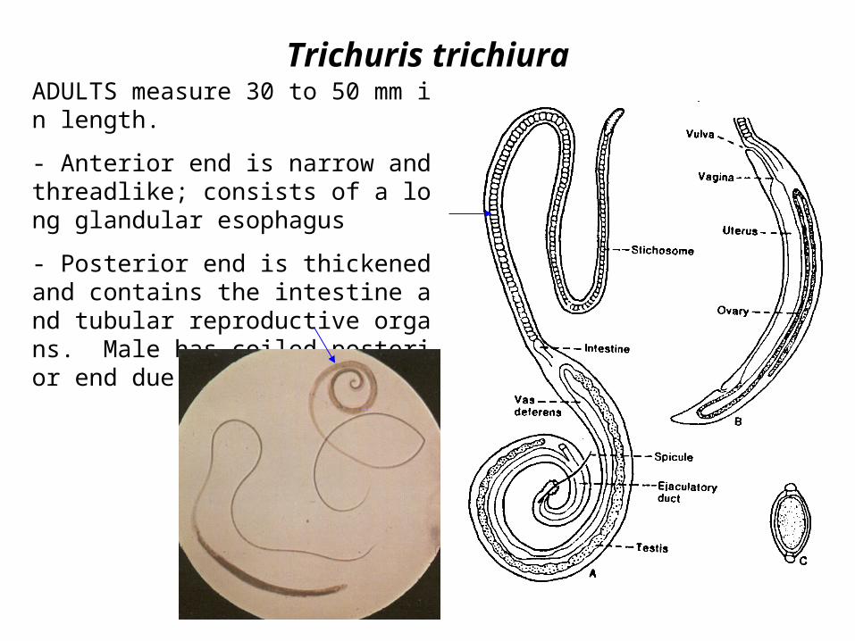
Trichuris trichiuraADULTS measure 30 to 50 mm in length.
- Anterior end is narrow and threadlike; consists of a long glandular esophagus
- Posterior end is thickened and contains the intestine and tubular reproductive organs. Male has coiled posterior end due to spicule.
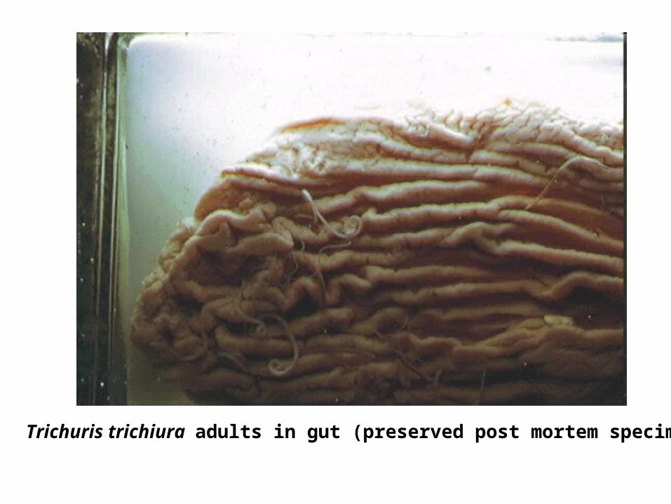
Trichuris trichiura adults in gut (preserved post mortem specimen)
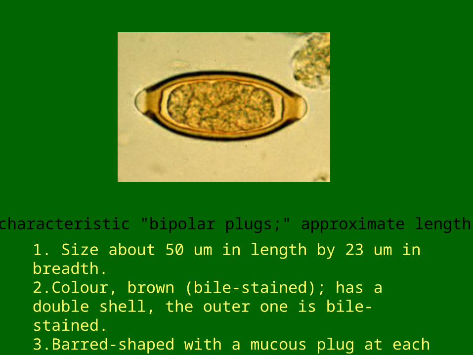
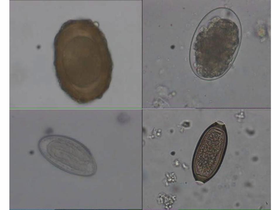
1. Size about 50 um in length by 23 um in breadth.2.Colour, brown (bile-stained); has a double shell, the outer one is bile-stained.3.Barred-shaped with a mucous plug at each pole.
Note the characteristic "bipolar plugs;" approximate length = 50 µm
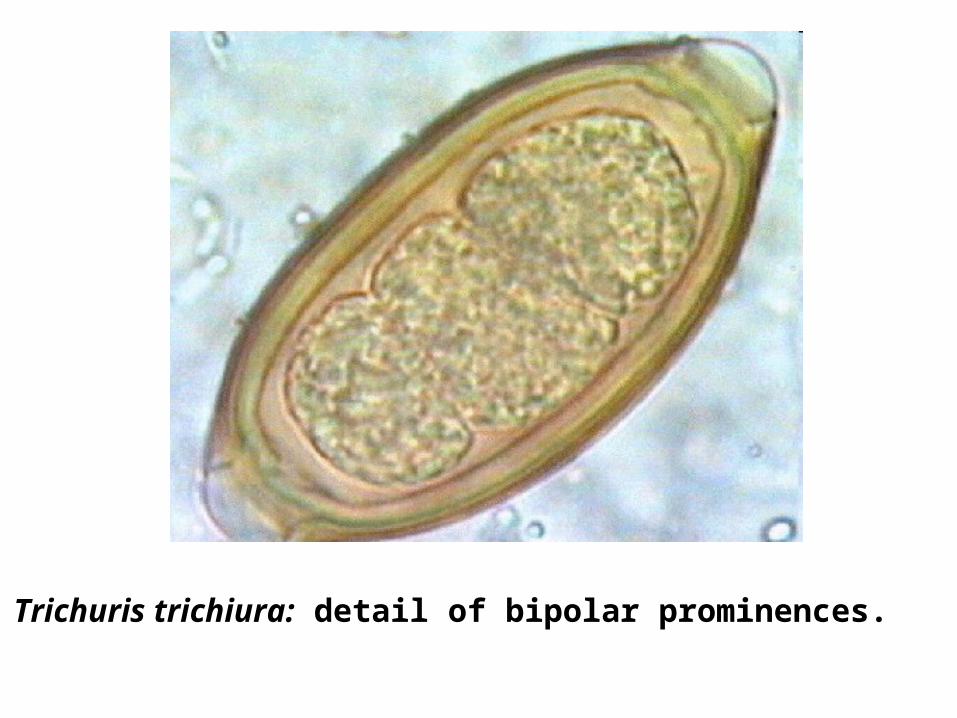
Trichuris trichiura: detail of bipolar prominences.
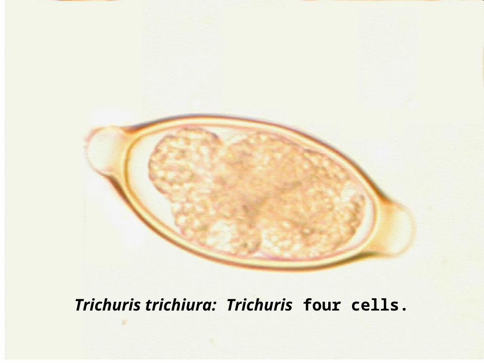
Trichuris trichiura: Trichuris four cells.
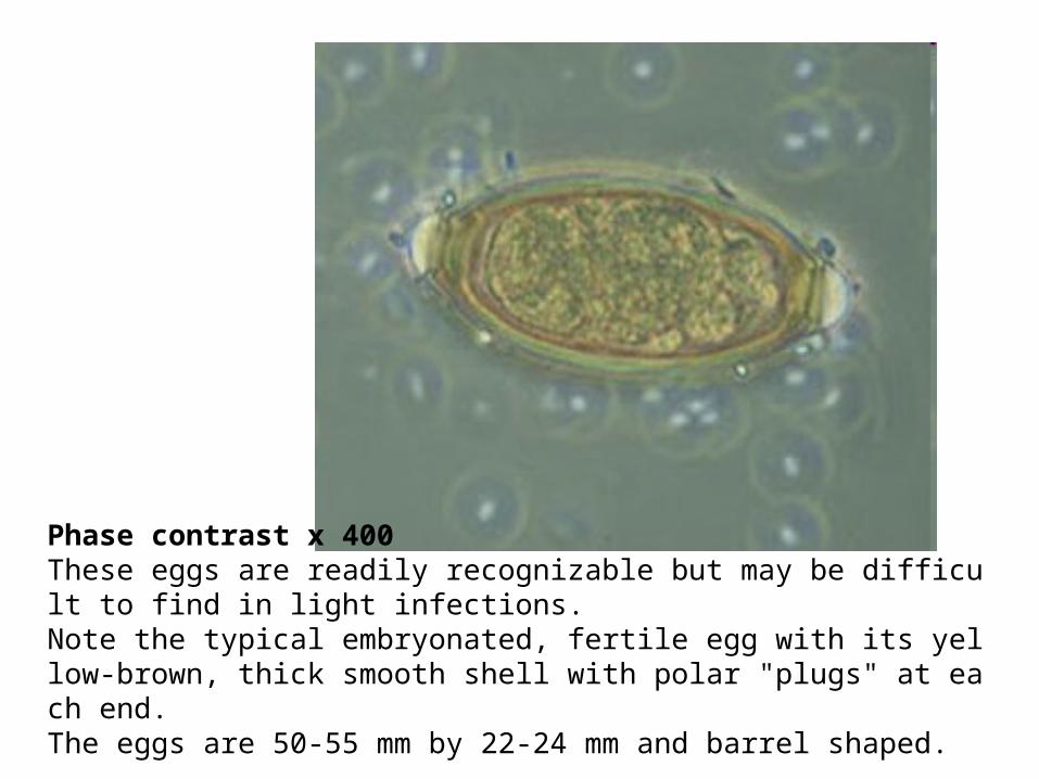
Phase contrast x 400These eggs are readily recognizable but may be difficult to find in light infections. Note the typical embryonated, fertile egg with its yellow-brown, thick smooth shell with polar "plugs" at each end. The eggs are 50-55 mm by 22-24 mm and barrel shaped.
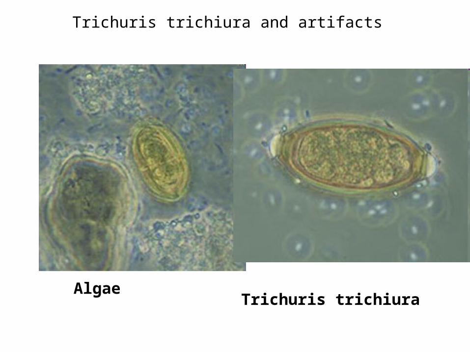
Algae Trichuris trichiura
Trichuris trichiura and artifacts

This could be confused with Trichuris ova, but the occurrence of chains suggests fungi.
Fungal conidia with yeast cells
ENTEROBIASIS&
Enterobius vermicularis (pinworm)
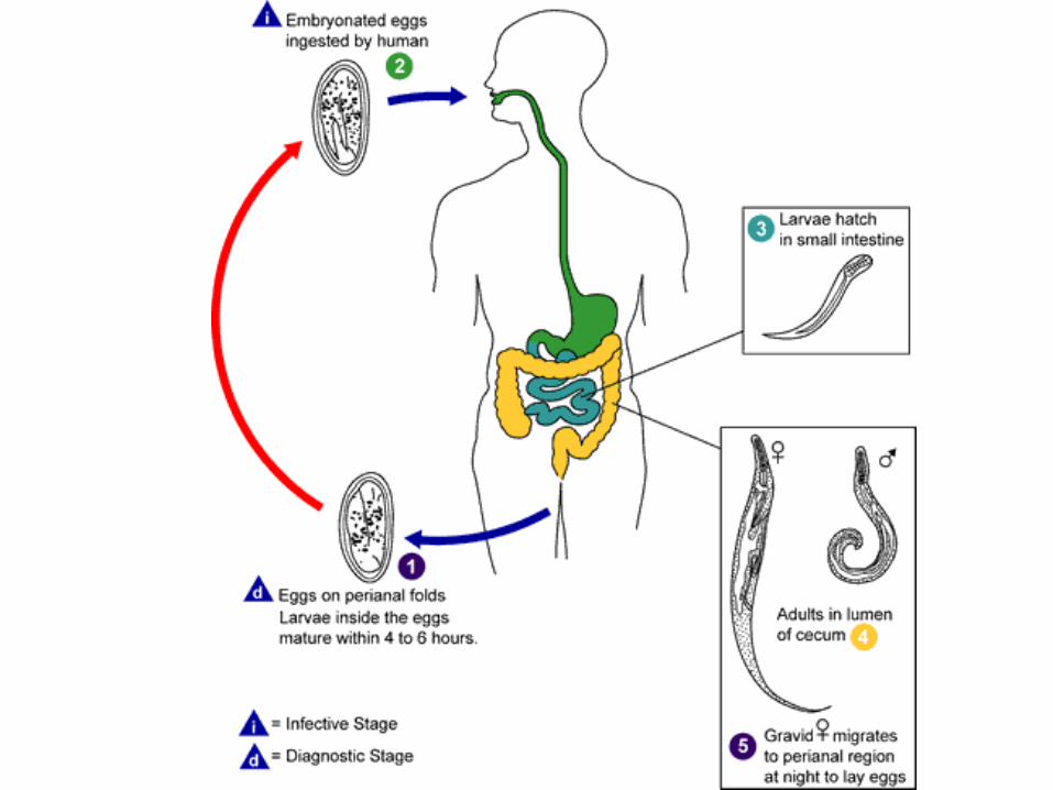
DiagnosisCellophane tape is used to obtain eggs.
collect eggs by a Swab tube (paddle coated with adhesive material), examined directly on bright field.
Finding adult worm or eggs in perianal area, particularly at night.
REPEAT !
CAUTION: INFECTION BY TOUCH !!
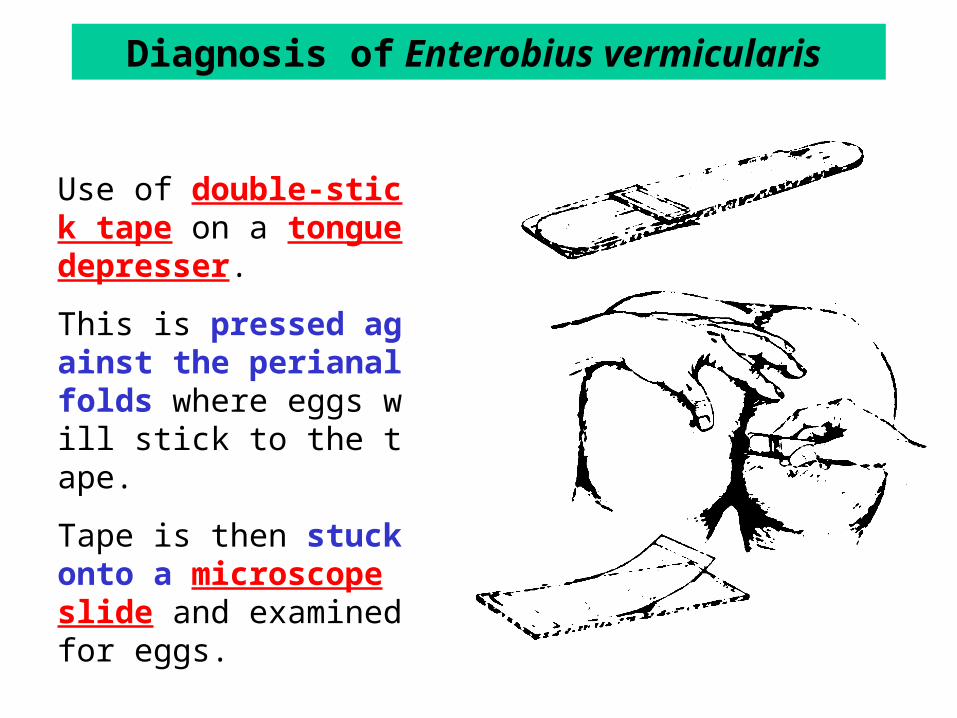
Diagnosis of Enterobius vermicularis
Use of double-stick tape on a tongue depresser.
This is pressed against the perianal folds where eggs will stick to the tape.
Tape is then stuck onto a microscope slide and examined for eggs.
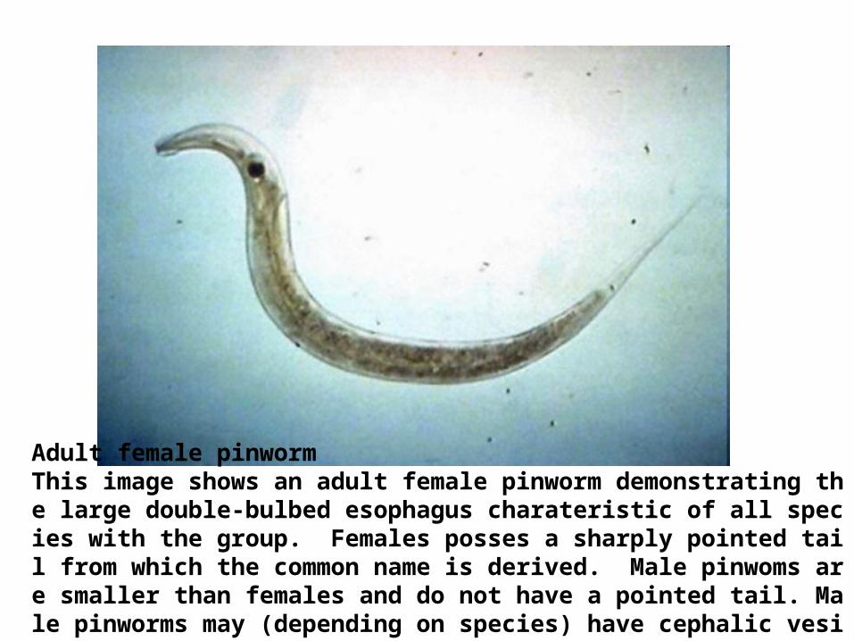
Adult female pinworm This image shows an adult female pinworm demonstrating the large double-bulbed esophagus charateristic of all species with the group. Females posses a sharply pointed tail from which the common name is derived. Male pinwoms are smaller than females and do not have a pointed tail. Male pinworms may (depending on species) have cephalic vesicles,
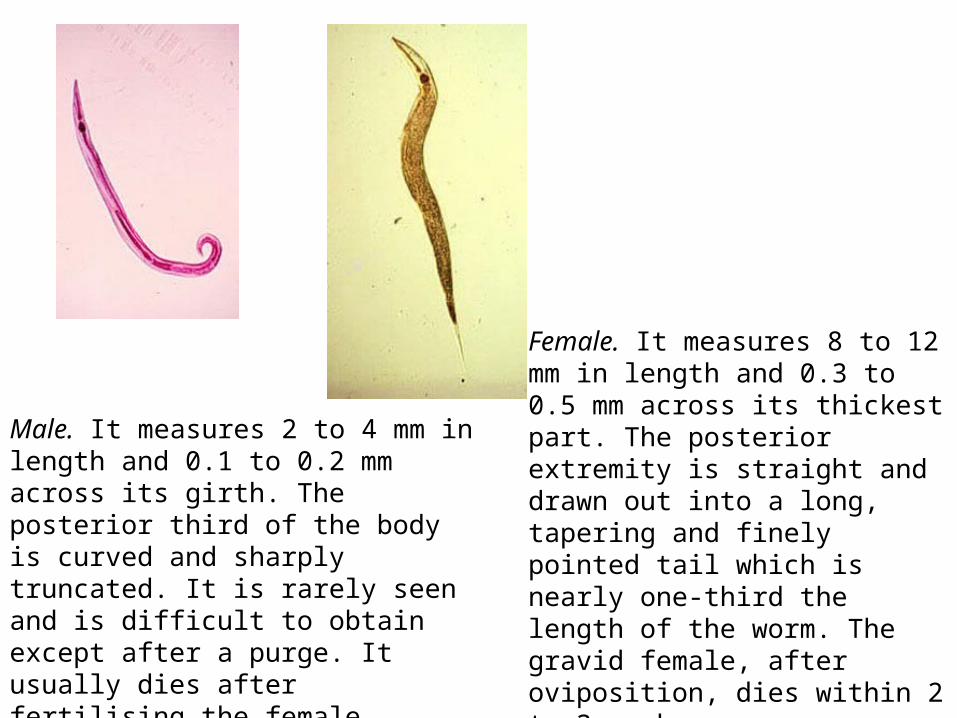
Male. It measures 2 to 4 mm in length and 0.1 to 0.2 mm across its girth. The posterior third of the body is curved and sharply truncated. It is rarely seen and is difficult to obtain except after a purge. It usually dies after fertilising the female.
Female. It measures 8 to 12 mm in length and 0.3 to 0.5 mm across its thickest part. The posterior extremity is straight and drawn out into a long, tapering and finely pointed tail which is nearly one-third the length of the worm. The gravid female, after oviposition, dies within 2 to 3 week.
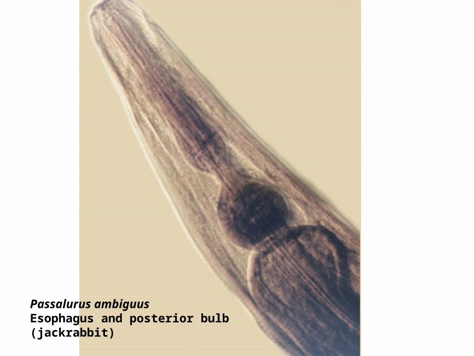
Passalurus ambiguusEsophagus and posterior bulb(jackrabbit)
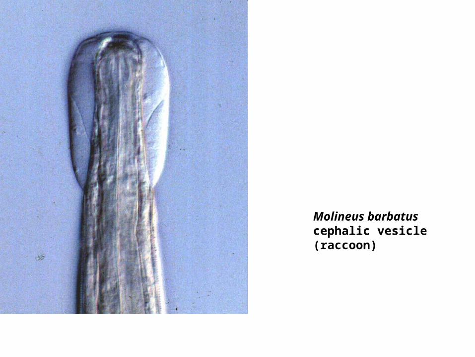
Molineus barbatuscephalic vesicle(raccoon)
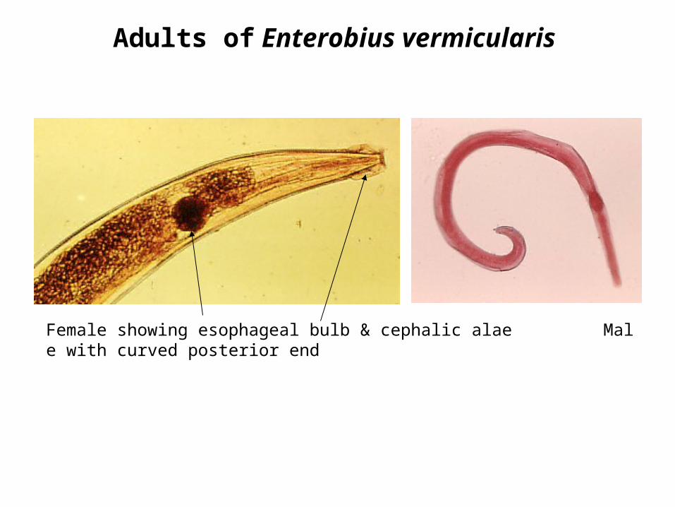
Adults of Enterobius vermicularis
Female showing esophageal bulb & cephalic alae Male with curved posterior end
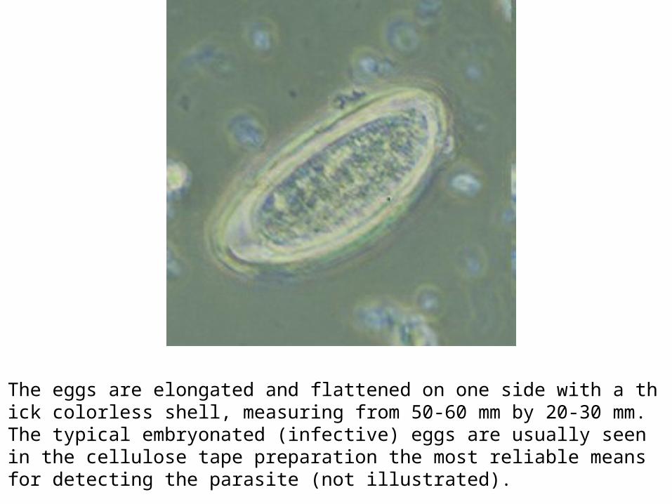
The eggs are elongated and flattened on one side with a thick colorless shell, measuring from 50-60 mm by 20-30 mm. The typical embryonated (infective) eggs are usually seen in the cellulose tape preparation the most reliable means for detecting the parasite (not illustrated).
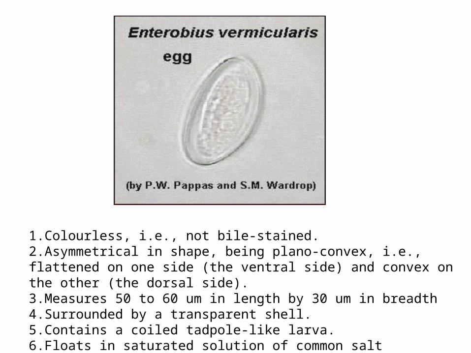
1.Colourless, i.e., not bile-stained.2.Asymmetrical in shape, being plano-convex, i.e., flattened on one side (the ventral side) and convex on the other (the dorsal side).3.Measures 50 to 60 um in length by 30 um in breadth4.Surrounded by a transparent shell.5.Contains a coiled tadpole-like larva.6.Floats in saturated solution of common salt
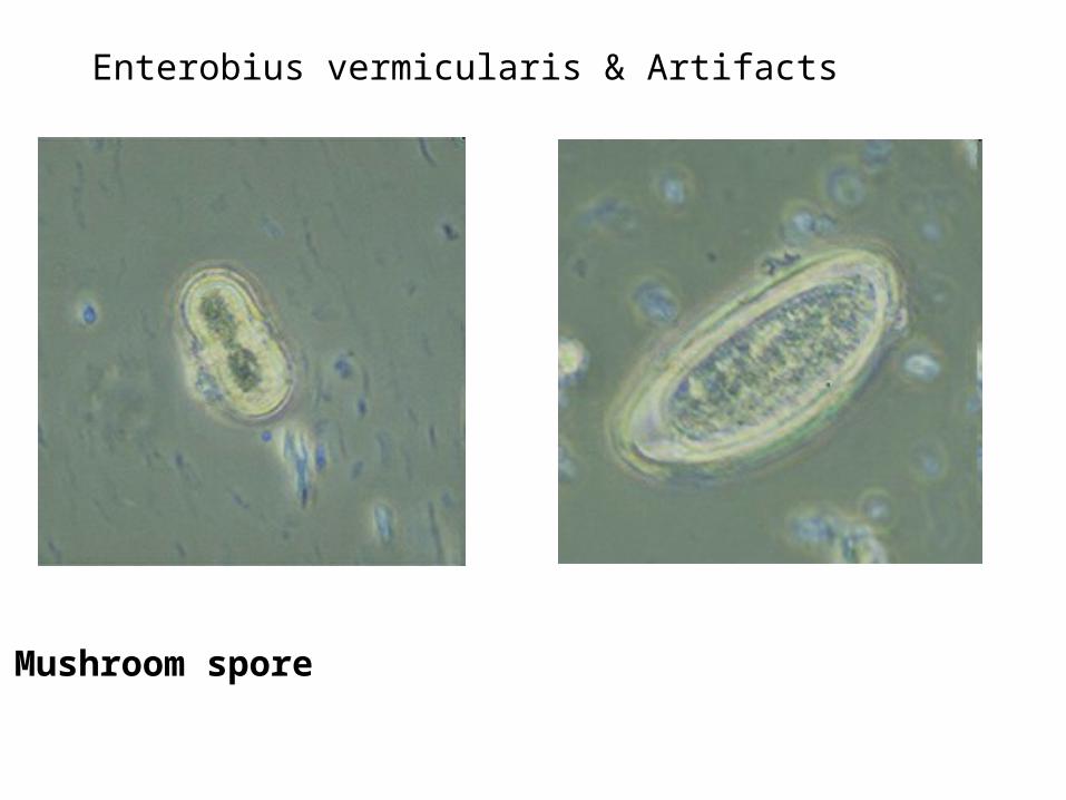
Mushroom spore
Enterobius vermicularis & Artifacts
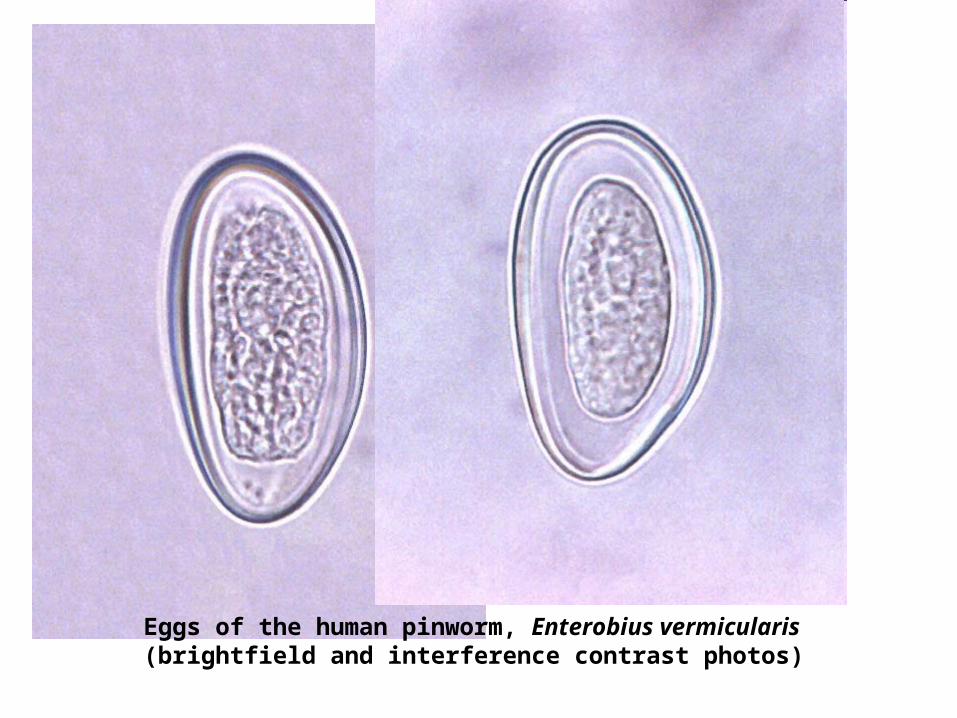
Eggs of the human pinworm, Enterobius vermicularis(brightfield and interference contrast photos)

HOOKWORM DISEASE
&
HOOKWORMS
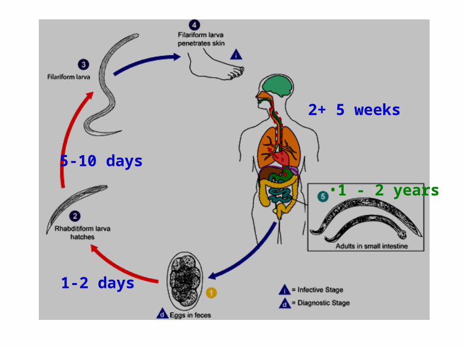
1-2 days
5-10 days
2+ 5 weeks
•1 - 2 years
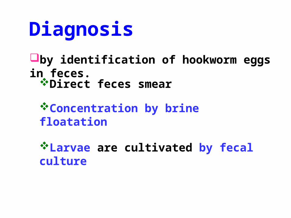
by identification of hookworm eggs in feces.
Diagnosis
Direct feces smear
Concentration by brine floatation
Larvae are cultivated by fecal culture
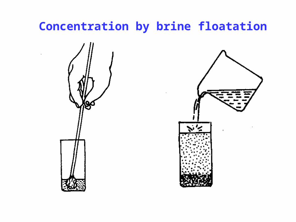
Concentration by brine floatation
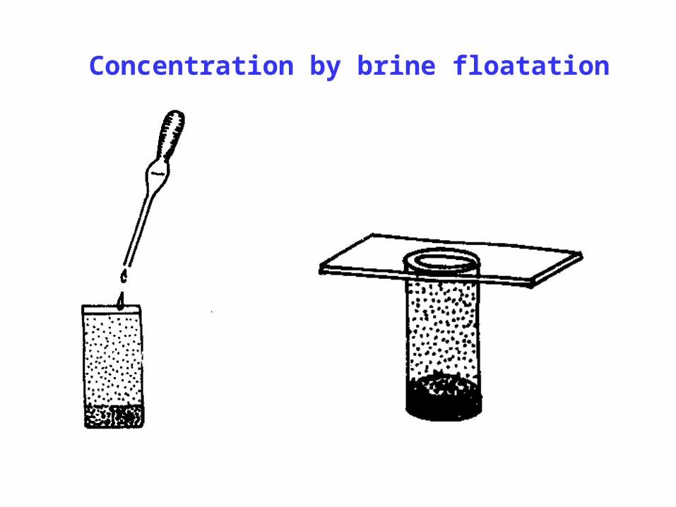
Concentration by brine floatation
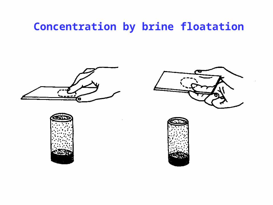
Concentration by brine floatation
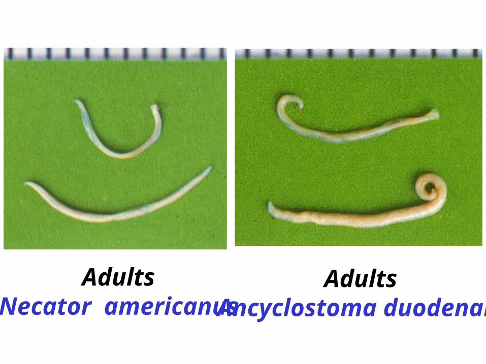
AdultsNecator americanus
AdultsAncyclostoma duodenale
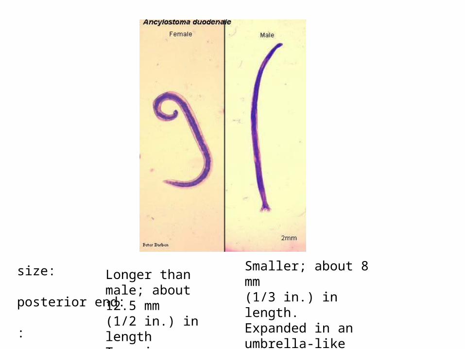
size: posterior end: :
Longer than male; about 12.5 mm(1/2 in.) in lengthTapering; no expanded bursa.
Smaller; about 8 mm(1/3 in.) in length.Expanded in an umbrella-like fashion (copulatory bursa).
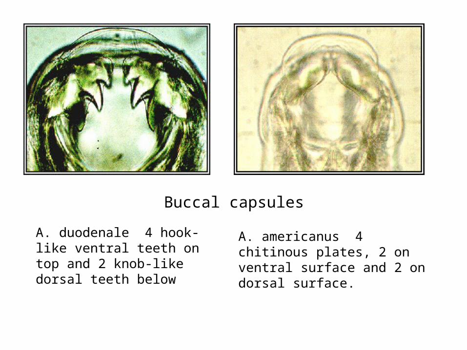
Buccal capsules
A. duodenale 4 hook-like ventral teeth on top and 2 knob-like dorsal teeth below
A. americanus 4 chitinous plates, 2 on ventral surface and 2 on dorsal surface.
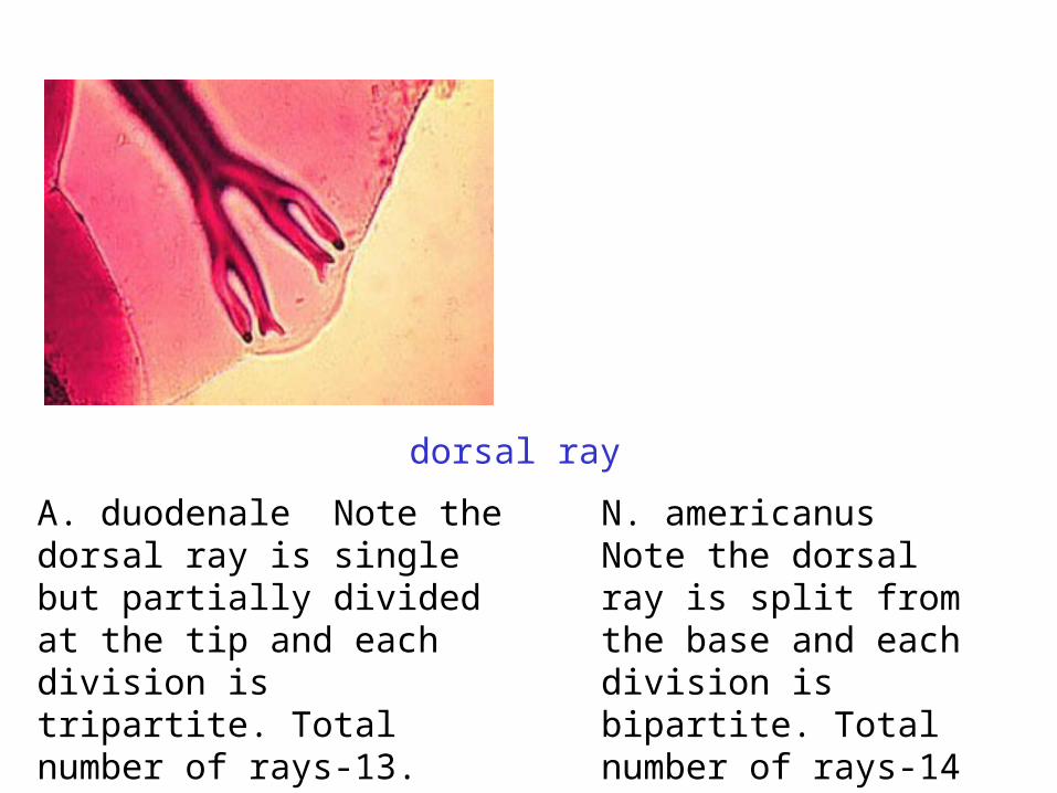
dorsal ray
A. duodenale Note the dorsal ray is single but partially divided at the tip and each division is tripartite. Total number of rays-13.
N. americanus Note the dorsal ray is split from the base and each division is bipartite. Total number of rays-14
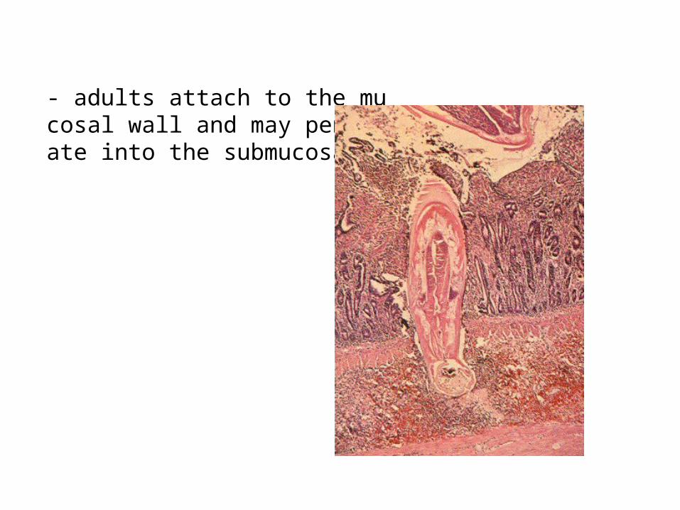
- adults attach to the mucosal wall and may penetrate into the submucosa
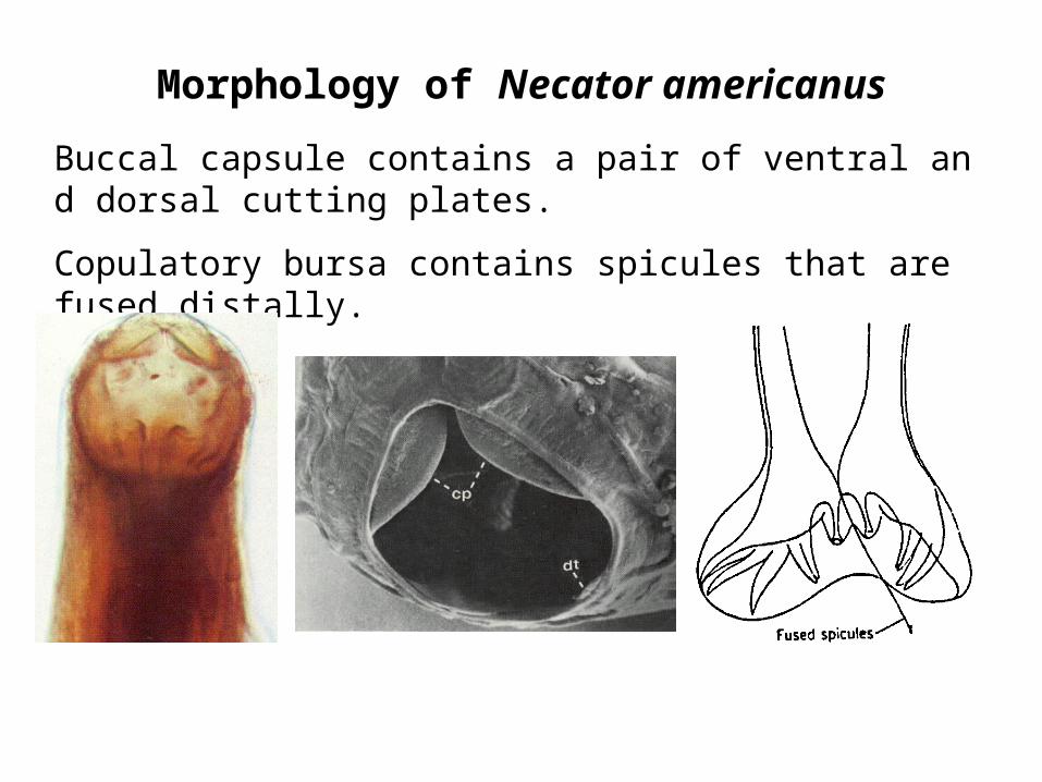
Morphology of Necator americanus
Buccal capsule contains a pair of ventral and dorsal cutting plates.
Copulatory bursa contains spicules that are fused distally.
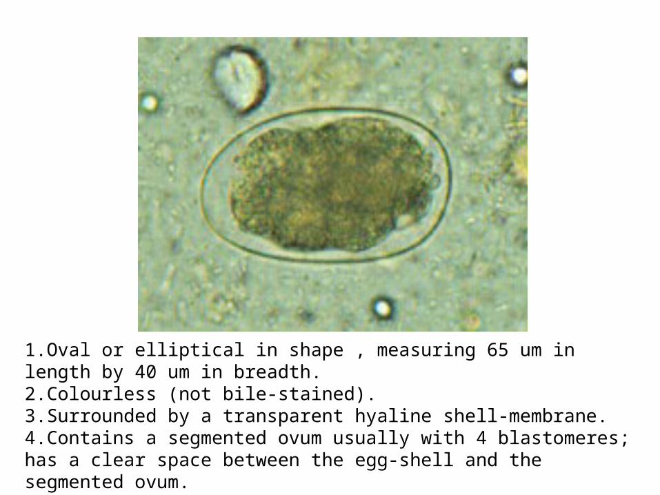
1.Oval or elliptical in shape , measuring 65 um in length by 40 um in breadth.2.Colourless (not bile-stained).3.Surrounded by a transparent hyaline shell-membrane.4.Contains a segmented ovum usually with 4 blastomeres; has a clear space between the egg-shell and the segmented ovum.5.Floats in saturated solution of common salt.
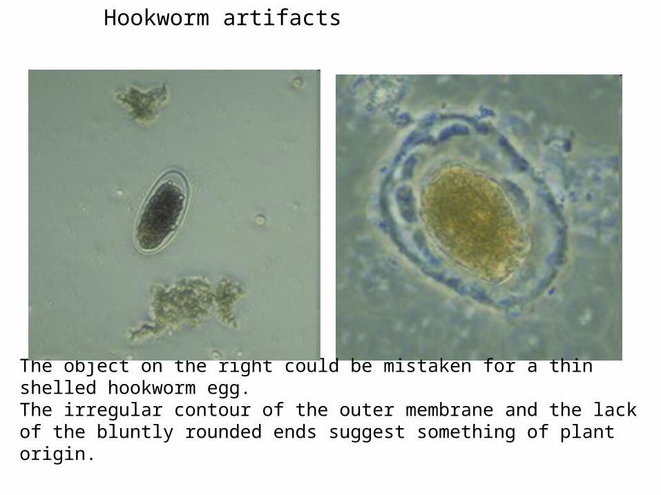
The object on the right could be mistaken for a thin shelled hookworm egg. The irregular contour of the outer membrane and the lack of the bluntly rounded ends suggest something of plant origin.
Hookworm artifacts
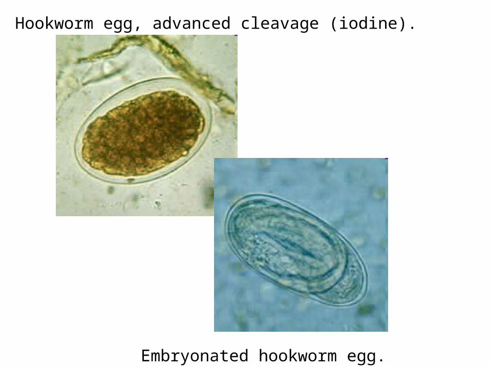
Hookworm egg, advanced cleavage (iodine).
Embryonated hookworm egg.
TRICHINOSIS TRICHINITRICHINOSIS TRICHINIASIS TRICHINELLOSIS ASIS TRICHINELLOSIS
&
Trichinella spiralisTrichinella spiralis
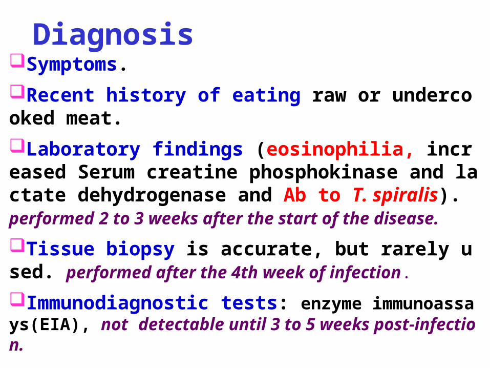
Symptoms.
Recent history of eating raw or undercooked meat.
Laboratory findings (eosinophilia, increased Serum creatine phosphokinase and lactate dehydrogenase and Ab to T. spiralis). performed 2 to 3 weeks after the start of the disease.
Tissue biopsy is accurate, but rarely used. performed after the 4th week of infection.
Immunodiagnostic tests: enzyme immunoassays(EIA), not detectable until 3 to 5 weeks post-infection.
Diagnosis
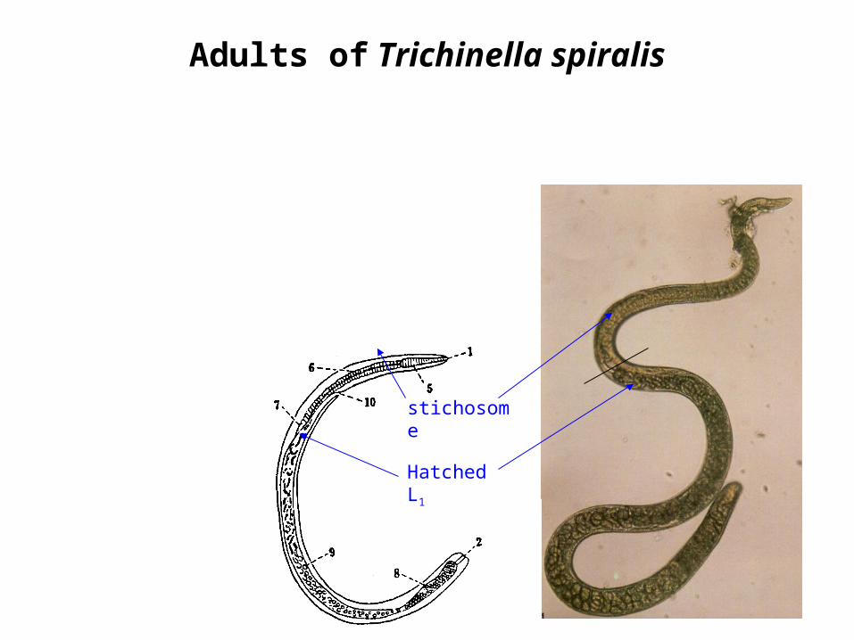
Adults of Trichinella spiralis
stichosome
Hatched L1
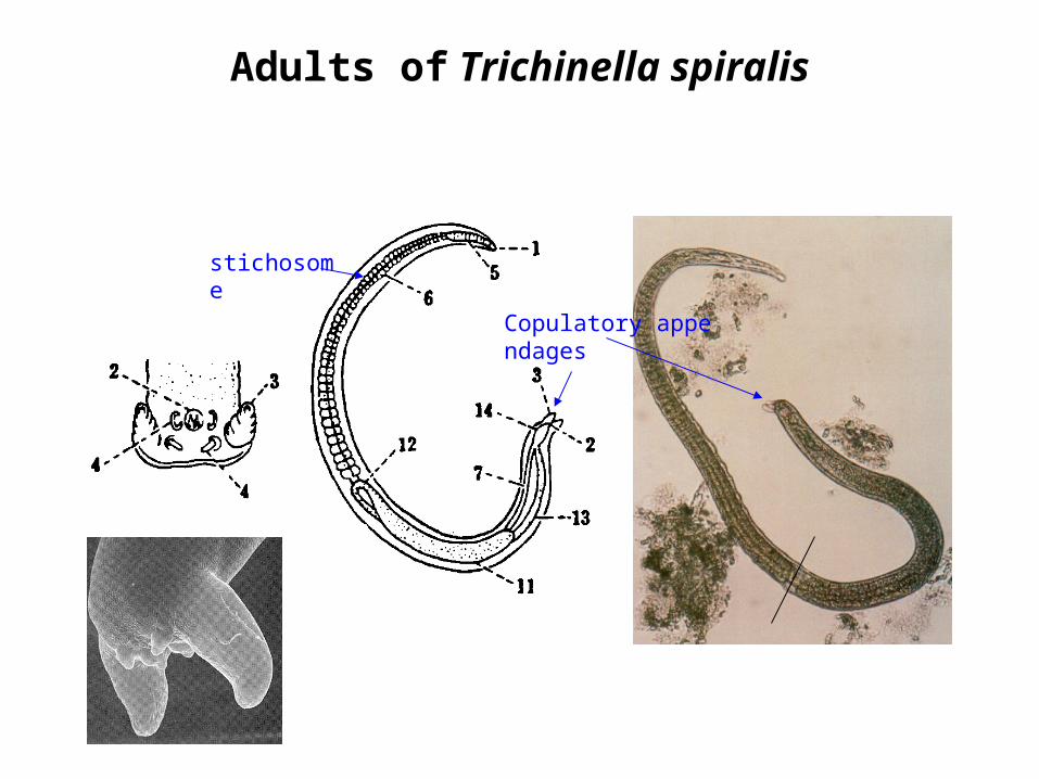
Adults of Trichinella spiralis
Copulatory appendages
stichosome
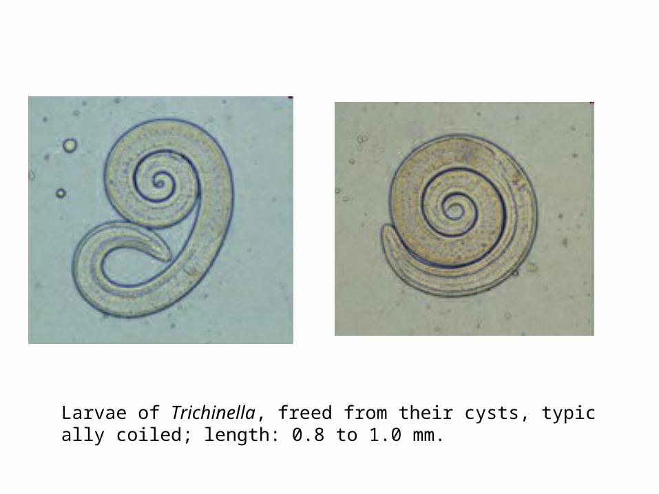
Larvae of Trichinella, freed from their cysts, typically coiled; length: 0.8 to 1.0 mm.
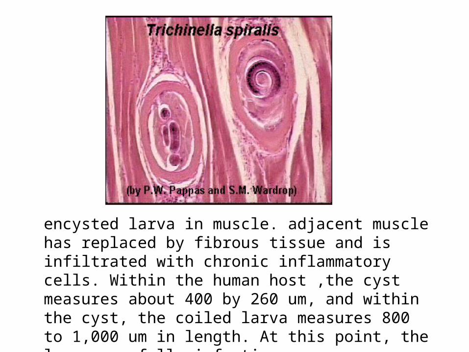
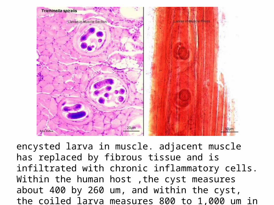
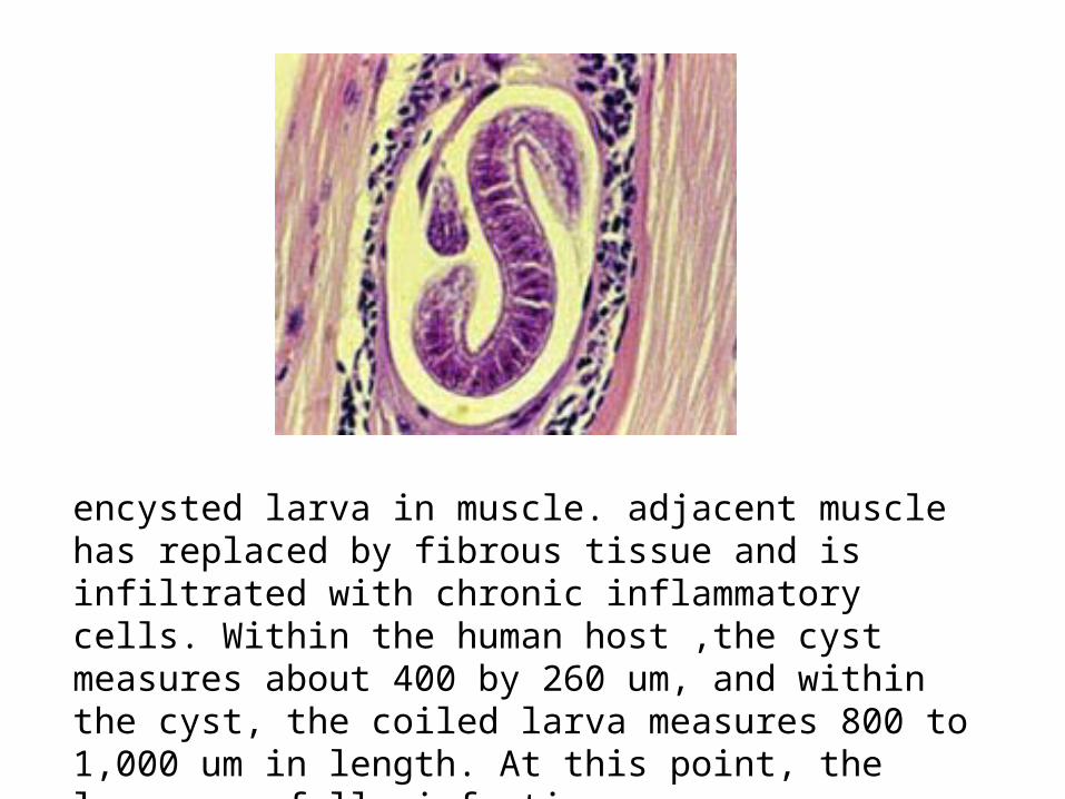
encysted larva in muscle. adjacent muscle has replaced by fibrous tissue and is infiltrated with chronic inflammatory cells. Within the human host ,the cyst measures about 400 by 260 um, and within the cyst, the coiled larva measures 800 to 1,000 um in length. At this point, the larva are fully infective.
encysted larva in muscle. adjacent muscle has replaced by fibrous tissue and is infiltrated with chronic inflammatory cells. Within the human host ,the cyst measures about 400 by 260 um, and within the cyst, the coiled larva measures 800 to 1,000 um in length. At this point, the larva are fully infective.
encysted larva in muscle. adjacent muscle has replaced by fibrous tissue and is infiltrated with chronic inflammatory cells. Within the human host ,the cyst measures about 400 by 260 um, and within the cyst, the coiled larva measures 800 to 1,000 um in length. At this point, the larva are fully infective.
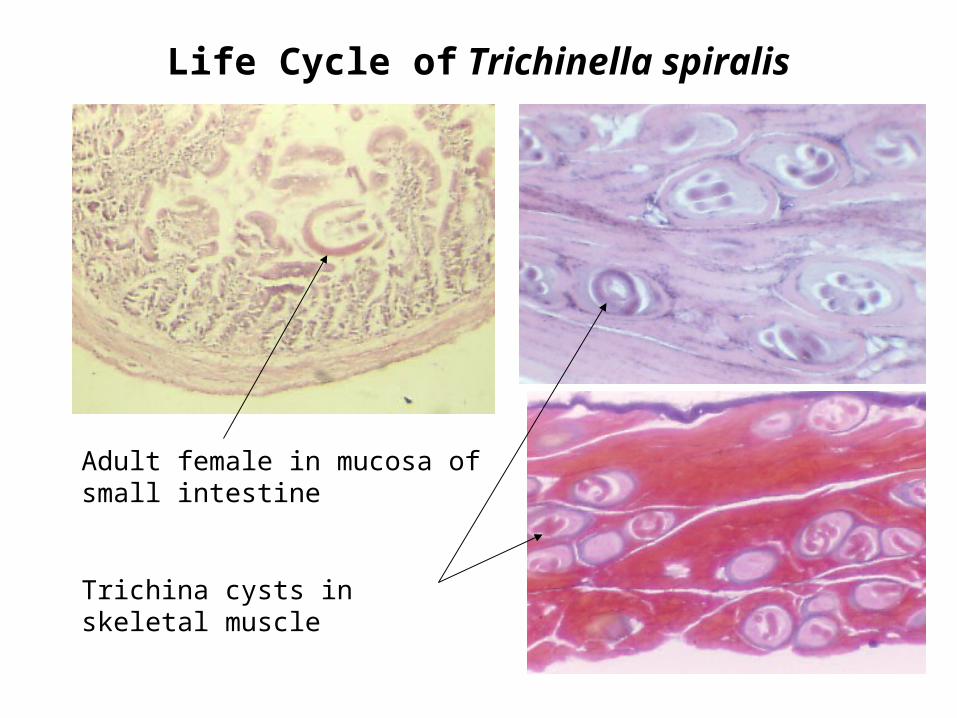
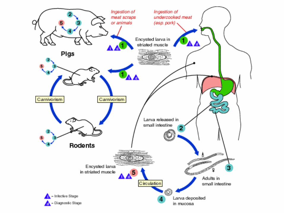
Life Cycle of Trichinella spiralis
Adult female in mucosa of small intestine
Trichina cysts in skeletal muscle
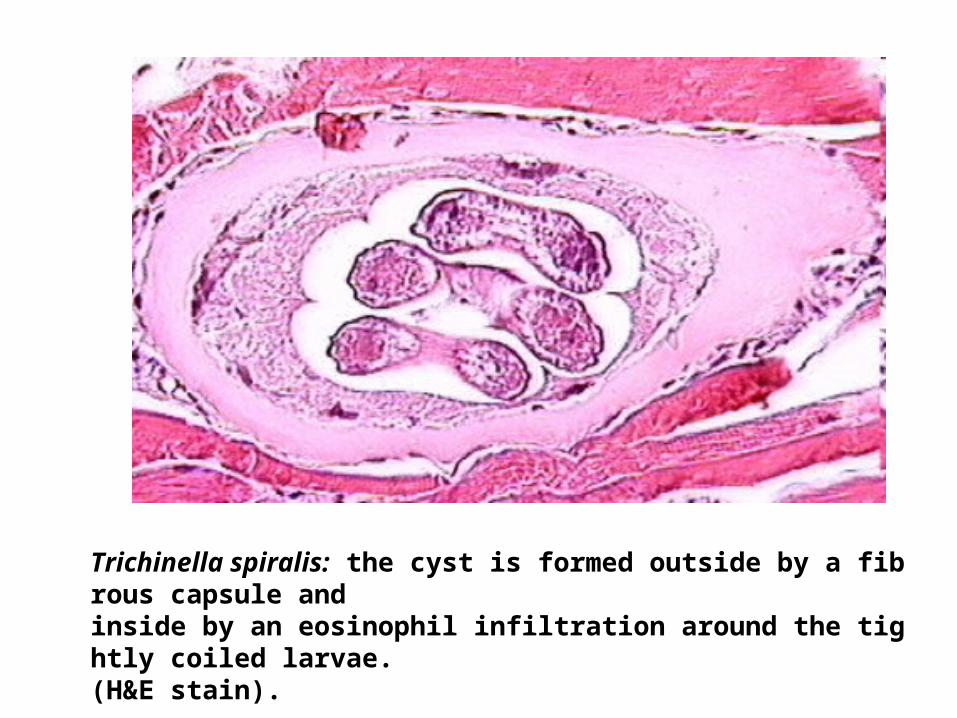
Trichinella spiralis: the cyst is formed outside by a fibrous capsule and inside by an eosinophil infiltration around the tightly coiled larvae. (H&E stain).
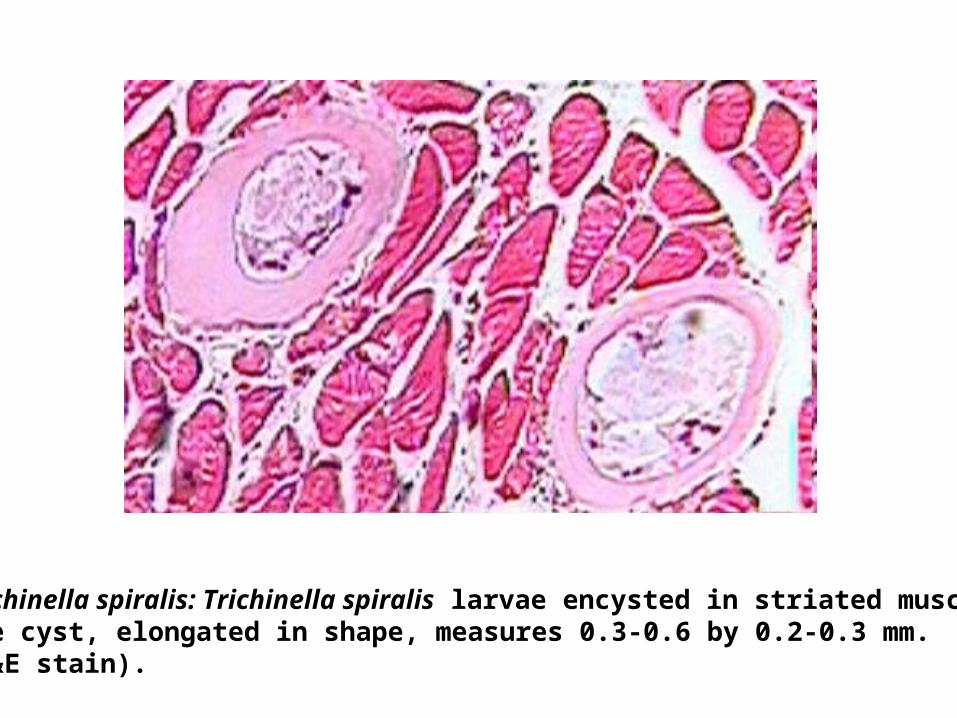
Trichinella spiralis: Trichinella spiralis larvae encysted in striated muscle. The cyst, elongated in shape, measures 0.3-0.6 by 0.2-0.3 mm. (H&E stain).
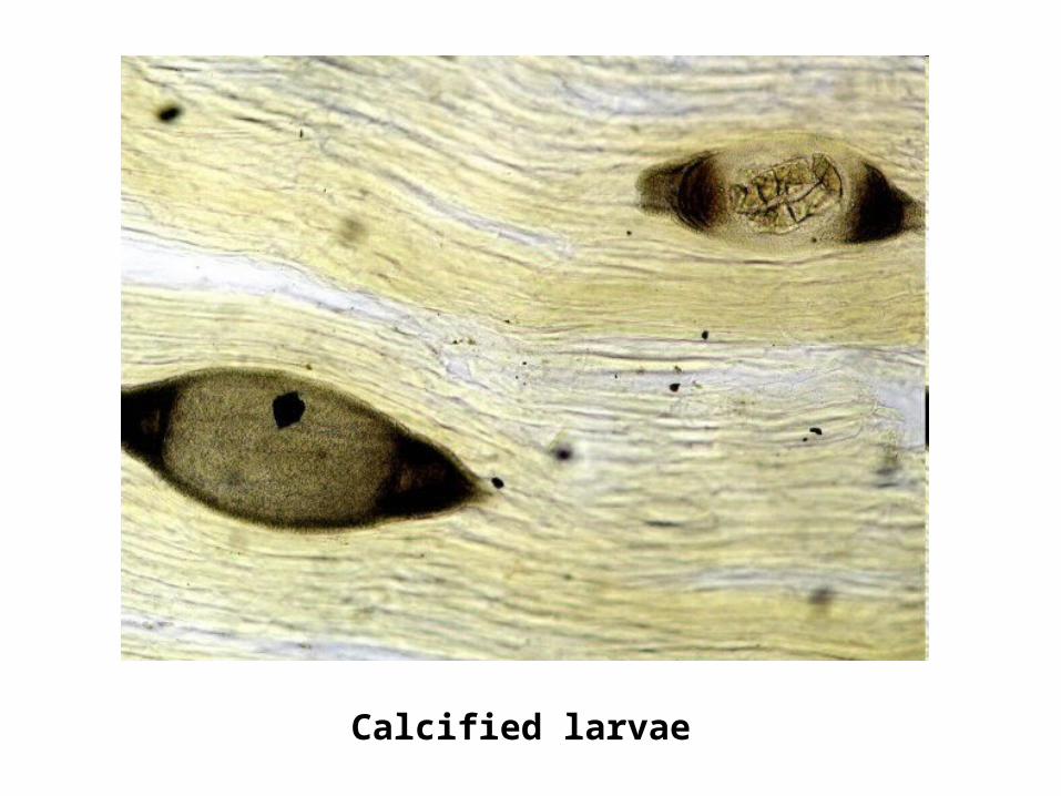
Calcified larvae
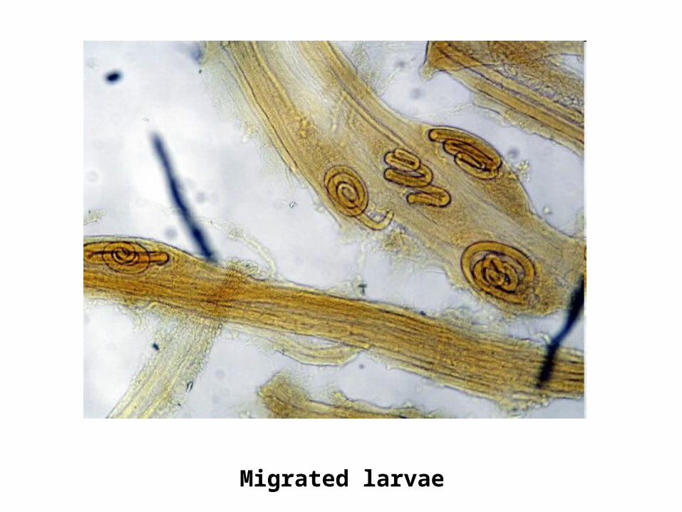
Migrated larvae
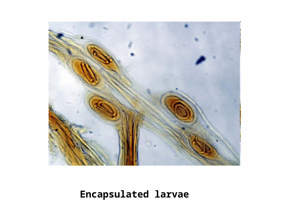
Encapsulated larvae
FILARIASIS&
FILARIAE
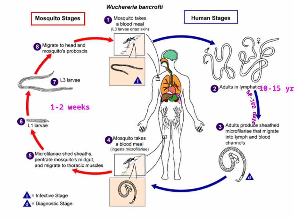
10-15 yrs
1-2 weeks
clinical findings,presence of microfilaria in bloodThick smears: stained with haematoxylin.Concentration method:centrifugation, microfilter
blood samples collected preferably at night---W. Bancrofti: 10 P.M. --- 2 A.M.B. malayi: 8 P.M. --- 4 A.M.
Serologic testing : IgE titers, antifilarial Ab, circulating Ag, eosinophilia.
Diagnosis
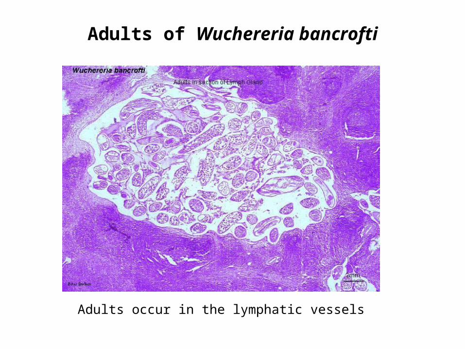
Adults of Wuchereria bancrofti
Adults occur in the lymphatic vessels
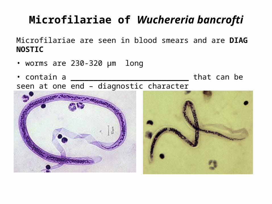
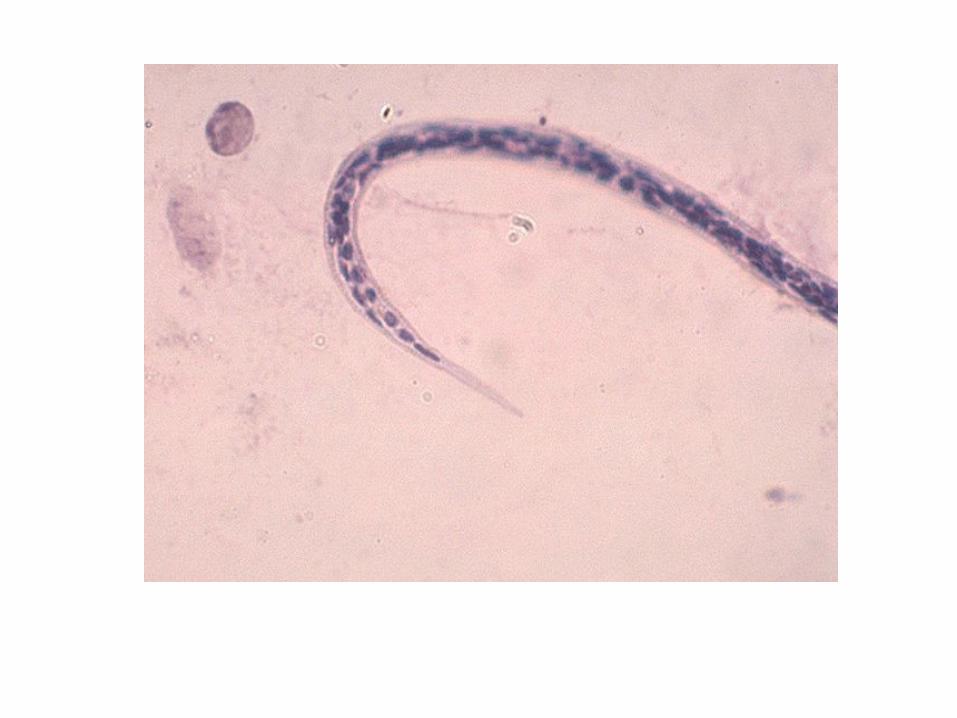
Microfilariae of Wuchereria bancrofti
Microfilariae are seen in blood smears and are DIAGNOSTIC
• worms are 230-320 µm long
• contain a __________________________ that can be seen at one end – diagnostic character
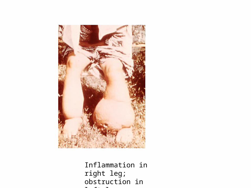
Inflammation in right leg; obstruction in left leg
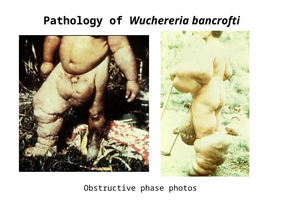
Pathology of Wuchereria bancrofti
Obstructive phase photos
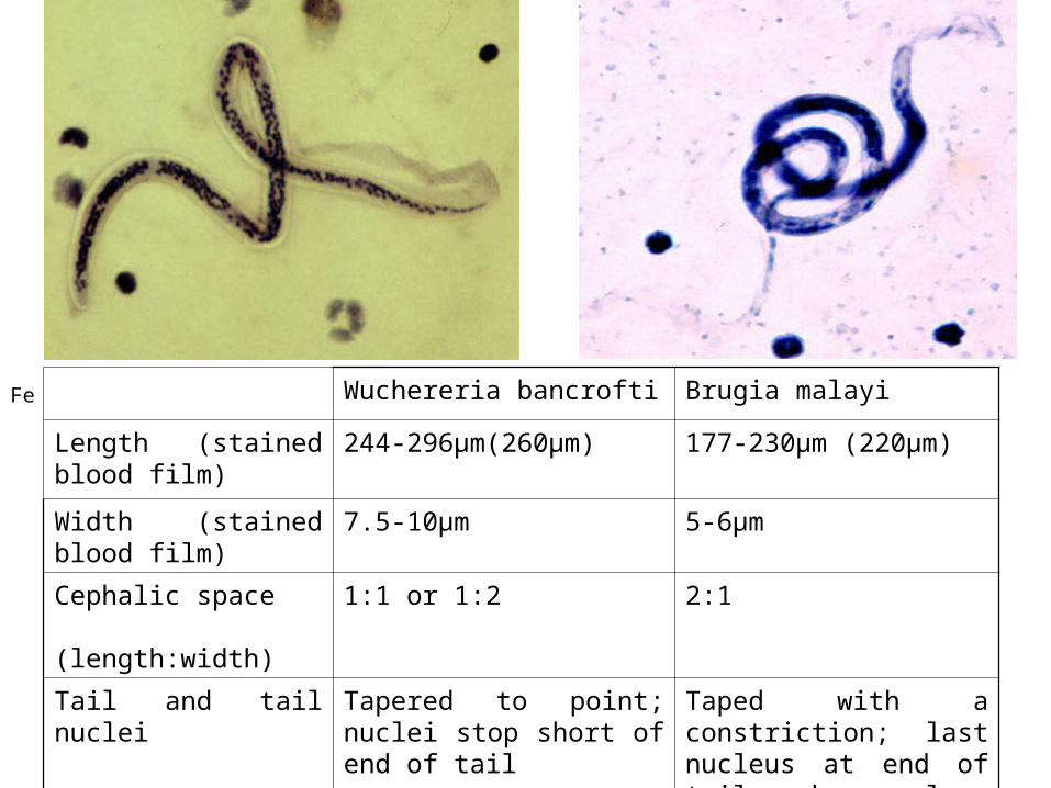
Wuchereria bancrofti Brugia malayi
Length (stained blood film)
244-296μm(260μm) 177-230μm (220μm)
Width (stained blood film)
7.5-10μm 5-6μm
Cephalic space (length:width)
1:1 or 1:2 2:1
Tail and tail nuclei Tapered to point; nuclei stop short of end of tail
Taped with a constriction; last nucleus at end of tail and a nucleus anterior to constriction
Fe