papillomavirus contain a novel transforming - journal of virology
TRANSCRIPT

JOURNAL OF VIROLOGY, Dec. 1994, p. 8365-83730022-538X/94/$04.00+0Copyright ( 1994, American Society for Microbiology
The Genomes of the Animal Papillomaviruses European ElkPapillomavirus, Deer Papillomavirus, and Reindeer
Papillomavirus Contain a Novel TransformingGene (E9) near the Early Polyadenylation Site
ANNIKA ERIKSSON,1 ANN-CHARLOTIE STEWART,2 JORGE MORENO-LOPEZAND ULF PETYIERSSON2*
Department of Veterinary Microbiology, Section of Virology, Swedish University ofAgriculturalScience,1 and Department of Medical Genetics, Uppsala University Biomedical Center,2
Uppsala, Sweden
Received 5 July 1994/Accepted 17 September 1994
We report that the genomes of reindeer papillomavirus (RPV), European elk papillomavirus (EEPV), anddeer papillomavirus (DPV) contain a short conserved translational open reading frame (ORF), E9, which islocated between the E5 ORF and the early polyadenylation site. In RPV, DPV, and EEPV, E9 ORFs have thepotential to encode extremely hydrophobic polypeptides of approximately 40 amino acids. In mouse C127 cellstransformed by EEPV and RPV, there exists a unique, abundant mRNA species of approximately 700nucleotides which has the capacity to encode an E9 polypeptide. This mRNA is transcribed from a previouslyunrecognized promoter at position 4030 in the EEPV genome. The EEPV E9 ORF exhibits weak transformingactivity in C127 cells and primary rat embryo fibroblasts. We also show that EEPV E5 is the major oncogene
in the EEPV genome when assayed in C127 cells, although it is less eflicient in transformation than the E5genes of bovine papillomavirus type 1, DPV, and RPV.
Papillomaviruses, the causative agents of infectious warts,infect a wide variety of vertebrates, including humans. They are
very host and tissue specific and transform basal layer cells insquamous epithelia or mucous membranes, resulting in theproliferation of infected cells (41). In lesions induced by thefibropapillomavirus subgroup of animal papillomaviruses, theproliferation of epidermal keratinocytes is also accompaniedby the proliferation of underlying dermal fibroblasts. Fibropap-illomaviruses are also efficient inducers of morphologic andtumorigenic transformation of cultured rodent fibroblasts, andthey can induce tumor formation in hamsters. Bovine papillo-mavirus type 1 (BPV-1) is the prototype fibropapillomavirusand has been subjected to detailed genetic analysis (for a
review, see reference 12). Other fibropapillomaviruses arereindeer papillomavirus (RPV), European elk papillomavirus(EEPV), and deer papillomavirus (DPV) (22, 35, 46). Incultured C127 cells transformed by BPV-1, the viral genome ismaintained as an autonomous nuclear plasmid with a copynumber of approximately 50 to 200 per cell (31). The earlyregion of the genome, containing open reading frames (ORFs)El through E8, is the only region transcribed in these cells.
In BPV-1, the E5 ORF has been shown to encode one of thetwo independent transforming proteins in the viral genome(11, 21, 44, 47). The BPV-1 E5 gene encodes a 7-kDahydrophobic polypeptide that localizes to the Golgi apparatus,endoplasmic reticulum, and plasma membranes (6, 7, 45). Itassociates with a 16-kDa cellular protein that has been identi-fied as a subunit of the vacuolar H+-ATPase (17, 18). BPV-1ES protein has also been shown to form a complex with theplatelet-derived growth factor (PDGF) receptor and to influ-ence its activity in addition to those of other growth factor
* Corresponding author. Mailing address: Department of MedicalGenetics, Biomedical Center, Box 589, S-751 23 Uppsala, Sweden.Phone: 46 18 17 45 80. Fax: 46 18 52 68 49.
receptors (33, 39, 40). The E5 ORF is highly conserved amongthe fibropapillomaviruses BPV-1, DPV, EEPV, and RPV andhas been shown to be the major transforming gene in thegenomes of BPV-1 and DPV when assayed in C127 cells (5, 7,28). The other transforming protein of BPV-1 is the E6 protein(43, 47), which is considerably less well conserved among theseviruses than the 43- to 44-amino-acid (aa) ES protein.The genomes of DPV, EEPV, and RPV, all of which infect
members of the Cervidae family, are more closely related toone another than they are to the BPV-1 genome. All theformer genomes are slightly larger (by approximately 200 to400 bp) than the BPV-1 genome, and they cross-hybridize toeach other even under stringent hybridization conditions, a
unique finding for papillomaviruses which infect differentspecies (35). Moreover, EEPV and RPV express mRNAclasses of similar sizes. A noteworthy feature is the presence ofa class of small viral transcripts, with a length of 600 to 700nucleotides (nt), in EEPV- and RPV-transformed C127 cells,which has no apparent counterpart in BPV-1-transformed cells(1, 35). In the case of EEPV-transformed cells, it was shownthat this small transcript contained sequences exclusively fromthe 3' end of the transforming region (1). These observationsprompted us to study this region of animal papillomavirusesfurther, and in the present communication, we present theDNA sequence of the corresponding region of the RPVgenome. In RPV, we identified an additional approximately380-bp region between ORFs E5 and L2, which appears to beabsent from the BPV-1 genome. Similar 3' early regionextensions exist in the EEPV and DPV genomes at equivalentlocations, accounting for the larger sizes of these genomesrelative to that of BPV-1. Moreover, comparing the sequencesof this region in EEPV, RPV, and DPV, we found that all threecontained a previously undiscovered ORF, E9, which has thepotential to encode an extremely hydrophobic peptide ofapproximately 40 aa. The previously discovered 700-nt tran-
8365
Vol. 68, No. 12
on Novem
ber 29, 2018 by guesthttp://jvi.asm
.org/D
ownloaded from
8366 ERIKSSON ET AL.
script in cells transformed by RPV and EEPV (1) is likely toencode the E9 polypeptide. In this report, we show that theEEPV genome contains a unique promoter, P4030, locatedimmediately downstream of the ES ORF. We present evidencethat this promoter is used to transcribe the 700-nt class ofEEPV mRNAs. We show also that the E9 polypeptide exhibitsweak transforming activity in murine C127 cells and primaryrat embryo fibroblasts (REF).
MATERIALS AND METHODS
Nucleotide sequence analysis. The nucleotide sequence wasobtained by the technique of Maxam and Gilbert (34) andanalyzed with University of Wisconsin Genetics ComputerGroup software (10).
Cells and virus. The EEPV-transformed C127 cell line usedfor RNA isolation was obtained after infection with purifiedEEPV virions (1). For transfection experiments, we used REF,prepared by trypsination of 14-day-old rat embryos, and mu-rine C127 cells. All cells were propagated in Dulbecco modi-fied Eagle medium supplemented with either 5% (transfectedC127 cells) or 10% (transfected REF and the EEPV-trans-formed C127 cell line) fetal calf serum. REF were treated withmedium supplemented with 0.4 mg of G418 sulfate (GibcoBRL) per ml.
Isolation of RNA. Cytoplasmic RNA was isolated and frac-tionated by oligo(dT)-cellulose chromatography essentially asdescribed by Maniatis et al. (32).
Northern (RNA) blot hybridization. Polyadenylated RNAwas electrophoresed on a 1% agarose-formaldehyde gel andblotted onto a nitrocellulose filter. Hybridization was per-formed under stringent conditions with subgenomic EEPVprobes (for details, see Fig. 5) labelled by random priming to aspecific activity of 108 cpm/,jg.
S1 nuclease mapping. Poly(A)-selected RNA (5 to 10 ,ug)was mixed with 5'-end-labelled DNA probe (5 x 10⁴ cpm) andethanol precipitated. It was then redissolved in a reactionmixture of 10 ,ul. Hybridization and S1 nuclease digestion wereperformed essentially as described by Favaloro et al. (15). Sidigestion products were analyzed on 6 or 8% polyacrylamidegels with 7.5 M urea together with radiolabelled DNA frag-ment size markers (for details, see Fig. 6).Primer extension analysis. Reactions were performed with
10 ,ug of poly(A)-selected cytoplasmic RNA essentially asdescribed by Baker and Howley (4). The resulting primerextension products were fractionated on 6 or 8% polyacryl-amide sequencing gels. One primer extension product was
excised from the gel (3) and sequenced by the technique ofMaxam and Gilbert (34). The oligonucleotide primer p4070Nwas complementary to the EEPV sequence between nt 4044and 4070.Plasmid constructions. RPV particles were isolated from a
cutaneous fibropapilloma. Viral DNA was inserted into theBamHI site of pBR322, and subclones were constructed as
described elsewhere (35).The EEPV E5- mutant was obtained by the insertion of a
linker (TTL) with translation stop codons in all three readingframes at the PvuII restriction cleavage site (nt 3933). TheEEPV E9- mutant was obtained by the insertion of a TTLlinker at the SspI restriction cleavage site (nt 4396).
In order to insert EEPV E9 and E9- regions under thecontrol of the murine sarcoma virus (MSV) long terminal repeatenhancer/promoter region, we first amplified EEPV wild-type(wt) and E9- DNA by PCR. Oligonucleotides of 30 nucleo-tides were synthesized, and one pair of oligonucleotides,E4361+ and E4640-, was used. PCR products were cloned
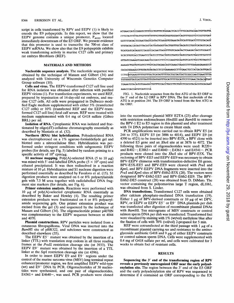
ATGAACCATCCGGGTCTTTTCCTGTTTCTGGGACTCACCTTTGCAGTACAACTGTTATTA
CTTGTATTTTTATTGTTTTTTTTTCTTGTGTGGTGGGATCAGTTTGGGTGTCGGTGTGAT
GGTTTTATACTGTAATTAGTCATACTCAAGGTGTAAATATTCATTTGATCTTTGTACAGT
TTTTATACCATATTTACATTATTATAGGTTACTGGTTGCTACATCCTGGTCATAGTCACA
TCATCATCATAGGTCTAGGTCACAATTAGGTTTGTCAGATACTCAAGACGACGTGGAATC
TCTCTTGTCACCTGAATCCTATCCTTTACTGTCCTATCCTATCCTATCTGCCTTTGTTTT
GTTATCCAAAGTCAGCAAGTGCCATCTTTCTCCAAGTGCAATGTCATCTGCCTGTAATCC
AAAAGCTGGTGTCATCCTTGTCAGTAGAACAGTCAAACTAAGCCTTTGAAAAGAAAGCCT
CACACGGAAACCTTGTATGTATACCTCGTGAAAAAGCTTTGAACTGCCACGACATTGACG
CCTG TGAAGTTTTGTTTACTCATATTTTTGCTGCTATTATTCGGCCAATTGAATTTTA
TGTGGGTTATCATTTTATTTGTATGGTTTGCATTTTTGCATTCTTTGAACTATAC GAT
TGAAATGTACATGTGAAGGCTGTTTCCACCTGCCTTCTGTCTCGCGTACACGTGCACAAT
AAACCCACCATGTCATCCCA
FIG. 1. Nucleotide sequence from the first ATG of the E5 ORF tothe 5' end of the L2 ORF in RPV DNA. The first nucleotide of theATG is at position 244. The E9 ORF is boxed from the first ATG inthe ORF.
into the recombinant plasmid MSV E2TA (23) after cleavagewith restriction endonucleases Hindlll and BamHI to removethe BPV-1 E2 to E5 region in this plasmid and after treatmentwith T4 DNA polymerase to create blunt ends.PCR amplifications were carried out to obtain RPV E5 (nt
244 to 375), EEPV ES (nt 3886 to 4014), and EEPV E9 (nt4390 to 4521) to be inserted into BPV-E5X, a BPV clone witha deleted E5 gene and an XhoI site at nt 3876 to 4072. Thefollowing three pairs of oligonucleotides were used: R220+and R402-; E3863+ and E4040-; E4361 + and E4541 -. PCRproducts were cloned into the XhoI site of BPV-E5X. Therecloning of BPV-EE5 and EEPV-EE9 was necessary to obtainBPV-EEPV chimeras with transformation-defective E6 genes.BPV-E5X-EE5 and BPV-EE9 were cleaved with PvuI andKpnI, and BPV-EEPV DNA fragments were inserted into thePvuI and KpnI sites of BPV-E6fs2-E5X (28). The vectors weredesignated BPV-E6fs2-EE5 and BPV-E6fs2-EE9. The BPV-E6fs2-DE5 construct (28) was obtained from D. DiMaio. Thevector containing the polyomavirus large T region, dL1061,was obtained from S. Linder.DNA transfections. Transformed C127 cells were obtained
after calcium phosphate-mediated DNA transfection (19).Either 1 ,ug of BPV-derived constructs or 10 ,ug of wt DPV,RPV, or EEPV or EEPV E5S or E9- DNA plasmids per dishwas transfected after digestion of recombinant plasmid DNAswith BamHI. Ten micrograms of MSV constructs or controlsalmon sperm DNA per dish was transfected. Transformed fociwere visualized by staining with 1% (wt/vol) methylene blue afterthe fixation of cells with 70% (vol/vol) 2-propanol for 5 min.REF were cotransfected at the third passage with 1 ,ug of a
recombinant plasmid carrying ras and resistance to the amino-glycoside antibiotic G418 and 9 ,ug of either EEPV constructsor control salmon sperm DNA. Cells were supplemented with0.4 mg of G418 sulfate per ml, and cells were cultivated for 3weeks to obtain foci of resistant cells.
RESULTS
Sequencing the 3' end of the transforming region of RPVreveals a previously unrecognized ORF near the early polyad-enylation site. The region between the 3' end of the E5 ORFand the early polyadenylation site of RPV was sequenced todetermine if it contained an ORF corresponding to the EX
J. VIROL.
on Novem
ber 29, 2018 by guesthttp://jvi.asm
.org/D
ownloaded from

E9 GENES OF EEPV, DPV, AND RPV 8367
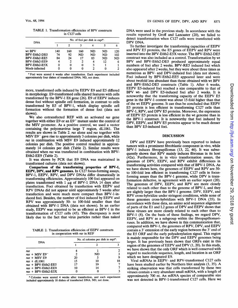
1 [EEEPV 2 El
3 E6
FE41] E3 -- I
4360 4518R E2-715 1D FCTJ FE L1 l
3865 4014 A
I A
0 1000 2000 3000 .4000 50008
11
6000 7000 8000 8095
1,==_===71I ] -W
P4030f0EEPV
DPV
RPV
[ -. E5:
BPV-1 v
FIG. 2. Organization of the EEPV genome. In the lower section, the region of the EEPV genome from the first ATG in the E5 ORF to thebeginning of the L2 ORF is compared with the corresponding regions of DPV, RPV, and BPV-1 (1, 2, 9, 14, 21, 35). The first methionine of eachORF is indicated by a line, and early polyadenylation signals are indicated by arrows.
ORF observed in the sequences of EEPV and DPV (3, 20).The established sequence (Fig. 1) was compared with se-
quences from the corresponding regions in EEPV, DPV, andBPV-1. The distance between the termination codon of the E5ORF and the polyadenylation signal, AATAAA, was 586 nt inRPV, compared with 547 nt in EEPV and 706 nt in DPV. InBPV-1, the corresponding distance is 170 nt. A sequence
comparison of the region which contains the candidate EXgene in EEPV and the corresponding regions in RPV andDPV revealed no significant homologies. In fact, several gapshad to be introduced into the RPV and EEPV sequences inorder to align them with the DPV sequence in this region (Fig.2). Moreover, the single ATGs in these candidate ORFs were
located in different positions in the three viral genomes. Fromthese results, we conclude that the EX ORF is unlikely torepresent a functional gene. However, when the three se-
quences were compared, we observed another short ORFimmediately upstream of the polyadenylation signal (Fig. 2).This ORF was designated E9 (Fig. 2).
Properties of the E9 ORF. Figure 2 shows that the E9 ORFand its initiation codon (ATG) are located in the same relativepositions in the genomes of EEPV, DPV, and RPV. The E9ORFs of RPV and EEPV extend for a considerable distanceupstream of this ATG. This extension might, however, befortuitous. The overall nucleotide sequence identities were 56
and 51% when the E9 ORF of RPV was compared with the E9ORFs of EEPV and DPV, respectively. The correspondingidentity for the EEPV and DPV E9 ORFs was 55%. Thecoding capacities of the E9 ORFs from the first ATG are 37 aafor RPV, 41 aa for DPV, and 43 aa for EEPV. The putative E9proteins of all three viruses are very hydrophobic, and theamino-terminal regions are extremely well conserved (Fig. 3and 4). Fifteen of 21 amino-terminal aa in the E9 proteins ofRPV and EEPV are identical, with most amino acid substitu-tions being conservative (Fig. 3). The carboxy termini of thethree E9 proteins are less well conserved (Fig. 3).
Northern blot analysis of EEPV mRNAs. Poly(A)-selectedRNA from EEPV-infected C127 cells was subjected to North-ern blot analysis with a series of subgenomic probes. In aprevious report, we showed that a probe covering the entireEEPV genome detected six classes of mRNA size, varying
DPV LLLL CL « IF. I.41EEPV LLLFLLLLLG NP HFT43RPV FIA..LF. NYE]...37FIG. 3. Comparison of the amino acid sequences of E9 proteins in
DPV, EEPV, and RPV, starting with the first methionine. Conservedamino acids are boxed.
7i= M-.\Z\,-,E9MMv
I ES L2
Iv
. m.l m
VOL. 68, 1994
on Novem
ber 29, 2018 by guesthttp://jvi.asm
.org/D
ownloaded from

J. VIROL.8368 ERIKSSON ET AL.
MPNLWFLLFLGLVAAMOLLLLLFLLLFFLVYWDHFECSCTGLPF3.69]
BPV-1 E5
MTGL FLLFLQMLF LFFFVFCRCNMQ
EEPV E5
BPV-1 E51
EEPV E9
DPV E9
RPV Eg
3.44.
0-1.23..
MTYGLLL FLGLTFGLOLMLLVFL LFFFLVWWDQFGCRCENMQL
-
MPLRI-tMLAVFLLLCGFDLFYI LYEVFSFVLVLLFVSFLLEFVFPLLRFWAV
MKYCLLL FLLLLLGQWNPMWVLLLI VWLS VLLFELHFVEHFT3.69
MKLWLLLLLLLLLGHWTPVWVIFCLLWSSFLFLLHSVFITF3.8f
MKFCLLIFLLLLFGQLNFMWVI ILFVWFAFLHSLNYT3.-
0-aDveFIG. 4. Kyte and Doolittle (30) hydrophobicity plots of the putative
E9 proteins of DPV, EEPV, and RPV. EEPV
E5 and ESb proteins are included for
from 700 to 5,300 nt (1). To make a more
six mRNA classes, we scanned the EEPV
different subgenomic probes. The results
are shown in Fig. A probe covering the
Fig. SB), as well as a probe spanning
part of the N-terminal region of El
detected the four previously described
mRNAs, with 2,400, 3,100, 3,700, and 5,200
to these classes, probe B also hybridized
1,900 nt that has not been described
ization signal was less intense than
mRNAs, indicating a low abundance of
small overlap between this mRNA and a
probe covering the middle portion of El
Fig. SB), we detected only the largest
the 1,900-nt transcript, suggesting the
in this region. Probes D and E (Fig.
identical patterns, with the exception
This transcript gave a much more intense
with probe E than with probe D. All
from the most 3' part of the early region, I,
hybridized to the major mRNA classes.
did not detect the smallest (700-nt)
the H and I probes did. None of the
region, J, K, or L, nor probe M from the URR was able todetect any mRNA species even after very long exposure (>3weeks). The results thus show that an abundant 700-nt mRNAwhich maps in the E9 region of the EEPV genome exists.A candidate mRNA for E9 protein. In order to map the 5'
end of the 700-nt mRNA, Si nuclease analysis was performedwith a 5'-end-labelled DNA probe derived from the 3' part ofthe early region of the EEPV genome. Autoradiograms of theSl mapping experiments are shown in Fig. 6A and B. When anEcoRV-SspI DNA fragment of 1,533 bp labelled at the SspIsite was hybridized to cytoplasmic poly(A)-selected RNA fromEEPV-infected C127 cells and subsequently digested with S1nuclease, six bands of 1,480, 1,400, 1,200, 950, 770, and 370 ntwere protected against Si. The smallest (approximately 370-nt) band was the most intense band protected in this experi-ment. The 370-nt band extended approximately to nt 4026 andmay represent the 5' end of the 700-nt mRNA. In order tofurther characterize the 5' end of the 700-nt mRNA, primerextensions were performed with an oligonucleotide with a 5'terminus at nt 4070. We obtained several primer extensionproducts with this primer. This was anticipated since thisprimer is located only shortly upstream of the early poly(A)site, which is likely to be utilized by all of the early regiontranscripts. The most prominent band, however, mapped just40 nt upstream of the 5' terminus of the oligonucleotide, at nt4030 (Fig.6C). To analyze the primary structure of this primerextension product, the band was excised from the gel andsequenced. The product was found to be colinear all the wayfrom nt 4030 to the 3' end of the primer. This extensionproduct was detected in many independent experiments, withfive different RNA preparations. No bands of similar size weredetected in nontransformed C127 cultures. It thus seems thatthe initiation site for the 700-nt transcript maps at position4030. We designate this new promoter P4030.When EEPV sequence upstream of P4030 was compared
with the corresponding region in the BPV-1 genome, some
homology upstream of nt 4096 was detected. An attempt was
made to search for a putative promoter in this region of theBPV-1 genome. Primer extension and Si analysis were per-
formed with oligonucleotides with 5' termini at nt 4178.However, we did not detect any primer extension or Sidigestion products mapping to this region of the BPV-1genome (data not shown).Transforming properties of EEPV E5 and E9 genes. In order
to examine a possible role of the E9 ORF in cell transforma-tion, C127 cells were transfected with EEPV wt,E5-, orE9-DNAs. The insertion of a linker with stop codons in the EEPVES gene (EEPV ES-) almost completely abolished the trans-
forming capacity of the EEPV genome (data not shown). Thus,we conclude that the ES gene is the major transforming gene
of EEPV, as is the case in BPV-1. Mock-transfected disheswere virtually devoid of foci (approximately 0.2 foci per dish),while the transformation of EEPVE5- DNA resulted in an
average of 2 foci per dish (data not shown). This resultsuggested that the EEPV genome contained a minor ES-independent transforming activity in C127 cells. Mutant viralDNA with a disrupted E9 ORF induced approximately 35 foci,compared with the wt EEPV genome that induced approxi-mately 40 foci per dish (data not shown). Taken together, theseresults suggested that the E9 ORF is not required for the
transformation of C127 cells but did not exclude an accessory
role for the E9 gene in cell transformation.Recently, it was reported that the DPVE5 gene is as
efficient as the BPVE5 gene in transforming C127 cells wheninserted into a BPV-1 vector with a transformation-defectiveE6 gene and a deleted ES gene (BPV-E6fs2-E5X) (28). It was
on Novem
ber 29, 2018 by guesthttp://jvi.asm
.org/D
ownloaded from

E9 GENES OF EEPV, DPV, AND RPV 8369
1 2 3 4 5 6*S 40 .,
.* 9
__H
- - T " , i)
- i
1' -7-.>_,
" IS 7LII9.
'9^
r. E 2 f1El EL Imzmz
E43EX . L I I
A
3000 4000
CAJ C'clC CED EF] DO
D'J E[ rEL:JZ3 rEL ::
r _ K_ 3 00=
FIG. 5. Mapping of EEPV transcripts in transformed C127 cells. (A) Northern blots were prepared as described in Materials and Methods.After blotting, membranes were cut in strips and hybridized to the following early region probes: whole EEPV genome probe (lane 1), probe A(lane 2), probe B (lane 3), probe C (lane 4), probe E (lane 5), and probe I (lane 6). The estimated sizes (in nucleotides) of mRNA classes are shownon the left. The locations of 28S and 18S rRNAs are shown on the right. (B) Schematic representation of the EEPV genome and the locationsof the subgenomic probes (shaded bars) used for Northern blot analysis, with major ORFs shown as open bars. The restriction enzymes used forthe preparation of subgenomic probes were BglII-NcoI, nt 14 to 337, for A; BanI-SmaI, nt 586 to 1024, for B; HindIII-BglII, nt 1412 to 2525, forC; BglII-EcoRV, nt 2525 to 2863, for D; EcoRV-XhoI, nt 2863 to 3224, for E; Sau3A-Sau3A, nt 3450 to 3667, for F; BsmI-BsmI, nt 3521 to 3767,for G; Hinfl-Hinfl, nt 3808 to 4158, for H; PvuII-SspI, nt 3933 to 4396, for I; BstNI-HindIII, nt 5022 to 5470, for J; HindIII-BglII, nt 5470 to 6681,for K; BglII-HindIII, nt 6681 to 7144, for L; and HindIII-HpaI, nt 7144 to 3, for M.
proposed that the difference in transforming potential betweenthe ES genes of DPV and BPV-1 was due to the poorexpression of ES from the wt DPV genome. In order toexamine the transforming potential of the putative E9 gene, itwas inserted into the BPV-E6fs2-E5X vector and the BPV-E6fs2-DE5 construct, containing the ES gene from DPV, was
used as a positive control. BPV-E6fs2-EE9 induced approxi-
mately 5 foci per dish of transfected C127 cells, compared withapproximately 0.8 foci per dish for nontransforming BPV-E6fs2-E5X DNA and mock-transfected dishes (Table 1). How-ever, the foci induced by BPV-E6fs2-EE9 appeared very lateafter transfection. About 8 weeks after transfection, EEPVE9-induced foci had grown to a size similar to that obtained byfoci induced by BPV-E6fs2-DE5 after 2 to 3 weeks. Further-
A
R
-28S
-1 8S
B|E6 1
ORFs ImE?7
Ir. E I
8095/1 1000 2000 5000
A
6000 7000
VOL. 68, 1994
on Novem
ber 29, 2018 by guesthttp://jvi.asm
.org/D
ownloaded from

8370 ERIKSSON ET AL.
M
1631 -
0
999 _930 w
752- 40
633- 4
517506-
344-St
296-AM
1 2
234-MM
22G221-I
154-_t
B
D
1: - -
, .
r - - - -.-F - \ 1: --I_ __.% - ._
V
_ - * A, B
-.4-
FIG. 6. Mapping the 5' ends of EEPV transcripts by Si analysis or primer extension. Poly(A)+ RNA isolated from EEPV-transformed C127cells (lanes 1) or total cytoplasmic RNA from uninfected C127 cells (lanes 2) was either hybridized to different 5'-end-labelled probes and digestedwith Si nuclease (A and B) or hybridized to a primer and extended with reverse transcriptase (C) before being analyzed on acrylamide-urea gels.Lanes M, end-labelled DNA fragment markers. (D) Schematic drawing of the early region of the EEPV genome, indicating the locations of theSi probe (with the labelled end indicated by an asterisk) and the oligonucleotide, p4070N, used for primer extension analysis. The probes usedin the autoradiograms shown were EcoRV-SspI (nt 2863 to 4396) (A and B) and oligonucleotide p4070 (nt 4041 to 4070) (C).
1 2 2M 1Wmm__
J. VIROL.
396 __
-e*
I
j _-1
_@
__I.nc"),C:' ..5,),
..I; on Novem
ber 29, 2018 by guesthttp://jvi.asm
.org/D
ownloaded from
E9 GENES OF EEPV, DPV, AND RPV 8371
TABLE 1. Transformation efficiencies of BPV constructsin C127 cells
No. of foci per dish in expt":DNA
1 2 3 4 5 6
wt BPV 140 160 160 ND ND 120BPV-E6fs2-DE5 74 92 ND ND ND 102BPV-E6fs2-EE5 84 24 17 83 ND NDBPV-E6fs2-EE9 4 2 2 4 12 6BPV-E6fs2-E5X 0 0 0 3 1 1Mock-infected 0 0 0 2 0 2
a Foci were scored 6 weeks after transfection. Each experiment includedapproximately four dishes of transfected DNA. ND, not done.
more, transformed cells induced by EEPV E9 and E5 differedin morphology. E9-transformed cells shared features with cellstransformed by the BPV-1 E6 gene (26). E9 of EEPV inducesdense foci without spindle cell formation, in contrast to cellstransformed by E5 of BPV-1, which display spindle cellformation without the formation of dense foci (data notshown).We also cotransfected REF with an activated ras gene
together with either E9 or an E9- mutant under the control ofthe MSV promoter. As a positive control, we used a vectorcontaining the polyomavirus large T region, d,1061. Theresults are shown in Table 2. ras alone and ras together withMSV E9- gave rise to approximately 3 colonies per dish, whileras in combination with MSV E9 induced approximately 12colonies per dish. The positive control resulted in approxi-mately 14 colonies per dish (Table 2). Similar results wereobtained when ras was transfected in combination with BPV-E6fs2-EE9 (Table 2).
It was shown by PCR that E9 DNA was maintained intransformed cultures (data not shown).Comparison of the transforming properties of BPV-1,
EEPV, DPV, and RPV genomes. In C127 focus-forming assays,BPV-1, EEPV, RPV, and DPV DNAs differ dramatically intransforming efficiencies. Approximately 100 foci appeared onplates transformed with 1 ,ug of BPV-1 DNA 14 days aftertransfection. Foci obtained by transfection with EEPV andRPV DNAs did not appear until approximately 5 weeks aftertransfection and were much more compact than BPV-1-in-duced foci. Besides, the number of foci induced by EEPV andRPV was approximately 50- to 100-fold smaller than thatobtained with BPV-1 DNA (data not shown). In an earlierstudy, EEPV was reported to be as efficient as BPV-1 in thetransformation of C127 cells (45). This discrepancy is mostlikely due to the fact that virus particles rather than naked
TABLE 2. Transformation efficiencies of EEPV constructsin cooperation with ras in REF
No. of colonies per dish in expta:DNA
1 2 3
ras 4 1 2ras + MSV E9- 2 ND 3ras + MSV E9 20 5 9ras + dL1061 15 10 18ras + BPV-E6fs2-EE5 17 19 7ras + BPV-E6fs2-EE9 10 14 7ras + BPV-E6fs2-E5X 0 0 3
a Colonies were scored 4 weeks after transfection, and each experimentincluded approximately 18 dishes of transfected DNA. ND, not done.
DNA were used in the previous study. In accordance with theresults reported by Groff and Lancaster (20), we failed todetect transformation when C127 cells were transfected withDPV DNA.To further investigate the transforming capacities of EEPV
and RPV E5 proteins, the E5 genes of EEPV and RPV wereinserted into the BPV-E6fs2-ESX vector. The BPV-E6fs2-DE5construct was also included as a control. Transformation by wtBPV and BPV-E6fs2-DE5 produced approximately equalnumbers of foci after 2 weeks. BPV-RE5 induced foci whichalso appeared after 2 weeks, but they were about three times asnumerous as BPV- and DPV-induced foci (data not shown).Foci induced by BPV-E6fs2-EE5 appeared later and wereabout twofold less abundant than those obtained with wt BPVand BPV-E6fs2-DE5 constructs (Table 1). After 6 weeks,EEPV E5-induced foci reached a size comparable to that ofBPV wt- and DPV E5-induced foci after 2 weeks. It isnoteworthy that the transforming capacity of the EEPV E5gene in the BPV-1 context was about 15-fold higher than thatof the wt EEPV genome. It can thus be concluded that EEPVE5 protein is less efficient in transforming C127 cells thanBPV-1, RPV, and DPV E5 proteins. Moreover, the expressionof EEPV E5 protein is less efficient in the wt genome than inthe BPV-1 construct. It is noteworthy that foci induced byDPV, EEPV, and RPV E5 proteins appear to be much denserthan BPV E5-induced foci.
DISCUSSION
DPV and EEPV have previously been reported to inducetumors with a prominent fibroblastic component in vivo, whileBPV-1 induces fibropapillomas (13, 22, 46). It was subse-quently shown that RPV mainly induces fibromas in its host(42a). Furthermore, in in vitro transformation assays, thegenomes of DPV, EEPV, and RPV exhibit differences intransforming activities compared with that of BPV-1 DNA. Aswe have shown here, the genomes of EEPV and RPV are 50-to 100-fold less efficient in transforming C127 cells in focus-forming assays than the BPV-1 genome, while DPV is trans-formation defective, in agreement with an earlier report (20).The genomes of DPV, EEPV, and RPV are more closelyrelated to each other than to the genome of BPV-1, and theyare slightly larger than the BPV-1 genome. DPV, EEPV, andRPV cross-hybridize under stringent conditions, while none ofthese genomes cross-hybridizes with BPV-1 DNA (35). Inaccordance with these data, an amino acid sequence alignmentof parts of the El and L2 genes of DPV and EEPV shows thatthese viruses are more closely related to each other than toBPV-1 (8). On the basis of these findings, we regard DPV,EEPV, and RPV as a subgroup within the fibropapillomavi-ruses. In addition, we have shown in this communication that,compared with BPV-1, the genomes of RPV, DPV, and EEPVcontain a 3' extension of the early region between the 3' end ofthe E5 ORF and the early polyadenylation signal. This regionis mainly responsible for the DPV and EEPV genomes beinglarger. It has previously been shown that ORFs exist in thisregion of the genomes of EEPV and DPV (1, 20). In this study,we have shown that the only ORF which is well conserved withregard to nucleotide sequence, length, and location is an ORFwhich we have designated E9.
Viral mRNAs in EEPV- and RPV-transformed C127 cellshave been studied earlier by Northern blot analysis (1, 35). Astriking observation was that cells transformed by these twoviruses contain a very abundant small mRNA, with a length ofapproximately 700 nt. An mRNA species of comparable sizewas not detected in BPV-1-transformed C127 cells. Here we
VOL. 68, 1994
on Novem
ber 29, 2018 by guesthttp://jvi.asm
.org/D
ownloaded from

8372 ERIKSSON ET AL.
have shown that the 700-nt class of transcripts was detectedonly with probes from the extreme 3' part of the early region(probes H and I in Fig. 5), and the 700-nt mRNA has beenshown to be expressed from a novel EEPV promoter, P4030,mapping immediately downstream of ORF ES. A colinearmRNA initiated at nt 4030 and polyadenylated at nt 4606would be approximately 770 nt long, including the poly(A) tail,and could, in theory, be used for the expression of two smallORFs, EX, which is located downstream of ORF E5, and E9.However, only ORF E9 contains a methionine at the 5' end,and thus it is the only ORF that could be expressed from thisclass of transcripts. The two very faint bands mapped down-stream of P4030 by Si analysis (Fig. 6B) could represent eitheralternative initiation sites or additional splice acceptors. In thelatter case, alternative forms of the 700-nt mRNA containingdifferent leaders at their 5' ends could be expressed.The smallness and pronounced hydrophobicity of the puta-
tive E9 proteins are features that E9 has in common with the43- to 44-aa E5 proteins encoded by BPV-1, RPV, EEPV, andDPV. The hydrophilic carboxy-terminal third of the BPV-1 ESprotein, however, does not have any equivalent in the putativeE9 proteins. The central hydrophobic region of BPV-1 ESprotein can be replaced by many amino acid sequences withoutthe loss of transforming activity, provided they are hydropho-bic and contain a glutamine or a histidine at position 17 (24, 25,29). BPV-1 ES protein forms a tricomponent complex with the16-kDa subunit of the vacuolar H+-ATPase and the PDGFreceptor, and this interaction is mediated through this hydro-phobic domain (16). In BPV-1, the glutamine at position 17has been shown to be important for the binding of ES proteinto the 16-kDa protein (17). A glutamine residue is conserved inthat position in the ES proteins of BPV-1, DPV, EEPV, andRPV. It is noteworthy that the amino acid residue at position15 in the putative E9 proteins of EEPV and RPV is aglutamine, while there is a histidine in DPV.
Interestingly, a new gene in the genome of BPV-1 immedi-ately upstream of the early polyadenylation signal has recentlybeen identified (36). This ORF, ESb (extending from nt 4013to 4170), codes for a hydrophobic 52-aa polypeptide that hasbeen found to induce an alteration in the processing of twohost cellular endoplasmic reticulum proteins previously de-scribed by O'Banion and Young (37). The hydrophobicitypattern of BPV ESb protein exhibits similarities to that of thethree putative E9 proteins, although they do not share anyregion of high amino acid sequence homology (Fig. 4). More-over, the E8 ORFs of BPV-3, -4, and -6 also have the potentialto encode small (42-aa) hydrophobic proteins (27). Theseputative E8 proteins exhibit a hydrophobicity profile similar tothat of the E9 proteins of EEPV, RPV, and DPV. The mostcarboxy-terminal ends of BPV-1 ESb protein, the three E9proteins, and the BPV-3, -4, and -6 E8 proteins are slightlyhydrophilic or neutral in an otherwise hydrophobic stretch ofamino acids. The BPV-4 E8 gene has been found to contributeto the transformed phenotype induced by E7, the main trans-forming gene in the BPV-4 genome, by inducing anchorageindependence (38). BPV-4 E8 protein has been detected in theperinuclear membrane, endoplasmic reticulum, and Golgiapparatus (38). Taken together, these results may indicate thatpapillomaviruses encode a family of short extremely hydropho-bic polypeptides, possibly with related functions.Both EEPV and DPV induce a different type of lesion in
their natural hosts than does BPV-1, with a prominent fibro-blastic component. It is possible that this phenotypic characteris related to the presence of the E9 gene. We have presentedevidence that the EEPV E9 region under the control of aheterologous promoter and also expressed from the BPV-1
genome inserted in place of the BPV E5 gene has weaktransforming activity in mouse C127 cells and REF.
Finally, we conclude that the E5 protein is the major oncogenein the EEPV genome since a nonsense mutation in the EEPVE5 gene almost completely abolished the transforming activityof the EEPV genome. However, EEPV E5 protein was foundto be less active in the transformation of C127 cells thanBPV-1, DPV, and RPV E5 proteins. A consistent finding wasthat foci appeared much later after transfection with E5 fromEEPV. The basis for this difference in transforming activity isunknown. BPV-1, DPV, EEPV, and RPV E5 proteins arehighly homologous, and 7 of the 8 aa essential for thetransforming capacity of BPV E5 protein are conserved inDPV, EEPV, and RPV (24). One unique structural property ofEEPV E5 protein is that it is composed of 43 aa, instead of the44 aa of the other fibropapillomaviruses sequenced so far. Thehydrophobic amino-terminal two-thirds of EEPV E5 protein is1 aa shorter than those of BPV-1, DPV, and RPV E5 proteins.This part of BPV-1 E5 protein is predicted to span themembrane and has been shown to interact with the 16-kDasubunit of the vacuolar H+-ATPase and the PDGF receptor(7, 18). The length of this hydrophobic part of these shortproteins might be of importance for interaction with themembrane and also with the subunit of the vacuolar H+-ATPase and PDGF receptor.
ACKNOWLEDGMENTS
Annika Eriksson and Ann-Charlotte Stewart made equal contribu-tions to this paper.We thank S. Linder for the gift of dL1061, D. DiMaio for the gift of
BPV-E6fs2-DE5, and G. Magnusson for helping us with the prepara-tion of REF.
This work was supported by a grant from the Swedish CancerSociety.
REFERENCES1. Ahola, H., P. Bergman, A.-C. Strom, J. Moreno-Lopez, and U.
Pettersson. 1986. Organization and expression of the transformingregion from the European elk papillomavirus (EEPV). Gene 50:195-205.
2. Ahola, H., A. Stenlund, J. Moreno-Lopez, and U. Pettersson. 1983.Sequences of bovine papillomavirus type 1 DNA-functional andevolutionary implications. Nucleic Acids Res. 11:2639-2650.
3. Ahola, H., A. Stenlund, J. Moreno-Lopez, and U. Pettersson. 1987.Promoters and processing sites within the transforming region ofbovine papillomavirus type 1. J. Virol. 61:2240-2244.
4. Baker, C. C., and P. M. Howley. 1987. Differential promoterutilization by the bovine papillomavirus in transformed cells andproductively infected wart tissues. EMBO J. 6:1027-1035.
5. Bergman, P., M. Ustav, J. Sedman, J. Moreno-Lopez, B.Vennstrom, and U. Pettersson. 1988. The E5 gene of bovinepapillomavirus type 1 is sufficient for complete oncogenic trans-formation of mouse fibroblasts. Oncogene 2:453-459.
6. Burkhardt, A., M. Willingham, C. Gay, K.-T. Jeang, and R.Schlegel. 1989. The E5 oncoprotein of bovine papillomavirus isoriented asymmetrically in Golgi and plasma membranes. Virol-ogy 170:334-339.
7. Burkhardt, A. L., D. DiMaio, and R. Schlegel. 1987. Genetic andbiochemical definition of the bovine papillomavirus E5 transform-ing protein. EMBO J. 6:2381-2385.
8. Chan, S.-Y., H.-U. Bernard, C.-K. Ong, S.-P. Chan, B. Hoffman,and H. Delius. 1992. Phylogenetic analysis of 48 papillomavirustypes and 28 subtypes and variants: a showcase for the molecularevolution of DNA viruses. J. Virol. 66:5714-5725.
9. Chen, E. Y., P. M. Howley, A. D. Levinson, and P. H. Seeburg.1982. The primary structure and genetic organization of the bovinepapillomavirus type 1 genome. Nature (London) 299:529-534.
10. Devereux, J., P. Haeberli, and 0. A. Smithies. 1984. A compre-hensive set of sequence analysis programs for the VAX. Nucleic
J. VIROL.
on Novem
ber 29, 2018 by guesthttp://jvi.asm
.org/D
ownloaded from
E9 GENES OF EEPV, DPV, AND RPV 8373
Acids Res. 12:387-395.11. DiMaio, D., D. Guralski, and J. T. Schiller. 1986. Translation of
open reading frame E5 of bovine papillomavirus is required for itstransforming activity. Proc. Natl. Acad. Sci. USA 83:1797-1801.
12. DiMaio, D., and K. Neary. 1990. The genetics of bovine papillo-mavirus type 1, p. 113-144. In H. Pfister (ed.), Papillomavirusesand human cancer. CRC Press, Inc., Boca Raton, Fla.
13. Dvoretzky, I., R Shober, S. K. Chattopadhyay, and D. R. Lowy.1980. A quantitative in vitro focus assay for bovine papillomavirus. Virology 103:369-375.
14. Eriksson, A., H. Ahola, U. Pettersson, and J. Moreno-Lopez. 1988.Genome of the European elk papillomavirus (EEPV). VirusGenes 12:123-133.
15. Favaloro, J. R, R Treisman, and R Kamen. 1980. Transcriptionmaps of polyoma virus-specific RNAs: analysis by two-dimensionalnuclease S1 mapping. Methods Enzymol. 65:718-749.
16. Goldstein, D. J., T. Andresson, J. J. Sparkowski, and R Schlegel.1992. The BPV-1 E5 protein, the 16 kDa membrane pore-formingprotein and the PDGF receptor exist in a complex that isdependent on hydrophobic transmembrane interactions. EMBO J.11:4851-4859.
17. Goldstein, D. J., R Kulke, D. Dimaio, and R Schlegel. 1992. Aglutamine residue in the membrane-associating domain of thebovine papillomavirus type 1 E5 oncoprotein mediates its bindingto a transmembrane component of the vacuolar H+-ATPase. J.Virol. 66:405-413.
18. Goldstein, D. J., and R Schlegel. 1990. The E5 oncoprotein ofbovine papillomavirus binds to a 16 kDa cellular protein. EMBOJ. 9:137-146.
19. Graham, F. E., and A. J. van der Eb. 1973. A new technique for theassay of infectivity of human adenovirus 5 DNA. Virology 52:456-467.
20. Grof, D. E., and W. D. Lancaster. 1985. Molecular cloning andnucleotide sequence of deer papillomavirus. J. Virol. 56:85-91.
21. Groff, D. E., and W. D. Lancaster. 1986. Genetic analysis of the 3'early region of bovine papillomavirus type 1. Virology 150:221-230.
22. Groff, D. E., J. P. Sundberg, and W. D. Lancaster. 1983. Extra-chromosomal deer fibromavirus DNA in deer fibromas and virus-transformed mouse cells. Virology 131:546-550.
23. Haugen, T. H., L. P. Turek, F. M. Mercurio, T. P. Cripe, B. J.Olson, R D. Andersson, D. Seidl, M. Karin, and J. Schiller. 1988.Sequence-specific and general transcriptional activation by thebovine papillomavirus-1 E2 domain. EMBO J. 7:4245-4253.
24. Horwitz, B. H., A. L Burkhardt, R Schlegel, and D. DiMaio. 1988.44-amino-acid E5 transforming protein of bovine papillomavirusrequires a hydrophobic core and specific carboxyl-terminal aminoacids. Mol. Cell. Biol. 8:4071-4178.
25. Horwitz, B. H., D. L. Weinstat, and D. DiMaio. 1989. Transform-ing activity of a 16-amino-acid segment of the bovine papilloma-virus E5 protein linked to random sequences of hydrophobicamino acids. J. Virol. 63:4515-4519.
26. Howley, P. M., and R Schlegel. 1987. Papillomavirus transforma-tion, p. 141-166. In N. P. Salzman and P. M. Howley (ed.), ThePapovaviridae, vol. 2. The papillomaviruses. Plenum Press, NewYork.
27. Jackson, M. E., W. D. Pennie, R E. McCaffey, K. T. Smith, G. J.Grindlay, and M. S. Campo. 1991. The B subgroup bovinepapillomaviruses lack an identifiable E6 open reading frame. Mol.Carcinog. 4:382-387.
28. Kulke, R, and D. DiMaio. 1991. Biological properties of the deerpapillomavirus E5 gene in mouse C127 cells: growth transforma-
tion, induction of DNA synthesis, and activation of the platelet-derived growth factor receptor. J. Virol. 65:4943-4949.
29. Kulke, R, B. H. Horwitz, T. Zibello, and D. DiMaio. 1992. Thecentral hydrophobic domain of the bovine papillomavirus E5transforming protein can be functionally replaced by many hydro-phobic amino acid sequences containing a glutamine. J. Virol. 66:505-511.
30. Kyte, J., and R F. Doolittle. 1982. A simple method for displayingthe hydropathic character of a protein. J. Mol. Biol. 157:105-132.
31. Law, M. F., D. R Lowy, I. Dvoretzky, and P. M. Howley. 1981.Mouse cells transformed by bovine papillomavirus contain onlyextrachromosomal viral DNA sequences. Proc. Natl. Acad. Sci.USA 78:2727-2731.
32. Maniatis, T., E. F. Fritsch, and J. Sambrook 1982. Molecularcloning: a laboratory manual. Cold Spring Harbor Laboratory,Cold Spring Harbor, N.Y.
33. Martin, P., W. C. Vass, J. T. Schiller, D. R Lowy, and T. J. Velu.1989. The bovine papillomavirus E5 transforming protein canstimulate the transforming activity of EGF and CSF-1 receptors.Cell 59:21-32.
34. Maxam, A., and W. Gilbert. 1977. A new method for sequencingDNA. Proc. Natl. Acad. Sci. USA 74:560-564.
35. Moreno-Lopez, J., H. Ahola, A. Eriksson, P. Bergman, and U.Pettersson. 1987. Reindeer papillomavirus transforming proper-ties correlate with a highly conserved E5 region. J. Virol. 61:3394-3400.
36. O'Banion, M. K., V. D. Winn, J. Settleman, and D. A. Young. 1993.Genetic definition of a new bovine papillomavirus type 1 openreading frame, ESB, that encodes a hydrophobic protein involvedin altering host-cell protein processing. J. Virol. 67:3427-3434.
37. O'Banion, M. K, and D. A. Young. 1991. Bovine papillomavirustype 1 alters the processing of host glucose- and calcium-modu-lated endoplasmic reticulum proteins. J. Virol. 65:3481-3488.
38. Pennie, W. D., J. Grindlay, M. Cairney, and M. S. Campo. 1993.Analysis of the transforming functions of bovine papillomavirustype 4. Virology 193:614-620.
39. Petti, L., and D. DiMaio. 1992. Stable association between thebovine papillomavirus E5 transformation protein and activatedplatelet-derived growth factor receptor in transformed mousecells. Proc. Natl. Acad. Sci. USA 89:6736-6740.
40. Petti, L., L. A. Nilsson, and D. Dimaio. 1991. Activation of theplatelet-derived growth factor receptor by the bovine papilloma-virus E5 transforming protein. EMBO J. 10:845-855.
41. Pfister, H. 1984. The biology and biochemistry of papillomavi-ruses. Rev. Physiol. Biochem. Pharmacol. 99:111-181.
42. Rehbinder, K Personal communication.43. Schiller, J. T., W. C. Vass, and D. R Lowy. 1984. Identification of
a second transforming region in bovine papillomavirus DNA.Proc. Natl. Acad. Sci. USA 81:7880-7884.
44. Schiller, J. T., W. C. Vass, K. H. Vousden, and D. R Lowy. 1986.E5 open reading frame of bovine papillomavirus type 1 encodes atransforming gene. J. Virol. 57:1-6.
45. Schlegel, R, M. Wade-Glass, M. S. Rabson, and Y.-C. Yang. 1986.The E5 transforming gene of bovine papillomavirus encodes asmall, hydrophobic polypeptide. Science 233:464-467.
46. Stenlund, A., J. Moreno-Lopez, H. Ahola, and U. Pettersson. 1983.European elk papillomavirus: characterization of the genome,induction of tumors in animals, and transformation in vitro. J.Virol. 48:370-376.
47. Yang, Y.-C., H. Okayama, and P. M. Howley. 1985. Bovinepapillomavirus contains multiple transforming genes. Proc. Natl.Acad. Sci. USA 82:1030-1034.
VOL. 68, 1994
on Novem
ber 29, 2018 by guesthttp://jvi.asm
.org/D
ownloaded from