paper i- february 2009 1. sources & fate of acetyl co a ... · paper i- february 2009 1....
TRANSCRIPT

Paper I- February 2009
1. Sources & fate of acetyl Co A. Explain the denovo synthesis of cholesterol
& its regulation? [Aug 2009 SN]
Other name: Animal sterol
Cholesterol means ‘solid bile alcohol’
It derives its name from Greek word ‘cholesterine’ means Bile solid..
It was 1st isolated from human gall stones
Site of occurence:
Primary site: 80 % in liver
Secondary sites: adrenal cortex, ovaries, testes, 10% intestine, 5% skin
Intracellular location: cytosol
Requires NADPH & ATP
• 4 Stages
– 1. Formation of mevalonate
– 2. Formation of activated 5 carbon intermediates (isoprenoids)

– 3. Formation of squalene
– 4. Formation of Lanosterol
Regulation:
– Inhibited by phosphorylation

2. Explain how pyruvate enters the Kreb’s citric acid cycle. How many ATPs are
produced in this pathway?[ Apr 2001, Aug 2004, Aug 2006 SN]
The citric acid cycle (Krebs cycle, tricarboxylic acid cycle) is a series of reactions in mitochondria that oxidize acetyl residues (as acetyl-CoA) and reduce coenzymes that upon reoxidation are linked to the formation of ATP.
Fig TCA cycle
The citric acid cycle is not only a pathway for oxidation of two-carbon units—it is also a major pathway for interconversion of metabolites arising from transamination and deamination of amino acids. It also provides the substrates for amino acid synthesis by transamination, as well as for gluconeogenesis and fatty acid synthesis. Because it functions in both oxidative and synthetic processes, it is amphibolic
II. Write short notes on (10*5=50)
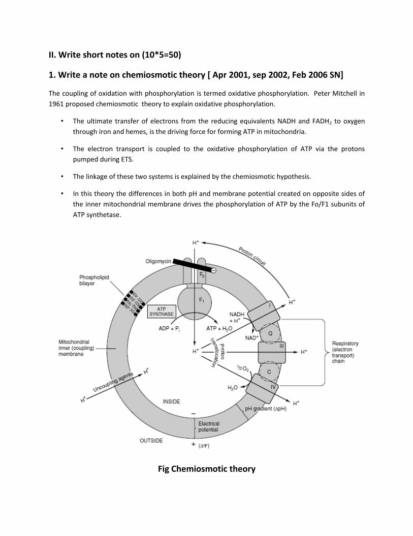
1. Write a note on chemiosmotic theory [ Apr 2001, sep 2002, Feb 2006 SN]
The coupling of oxidation with phosphorylation is termed oxidative phosphorylation. Peter Mitchell in
1961 proposed chemiosmotic theory to explain oxidative phosphorylation.
• The ultimate transfer of electrons from the reducing equivalents NADH and FADH2 to oxygen
through iron and hemes, is the driving force for forming ATP in mitochondria.
• The electron transport is coupled to the oxidative phosphorylation of ATP via the protons
pumped during ETS.
• The linkage of these two systems is explained by the chemiosmotic hypothesis.
• In this theory the differences in both pH and membrane potential created on opposite sides of
the inner mitochondrial membrane drives the phosphorylation of ATP by the Fo/F1 subunits of
ATP synthetase.
Fig Chemiosmotic theory
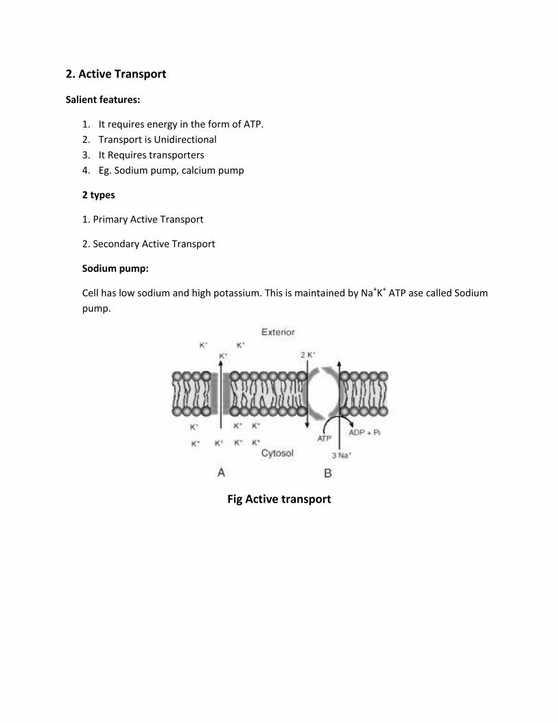
2. Active Transport
Salient features:
1. It requires energy in the form of ATP.
2. Transport is Unidirectional
3. It Requires transporters
4. Eg. Sodium pump, calcium pump
2 types
1. Primary Active Transport
2. Secondary Active Transport
Sodium pump:
Cell has low sodium and high potassium. This is maintained by Na+K+ ATP ase called Sodium
pump.
Fig Active transport

3. Uronic acid pathway
Importance:
It provides UDP glucuronic acid (active form of glucuronic acid)
UDP glucuronic acid is used for
Conjugation of bilirubin
Conjugation of steroids
Conjugation of various drugs which will make them more soluble and hence easily
excretable.
Glycosamino glycan Synthesis
Fig uronic acid pathway

4. Insulin:
Other name: Hypoglycemic hormone
The word insulin is derived from latin, insula means islet. It is hormone with 2 polpeptide
chains.
A chain carries 21 amino acids
B chain carries 30 amino acids. So, total 51 amino acids.
Inter chain disulphide bridge:
A7 – B7 A20 – B19 Intra chain disulphide bridge:
A6 – A11
Synthesis:
It is synthesized as prohormone “proinsulin “by beta cells of islet of langerhans of pancreas.
Actions of insulin:
Uptake of glucose by tissues
Stimulating glycolysis
Promotes glycogenesis by activating glycogen synthase.
Promotes fatty acid synthesis ( lipogenesis) and inhibit lipolysis in adipose tissue by
inhibiting hormone sensitive lipase
Gluconeogenesis is inhibited by insulin
It also inhibits glycogenolysis by inactivating glycogen phosphorylase.
It depresses HMG Co A synthase and so ketogenesis is inactivated.

5. Wald’s visual cycle:[ Feb 2005 SN]
Rhodopsin plays the pivotal role in vision. It is the membrane protein found in the
photo receptor cells of the retina.
Rhodopsin is made up of the protein opsin and 11 cis retinal.
When light falls on the retina, 11 cis retinal isomerizes to all trans retinal.
Wald’s Visual Cycle
Photo
receptor
Rhodopsin
All –trans retinal
Light
Opsin11 cis retinal
Retinal
epitheliumtrans – retinalCis- retinal
Retinal
isomerase
Blood
trans -retinal
trans – retinol
NADH
NAD
ADH
Cis retinol
Cis – retinalLiver
NADH
NAD
ADH
• A single photon can excite the rod cell. The photon produces immediate conformational
changes.
• The unstable intermediates produced are rhodopsin bathorhodopsin
lumirhodopsin metarhodopsin and finally opsin + all trans retinal.
• The all- trans retinal is then released from the protein and transported out of the retinal
epithelium by an ABC protein. The all- trans retinal is isomerised to 11-cis retinal in the
retina itself in the dark by the enzyme retinal isomerase.This reaction takes place in the
retinal pigment epithelium.
• The 11 cis –retinal combines with opsin to generate rhodopsin. Alternatively the all –
trans retinal is transported to liver and then reduced to all –trans retinol by alcohol
dehydrogenase(ADH ).
• The all-trans retinol is isomerised to 11cis- retinol and then oxidised to 11 cis –retinal in
liver. This is then transported to retina. This completes the Wald’s visual cycle.

6. Collagen
The major structural protein found in connective tissue is collagen.
It is a Greek word means the substance to produce glue.
About 25 -30 % of the total weight of protein in the body is collagen.
Synthesis:
It is synthesized by fibroblasts as procollagen (MW 360 KD). Then it is cleaved by specific
peptidases to form tropocollagen. Hydroxylation of proline and lysine residues of
collagen (post translational modification) is done and finally form collagen.
Functions:
Serves to hold together the cells in tissues
Major fibrous element of tissues like bone, teeth, tendons, cartilage and blood
vessels.
To support the organs
To provide alignment of cells, so that anchoring is possible.
Helps in proliferation and differentiation of cells.
In blood vessels, if collagen is exposed, platelets adhere and thrombus formation
is initiated.
Abnormalities in collagen:
1. Osteogenesis imperfect
2. Ehlers Danlos syndrome
3. Homocystinuria
4. Deficiency of Vit C
5. Lathyrism.
Fig. Structure of collagen

6. Glysaminoglycans
Glycosamino glycans are heteropolysaccharides, contains uronic acid & amino sugars.
Examples: Hyaluronic acid:
Anionic, non-sulfated glycosamino glycan.
Present in connective tissues, tendons, synovial fluid, etc. .
Composed of D-glucuronic acid & N-acetyl glucosamine, linked by β-1,4 & β-1,3 glycosidic bonds
Fig Hyaluronic acid
Heparin:
Highly sulfated glycosamino glycan.
An anticoagulant, present in liver.
Produced mainly by mast cells of liver.
STRUCTURE:
Fig Heparin
IMPORTANCE: Acts as anticoagulant.

7. Chromatography (nov 2001)
ANS: The term is derived from the Greek word chroma, means color. The method
was first employed by Tswett, a botanist in 1903, for the separation of plant
pigments using a column of alumina. Now a days HPLC is used to separate almost all
biological substances including proteins, carbohydrates, lipids and nucleic acids.
Types of chromatography
I) Adsorption chromatography
In this technique the separation is based on differences in adsorption at the
surface of a solid stationary medium. The common adsorbing substances are
alumina, silicates or silica gel. These are packed into columns and the
mixture of proteins to be separated is applied in a solvent on the top of the
column.
II) Partition chromatography
This includes different types depending on the phases between which the
components are partitioned, e.g. solid-liquid, liquid –liquid, gas-liquid etc.
Used for the separation of mixtures of amino acids and peptides. The
components of mixture to be separated are partitioned between the two
phases depending on the partition co-efficient (solubility) of the particular
substances.
i) Paper chromatography
The stationary phase is water held on a solid support of filter paper
(cellulose). The mobile phase-mixture of immiscible solvents like water,
non polar solvent and an acid or base. Chromatography can be done with
the mobile phase applied from top is (descending) or bottom
(descending).
ii) Thin layer chromatography
This is liquid-liquid chromatography; thin layer of silica gel is applied on
a glass plate sample applied as small spots
The plates placed in a trough solvent. It takes 3-4 hours
iii) Visualization chromatography

Some common location reagents are ninhydrin for aminoacids and
proteins, sulphuric acid for phospholipids.
iv) Importance of Rf value
Rf value is the ratio of distance travelled by substance distance travelled
by the solvent.
III) Ion exchange chromatography
The separation is based on electro static attraction between charged
biological molecules to oppositely charged groups on ion exchange resins.
IV) Gel filtration chromatography
It is also called molecular sieving.hydrophillic cross linked gels like
acrylamide, agarose, and dextran used for the separation of based on their
size
V) Affinity chromatography
It is based on the high affinity of specific proteins for specific chemical
groups. Co enzyme can be used to purify enzymes.
VI) HPLC
It is used for the separation of all compounds.

9. Levels of organization of proteins.
Proteins have different levels of structural organization
1. Primary
2. Secondary
3. Tertiary
4. Quaternary
Primary structure:
1. It denotes the number & sequence of amino acids in the protein.
2. Primary structure is maintained by peptide bond (amide bond or CONH bond)
3. eg insulin
Secondary structure:
1. It denotes the configurational relationship between residues which are about 3-4 amino
acids apart in linear structure.
2. Two types:
i) alpha helix eg. Alpha keratin
ii) beta pleated sheet: eg. Carbonic anhydrase
Tertiary structure:
1. It denotes the 3 dimensional structure of whole protein.
2. It defines the stearic relationship of amino acids which are far apart from each other
in linear structure, but are close in the 3 dimensional aspect.
3. Both secondary and tertiary and quaternary structure is maintained by hydrogen
bonds, electrostatic bonds, hydrophobic bonds and weak van der waals forces.
4. Eg. Myoglobin.
Quaternary structure:
1. Polypeptide aggregates to form quaternary structure
2. Eg. Haemoglobin.
10. Calcium Homeostasis.
Maintaining constant concentrations of calcium in blood requires frequent
adjustments.
3 organs participate in supplying calcium to blood and removing it from blood when
necessary
Three hormones regulates the Blood Calcium level
Vitamin D
PTH
Calcitonin
PARATHYROID HORMONE
PTH has 3 major sites of action -Intestine, Bone & Kidney
In kidney: Stimulates production of the biologically-active form of vitamin D
(calccitriol) in the kidney which increases the absorption of calcium.
In bone: Facilitates mobilization of calcium from bone by increasing the number of
Osteclasts & activating pyrophosphatase in osteoclasts.
In intestine: Maximizes tubular reabsorption of calcium within the kidney thus
results in minimal losses of calcium in urine.
VITAMIN D
Vitamin D (calcitriol) induces the synthesis of carrier protien (calbindin) in the
intestinal mucosa, which increases the absorption of calcium.
CALCITONIN
Calcitonin is a hormone that reduces the blood calcium levels by
Suppression of renal tubular reabsorption of calcium (ie) Calcitonin enhances
excretion of calcium into urine.
Inhibition of bone resorption, which would minimize fluxes of calcium from bone
into blood.

III. Short Answer Questions. (10*2=20)
1. Key enzymes of glycolysis.[March 2002 essay]
Glucokinase- It phosphorylates glucose to glucose 6 phosphate. It has a higher km
for glucose than hexokinase.
Phosphofructokinase (PFK) – It converts fructose 6 phosphate to fructose 1, 6
bisphosphate
Pyruvate kinase – It converts PEP to pyruvate.

2. Fatty liver:[sep 2002 SN]
Fatty liver refers to the deposition of excess TGL in the liver cells.
Causes of fatty liver:
I. Increased Mobilization of non esterified fatty acids from adipose tissue.
II. Increased lipolysis in adipose tissue in diabetes and starvation.
III. More synthesis of fatty acid from glucose.
IV. Decreased oxidation of fat by hepatic cells.
V. Toxic injury to liver due to poisoning by carbon tetra chloride, arsenic, lead
compounds
VI. Hepatitis B infection
VII. Obesity
VIII. Protein energy malnutrition causes reduced apoprotein synthesis and hence
fatty liver.
IX. Alcholism

3. Lipid peroxidation:
PUFA present in cell membrance are easily destroyed by peroxidation. Intiation phase:
o Primary event is the production of R’ (carbon centered radical) or ROO’ (lipid
peroxide radical)
o RH + OH’---------------------> R’ + H2O
o ROOH --------------------------> ROO’ + H+
Propagative phase:
o R’ reacts with oxygen forms peroxyl radical which attack another PUFA.
o R’ + O2 -------------------------------> ROO’
Termination phase:
o The above reaction proceeds until one peroxyl radical combines with another
peroxyl radical.
o ROO’ + ROO’--------------------------> ROOR + O2
4. Zymogens.
The inactive form of enzyme is known as zymogen. For eg, the inactive form of
chymotrypsin is chymotrypsinogen.

5. BMR (Basal Metabolic Rate):[Aug 2005 SN]
The energy required by a awaken individual during physical, emotional and digestive
rest.
It is the minimum amount of energy required to perform vital functions such as
circulation, respiration, working of heart etc.
Normal Value: Men - 34-37 k cal/m2/hr
Women - 30-35 k cal/m2/hr
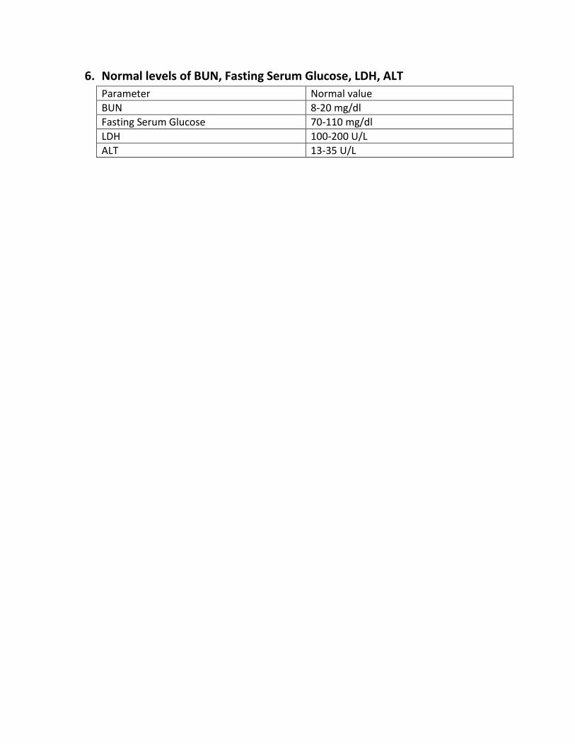
6. Normal levels of BUN, Fasting Serum Glucose, LDH, ALT
Parameter Normal value
BUN 8-20 mg/dl
Fasting Serum Glucose 70-110 mg/dl
LDH 100-200 U/L
ALT 13-35 U/L
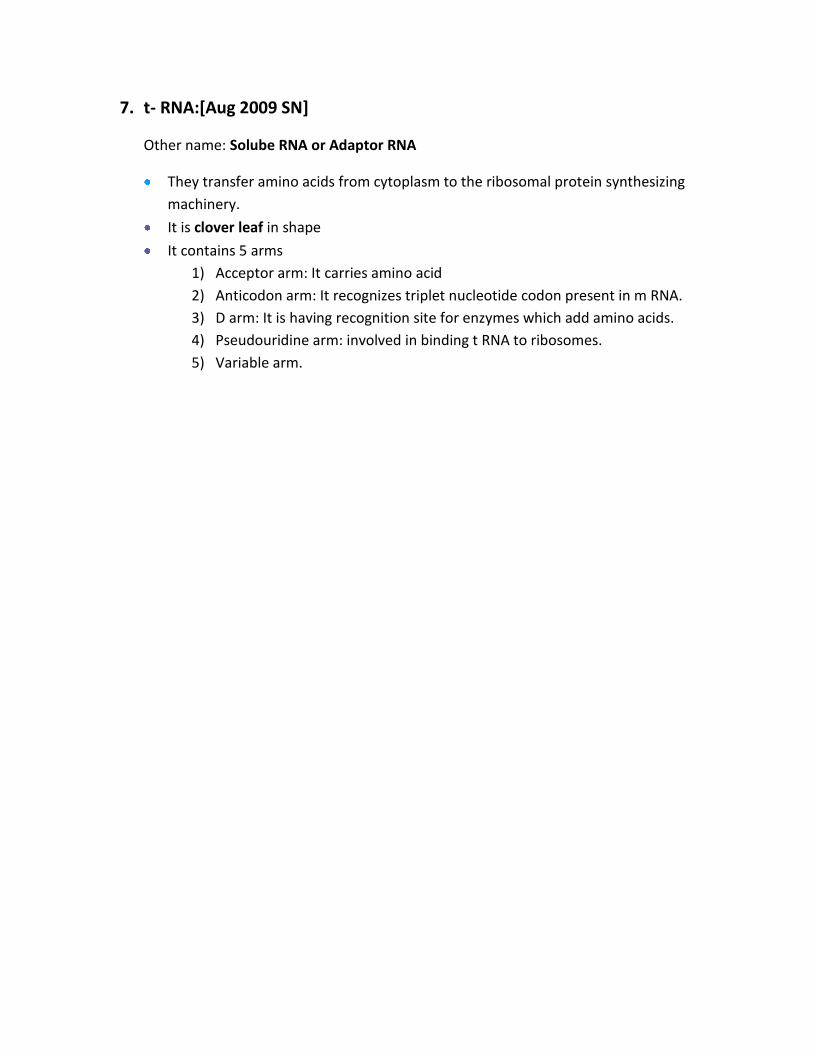
7. t- RNA:[Aug 2009 SN]
Other name: Solube RNA or Adaptor RNA
They transfer amino acids from cytoplasm to the ribosomal protein synthesizing
machinery.
It is clover leaf in shape
It contains 5 arms
1) Acceptor arm: It carries amino acid
2) Anticodon arm: It recognizes triplet nucleotide codon present in m RNA.
3) D arm: It is having recognition site for enzymes which add amino acids.
4) Pseudouridine arm: involved in binding t RNA to ribosomes.
5) Variable arm.
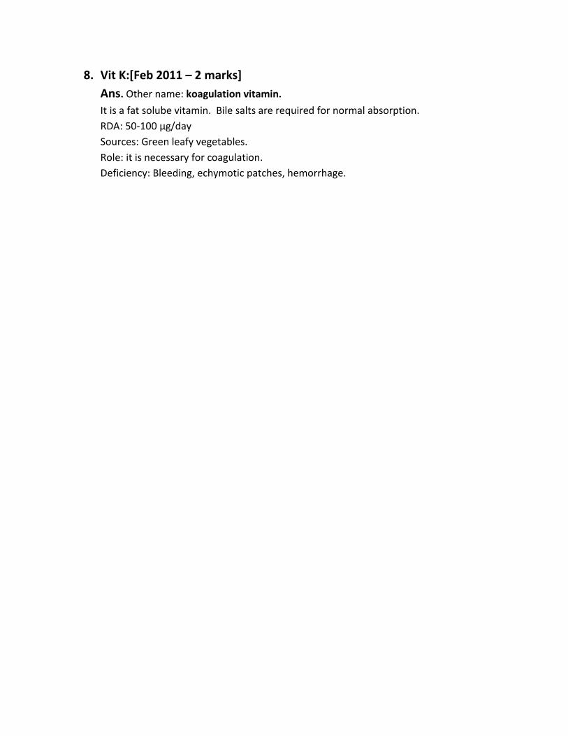
8. Vit K:[Feb 2011 – 2 marks]
Ans. Other name: koagulation vitamin.
It is a fat solube vitamin. Bile salts are required for normal absorption.
RDA: 50-100 µg/day
Sources: Green leafy vegetables.
Role: it is necessary for coagulation.
Deficiency: Bleeding, echymotic patches, hemorrhage.

9. Limiting amino acids:
Ans. Certain proteins are deficient in one or more essential amino acids. If this
particular protein is fed to a rat, it fails to grow. This amino acid is said to be the
limiting amino acids.
Protein Limiting amino acids Has to be supplemented
Rice Lys, thr Pulse proteins
Zein Tyr, lys Meat
Tapioca Phe, tyr Fish
Bengal gram Cys, met cereals
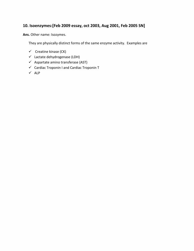
10. Isoenzymes:[Feb 2009 essay, oct 2003, Aug 2001, Feb 2005 SN]
Ans. Other name: Isozymes.
They are physically distinct forms of the same enzyme activity. Examples are
Creatine kinase (CK)
Lactate dehydrogenase (LDH)
Aspartate amino transferase (AST)
Cardiac Troponin I and Cardiac Troponin T
ALP