pakistan journal of plastic surgery · dr. naheed ahmed ch. dr. nasir uddin khan members ... arshi...
TRANSCRIPT
N OO FI T PLAI A
C STOS ICS SA U
N R
A GT ESI O
K N
A SP
1994
Official Publication of
Pakistan Association of Plastic Surgeons www. pjplasticsurgery.com
ISSN #: 2307-213X
PAKISTAN JOURNAL OF PLASTIC SURGERY Volume 2 Number 1
March 2013
House No. 557-B II, Model Town-B, Multan Road, Bahawalpur. Clinic: 062-2883456 Fax: 062-2884566 Mobile: 0300-9680481, 0321-6815009


Dr. Saleem Akhtar MalikDiplomate American Board (Plastic Surgery)
PATRON
CHIEF EDITORMohammad Mughese AminAssociate Professor Plastic Surgery Quaid-e-Azam Medical College Bahawalpur
EDITORDr. Shahab Ghani
PRESIDENT PAPS
Dr. Tahmeed Ullah
Dr. Mauzzam Nazeer Tarar
GENERAL SECRETARY
Dr. Saeed Ashraf CheemaDr. EhtishamDr. Zia-ul-Islam KahachiDr. Obaid UllahDr. Tahir SheikhDr. Naheed Ahmed Ch.Dr. Nasir Uddin Khan
MEMBERS
CONTENTS
INTERNATIONAL FACULTY
Dr. Riaz Malik
Dr. Muhammad Adil Abbas Khan(HULL U.K)
(HULL U.K)
EDITORIAL BOARD
PUBLISHERPakistan Journal of PlasticSurgery
PAKISTAN JOURNAL OF PLASTIC SURGERY ISSN #: 2307-213X Volume 2 Number 1 March 2013
N OO FI T PLAI A
C STO
S ICS SA U
N R
A GT ESI O
K N
A SP
1994
01
06
10
15
22
25
Mohan AKhan ASrinivasan KRoberts J
Gynaecomastia correction: A review of our experience
Head and Neck Squamous Cell Carcinoma a5-year Experience at a Tertiary Care Hospital in Bahawalpur
Coverage of open Tibial Fractures: what to Do? Prof. Moazzam Nazeer TararDr. Ata Ul Haq
M. Hamid ChaudharySamina ShabbirM Shams ul AlamEhsan UllahKhalid Usman
Study of Beliefs of Parents About The Etiology and Causes of Cleft Lip and Palate in Their ChildrenAdeel Haq Chowdry, Saad Aahmed, Noman Karim, Afshan Shaikh, Tooba Khalid, Rasheeqa Mehmood,Hafshah Siddiqui, Rabia Sana, Mariam Mustafa, Asma Mazhar, Sidra Zaheer, Syeda Aimen Anis, Asif Iqbal, Arshi Imran, Tehmina Badar,Muhammad Ashraf Ganatra
Sturge-Weber Syndrome – Case Series
Dr. Ghulam Ali MemonLiaquat University of Medical & Health Sciences, Jamshoro
INSTRUCTION TO AUTHORS 28
Composite Helix Chondrocutaneous Grafts for Reconstruction of the Soft Triangle
Obaidullah, M. AslamRehman Medical Institute, Peshawar
ASSISTANT EDITOR
Dr. Balal Afzal Sheikh

Introduction:Gynaecomastia is a common problem in the male population, particularly in young adults, with a
1reported prevalence of up to 36% . The term refers to a benign female-like enlargement of the male breast resulting from an increase in ductal tissue, stroma, and/or fat. Enlarged breasts can cause anxiety, self-consciousness and embarrassment, functional problems and psychosocial discomfort and fear of malignancy. It is not surprising therefore, that gynaecomastia is the most common cause for seeking medical advice for a breast condition in men. Surgical options for gynaecomastia include liposuction, open resection, and resection with skin reduction. Outcome studies of surgical correction have generally shown high levels of
2,3satisfaction . However, Ridha et al demonstrated only a 62.5% of patients within a cohort of 74
4patients were 'satisfied' or 'very satisfied' with their surgery . Surgery is, therefore, not a decision to be taken without careful patient assessment. Various techniques have been described over the years but no technique has yet gained universal acceptance. We aimed to review all gynaecomastia patients operated on under the care of one consultant in a regional unit over a 7-year period to assess the morbidity and complication rates associated with the procedure and to determine whether certain surgical techniques produced improved outcomes.
Key Words: Gynaecomastia, Liposuction, Open Resechion.
ORIGINAL ARTICLE
assess the morbidity and complication rates associated with the procedure.Clinical notes and outpatient records of all patients who underwent gynaecomastia correction at University Hospital North Staffordshire between 01/10/2001 to 01/10/2009 were retrospectively reviewed. A modified version of the Breast Evaluation Questionnaire was used to assess patients satisfaction with the procedure. Twenty-nine patients and a total of 53 breasts were operated on during the study period. Patients underwent either liposuction alone (6 breasts – 11.3%), excision alone (37 breasts – 69.8%) or both excision and liposuction (10 breasts – 18.9%). Twelve operated breasts (22.6%) experienced some form of complication. Minor complications included seroma (2 patients), superficial wound dehiscence (2 patients) and minor bleeding not requiring theatre (3 patients). Two patients developed haematomas requiring evacuation in theatre. No cases of wound
Arvind MohanPlastic Surgery Trainee; University Hospital North Staffordshire *as above ([email protected])Muhammad Adil Abbas KhanPlastic Surgery Registrar; Leeds Royal Infirmary*University Hospital North Staffordshire([email protected])Karthik SrinivasanPlastic Surgery Registrar; University Hospital Birmingham *University Hospital North Staffordshire ([email protected])Jeremy RobertsPlastic Surgery Consultant; University Hospital North Staffodshire *as above ([email protected]) *affiliation where work was carried out
9 Chadstone Close Monkspath Solihull United KingdomEmail: [email protected]: +447952247315
Mohan A, Khan A, Srinivasan K, Roberts J
01PAKISTAN JOURNAL OF PLASTIC SURGERY Volume 2 Number 1 March 2013
Gynaecomastia correction: A review of our experience
Gynaecomastia is a common problem in the male population with a reported prevalence of up to 36%. Various treatment techniques have been described but none have gained universal acceptance. We reviewed all gynaecomastia patients operated on under the care of one consultant over a 7-year period to

Mohan A, Khan A, Srinivasan K, Roberts JGynaecomastia correction: A review of our experience
infection, major wound dehiscence or revision surgery were encountered. Twenty-six patients (89.7%) returned the patient satisfaction questionnaire. Patients scored an average 4.12 with regards comfort of their chest in different settings, 3.98 with regards chest appearance in different settings, and 4.22 with regards satisfaction levels for themselves and their partner/family. Overall complication rate was 22.6%. Grade III patients experienced the highest complication rate (35.7%), followed by grade II (22.7%) and grade I (17.6%). Overall complication rates among the excision only group was the highest (29.8%) followed by the liposuction only group (16.7%) and the liposuction and excision group (10.0%). There were high satisfaction rates amongst both patients and surgeon. Eleven patients (37.9%) had their outcome classified as 'excellent' by the operating surgeon, 16 patients (55.2%) as 'good', 1 (3.4%) as 'satisfactory' and 1(3.4%) as 'poor'.Gynaecomastia is a complex condition which poses a significant challenge to the plastic surgeon. Despite the possible complications our case series demonstrates that outcomes of operative correction can be favourable and yield high levels of satisfaction from both patient and surgeon.
MethodsOperating procedure notes, clinical notes and outpatient records of all patients who underwent gynaecomastia correction at University Hospital North Staffordshire during the the period 01/10/2001 to 01/10/2009 were retrospectively reviewed. For the purpose of this study, we considered each operated breast as an individual case. The grade of gynaecomastia, the presence of skin excess, causative factors, duration of symptoms and surgical procedure were recorded. Short and long term minor and major complications, poor results and revision rates were recorded and analysed.
No val idated outcome assessment questionnaire exists specifically for gynaecomastia correction. We therefore created a three item questionnaire which was sent to all patients who underwent surgery to ascertain their satisfaction with the procedure. This was based on the more comprehensive 55 item Breast Evaluation
5Questionnaire which is a validated assessment questionnaire designed to assess patient satisfaction with breast and quality-of-life outcomes following a variety of breast surgery procedures. A similar proforma was
4used by Ridha et al . The proforma asked patients to rank their satisfaction levels with their surgery in relation to three factors. The first question related to patients' comfort with their breast/chest in different settings (intimate, social and professional). The second question related to the degree of comfort with their breast/chest appearance. The third question asked patients to rank the satisfaction level for themselves and their partner/family. Patients were asked to respond on a 5-point Likert scale (1=very dissatisfied; 2=dissatisfied; 3=neither; 4=satisfied; 5=very satisfied). Patients were classified as having either mild, moderate, or gross gynaecomastia as per
6Simon's classification and the presence of skin excess was noted. Twenty-six patients returned the questionnaire (89.6%).
ResultsTwenty-nine patients and a total of 53 breasts were operated on during the study period. Patients were referred from a variety of sources. General practitioners referred 24(82.8%) patients, 4(13.8%) were referred by the general surgical team and 1(3.4%) from the paediatric team. Twenty-eight patients cited emotional problems as the reason for them seeking help whereas one complained of pain and discomfort. The cohort characteristics, outcomes and morbidity are illustrated in Table 1.
02 PAKISTAN JOURNAL OF PLASTIC SURGERY Volume 2 Number 1 March 2013

The average time from the first outpatient clinic appointment to surgery was 25.3 weeks (range 5-156). Conservative management was attempted in 7 (24.1%) patients before they were listed for surgery. Patients underwent either liposuction alone (6 breasts – 11.3%), excision alone (37 breasts – 69.8%) or both excision and liposuction (10 breasts – 18.9%). All but 5(17.2%) patients had drains inserted which were removed 1-3 days post-operatively. Twenty-six patients (89.7%) wore some form of support garment post-operatively, with 18(62.1%) wearing an abdominal binder. Support garments were worn for an average of 4.6 weeks (range 3-6) following surgery. Twelve opera ted b reas t s (22 .6%) experienced some form of complication. Minor complications included seroma (2 patients), superficial wound dehiscence treated conservatively (2 patients) and minor bleeding not requiring theatre (3 patients).
The only acute major complication encountered were haematomas requiring evacuation in theatre (2 patients). There were no cases of wound infection or major wound dehiscence documented within our patient group. Although one patient was noted to have skin excess post-operatively that may have benefited from revision surgery, this was not possible due to hypertrophic scarring. Patients were followed up for an average of 6.0 months (range 1-11). One patient did not attend again after their first post-operative appointment. Twenty-six patients (89.7%) returned the patient satisfaction questionnaire. Patients scored an average 4.12 with regards comfort of their chest in different settings, 3.98 with regards chest appearance in different settings, and 4.22 with regards satisfaction levels for themselves and their partner/family. Table 1: Patient cohort characteristics, outcomes
and morbidity
I
II
III
17
22
14
Patient details
Age at surgery; mean (range)
Patient weight; mean (range)
Patient BMI; mean (range)
24.5 years (13-39)
82.7 kg (60-104)
27.1 (20-35.1)
Duration of symptoms; mean (range) 5.3 years (1-20)
Grade of gynaecomastia Operated breasts
Side involved
Left
Right
Bilateral
No. of patients
5
0
24
Mohan A, Khan A, Srinivasan K, Roberts JGynaecomastia correction: A review of our experience
03PAKISTAN JOURNAL OF PLASTIC SURGERY Volume 2 Number 1 March 2013

a (Seromas, bleeding not requiring theatre, superficial wound dehiscence) b (Haematomas, wound dehiscence requiring theatre, wound infection). All of these cases were taken back to theatre on the same admissionc Revision surgery not possible due to hypertrophic scarring
DiscussionAs discussed earlier, gynaecomastia has peaks in incidence within three age groups. Although the highest prevalence is among middle aged and older men (50-80 years old), the oldest patient in our cohort was 39 years old. This may relate to the fact that the most common trigger for surgery was emotional distress and middle aged/older men may be less affected by this stimulus compared to the younger age group. Overall complication rates for gynaecomastia surgery have been reported to be 15.5%, with
7the highest rate in grade I patients (21.6%) . Our overall complication rate was slightly
higher than this (22.6%). However, these were mainly minor acute complications that did not significantly affect the final result. There were no cases of nipple-areola complex necrosis or areolar tethering. Only two patients returned to theatre for evacuation of a haematoma. In our series, grade III patients experienced the highest complication rate (35.7%), followed by grade II (22.7%) and grade I (17.6%). Previous studies have quoted overall revision surgery rates as 17.4%, with the highest rate in grade
7II patients (34.8%) . None of the patients in our series underwent revision surgery although one may have benefitted from this but could undergo surgery due to hypertrophic scarring.Complication rates between different surgical techniques also varied significantly. Overall complication rates among the excision only group was the highest (29.8%) followed by the liposuction only group (16.7%) and the liposuction and excision group (10.0%).
Mohan A, Khan A, Srinivasan K, Roberts JGynaecomastia correction: A review of our experience
Operative time; mean (range)
76 minutes (30-180)
Operative technique
Liposuction only
Excision only
Liposuction & excision
Operated breasts
6
37
10
Weight of tissue removed; mean (range)
Hospital stay; mean (range)
155 grams (10-346)
1.6 days (1-4)
Binder use 18 patients
Morbidity
Minor complications
Acute major complications
Unsatisfactory result
7/53a
4/53b
1/53c
04 PAKISTAN JOURNAL OF PLASTIC SURGERY Volume 2 Number 1 March 2013

8Sophocles et al found that the weight of the specimen excised was not a significant predictor of minor or acute major complications. This is also confirmed by our series of patients. It is not possible to examine whether any factors contribute to a poor cosmetic result within our series as only one patient had an unsatisfactory result. Outcome studies of gynaecomastia correction have shown varying levels of satisfaction with the results of surgery with
2Fruhstorfer et al showing high levels of 4 satisfaction while Ridha et al showed much
lower levels. Our series demonstrated generally high satisfaction rates amongst both patients and surgeon. Eleven patients (37.9%) had their outcome classified as 'excellent' at their second follow up appointment by the operating surgeon, 16 patients (55.2%) as 'good', 1 (3.4%) as 'satisfactory' and 1(3.4%) as 'poor'. Patients too were generally 'satisfied' with their outcome with regards comfort and appearance.
ConclusionGynaecomastia is a complex condition which poses a significant challenge to the plastic surgeon. The initial treatment should aim to correct any underlying abnormality or discontinuing any medications that may be contributing to the condition. Although the efficacy of medical treatment has not yet been well established, conservative measures should be considered prior to surgery.Gynaecomastia present for more than 2 years is unlikely to regress spontaneously or with medical treatment due to the tissue becoming
3irreversibly fibrotic . In these cases surgery remains the mainstay of treatment. Despite many operative techniques being described, the principal aims of surgery remain to correct the deformity, restore normal body contour and image, maintain the viability of the nipple-areola complex and avoid
8excessive scarring . Although there are significant possible complications associated with surgery, our case series demonstrates that with careful
planning and shrewd patient selection, outcomes of operative correction can be favourable and yield high levels of satisfaction from both patient and surgeon.
Conflict of interest statementWe declare there is no financial or personal relationship with other people or organisations that could inappropriately influence this work.
References1. Nuttal FQ. Gynaecomastia as a physical
finding in normal men.Journal of Clinical Endocrinology Metabolism 1979;48:338-40.
2. Fruhstorfer BH, Malata CM. A systematic
approach to the surgical treatment of
gynaecomastia. British Journal of Plastic
Surgery 2003;56:237-46.
3. Wiesman IM, Lehman Jr JA, Parker MG, et
al. Gynaecomastia outcome analysis.Annals of Plastic Surgery 2004;53:97-101.
4. Ridha H, Colville RJI, Vesely MJJ. How
happy are patients with their gynaecomastia
reduction surgery? Journal of Plastic
Reconstructive and Aesthetic Surgery
2009;62:1473-78.
5. Anderson RC, Cunningham B, Tafesse E, et
al. Validation of the breast evaluation
questionnaire for use with breast surgery
patients. Plastic Reconstructive Surgery
2006;118:597-602.
6. Simon BE, Hoffman S, Kahn S.
Classification and surgical correction of
gynaecomastia.Plastic Reconstructive Surgery 1973;51(1):48-52
7. Colombo-Benkmann M, Buse B, Stern J,
Herfarth C. Indications for and results of
surgical therapy for male gynaecomastia.
American Journal of Surgery
1999;178(1):60-3.
8. Sophocles L, Starren E, Read J et al.
Surgical management of gynaecomastia:
outcomes from our experience. The Breast
2008(17);596-603
Mohan A, Khan A, Srinivasan K, Roberts JGynaecomastia correction: A review of our experience
05PAKISTAN JOURNAL OF PLASTIC SURGERY Volume 2 Number 1 March 2013

Introduction:Standards for management of open tibial fractures have been set and practiced around the globe1. Our trauma burden is one of the highest in the world. According to W.H.O report on road safety published in 2013 there are 30,131 deaths per year due to RTA in Pakistan2 and for every death there are 20 people injured. It means approximately 602,640 injuries per year due to road traffic accidents only. We have an array of other accidents like collapsed buildings, electrocution, machine injuries, agricultural injuries and last but not the least blasts due to the wave of terrorism in addition to RTA, that makes the number more formidable. Although no reliable national figures are available but we can get some estimate from Rescue 1122(rescue service of Punjab) data3. They transferred 75,265 injured persons per year to different hospitals.
Key Words: Tibial Fractures, Fasciocutaneous, Sural art hap.
ORIGINAL ARTICLE
surgery needed for bony problems. Also donor site is usually cosmetically unsightly. Distal necrosis rate is approximately15- 20% in local flaps4,5,6 that is unacceptably high compared to the free flap surgery. These figures reflect that flap is compromised at the end where it is most needed hence should equate the total flap failure. Another argument against the local flaps is that if patient ends up with below knee amputation due to any reason then stump formation will be compromised. This is particularly true in case of distal 3rd fractures. Local Fasciocutaneous flaps used to cover distal 3rd of tibia borrow the skin which is normally used for stump closure in case of Below Knee Amputation BKA. This means using skin graft or another flap (usually free flap) for stump coverage Free flaps proponents claim free flap success rate of more than 95%7,8, a clear winner over local flaps. It imports tissue to the injured leg thus not compromising already injured leg. Donor site is usually distant and hidden so cosmetic result is usually acceptable. In our special circumstances of massive work load versus minimum manpower and resources, we should keep our options open. Local flaps should be used for coverage of small wounds and that too of low energy type.
Prof. Moazzam Nazeer TararDr. Ata Ul Haq Correspondence: Dr Ata Ul Haq Senior Registrar Plastic Surgery Department, Jinnah Hospital Lahore. Email: [email protected].
Being a plastic surgeon of a society with massive trauma burden, every one of us is faced with patients requiring soft tissue coverage of open tibial fracture. Despite being such a common problem, still there is no consensus regarding treatment options. There are certain management issues that are settled i.e. timing of surgery but there are two major queries regarding flap selection for tibial coverage. 1. Whether to use a local or free flap? 2 . W h i c h o n e i s m o r e s u i t a b l e , fasciocutaneous or muscle flap?
We have tried to find answers for these questions keeping in mind our circumstances. In literature both local and free flaps have their advocates. Arguments in favor of local flaps include: shorter operating time, technical ease and usually replacement of like with like. The arguments against the use of local flaps include: zone of trauma extending into the tissue to be used, hence not very reliable, usually compromise the secondary
Coverage of open Tibial Fractures: what to Do?
Prof. Moazzam Nazeer Tarar, Dr. Ata Ul Haq
06 PAKISTAN JOURNAL OF PLASTIC SURGERY Volume 2 Number 1 March 2013
For large and high energy trauma we should go for free flaps because local tissue is usually injured and not suitable for fracture coverage. Our dictum is when in doubt go for free flap. For difficult defects, free flap can be an easy option. Now we come to the second question of muscle vs. fasciocutaneous flaps. Generally it was agreed that muscle flaps contribute greater vascularity than fasciocutaneous counterparts9-13. Based on this fact muscle flaps were considered a better option for wound healing (bone union) and control of infection14. This concept has been challenged in recent years. 15 A recent study by James K.-K. Chan, Jagdeep Nanchahal and Lorraine Harry stats that fasciocutaneous flaps have even better blood supply per unit area than muscle flaps once transferred. But Muscle provides a bone anabol ic environment through the expression of members of the transforming growth factor -β super family of growth and differentiation factors, including the bone morphogenetic proteins. This results in better bone healing16. Another factor to be kept in mind while deciding the flap for tibial coverage is the dead space. We feel that if there is dead space it should be obliterated and muscle flap is the b e t t e r c h o i c e i n t h i s s i t u a t i o n . Fasciocutaneous flap is usually tented over the dead space not obliterating it. This leads to the accumulation of exudate under the flap which ends up in infection in most of the cases. As we have already mentioned that revisional surgery is relatively easier in case of fasciocutaneous flap than the muscle flaps. Also muscle flaps have high metabolic rate than fasciocutaneous ones and thus need more blood supply for survival than their counterparts. It means that fasciocutaneous flaps tolerate ischemia better than muscle flaps resulting in higher survival rate of fasciocutaneous flaps than muscle flaps. So fasciocutaneous flap is preferred choice if it
can serve the purpose. We tend to ignore cosmesis and take it as non issue in lower limb but this is an issue and once healing is achieved, patients do complain about appearance of the limb. There is consensus that fasciocutaneous flaps have better cosmetic outcome compared to muscle flaps. However local fasciocutaneous flaps do create unsightly donor sites. Usually there are dog ears and persistent edema resulting in poor cosmesis. When we consider our circumstances following are the facts. Most of the plastic surgery units in the country have skills to perform microsurgical procedures so facilities are not an excuse any more. We believe in this time and age every trained plastic surgeon should be capable of microsurgery. Workload is main problem in our set up. It's not always possible to do free flaps for every case. Sometimes despite all your intentions you find it hard to perform microsurgery because of heavy workload and you go for alternative options. This should be decided on merit because a failed local flap done inappropriately will land the patient in more trouble. However Local flaps cannot be condemned. They have their place and require more technical skill and knowledge to achieve good results. Also Adhoc perforator flaps are coming forward and sometimes a local perforator flap gives as good reconstruction as free flap. Most of the patients present late with infection already there so eradication of infection is almost always an issue in our set up. Our recommendations are In case of small defects due to low energy trauma, local flaps are the preferred choice. Large defects and high energy trauma wounds usually need free tissue transfer. If there is no dead space and fracture has no adverse features then fasciocutaneous flaps are the preferred choice.
COVERAGE OF OPEN TIBIAL FRACTURES: WHAT TO DO? Prof. Moazzam Nazeer Tarar, Dr. Ata Ul Haq
07PAKISTAN JOURNAL OF PLASTIC SURGERY Volume 2 Number 1 March 2013

In case of dead space or the fracture itself has difficult configuration, muscle flaps should be considered as the first choice.
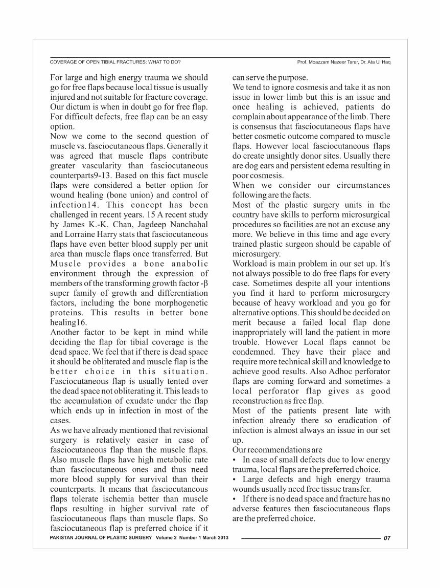
Here are few representative cases: FIG. 1 (A through C) Coverage of exposed ankle joint with Pedicled fasciocutaneous flap (reverse flow sural artery flap).
FIGURE 2 ( A Through C) : Coverage of open tibial fracture with free Anterolateral Thigh Flap.
FIG. 2 (A)
FIG. 2 (B)
FIG. 2 (C)
FIG. 1(A)
FIG. 1(B)
FIG. 1(C)
COVERAGE OF OPEN TIBIAL FRACTURES: WHAT TO DO? Prof. Moazzam Nazeer Tarar, Dr. Ata Ul Haq
08 PAKISTAN JOURNAL OF PLASTIC SURGERY Volume 2 Number 1 March 2013

FIG. 3 (A through C): Coverage of open tibial fracture of lower 1/3rd with distally based hemisoleus muscle flap.
References 1. � Nanchahal J, Nayagam S, Khan U, Moran C, Barrett S,
Sanderson F et al. Standards of management of Open Fractures of Lower Limb: A short guide. London: BAPRAS and BOA; 2009
2. � World Health Organization. Global status report on road safety 2013, supporting a decade of action.luxembourg 2013. 318p.
3. � Rescue 1122 (Pakistan). Consolidated Report of Emergency Calls and Rescue Operations in Punjab. 2013 Jun.
4. � Ajmal S, Khan MA, Khan RA, , Shadman M, Yousaf K, Iqbal T. Distally based sural fasciocutaneous flap for soft tissue reconstruction of distal leg, ankle and foot defects. J Ayub Med Coll. 2009; 21(4)
5. � Hallock GG. Complications of 100 consecutive local fasciocutaneous flaps. Plast Reconstr Surg. 1991;88:264–268.
6. � Gir P, Cheng A, Oni G, Mojallal A, Saint-Cyr M. Pedicled-perforator (propeller) flaps in lower extremity defects: a systematic review. J Reconstr Microsurg. 2012 Nov; 28(9):595-601.
7. � Ducic I, Brown BJ, Rao SS. Lower extremity free flap reconstruction outcomes using venous coupler. Microsurgery. 2011 Jul; 31(5): 360-64.
8. � Basheer MH, Wilson SM, Lewis H, Herbert K. Microvascular free tissue transfer in reconstruction of the lower limb. J Plast Reconstr Aesthet Surg. 2008;61:525–28.
9. � Gothman L. Local arterial changes associated with diastasis in experimental fractures of the rabbit's tibia t r e a t e d w i t h i n t r a m e d u l l a r y n a i l i n g : A microangiographic study. Acta ChirScand. 1962;123:104–110.
10. �Trueta J, Buhr AJ. The vascular contribution to osteogenesis:V. The vasculature supplying the epiphysial cartilage in rachitic rats. J Bone Joint Surg Br.1963;45:572–581.
11. �Rhinelander FW. The normal microcirculation of diaphyseal cortex and its response to fracture. J Bone Joint Surg Am.1968;50:784–800.
12.� Holden CE. The role of blood supply to soft tissue in the healing of diaphyseal fractures: An experimental study. J Bone Joint Surg Am. 1972;54:993–1000.
13. �Whiteside LA, Ogata K, Lesker P, Reynolds FC. The acute effects of periosteal stripping and medullary reaming on regional bone blood flow. Clin Orthop Relat Res. 1978;131: 266–272.
14. �Anthony JP, Mathes SJ, Alpert BS. The muscle flap in the treatment of chronic lower extremity osteomyelitis: Results in patients over 5 years after treatment. Plast. Reconstr. Surg. 1991 Aug; 88(2): 311-18.
15. �Harry LE, Sandison A, Pearse MF, Paleolog EM, Nanchahal J. Comparison of the vascularity of fasciocutaneous tissue and muscle for coverage of open tibial fractures. Plast Reconstr Surg. 2009;124:1211–1219.
16. �Chan JKK, Harry L, Williams G, Nanchahal J. Soft-Tissue Reconstruction of Open Fractures of the Lower Limb: Muscle versus Fasciocutaneous Flaps. Plast Reconstr Surg. 2012;130(2):284e–295e.
FIG. 3(A)
FIG. 3(B)
FIG. 3(C)
COVERAGE OF OPEN TIBIAL FRACTURES: WHAT TO DO? Prof. Moazzam Nazeer Tarar, Dr. Ata Ul Haq
09PAKISTAN JOURNAL OF PLASTIC SURGERY Volume 2 Number 1 March 2013

Background: Squamous cell carcinoma is the commonest head and neck malignancy which accounts for approximately 20% of the cancer burden in Asian countries. Major risk factors include tobacco smoking thus a rendering it a preventable disease. Frequencies and incidence rates of site-specific head and neck squmaous cell carcinoma have been reported regularly in different studies from various parts of the country. Current study aims at contributing the similar data from Bahawalpur region. Methods: It was a descriptive study including 184 biopsy proven cases of squamous cell carcinoma from head and neck region reported by Department of Pathology, Quaid-e-Azam Medical College Bahawalpur during January 2008 and December 2012. Data was acquired from hospital and lab records and analysed using SPSS version 18. Results: Mean age of the patients was 55.76±7.21 (median: 50) years. Male to female ratio was almost 2:1. History of smoking was positive in 72.28% of patients. The most common affected sites in order of frequency were larynx (n=79, 42.93%), hypopharnx (n=36, 19.56%), oral cavity (n=29, 15.76%), face/skin (n=24, 13.04%), lips (n=10, 5.43%) and tongue (n=6, 3.26%). More than a half of the tumours were histologically classified as well-differentiated squamous cell carcinomas (n=94, 51.09%).
thConclusions: Head and neck squamous cell carcinoma has a peak age in 6 decade of life and twice common in men as compared to women. Most frequent site of HNSCC in Bahawalpur region is larynx.
Key Words: Head and neck, cancer, squamous cell carcinoma, larynx, pharynx, oral cavity, Bahawalpur
ORIGINAL ARTICLE
burden in Asian countries. Major risk factors include tobacco smoking thus a rendering it a preventable disease. Head and neck cancers, ICD-10 (International Classification of Diseases 10th Revision) categories C00-C14 (cancer of the lip, oral cavity and pharynx) and C32 (larynx) are categorized amongst the top ten malignancies globally. Head and neck cancers are grouped together with the justification of similar natural history, epidemiology, risk factors, morphology, and
3control measures. HNC comprise of soft tissue neoplasms of oral cavity including lips, nasal cavity and paranasal sinuses (PNS),
4pharynx, larynx and salivary glands. More than 5% of all malignant tumors worldwide are head and neck cancer, with more than 100,000 cases diagnosed in Europe
5each year.
Authors: M. Hamid Chaudhary , Samina Shabbir, M. Shams ul Alam , Ehsan Ullah , Khalid UsmanAuthors Affiliations:
th4 Year MBBS students, Quaid-e-Azam Medical College BahawalpurDepartment of Pathology, Quaid-e-Azam Medical College BahawalpurFor Correspondence: Dr. Ehsan UllahDepartment of Pathology, Quaid-e-Azam Medical College BahawalpurEmail: [email protected], Phone: +92-333-6390765
Introduction:Head and neck cancer is a major health problem worldwide. It is a major global health issue, with about half a million new cases diagnosed per year, and their incidence appears to be increasing in developing countries.,2 Squamous cell carcinoma is the commonest head and neck malignancy which accounts for approximately 20% of the cancer
Head and Neck Squamous Cell Carcinomaa5-year Experience at a Tertiary Care Hospital in Bahawalpur
M. Hamid Chaudhary, Samina Shabbir, M Shams ul Alam, Ehsan Ullah, Khalid Usman
10 PAKISTAN JOURNAL OF PLASTIC SURGERY Volume 2 Number 1 March 2013

The risk increases in proportion to the intensity and duration of the exposure to each carcinogen. Yet, individual susceptibilities to these risk factors vary within the general population. The basis for this susceptibility may be inborn or acquired, which is still under investigation. Head and neck cancers are mainly seen in the low socioeconomic strata.()
Many factors that are implicated for its causation are consumption of tobacco in its various forms, alcohol, smoking habits, lack
6of awareness, and lack of proper nutrition.
The etiology of head and neck squamous cell cancer is multifactorial with alcohol and tobacco consumption considered to be the main risk factor. Other factors include irradiation, oncogenic virus infections like Epstein Barr virus (EBV) and Human Papilloma Virus (HPV). EBV is commonly associated with nasopharyngeal carcinoma and HPV has been associated with laryngeal
3,7carcinoma.
The incidence of head and neck cancer increases with age, especially after 50 years of age. Although most patients are between 50 and 70 years old, head and neck cancer does occur in younger patients. There are more women and fewer smokers in the younger
8 patient group. Mean age of presentation lies in the 5th-6th decade for the Asian population as compared to 7th-8th decade among the North American population. Incidence of oral cancer in South-East Asia and of oral cavity plus nasopharyngeal cancer in East Asia
7follows the global HNC pattern.
In the Western world, squamous cell carcinoma of the head and neck (HNSCC) accounts for more than 90% of all head and neck cancers. The 5-y survival rate of less than 30% is due to a high lymphogenic metastatic tendency, a high recurrence rate, and an increased occurrence of
5secondary tumors.
The commonest epithelium covering the head
and neck mucosal surfaces is squamous epithelium and this may explains the
9domination of squamous cell carcinomas.
SCC has a focal invasion and its behavior depends on the region that it originates. Each anatomic area has its own growth patterns and prognosis .Head and neck squamous cell carcinoma (HNSCC) has been had a challenging treatment for a long time because of the high rates of recurrence and its
10 advanced disease at the time of diagnosis.Frequencies and incidence rates of site-specific head and neck squmaous cell carcinoma have been reported regularly in different studies from various parts of the country. Current study aims at contributing the similar data from Bahawalpur region.
Methods:It was a retrospective cross sectional descriptive study. All 184 biopsy proven cases of squamous cell carcinoma from head and neck region reported by Histopathology Section of Department of Pathology, Quaid-e-Azam Medical College Bahawalpur in a period of 5-years i.e. from January 2008 to December 2012 were included in the study. Data about age, gender, exact site of tumour, history of tobacco smoking, histological diagnosis and tumour grade was acquired from hospital and lab records. Data was entered and analysed using SPSS version 18. Means were calculated for quantitative and percentages were drawn for qualitative variables.
Results:A total of 184 biopsy proven cases of head and neck squamous cell carcinoma were reported at Department of Pathology, Quaid-e-Azam Medical College from January 2008 to December 2012. Mean age of the patients was 55.76±7.21 years ranging from 23 to 85 years. Median age was 50 years. Male to female ratio was almost 2: 1 (126 males: 58 females). Females were affected at little younger age
Head and Neck Squamous Cell Carcinoma M. Hamid Chaudhary, Samina Shabbir, M Shams ul Alam, Ehsan Ullah, Khalid Usman
11PAKISTAN JOURNAL OF PLASTIC SURGERY Volume 2 Number 1 March 2013

53.15±6.90 years as compared to their male counterparts (58.33±7.85 years). Majority of patients were village dwellers (n=141, 76.63%) as shown in Figure 1. History of current active tobacco smoking was positive in a large proportion (n=133, 72.28%) of patients as shown in Figure 2. The most common affected sites in order of frequency were larynx (n=79, 42.93%), hypopharnx (n=36, 19.56%), oral cavity (n=29, 15.76%), face/skin (n=24, 13.04%), lips (n=10, 5.43%) and tongue (n=6, 3.26%). Just more than a half of the tumours were h i s to log ica l ly c l a s s i f i ed as we l l -differentiated squamous cell carcinomas (n=94, 51.09%) on the basis of presence of keratin, intercellular bridges and low mitotic activity while other tumours were graded as moderately and poorly differentiated as shown in Table 1.
Discussion:Head and neck malignancies are common in several regions of the world where tobacco use and alcohol consumption is high. The highest rate of oral cancer is found in the developing world where oral cancer with pharynx combined is the fourth commonest
1site of cancer. In the Western world, squamous cell carcinoma of the head and neck (HNSCC) accounts for more than 90%
11of all head and neck cancers. Pakistan falls into a high risk head and neck cancer geographical zone, presentation is late and
3treatment is not optimum. Lack of national tumor registry in our country is the main reason for lack of accurate statistical data
12about prevalence and incidence of cancer.However, regional and institution-based registry systems from different centers are providing scattered but useful information regarding the prevalence of cancer. Centers like AFIP Rawalpindi, IRNUM Peshawar, PMC Faisalabad, QMC Bahawalpur, D.I. Khan, KEMC Lahore, JPMC Karachi and the well-organized Karachi tumor registry are
12serving the purpose to a great extent.
Present study focuses on cases of head and neck squamous cell carcinomas in one of the largest tertiary care hospitals in Southern-Punjab.Majority of the patients suffering from head and neck squamous cell carcinoma belonged to rural areas (>75%).a significant number of patients (>70%) had a known active history of tobacco smoking. Our results were consistent with similar findings all over the world. A research conducted by Basu et al. in India suggested that a tobacco habit was significantly correlated with the incidence of HNSCC and persons with current addiction had a 2.17 fold increased risk of cancer
1 3development. A case-control study conducted in Germany showed that out of 200 patients suffering from HNSCC, 95.5% were
14having a smoking history.
Mean age:In a research conducted by Abdul-Hamid and colleagues mean age of presentation was found to be 51.3 ± 14.9 years (range: 3 – 82 years). And there was significant difference between the mean age of male (49.5 ± 15.1 years) and female (45.4 ±16.3 years) patients with head and neck cancers. Abuidris and colleagues conducted a study of 314 head and neck cancer cases in Sudan and found that mean age of presentation was 48.79 and
9median age of 50 years. In our study mean age of the patients was 55.76±7.21 years ranging from 23 to 85 years. Median age was 50 years. Another study from Lahore, Pakistan also revealed similar peak age for
12head and neck malignancy.
Male to female ratio:The male to female ratio of head and neck
7cancer in Nairobi was found to be 2:1. In a study conducted in India the ratio was found
6to be 2.9:1. Our study also suggests a similar male to female ratio of almost 2:1. Discussion about site-wise frequency of head and neck squamous cell carcinoma is as follows;
Head and Neck Squamous Cell Carcinoma M. Hamid Chaudhary, Samina Shabbir, M Shams ul Alam, Ehsan Ullah, Khalid Usman
12 PAKISTAN JOURNAL OF PLASTIC SURGERY Volume 2 Number 1 March 2013

Larynx:Our research showed that the most common affected site in order of frequency was larynx (42.3%). In a study of 89 cases of head and neck cancers in a tertiary care hospital in Nigeria found that larynx cancer constituted
154.5% of cancers. However, Aziz et al. from within the country showed larynx as the most frequent site of head and neck cancer, which was hstologically squamous cell carcinoma, a
12 finding consistent with our results.
HypopharynxIn our research second most common site was hypophayrnx (19.56%). Siddiqui and colleagues found that in Indian state of Bihar hypopharynx carcinoma was the third most common cancer predominately of squamous
4cell carcinoma type. A study conducted in Nepal found that 10.3% of cases were
15identified as hypophayrnx SCC. Our study shows significantly higher incidence of hypophyranx SCC it may be due to the reason that the sample size of research conducted by other colleagues is considerably larger.
Oral cavity & tongue: In our study frequency of oral cavity tumors were the third most common site (15.76%) and tongue as a site of SCC had only 3.26 % frequency. These results are comparable to an Indian research conducted by Bhattacharjee and colleagues in which 32.67% out of TBM were in tounge region and most of them were of SCC type. Other researches also show oral cavity cancers to be a common site of head
. (16-20)and neck squamous cell carcinoma Another large series of 5865 patients from
thGulf region showed that oral cancer was 8 commonest and carcinoma arising in pharynx
thwas 7 commonest malignancy and both tumours were relatively preponderant in males with gender ratios of 1.65:1 and 2:1
21respectively. Thus, our findings regarding head and neck squamous cell carcinoma in general and site-wise frequencies of squamous cell carcinoma along with its peak
age and gender distribution are well incoherence with the published reports from within the country and other Asian countries.
Conclusions:Head and neck squamous cell carcinoma has
tha peak age in 6 decade of life and twice common in men as compared to women. Most frequent site of HNSCC in Bahawalpur region is larynx.
References:1. Abdul-Hamid G, Saeed NM, Al-Kahiry W,
Shukry S. Pattern of head and neck cancer in Yemen. Gulf J Oncolog 2010;1(7):21-24.
2. Johnson NW, Warnakulasuriya S, Gupta PC, Dimba E, Cihindia M, Otoh EC, et al. Global oral health inequalities in incidence and outcomes for oral cancer causes and solutions. Adv Dent Res 2011;23:237-246.
3. Bhurgri Y, Bhurgri A, Usman A, Pervez S, Kayani N, Bashir I, et al. Epidemiological review of head a n d n e c k c a n c e r s i n K a r a c h i . AsianPacJCancerPrev 2006;7:195-200.
4. Siddiqui MS, Chandra R, Aziz A, Suman S. Epidemiology and histopathological spectrum of head and neck cancers in Bihar, a state of Eastern India. AsianPacJCancerPrev 2012;13:3949-3953.
5. Nothelfer EM, Zitzmann-Kolbe S, Garcia-Boy R, Krämer S, Herold-Mende C, Altmann A, et al. Identification and Characterization of a Peptide with Affinity to Head and Neck Cancer. J Nucl Med 2009;50:426-434.
6. Bhattacharjee A, Chakraborty A, Purkaystha P. Prevalence of head and neck cancers in the north east-An institutional study. IndianJOtolaryngol HeadNeckSurg 2006;58:15-19.
7. Gathere S MG, Korir A, Musibi A. Head and Neck Cancers four year trend at the Nairobi Cancer Registry. Afr J Health Sci 2011;19:30-35.
8. Ridge JA, Glisson BS, Horwitz, EM, Meyers MO. Head and neck tumors. In: Haller DG, Wagman LD, Camphausen KA, Hoskins WJ (ed). Cancer Management: A Multidisciplinary Approach. Medical, Surgical, and Radiation Oncology. Online Edition, Updated 2013. Available from URL:
9. Abuidris DO EA, Eltayeb EA, Elgayli EM and El Mustafa OM. Pattern of Head and neck malignancies in Central Sudan-(study of 314 cases) Sudan J Med Sci 2008;3:105-109.
Head and Neck Squamous Cell Carcinoma M. Hamid Chaudhary, Samina Shabbir, M Shams ul Alam, Ehsan Ullah, Khalid Usman
13PAKISTAN JOURNAL OF PLASTIC SURGERY Volume 2 Number 1 March 2013

10. Thomas GR, Nadiminti H, Regalado J. Molecular predictors of clinical outcome in patients with head and neck squamous cell carcinoma. Int J Exp Pathol 2005; 86: 347-63.11. Reuter CW, Morgan MA, Eckardt A. Targeting
EGF-receptor-signalling in squamous cell carcinomas of the head and neck. Br J Cancer. 2007;96:408-416.
12. Aziz F, Ahmed S, Malik A, Afsar A, Yusuf NW. Malignant tumors of head and neck region- A retrospective analysis. J Coll Phy Surg Pak 2001;11:287-290.
13. Basu R, Mandal S, Ghosh A, Poddar TK. Role of tobacco in the development of head and neck squamous cell carcinoma in an eastern Indian population. AsianPacJCancerPrev 2008;9:381-386.
14. Maier H, Dietz A, Gewelke U, Heller WD, Weidauer H. Tobacco and alcohol and the risk of head and neck cancer. Clin Investig 1992;70:320-327.
15. Ologe FE, Adeniji KA, Segun-Busari S. Clinicopathological study of head and neck cancers in Ilorin, Nigeria. Trop Doct 2005;35(1):2-4.
16. Lasrado S, Prabhu P, Kakria A, Kanchan T, Pant S, Sathian B, et al. Clinicopathological profile of head and neck cancers in the Western development region, Nepal: a 4-year snapshot. AsianPacJCancerPrev 2012;13:6059-6062.
17. da Lilly-Tariah OB, Somefun AO, Adeyemo WL. Current evidence on the burden of head and neck cancers in Nigeria. Head Neck Oncol 2009; 1:1-8.
18. J Kalavathy Elango, P Gangadharan, S Sumithra, M A Kuriakose, Trends of Head and Neck Cancers in Urban and Rural India. Asian Pac J Cancer Prev 2006;7:108-112
19. Petti S. Life style risk factors for oral cancers. Oral Oncol 2009; 45:340-350
20. Vora AR, Yeoman CM, Hayter JP. Alcohol tobacco and pan use and understanding of oral cancer risk among Asian mails in Leicester. Br Dent J, 2000;188:444-451
21. Halboub, ES, Al-Anazi YM, Al-Mohaya MA. Characterization of Yemeni patients treated for oral and pharyngeal cancers in Saudi Arabia. Saudi Med J 2011;32:1177-1182.
Figure 1. Rural-Urban Distribution of Patients of HNSCC
Figure 2. Smoking Status of Patients of HNSCC
Table 1. Histological Grades of HNSCC
Histological Grade
Grade-1 Grade-2 Grade-3 Grade-4
Total
Number of Cases
94 37 49 04
184
Percentage
51.1% 20.1% 26.6% 2.2% 100%
Head and Neck Squamous Cell Carcinoma M. Hamid Chaudhary, Samina Shabbir, M Shams ul Alam, Ehsan Ullah, Khalid Usman
34,19%
7,4%
141,77%
29,16%
19,11%
133,73%Smokers
Non-smokers
Unknown
Rural
Urban
Unknown
14 PAKISTAN JOURNAL OF PLASTIC SURGERY Volume 2 Number 1 March 2013

AbstractThe purpose of the study was to investigate about the perceptions of parents regarding the etiology and causes of cleft lip and palate in their children and the impact of these beliefs on medical consultation. The study was conducted in Civil Hospital Karachi and Jinnah Post Graduate Medical Center. A questionnaire was used to collect the required data. The parents of patients under 10 year were investigated about the possible causes. Total 73 parents and grand parents were interviewed out of which 44 (60.3%) mothers belief on solar/lunar eclipse during pregnancy causing this, while 11 (15.1%) on some spiritual involvement. Fathers were also questioned and result were 30 (41.1%) solar lunar eclipse , while 23(31.5) spiritual involvement and the results of grand mothers beliefs were 58 (78.1%) solar/lunar eclipse while 6 (8.2%) punishment of false deeds. The majority of the parents consider solar/lunar eclipse as the main causing factor of the anomaly. Family history of disorder was found positive in 42 (57.5%) cases and 43 (58.9%) marriages were found to be consanguineous. 35 (47.9%) mothers and 56 (76.1%) of the fathers were found to have some sort of addiction.
Key Words: Cleft Lip & Palate, Etiology.
ORIGINAL ARTICLE
more common in males, while isolated cleft palate is more common in females.2 World wide prevalence rate is 1 in 700 live births3. They are frequently associated with Congenital Heart Diseases. Problems are cosmetic, dental, speech, swallowing, hearing, facial growth, emotional. Cleft lip occurs when an epithelial bridge fails to unite. Clefts of primary palate occur anterior to incisive foramen while Clefts of secondary palate occur posterior to incisive foramen. Cleft Lip/Palate are an associated feature of over 300 Syndromes. Management of a child with a cleft lip (CL) or palate (CP) necessitates a team effort as these children have multiple problems. They require the skills of a plastic surgeon, speech-language pathologist, pediatric dentist, orthodontist, otolaryngologist, pediatrician, geneticist and an audiologist4.If the cleft does not affect the palate structure of the mouth it is referred to as cleft lip. Cleft lip is formed in the top of the lip as either a small gap or an indentation in the lip (partial or incomplete cleft) or it continues
Adeel Haq Chowdry, Saad Aahmed, Noman Karim, Afshan Shaikh, Tooba Khalid, Rasheeqa Mehmood,Hafshah Siddiqui, Rabia Sana, Mariam Mustafa, Asthma Mazhar, Sidra Zaheer, Syeda Aimen Anis, Asif Iqbal, Arshi Imran,Tehmina Badar,Muhammad Ashraf Ganatra
IntroductionCleft lip and palate is one of the most common congenital anomaly of oro- facial structures. Cleft lip (cheiloschisis) and cleft palate (palatoschisis), which can also occur together as cleft lip and palate, are variations of a type of clefting congenital deformity caused by abnormal facial development during gestation. A cleft is a fissure or opening a gap. It is the non-fusion of the body's natural structures that form before birth. Cleft Lip & Cleft Palate may occur individually, but frequently both are found in association with each other and other congenital anomalies. 1Isolated cleft lip and cleft lip with palate is
Study of Beliefs of Parents About The Etiology and Causes of Cleft Lip and Palate in Their Children
Adeel Haq Chowdry, Saad Aahmed, Noman Karim, Afshan Shaikh, Tooba Khalid, Rasheeqa Mehmood,Hafshah Siddiqui, Rabia Sana, Mariam Mustafa, Asma Mazhar, Sidra Zaheer, Syeda Aimen Anis,
Asif Iqbal, Arshi Imran,Tehmina Badar,Muhammad Ashraf Ganatra
15PAKISTAN JOURNAL OF PLASTIC SURGERY Volume 2 Number 1 March 2013

into the nose (complete cleft). Lip cleft can occur as a one sided (unilateral) or two sided (bilateral). It is due to the failure of fusion of the maxillary and medial nasal processes (formation of the primary palate) Cleft palate is a condition in which the two plates of the skull that form the hard palate (roof of the mouth) are not completely joined. The soft palate is in these cases cleft as well. In most cases, cleft lip is also present. Palate cleft can occur as complete (soft and hard palate, possibly including a gap in the jaw) or incomplete (a 'hole' in the roof of the mouth, usually as a cleft soft palate). When cleft palate occurs, the uvula is usually split. It occurs due to the failure of fusion of the lateral palatine processes, the nasal septum, and/or the median palatine processes (formation of the secondary palate).The hole in the roof of the mouth caused by a cleft connects the mouth directly to the nasal cavity Prevalence in Pakistan is 1.91 per 1000 live births (1 in 523). 42% cases are isolated cleft lip, 24% cases are isolated cleft palate w h i l e 3 4 % c a s e s a r e c o m b i n e d Anomalies.5.In Pakistan Every year more than 9000 children are born with clefts out of which 2000 get their Surgeries done. So each year the load of about 7000 Untreated Cleft Lip\Palate Patient add to the Population. After China, India & Indonesia, Pakistan is at the 4rth place in the occurrence of this anomaly, followed by USA.Besides the Medical & Scientific Causes of Cleft Lip/Palate, There exist some believes of Parents about many non-Medical causes of Cleft Lip/Palate. Our study is based to find ratio of the Parents Believing on such Scientific or Unscientific causes. Community believes on many cultural attributes towards the causation of disorder which are solar lunar eclipse, god's will, punishment of the sins committed in past life and black magic done by their enemies. These beliefs effect the psychosocial lives of patients and the families. Similar studies were taken place in
Nigeria and they found the prevalence of theses non scientific beliefs in the society.6 Studies were also underwent in rural India and the results found to be 84% parents believe on “God's will” as the leading cause behind this condition7. Statistical data about the perception in Pakistani community was lacking on this topic.It has been reported that the condition is surgically treatable with good prognosis and approximately 3% to 25% children treated will develop absolutely normal speech after primary surgery where as others require multiple interventions throughout the c h i l d h o o d a n d a d o l e s c e n c e 8 .It has been reported that the leading cause of the CL/P is maternal smoking and alcohol abuse9.While unnecessary use of drugs and nutritional deficiency during the gestation has also been reported. Studies have shown that supplementation therapy of vitamins and folate during pregnancy could have a protective effect on the congenital anomaly.10Parents of clefted children avoid any particular antenatal care and after birth treatment in few cases as they regard it supernatural and beyond cure. They consider a divine power giving them a clefted child and in some cases blame themselves or each other for this and consider it punishment of their sins in past lives or regard it as “God's will”11. It is reported that parents want to know from the healthcare professionals about the disorder and its consequences, about the feeding of these babies and the causes of the cleft and they also want to be told that their child is not in pain due to this condition and this anomaly is not due to their fault.12 By public awareness programs these false beliefs can be corrected which can help the parents and children to get damage repaired and lead a healthy productive life .
Study of Beliefs of Parents About The Etiology and Causes of Cleft Lip and Palate in Their Children Adeel Haq Chowdry, Saad Aahmed
16 PAKISTAN JOURNAL OF PLASTIC SURGERY Volume 2 Number 1 March 2013

MethodologyAims of studyThe aim of the study were to1. Find out the ratio of the parents of clefted
children who believe on un scientific or super natural causes of the anomaly
2. Find out the ratio of grand mothers believing on these causes
3. Obtain information from the mother about the pre natal causes that might had lead to anomaly
4. Inquire about the smoking and other addictions in mother and father of children with cleft
5. Find out how educational level of mother and father are related to their perception
6. Inquire about family history of the disease.
Study designAn exploratory-descriptive, quantitative research design was employed, which involved the use of individual interviews.
Inclusion and exclusion criteriaParents of the children with cleft lip or cleft palate or combined cleft lip and palate not more than 10 years of age at the time of interview were included.
Sampling techniquePurposive sampling
QuestionnaireInterview was taken from parents, after informed consent via questionnaire.Questions were regarding perception of Mother, Father and Grand Mother about the cause of this Anomaly.Areas investigated are: Family History of Disease. Addiction of Father and Mother. Gestational Age. Nutritional Status. Drug History. Educational level of Parents. Brief physical Examination was done to
assess the Nature, Severity and extent of Anomaly.
Place of studyCivil Hospital KarachiJinnah Postgraduate Medical Center Al Mustafa trust Hospital.
Sample sizeA total of 73 parents of children with cleft lip and palate were investigated after informed consent via questionnaire.
Data analysisClosed-ended questions were tabulated and analyzed using SPSS. version .14. results are shown with the help of charts , bar diagrams , multiple bar diagrams and pie charts.
RESULTSA total of 73 parents were investigated at public sector, tertiary care hospital in Karachi. Out of 73 anomalies 47 were isolated cases comprising of 30 cleft lip and 17 cleft palate while other 26 cases having combined anomaly with clefted lip and
palate. There were total 47 unilateral cases and 16 were right sided and 31 were left sided and another 26 cases were found to have bilateral involvement. Parents investigated were having 47(64.4%) male children and 36(35.6%) females. As reported earlier anomaly is more common in males with a ratio of 2:1. 13. 10 years of age were taken cut off for effected children; more than 85% cases were 1-4 years of age with mean of 3-4year age group.
Frequency PercentValid
PercentCumulative Percent
Valid Solar/Lunar eclipseCousin marriagePunishment of false deedsGod's willGeneticsBlack magic / supernaturalTotal
4428
1153
73
60.32.7
11.0
15.16.84.1
100.0
60.32.7
11.0
15.16.84.1
100.0
60.363.074.0
89.095.9
100.0
Study of Beliefs of Parents About The Etiology and Causes of Cleft Lip and Palate in Their Children Adeel Haq Chowdry, Saad Aahmed
17PAKISTAN JOURNAL OF PLASTIC SURGERY Volume 2 Number 1 March 2013

Consanguinity of Marriage
Type of pre marital relation between father and mother were found to be 59% c o n s a n g u i n e o u s a n d r e s t n o n -consanguineous.
Perception of Mothers
Mothers were questioned about their beliefs on etiology of cleft lip and palate in their children and the results were:
What do you (Mother) think is the cause of this disorder in your child?
Table .1
Family HistoryFamily history of anomaly was found positive in 42-57.5% cases. 19 (26%) of total 73 mothers of effected children took folate and/or multivitamins during gestation 12(16%) took some other medications and 31(42.5%) did not took any medication and
rest 11(15.1%) don't remember any such history. 9 out of 73 mothers(12.3%) mothers were found to be addicted to smoking cigarette/beenhi and another 26 (35.6%) mothers used to have paan, chaalia and ghutka. 39(53.4%) Mothers were completely uneducated and 20 mothers (27.4%) attended primary schools while other 14(19.2%) attended secondary and above education. It was found that mothers who are more educated used to think more scientifically than uneducated ones , results are depicted in figure 1.
Fig.1Education level of mothers vs. beliefs
what do you think is the cause of this disorder in your child?
Perception of FathersFathers were investigated regarding their believes on the etiology of anomaly and the results were found to be :
70
60
50
40
30
20
10
0
60.3
2.7
1115.1
6.8 4.1
Sol
ar/L
unar
Ecl
ipse
Cou
sin
Mar
riag
e
Pun
ishm
ent
of F
alse
Dee
ds
God
’s W
ill
Gen
es
Bla
ck M
agic
/Sup
erna
tura
l In
flue
nce
30
20
10
0Cou
nt
Solar/L
unar eclipse
Cousin m
arriage
Punishm
ent of false
Spiritual involvem
en
Gen
etics
Black m
agic
Education level of m
Un-educatedPrimaryMatricIntermediateGraduation
Study of Beliefs of Parents About The Etiology and Causes of Cleft Lip and Palate in Their Children Adeel Haq Chowdry, Saad Aahmed
18 PAKISTAN JOURNAL OF PLASTIC SURGERY Volume 2 Number 1 March 2013

45
40
35
30
25
20
15
10
5
0
41.1
6.8 5.5
31.5
11
4.1
Bla
ck m
agic
Gen
etic
s
Spir
itual
inv
olve
men
t
Pun
ishm
ent
of
fals
e de
eds
Cous
in m
arri
age
Sola
r/L
unar
ecl
ipse
Being a father, what do you think is the actual cause of this disorder?
Table 2.
30(41.1%) out of 73 fathers were found to be smokers while another 26(37.6%) used to take paan, chalia ghutka or maenpuri. 24 (32.9%) fathers were completely un educated while 17(23.3%) and 32(44.8%) attended primary and secondary or above educations. Same trend of more scientific beliefs with
increasing level of education were also found in fathers this comparison is depicted in figure.2
Socio Economic GroupsMost of the families investigated were belong to lower socio economic group 45 (61.6%) and lower middle socioeconomic group 23 (31.5%) while only 5 (6.8%) belong to middle class or well to do families. It can be ascertained from the result that as financial status of family effects antenatal and post natal care, uses of folates or multivitamins and awareness about hazard of smoking , it may be indirectly related to high incidence of congenital anomalies.
Fig .2Education level of father vs. beliefs
Perception of Grand MothersGrand mothers were also inquired where present or asked from mother or father about grand mother's perception on etiology of CL/P and the results found are depicted in Table.3.
Frequency PercentValid
PercentCumulative Percent
Valid Solar/Lunar eclipseCousin marriagePunishment of false deedsSpiritual involvementGeneticsBlack magicTotal
3054
23
83
73
41.16.85.5
31.5
11.04.1
100.0
41.16.85.5
31.5
11.04.1
100.0
41.147.953.4
84.9
95.9100.0
Solar/Lunar eclipse
Cousin marriage
Punishment of false deeds
Spiritual involvement
Genetics
Black magic
14
12
10
8
6
4
2
0Cou
nt
Un-educated Primary
Secondary
MatricIntermediate
Graduation
Education level of the father?
Being a father, what
Solar/Lunar eclipseCousin marriagePunishment of false deedsSpiritual involvementGeneticsBlack magic
Study of Beliefs of Parents About The Etiology and Causes of Cleft Lip and Palate in Their Children Adeel Haq Chowdry, Saad Aahmed
19PAKISTAN JOURNAL OF PLASTIC SURGERY Volume 2 Number 1 March 2013

Being a grand mother, what do you think is the cause of this disorder in your grand child?
Table .3
Discussion Our study provides statistical data about
the perception of community especially parents of children with cleft lip and palate.
According to results majority of the parents consider solar/lunar eclipse , God's will and supernatural causes
Similar study was done in Nigeria and the results were God's will, supernatural causes and self blame in order of occurrence.14
According to a study in India they found 84% due to God's will and 10% sins committed , as a social perception.15
Although scientific studies proved the genetic and environmental causes behind the anomaly.
It is also evident through our study that more of the Grand mothers beliefs on such scientific causes followed by mothers and then fathers, so awareness is increasing with time.
Due to these false beliefs a feeling of self guilt is found in some mothers and fathers w h i c h h a s i t s p s y c h o l o g i c a l consequences.
Recommendations It is observed that more educated parents
used to think more scientifically. Mass education programs should be
started to educate community about true causes.
Mothers should be educated about smoking as a leading cause in anomaly.
Stress should be given to nutritional adequacy during pregnancy.
Use of folates/ multivitamins should be encouraged by mothers.
Parents of patients should be counseled about availability of surgical treatments.
Psychological counseling of self blaming parents.
Public sector arrangements should be increased to surgically correct the condition.
More emphasis should be laid on rural population.
ConclusionOur study provide statistical prove of the non scientific beliefs found in parents of the patients of cleft lip and palate, it is a avoidable condition and we can help population by working on the recommendation made above.
LimitationsQuestionnaire based interviews were taken from 73 parents of children with cleft lip or palte or both. In some cases both parents were not available so we had to rely on one parent, we asked the available one about the perception of other. In the same way grand mothers were present only in few cases so we took her view directly but in other cases mother or father were asked to tell grand mothers view on the anomaly. Study duration was short so could not got the desired number of sample size. Some information inquired was from past history of mother or father and there may be a recall bias. Study on perception of parents on cleft lip or palate were not taken place in Pakistan so we can't compare our study with other studies with in our local dynamics. These were few limitations in our study design and conduction.
Frequency PercentValid
PercentCumulative Percent
Valid Solar/Lunar eclipsePunishment of false deedsGod's willBlack magic /supernaturalTotal
57
664
73
78.1
8.28.25.5
100.0
78.1
8.28.25.5
100.0
78.1
86.394.5
100.0
Study of Beliefs of Parents About The Etiology and Causes of Cleft Lip and Palate in Their Children Adeel Haq Chowdry, Saad Aahmed
20 PAKISTAN JOURNAL OF PLASTIC SURGERY Volume 2 Number 1 March 2013

REFERENCES
1: Malik S, Kakar N, Hasnain S, Ahmad J, Wilcox ER, Naz S. Epidemiology of Vander Woude syndrome from mutational analyses in affected patients from Pakistan.Clin Genet. 2010 Sep;78(3):247-56. Epub 2010 Feb 10. PubMed PMID: 20184620
2: Elahi MM, Jackson IT, Elahi O, Khan AH, Mubarak F, Tariq GB, Mitra A.Epidemiology of cleft lip and cleft palate in Pakistan. Plast Reconstr Surg. 2004May;113(6):1548-55. PubMed PMID: 15114113.
3: "Statistics by country for cleft palate". WrongDiagnosis.com. Retrieved 2007-04-24
4: Austin PW. Speech, language, and hearing management ofthe child with cleft palate. Med Health R I 2001;84:401-2.
5: Elahi MM, Jackson IT, Elahi O, Khan AH, Mubarak F, Tariq GB, Mitra A.Epidemiology of cleft lip and cleft palate in Pakistan. Plast Reconstr Surg. 2004May;113(6):1548-55. PubMed PMID: 15114113.
6: Fadekemi O. Oginni, Malachy E. Asuku, Ayodeji O. Oladele, Ozoemene N. Obuekwe, Richard E. Nnabuko (2010) Knowledge and Cultural Beliefs About the Etiology and Management of Orofacial Clefts in Nigeria's Major Ethnic Groups. The Cleft Palate-Craniofacial Journal: July 2010, Vol. 47, No. 4, pp. 327-334
7: R. C. A. Weatherley-White, William Eiserman, Marie Beddoe, Richard Vanderberg (2005) Perceptions, Expectations, and Reactions to Cleft Lip and Palate Surgery in Native Populations: A Pilot Study in Rural India. The Cleft Palate-Craniofacial Journal: September 2005, Vol. 42, No. 5, pp. 560-564
8: Sell D, Grunwell P, Mildinhall S, Murphy T, Cornish TA, Bearn D, et al. Cleft lip and palate care in the United Kingdom—the Clinical
Standards Advisory Group (CSAG) Study. Part 3: speech outcomes. Cleft Palate Craniofac J 2001;38:30-7.
9: Ericson A, Källén B, Westerholm P. Cigarette smoking as an etiologic factor in cleft lip and palate. Am J Obstet Gynecol. 1979 Oct 1;135(3):348-51. PubMed PMID:484624
10: HERBERT convay M.D newyork N.Y :effect of supplemental vitamin therapy on limitation of cleft lip and palate incidence.
11: R. C. A. Weatherley-White, William Eiserman, Marie Beddoe, Richard Vanderberg (2005) Perceptions, Expectations, and Reactions to Cleft Lip and Palate Surgery in Native Populations: A Pilot Study in Rural India. The Cleft Palate-Craniofacial Journal: September 2005, Vol. 42, No. 5, pp. 560-564
12: S.R Turner, N.Rumsey and J.R Sandy: psychological aspects of cleft lip and palate. European journal of orthodontics b, volume 20. Volume20, Issue4 Pp. 407-415
13: Elahi MM, Jackson IT, Elahi O, Khan AH, Mubarak F, Tariq GB, Mitra A.Epidemiology of cleft lip and cleft palate in Pakistan. Plast Reconstr Surg. 2004May;113(6):1548-55. PubMed PMID: 15114113.
14: Olasoji HO, Ugboko VI, Arotiba GT. Cultural and religious components in Nigerian parents' perceptions of the aetiology of cleft lip and palate: implications for treatment and rehabilitation. Br J Oral Maxillofac Surg. 2007Jun;45(4):302-5. Epub 2006 Oct 23. PubMed PMID: 17056161
15: R. C. A. Weatherley-White, William Eiserman, Marie Beddoe, Richard Vanderberg (2005) Perceptions, Expectations, and Reactions to Cleft Lip and Palate Surgery in Native Populations: A Pilot Study in Rural India. The Cleft Palate-Craniofacial Journal: September 2005, Vol. 42, No. 5, pp. 560-564
Study of Beliefs of Parents About The Etiology and Causes of Cleft Lip and Palate in Their Children Adeel Haq Chowdry, Saad Aahmed
21PAKISTAN JOURNAL OF PLASTIC SURGERY Volume 2 Number 1 March 2013

SUMMARY. Objective: The author reports six cases of Sturge-Weber Syndrome.
Setting: Plastic Surgery Ward 18, Liaquat University Hospital, Jamshoro.
Method: Descriptive presentation of six cases of Sturge-Weber Syndrome characteristics of the patients, surgical procedures and outcome noted.
Results: Results of this syndrome are usually dependent upon case to case.
Conclusion: This is a very rare congenital anomaly and hence its awareness to the plastic surgeons, neurosurgeons and eye surgeons is important.
Key Words: Sturge-Weber Syndrome, port wine stain, eye problems.
ORIGINAL ARTICLE
frequent and severe with age. A form of paralysis (hemiparesis or hemiplegia) occurs in 30% of patients. Mental disturbances occur in fifty to sixty per cent of patients.Seventy per cent of patients have associated eye problems on the same side of the face as the port wine stain and clumps of blood vessels (leptomeningeal angiomatosis) with intracranial calcifications. The eye problems do not tend to occur in Sturge-Weber patients who have no port wine stains. In many cases, the eye lesion is a glaucoma, typically present at birth and accompanied by the enlargement of the eyeball (buphthalmos), but it may begin
3anytime before or after the age of two years . The exact cause of SWS is not known. In some, it is believed to be an autosomal dominant hereditary disorder. It may also be caused by trauma or viral infection sustained during early gestation.
Patients & MethodsSix cases of Sturge-Weber Syndrome, three males and three females are presented. All the cases were above the age of 30 years. Five cases presented with oro-facial port wine stain angiomatosis with cutaneous and mucous lesions localised in the area of distribution of the first and second branch of trigeminal nerve in association with right upper and lower lip and cheek hypertrophy.
Professor Dr. Ghulam Ali Memon DSS, FCPSProfessor of Surgery and Plastic Surgery Dean Faculty of Surgery and Allied SciencesLiaquat University of Medical and Health Sciences,JamshoroCorrespondence to author
thPaper received 11 January 2004 thAccepted 28 January 2004.
Nevus flammeus is the discoloration of the face which mimics the red colour of port wine. In Sturge-Weber Syndrome (SWS), the port wine stain is noted at birth and generally occurs on the same side as the excessive blood vessel growth (leptomeningeal angiomatosis) in the brain accompanied by the accumulation of calcium (intracranial calcifications). The port wine stain primarily occurs along the distribution of the trigeminal nerve on the face, though in some cases it may not appear at all. Although the discolouration usually affects only one side of the face, a slight extension over the midline occurs in fifty per cent of the cases. The port wine stain tends to deepen in colour with age, and nodular elevations may also develop. Port wine stains on lips and mucous membrane lining of the mouth are present in approximately 25 per
1cent of the patients .Seizures occur in more than half of the patients, usually beginning during the first
2year of life . These tend to become more
Sturge-Weber Syndrome – Case Series
Liaquat University of Medical & Health Sciences, Jamshoro
Dr. Ghulam Ali Memon
22 PAKISTAN JOURNAL OF PLASTIC SURGERY Volume 2 Number 1 March 2013

All cases had drooped cheeks and lower lips. Of these five cases, one male patient presented with secondary glaucoma ending in blindness of one eye. One case presented with forehead angiomatosis and history of seizures. Patients with blindness and seizures were treated conservatively.
Surgical treatment was offered in four cases. In three patients, two females and one male, with excessive drooping of the lower lip and cheek, excessive tissues of the lower lip along with the cheek were excised and the defects closed by primary reconstruction. One female p a t i e n t , p r e s e n t i n g w i t h n o d u l a r papillomatosis of cheek was treated by 95% excision of the lesion and reconstruction of the cheek by advancement of a rotation flap.Case 1: (Fig. 1 & 2) Female with lower lip hypertrophy, right cheek and right neck port wine stain. Treated with lip reduction.Case 2: (Fig. 3) Female with lower lip hypertrophy and left cheek port wine stain. Treated by lip reduction.Case 3: (Fig. 4) Male with lower lip hypertrophy, right cheek, neck and upper chest port wine stain. Treated by lip reduction.Case 4: (Fig. 5 & 6) Female with nodular papillomatosis of the left lower lid, infra-orbital area and left temple. Treated by excision and cheek rotation flap.Case 5: (Fig. 7 & 8) Male with upper lip hypertrophy and port wine stain of the left cheek. Associated glaucoma causing blindness of the left eye.Case 6: (Fig. 9) Male with left forehead angiomatosis and history of seizures. Treated medically for epilepsy.
ResultsFour patients operated in this series presented with hypertrophy of the upper and lower lips and cheek leading to facial asymmetry and dento-skeletal malocclusion. At surgery, the excessive tissue was excised and the defect
was closed primarily in three cases while in one patient reconstruction of the cheek had to be carried out by the advancement of a rotation flap. At follow-up, all patients had improved looks with resultant elation of mood and enhanced self-confidence. Reduction in the drooping of the cheek and lips resulted in a competent oral sphincter and complete cessation of the dribbling of the saliva. It also theoretically prevented the development of future complications like mucosal u lcera t ion, infect ion and haemorrhage.One patient in this series presented with associated seizures which was treated conservatively by the neurosurgeon with good response.
DiscussionThe Sturge-Weber syndrome consists of a unilateral port-wine haemangioma of the skin along the trigeminal distribution and is a c c o m p a n i e d b y a n i p s i l a t e r a l leptomeningeal angioma causing seizures. Glaucoma is present in approximately half of
4the cases . In our series of six patients, four patients presented with mucocutaneous lesions with no associated ocular problems or seizures. Of the remaining two cases, one patient presented with glaucoma leading to blindness and one presented with seizures. The advent of the pulse dye laser therapy has dramatically improved the outcome in patients with port wine stain. However, patients with Sturge-Weber syndrome present with bulky haemangiomas of the cheeks and lips which require debulking surgery to achieve cosmetic and functional improvement. Associated glaucoma has been widely reported to be present in 50% of the patients with Sturge-Weber syndrome. This is quite contrast to our series where only one patient presented with eye problem. This can be related to the small number of cases in this study.
Sturge-Weber Syndrome – Case Series Dr. Ghulam Ali Memon
23PAKISTAN JOURNAL OF PLASTIC SURGERY Volume 2 Number 1 March 2013

Patients with Sturge-Weber syndrome often present with seizures during the first year of life. Currently, only patients with clinically significant seizures who do not respond to medical treatment are candidates for early
5epileptic surgery . The only case in this series with associated seizures, however, presented late and responded to conservative treatment.
References1. Gasparini G, Perugini M, Vetrano S, Cassoni A,
Fini G. Angiodysplasia with osteohypertrohy affecting the oromaxillofacial area: clinical findings. J Craniofac Surg. 2001; 12(5): 485-489.
2. Saneto RP, Wyellie E. Epilepsy surgery in infancy. Semin Pediatr Neurol. 2000; 7(3): 187-193
3. Amirika A, Scott IU, Murray TG. Bilateral diffuse haemangiomas with unilateral facial nevus flammeus in Sturge-Weber syndrome. Am J Ophthalmol. 2000; 130(3): 362-364.
4. Lee H, Choi SS, Kim SS, Hong YJ. A case of glaucoma associated with Sturge-Weber syndrome and Nevus of Ota. Korean J Ophthalmol. 2001; 15(1): 48-53.
5. Kramer U, Kahana E, Shorer Z, Ben-Zeev B. Outcome of infants with unilateral Sturge-Weber syndrome and early onset seizures. Dev Med Child Neurol. 2000; 42(11): 756.
Sturge-Weber Syndrome – Case Series Dr. Ghulam Ali Memon
24
Fig 1
Fig 2
Fig 3
Fig 4PAKISTAN JOURNAL OF PLASTIC SURGERY Volume 2 Number 1 March 2013

SUMMARY: Nasal tip reconstruction is a complex undertaking. However, the restoration of soft triangle to mimic the opposite side is extremely difficult. Small nasal defects especially resulting from human bite do not warrant staged forehead flaps. Local flaps are usually not sufficient. We present our experience of six cases in which we used composite grafts consisting of small pieces of cartilage wrapped in its cutaneous envelope for reconstruction of the tip of nose. Four of the grafts successfully took while one was totally lost. One patient required some secondary augmentation. The resulting donor defect was primarily closed with an acceptable scar.
Key Words: chondrocutaneous grafts, nose reconstruction, helical grafts, soft triangle
ORIGINAL ARTICLE
helix of the ear. The under surface of the graft was delicately stitched to the nasal lining with continuous 6/0 chromic catgut. However, the skin stitches were placed half buried in the graft and tying them on the donor skin side to provide maximum contact without any compromise tension on the graft (buried horizontal mattress). The graft was further stabilised with steristrips. The procedure was carried out as a day case. Postoperatively the patients were asked to remain indoors. Air conditioning or drought from fans was prohibited and they were encouraged to drink plenty of hot liquids so as to avoid cooling the graft as we believe that a warm environment increases circulation of the donor area and helps the graft vascularise early. This course was continued for five days. Sutures were removed on the fifth postoperative day.
ResultsThis study was carried out over a period of about three years from January 2001 to October 2003. A total of six patients were treated. Five of them were adult males. The only female patient happened to be a child of 12 years. All the male patients had injury caused by human bites. None of them could produce the missing part at the time of presentation to plastic surgery services. The
Mohammad Aslam Obaidullah FRCS, Consultant Plastic SurgeonRehman Medical Institute, PeshawarCorrespondence to Dr. Obaidullah.Paper received November 2003.Accepted 31 December 2003
Technique and patients Patients were selected on the basis of the size and shape of the defect of the tip of the nose. All defects with transverse measurements less than 15 mm and maximum length less than 20 mm were chosen for the study. All smokers were excluded from selection for this procedure. The procedure was carried out under local anaesthesia in adults using a regional block rather than local infiltration to avoid distortion of the defect site. The defect was debrided to healthy tissue even if it required trimming of the alar cartilages. The aim was to create a well vascularised tissue bed for the graft. A tracing of the contralateral side of the nose was made on a sterile foil from a suture pack. The template for the graft required was made by turning it over to create a mirror image of the defect.A slightly oversized composite graft was harvested atraumatically from the root of the
Composite Helix Chondrocutaneous Grafts for Reconstruction of the Soft Triangle
Obaidullah, M. AslamRehman Medical Institute, Peshawar
25PAKISTAN JOURNAL OF PLASTIC SURGERY Volume 2 Number 1 March 2013

only child had attributed the defect to some kind of infection in early childhood. Three of the five male adults had surgery done elsewhere once or more times with unsatisfactory results. Two had come in the acute phase. The defects were recreated in patients who had healed wounds or previously repaired defects. In all cases, ipsilateral root of helix was used as a donor site. In five patients the donor site was primarily closed along the length of the defect. This resulted in reduction of the root of helical rim but this was perfectly acceptable to the patients. One early patient had his donor defect repaired transversely which this resulted in slight cupping of the auricle though later the scar stretched. The graft in the child was completely lost. She remained in the hospital for five days but these were summer days and the hospital central air-conditioning was producing chilling temperature. We have no proof that room temperature had any adverse effect on the take of the graft.One patient had some loss of the graft. He wanted a repeat surgery. He is waiting to be operated sometime next year. Four patients were very happy with the results. One patient had hyperpigmentation of the graft but the shape was perfectly symmetrical and gave good appearance. Two of these three patients are presented with pre and post operative photographs (Figures 1 – 6).
Discussion Reconstruction of defects of the tip of the
1nose is always challenging . However, to recreate the soft triangle and the alar rim is extremely difficult. Although the former consists of only two layers of skin and a very
2thin layer of areolar tissue , reconstruction with cartilaginous helical rim of the external ear gives a satisfactory result. The normal shape of the alar-columellar junction is a rounded smooth curve, which is visible from not only the frontal view but also from the
3lateral aspect . The root of helical rim of external auricle presents perfect shape for the reconstruction of soft triangle of the tip of the nose, including some of the alar margin. The
4colour match is also satisfactory . We feel that careful handling of the graft during harvest and suturing adds to safety of the take. Various methods have been described to stabilise the graft including tie over dressing
5and splints from dental material . We, on the contrary, used simple steristrips to stabilise on top of careful atraumatic suturing. Although
Fig. 1, Patient I, frontal view Fig. 2, Patient I, lateral view before repair
Fig. 3, Patient I, frontal view 3m postop
Fig. 4, Patient I, worms eye view 3m postop
Fig. 5, Patient II, defect before surgery
Fig. 6, Patient II, 5 days after surgery
Composite Helix Chondrocutaneous Grafts for Reconstruction of the Soft Triangle Obaidullah, M. Aslam
26 PAKISTAN JOURNAL OF PLASTIC SURGERY Volume 2 Number 1 March 2013

some authors described cooling of the graft 6with ice packs , we differ in this respect that it
also hampers revascularisation. This has not adversely affected outcome in our series as we continued with keeping the patient indoor and warm. This series is too small to arrive at any conclusion about the effects of the environmental temperature. However, we continue to practice keeping our patients hypervascularised by high fluid intake and confinement to indoors.
References1. Zilinsky I, Winkler E, Jacobs DI, Josef H, Jermy T,
Orenstein A. Turnover forehead flap combined with composite crus of helix graft for partial nasal reconstruction. Plast Reconstr Surg. 1999; 103(1): 192-196.
2. Ali-Salaam P, Kashgarian M, Persing J. The Soft Triangle Revisited. Plast Reconstr Surg. 2002; 110: 14-16.
3. Rohrich RJ, Raniere Jr. J, Ha RY. The Alar Contour Graft: Correction and Prevention of Alar Rim Deformities in Rhinoplasty. Plast Reconstr Surg. 2002; 109: 2495-2505.
4. Kobayashi S, Haramoto U, Ohmori K. Correction of the hypoplastic nasal ala using an auricular composite graft. Ann Plast Surg. 1996; 37(5): 490-494.
5. Gregory G, Das Gupta R, Morgan B, Bounds G. Polyvinylsiloxane dental bite registration material used to splint a composite graft of the nasal rim. Br J Oral Maxillofac Surg. 1999; 37(2): 139-141.
6. Zilinsky I, Winkler E, Jacobs DI, Josef H, Jermy T, Orenstein A. Turnover Forehead Flap Combined with Composite Crus of Helix Graft for Partial Nasal Reconstruction. Plast Reconstr Surg. 1999; 103(1): 192-196.
Composite Helix Chondrocutaneous Grafts for Reconstruction of the Soft Triangle Obaidullah, M. Aslam
27PAKISTAN JOURNAL OF PLASTIC SURGERY Volume 2 Number 1 March 2013

All material submitted for publication should be sent exclusively to the Pakistan Journal of Plastic Surgery. Work that has already been reported in a published paper or is described in a paper sent or accepted elsewhere for publication should not be submitted. Multiple or duplicate submission of the same work to other journal should be avoided as this fall into the category of publication fraud and are liable for disciplinary consequences, including reporting to Pakistan Medical & Dental Council and Higher Education Commission. A complete report following publication of a preliminary report, usually in the form of an abstract, or a paper that has been presented at a scientific meeting, if not published in full in a proceedings or similar publication, may be submitted. Press reports of meetings will not be considered as breach of this rule, but additional data or copies of tables and illustrations should not amplify such reports. In case of doubt, a copy of the published material should be included with a manuscript to help the editors decide, how to deal with the matter. Authors can submit their articles by post or by E-mail: [email protected] to the Managing Editor, Pakistan Journal of Plastic Surgery. Article can also be submitted by post or by hand on a Compact Disc (CD) with three hard copies (laser copies or inkjet, photocopies are not accepted). Articles submitted by E-mail do not require any hard copy or CD.
General archival and linguistic instructions.
The author should submit the manuscript typed in MS Word. Manuscripts should be written in English in British or American style/format (same style should be followed throughout the whole text), in past tense and third person form of address. Sentences should not start with a number or figure. Any illustrations or photographs should also be sent in duplicate. Components of manuscript should be in the following sequence: a title page (containing names of authors, their postal and Email addresses, fax and phone numbers, including mobile phone number of the
corresponding author), abstract, key words, text, references, tables (each table, complete with title and footnotes) and legends for illustrations and photographs. Each component should begin on a new page. The manuscript should be typed in double spacing as a single column on A4 (8-1/2" x 11" or 21.5 cm x 28.0 cm), white bond paper with one inch (2.5 cm) margin on one side.
Material for Publication.The material submitted for publication may
be in the form of an Original research (Randomized control led t r ial - RCT, Metaanalysis of RCT, Quasi experimental study, Case Control study, Cohort study, Observational Study with statistical support etc), a Review Article, Commentary, a Case Report, Recent Advances, New techniques, Debates, Adverse Drug Reports, Current Practices, Clinical Practice Article, Short Article, KAP (Knowledge, Attitudes, Practices) study, An Audit Report, Evidence Based Report, Short Communication or a Letter to the Editor. Ideas and Innovations can be reported as changes made by the authors to an existing technique or development of a new technique or instrument. A mere description of a technique without any practical experience or innovation will be considered as an update and not an original article. Any study ending four years prior to date of submission is judged by Editorial Board for its suitability as many changes take place over the period of time, subject to area of the study. Studies more than four years old are not entertained. JCPCP also does not accept multiple studies/multiple end publications gathered/derived from a single research project or data (wholly or in part) known as 'salami slices'.
Original articles should normally report original research of relevance to clinical medicine. The original paper should be of about 2000-2500 words excluding abstract and references. It should contain a structured abstract of about 250 words. Three to 10 keywords should be given for an original article as per MeSH (Medical Subject Headings). There should be no
Instruction to Authors
INSTRUCTION TO AUTHORS
28 PAKISTAN JOURNAL OF PLASTIC SURGERY Volume 2 Number 1 March 2013

Instruction to Authors
more than three tables or illustrations. The data should be supported with 20 to 25 references, which should include local as well as international references. Most of the references should be from last five years from the date of submission.
Clinical Practice Article is a category under which all simple observational case series are entertained. The length of such article should be around 1500 - 1600 words with 15 - 20 references. The rest of the format should be that of an original article. KAP studies, Audit reports, Current Practices, Survey reports and Short Articles are also written on the format of Clinical Practice Article. Evidence based reports must have at least 10 cases and word count of 1000-1200 words with 10 - 12 references and not more than 2 tables or illustrations. It should contain a non-structured abs t rac t o f about 150 words . Shor t communications should be of about 1000 words, having a nonstructured abstract of about 150 words with one table or illustration and not more than five references. Clinical case reports must be of academic and educational value and provide relevance of the disease being reported as unusual. Brief or negative research findings may appear in this section. The word count of case report should be 1200-1500 words with a minimum of 3 key words. It should have a non-structured abstract of about 100-150 words (case specific) with maximum of 10 references.
Review article should consist of critical overview/analysis of some relatively narrow topic providing background and the recent development with the reference of original literature. It should incorporate author's original work on the same subject. The length of the review article should be of 2500 to 3000 words with minimum of 40 and maximum of 60 references. It should have non-structured abstract of 150 words with minimum 3 key words. An author can write a review article only if he/she has written a minimum of three original research articles and some case reports on the same topic.
Letters should normally not exceed 400 words, with not more than 5 references and be signed by all the authors-maximum 3 are allowed. Preference is given to those that take up points made in contributions published recently in the journal. Letters may be published with a response
from the author of the article being discussed. Discussions beyond the initial letter and response will not be entertained for publication. Letters to the editor may be sent for peer review if they report a scientific data. Editorials are written by invitation. Between 3 to 10 key words should be given for all the category of manuscripts under the abstracts as per mesh [medical subject heading].
Dissertation / Thesis Based Article.An article, based on dissertation, approved by
REU, submitted as part of the requirement for a Fellowship examination of the PJPS, can be sent for publication provided the data is not more than four years old. Approval of REU is not required for an article submitted for second fellowship examination in lieu of dissertation. The main difference between an article and a dissertation is the length of the manuscript, word count, illustrations and reference numbers. Dissertation based article should be re-written in accordance with the journal's instructions to the author guidelines. Such articles, if approved, will be published under the category of Dissertation based article.
Ethical Considerations.If tables, illustrations or photographs, which
have already been published, are included, a letter of permission for re-publication should be obtained from author (s) as well as the editor of the journal where it was previously published. Written permission to reproduce photographs of patients, whose identity is not disguised, should be sent with the manuscript; otherwise the eyes will be blackened out. If a medicine is used, generic name should be used. The commercial name may, however, be mentioned only within brackets, only if necessary. In case of medicine or device or any material indicated in text, a declaration by author/s should be submitted that no monetary benefit has been taken from manufacturer/importer of that product by any author. In case of experimental interventions, permission from ethical committee of the hospital
29PAKISTAN JOURNAL OF PLASTIC SURGERY Volume 2 Number 1 March 2013

Instruction to Authors
should be taken beforehand. Any other conflict of interest must be disclosed. All interventional studies submitted for publication should carry Institutional Ethical & Research Committee approval letter.
Ethical consideration regarding the intervention, added cost of test, and particularly the management of control in casecontrol comparisons of trials should be addressed: multicentric authors' affiliation may be asked to be authenticated by provision of permission letters from ethical boards or the heads of involved institutes.
Tables and Illustrations.Legends to illustrations should be typed on
the same sheet. Tables should be simple, and should supplement rather than duplicate information in the text; tables repeating information will be omitted. Each table should have a title and be typed in double space without horizontal and vertical lines on an 8-1/2" x 11" (21.5 x 28.0 centimeters) paper. Tables should be numbered consecutively with Roman numerals in the order they are mentioned in the text. Page number should be in the upper right corner. If abbreviations are used, they should be explained in footnotes. When Graphs, scatter grams, or histograms are submitted, the numerical data on which they are based should be supplied. All graphs should be made with MS Excel and other Windows/Macintosh compatible software such as SAS and be sent as a separate Excel file, even if merged in the manuscript.
S.I. Units.System International (S.I) Unit measurement
should be used. Imperial measurement units like inches, feet etc are not acceptable.
Figures and Photographs.Photographs, X-rays, CT scans, MRI and
photomicro-graphs should be sent in digital format with a minimum resolution of 3.2 mega pixels in JPEG compression. Photographs must be sharply focused. Most photographs taken with a mobile phone camera do not fulfill the necessary
requirements and, therefore, not acceptable for printing. The background of photographs must be neutral and preferably white. The photographs submitted must be those originally taken as such by a camera without manipulating them digitally. The hard copy of the photographs if sent, must be unmounted, glossy prints, 5" x 7" (12.7 x 17.3 centimeters) in size. They may be in black and white or in color. Negatives, transparencies, and X-ray films should not be submitted. Numerical number of the figure and the name of the article should be written on the back of each figure/photograph. Scanned photographs must have 300 or more dpi resolution. The author must identify the top of the figure. These figures and photographs must be cited in the text in c o n s e c u t i v e o r d e r . L e g e n d s f o r photomicrographs should indicate the magnification, internal scale and the method of staining. Photographs of published articles will not be returned. If photographs of patients are used, either they should not be identifiable or the photographs should be accompanied by written permission to use them.
References:References should be numbered in the order
in which they are cited in the text. At the end of the article, the full list of references should give the names and initials of all authors (if there are more than six, only the first six should be given followed by et al). The authors' names are followed by the title of the article; title of the journal, abbreviated according to the style of the Index Medicus (see "List of Journals Indexed," printed yearly in the January issue of Index Medicus); year, volume and page number; e.g.: Hall RR. The healing of tissues by C02 laser. Br J Surg 1971; 58:222-225 (Vancouver style). Reference to books should give the names of editors, place of publication, publisher, year and page numbers. The author must verify the references against the original documents before submitting the article. The Editorial Board may ask authors to submit either soft or hard copy (full length) of all the articles cited in the reference part of the manuscript.
30 PAKISTAN JOURNAL OF PLASTIC SURGERY Volume 2 Number 1 March 2013

Instruction to Authors
AbstractAbstract of an original article should be in
structured format with the following subheadings:i. Objective. ii. Design. iii. Place & duration of study. iv. Patients & Methods. v. Results. vi. Conclusion.
Four elements should be addressed: why was the study started, what was done, what was found, and what did it mean? Why was the study started is the objective. What was done constitutes the methodology and should include patients or other participants, interventions, and outcome measures. What was found is the results, and what did it mean constitutes the conclusion. Label each section clearly with the appropriate subheadings. Background is not needed in an abstract. The total word count of abstract should be about 250 words. A minimum of 3 Key words as per MeSH (Medical Subject Headings) should be written at the end of abstract. A non structured abstract should be written as case specific statement for case reports with a minimum of three key words.
Introduction.This section should include the purpose of the
article after giving brief literature review strictly related to objective of the study. The rationale for the study or observation should be summarized. Only strictly pertinent references should be cited and the subject should not be extensively reviewed. It is preferable not to cite more than 10 references in this segment. Pertinent use of reference to augment support from literature is warranted which means, not more than 2 to 3 references be used for an observation. Data, methodology or conclusion from the work being reported should not be presented in this section. It should end with a statement of the study objective.
Methods.Study design and sampling methods should
be mentioned. Obsolete terms such as
retrospective studies should not be used. The selection of the observational or experimental subjects (patients or experimental animals, including controls) should be described clearly. The methods and the apparatus used should be identified (with the manufacturer's name and address in parentheses), and procedures be described in sufficient detail to allow other workers to reproduce the results. References to established methods should be given, including statistical methods. References and brief descriptions for methods that have been published but are not well-known should be provided; only new or substantially modified methods should be described in detail, giving reasons for using them, and evaluating their limitations. All drugs and chemicals used should be identified precisely, including generic name (s), dose(s), and route(s) of administration. For statistical analysis, the specific test used should be named, preferably with reference for an uncommon test. Exact p-values and 95% confidence interval (CI) limits must be mentioned instead of only stating greater or less than level of significance. All percentages must be accompanied with actual numbers. SPSS output sheet must be attached with manuscript to clarify results (p-values).
Results.These should be presented in a logical
sequence in the text, tables, and illustrations. All the data in the tables or illustrations should not be repeated in the text; only important observations should be emphasized or summarized with due statement of demographic details. No opinion should be given in this part of the text.
Discussion.This section should include author's comment
on the results, supported with contemporary references, including arguments and analysis of identical work done by other workers. Study limitations should also be mentioned. A summary is not required. PJPS does not publish any
31PAKISTAN JOURNAL OF PLASTIC SURGERY Volume 2 Number 1 March 2013

Instruction to Authors
acknowledgement to the work done. Any conflict of interest, however, must be mentioned at the end of discussion in a separate heading.
Conclusion.Conclusion should be provided under
separate heading and highlight new aspects arising from the study. It should be in accordance with the objectives. No recommendations are needed under this heading.
Peer ReviewEvery paper will be read by at least two staff
editors of the Editorial Board. The papers selected will then be sent to two external reviewers. If statistical analysis is included, further examination by a staff statistician will be carried out. The staff Bibliographer also examines and authenticates the references.
Assurances.Authors should provide the following
information in appropriate places in the manuscript:
A statement that the research protocol was approved by the relevant institutional review boards or ethics committees and that all the participants gave written informed consent, if applicable,
The identity of those who analyzed the data.
Authors of original research articles are not required to submit a formal Financial Disclosure Form at the time of submission. The journal's editor shall request it later, if necessary. However, authors should notify major conflicts of interest or the source of funding in their covering letter.
Conflict of Interest.Authors of research articles should disclose at
the time of revision any financial arrangement they may have with a company whose product is pertinent to the submitted manuscript or with a company making a competing product. Such information will be held in confidence while the paper is under review and will not influence the
editorial decision, but if the article is accepted for publication, a disclosure statement will appear with the article. Because the essence of reviews and editorials is selection and interpretation of the literature, the Journal expects that authors of such articles will not have any significant financial interest in a company (or its competitor) that makes a product discussed in the article.
Abbreviations.Except for units of measurement, the first
time an abbreviation appears, it should be preceded by the words for which it stands.
Drug Name.Generic names should be used. When
proprietary brands are used in research, include the brand name and the name of the manufacturer in parentheses after first mentioning of the generic name in the Methods section
Authorship Criteria.As stated in the Uniform Requirements, credit
for authorship requires substantial contributions to (a) the conception and design or analysis and interpretation of the data, (b) the drafting of the article or critical revision for important intellectual content, critical appraisal of findings with literature search and actual write up of manuscript. and c) final approval of the version to be published. Each author must sign a statement attesting that he or she fulfills the authorship criteria of the Uniform Requirements.
PJPS strongly discourages gift authorship. Mere supervision, collection of data, statistical analysis and language correction do not grant authorship rights. Ideally all authors should belong to same department of an institute, except for multi-centre and multi-specialty studies. The Journal discourages submission of more than one article dealing with related aspects of the same study.
Reprints.Three copies of the journal will be sent to the
corresponding author.
32 PAKISTAN JOURNAL OF PLASTIC SURGERY Volume 2 Number 1 March 2013

Instruction to Authors
Copyright.The Journal of Pakistan Journal of Plastic Surgery is the owner of all copyright to any work published by the journal. Authors agree to execute copyright transfer of their Forms–ACP (Authors Certification Proforma) as requested with respect to their contributions accepted by the journal. Material printed in this journal being the copyright of the PJPS, may not be reproduced without the permission of the editors or publisher. Instructions to authors appear on the last page of each issue. Prospective authors should consult these before submitting their articles and other material for publication. The PJPS, accepts only
original material for publication with the understanding that except for abstracts, no part of the data has been published or will be submitted for publication elsewhere before appearing in this journal. The Editorial Board makes every effort to ensure the accuracy and authenticity of material printed in the journal. However, conclusions and statements expressed are views of the authors and do not necessarily reflect the opinions of the Editorial Board or the PJPS. Publishing of advertising material does not imply an endorsement by the PJPS.
33PAKISTAN JOURNAL OF PLASTIC SURGERY Volume 2 Number 1 March 2013

Bosch
Declared No. 1in term of prescriptions
in Pakistan 2011(Executive Summery Report Pakistan Pharma Msood)
st1 and Only Manufacturing Unit PLANT - I PLANT - IIPenicillin
Cephalosporin
Quinolone
Psychotropics
General Specialities
Lyophilization
Biotech
Parenteral Infusion
Lyophilization
Ampoule
Ophthalmic
Ointment / Cream
Plantswith 2
12&
Dedicated
Facilities Manufacturing
cGMP APPROVED
Bosch Bosch Pharmaceuticals (Pvt) Ltd.Head Office: 8,Modern Society, Tipu Sultan Road, Karachi 75360 Pakistan.Plant: Bosch House, 221, Sector 23, Korangi Industrial Area, Karachi (Pakistan)Web: www. bosch-pharma.com, email: [email protected]