paediatric caudal anaesthesia
TRANSCRIPT
8/3/2019 Paediatric Caudal Anaesthesia
http://slidepdf.com/reader/full/paediatric-caudal-anaesthesia 1/5
Paediatric caudal anaesthesia
O Raux, C Dadure, J Carr, A Rochette, X CapdevilaCorrespondence Email: [email protected]
AnaesthesiaUpdate in
Summary
Caudal anaesthesia (CA)is epidural anaesthesia o the cauda equina roots inthe sacral canal, accessedthrough the sacral hiatus.
CA is a common paediatricregional technique that isquick to learn and easy to
perorm, with high successand low complication rates.
CA provides high quality
intraoperative and earlypostoperative analgesiaor sub-umbilical surgery.
In children, CA is mosteectively used as adjunct
to general anaesthesiaand has an opioid-sparing
eect, permitting aster andsmoother emergence rom
anaesthesia.
INDICATIONS FOR CAUDAL ANAESTHESIA
Te indications or single shot CA are abdominal,urologic or orthopaedic surgical procedures locatedin the sub-umbilical abdominal, pelvic and genitalareas, or the lower limbs, where postoperative paindoes not require prolonged strong analgesia. Exampleso appropriate surgery include inguinal or umbilicalherniorrhaphy, orchidopexy, hypospadias and club ootsurgery. CA is useul or day case surgery, but opioidadditives to the local anesthetic agent should be avoidedin this setting. When CA is used, requirement or mildor intermediate systemic analgesia must be anticipatedto prevent pain resurgence at the end o caudal block.Catheter insertion can extend the indications to includesurgical procedures located in the high abdominalor thoracic areas, and to those requiring prolongedeective analgesia.
CONTRAINDICATIONSTe usual contraindications to regional anaesthesiasuch as coagulation disorders, local or general inection,progressive neurological disorders and patient orparental reusal apply to CA. Furthermore, cutaneousanomalies (angioma, hair tut, naevus or a dimple) nearthe puncture point require radiological examination(ultrasound, C or MRI), in order to rule outunderlying spinal cord malormation such as a tetheredcord.23 A Mongolian spot is not a contraindication toCA.
ANATOMY
Anatomical landmarks (Figure 1)
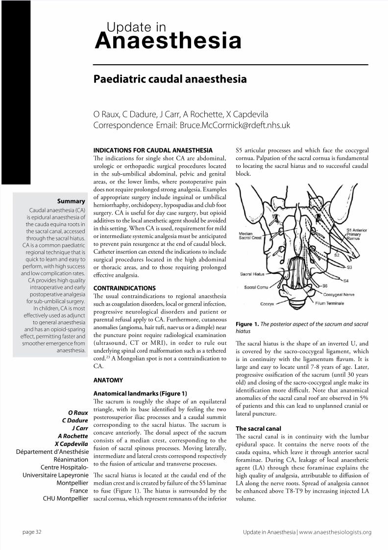
Te sacrum is roughly the shape o an equilateraltriangle, with its base identied by eeling the twoposterosuperior iliac processes and a caudal summitcorresponding to the sacral hiatus. Te sacrum isconcave anteriorly. Te dorsal aspect o the sacrumconsists o a median crest, corresponding to theusion o sacral spinous processes. Moving laterally,intermediate and lateral crests correspond respectively to the usion o articular and transverse processes.
Te sacral hiatus is located at the caudal end o the
median crest and is created by ailure o the S5 laminaeto use (Figure 1). Te hiatus is surrounded by thesacral cornua, which represent remnants o the inerior
S5 articular processes and which ace the coccygealcornua. Palpation o the sacral cornua is undamentalto locating the sacral hiatus and to successul caudalblock.
O Raux
C Dadure
J Carr
A Rochette
X Capdevila
Départementd’Anesthésie
Réanimation
CentreHospitalo-
UniversitaireLapeyronie
MontpellierFrance
CHUMontpellier
Figure 1. The posterior aspect of the sacrum and sacral
hiatus
Te sacral hiatus is the shape o an inverted U, andis covered by the sacro-coccygeal ligament, whichis in continuity with the ligamentum avum. It islarge and easy to locate until 7-8 years o age. Later,progressive ossication o the sacrum (until 30 years
old) and closing o the sacro-coccygeal angle make itsidentication more difcult. Note that anatomicalanomalies o the sacral canal roo are observed in 5%o patients and this can lead to unplanned cranial orlateral puncture.
The sacral canal
Te sacral canal is in continuity with the lumbarepidural space. It contains the nerve roots o thecauda equina, which leave it through anterior sacraloraminae. During CA, leakage o local anaestheticagent (LA) through these oraminae explains thehigh quality o analgesia, attributable to diusion o
LA along the nerve roots. Spread o analgesia cannotbe enhanced above 8-9 by increasing injected LA volume.
C l i n i c a l O v e r v i e w A r t i c l e s
page 32 Update in Anaesthesia | www.anaesthesiologists.org

8/3/2019 Paediatric Caudal Anaesthesia
http://slidepdf.com/reader/full/paediatric-caudal-anaesthesia 2/5
Te dural sac (i.e. the subarachnoid space) ends at the level o S3 ininants and at S2 in adults and children. It is possible to puncturethe dural sac accidentally during CA, leading to extensive spinalanaesthesia. Tereore the needle or cannula must be cautiously
advanced into the sacral canal, ater crossing the sacro-coccygealligament. Te distance between the sacral hiatus and the dural sacis approximately 10mm in neonates. It increases progressively withage (>30mm at 18 years), but there is signicant inter-individualvariability in children.1 Te contents o sacral canal are similar to thoseo lumbar epidural space, predominantly at and epidural veins. Inchildren, epidural atty tissue is looser and more uid than in adults,avoring LA diusion.
TECHNIQUE
Preparation
Obtain consent or the procedure either rom the patient or, i appropriate, rom the parents. Ater induction o general anaesthesiaand airway control, the patient is positioned laterally (or ventrally), with their hips exed to 90° (Figure 2). Skin disinection should beperormed careully, because o the proximity to the anus. Aseptictechnique should be maintained.
is between 5 and 15mm, depending on the child’s size. Te sacro-coccygeal ligament gives a perceptible ‘pop’ when crossed, analogousto the ligamentum avum during lumbar epidural anaesthesia. Atercrossing the sacro-coccygeal ligament, the needle is redirected 30° tothe skin surace, and then advanced a ew millimeters into sacral canal.I in contact with the bony ventral wall o sacral canal, the needle mustbe moved back slightly.
Figure 2. Preparation of patient - lateral position with the surgical sitedown
According to the child’s size, needle diameter and length arerespectively between 21G and 25G, and 25mm and 40mm. A shortbevel improves the eeling o sacrococcygeal ligament penetrationand decreases risk o vascular puncture or sacral peroration.2 Use o
a needle with a stylet avoids risk o cutaneous tissue coring, and the(theoretical) risk o epidural cutaneous cell grat. I a styletted needleis not available, a cutaneous ‘pre-hole’ can be made with a dierentneedle prior to puncture with the caudal needle. Another solution is topuncture with an IV catheter, the hollow needle o which is removedbeore injection through the sheath.
Puncture (Figures 3, 4 and 5)
Figure 3. Bony landmarks
Ater dening the bony landmarks o the sacral triangle, the two sacralcornuae are identied by moving your ngertips rom side to side.Te gluteal clet is not a reliable mark o the midline. Te punctureis perormed between the two sacral cornuae. Te needle is oriented
60° in relation to back plane, 90° to skin surace. Te needle bevelis oriented ventrally, or parallel to the bers o the sacro-coccygealligament. Te distance between the skin and sacro-coccygeal ligament
Figure 4. Puncture - orientation of the needle and reorientation after
crossing the sacro-coccygeal ligament.
Figure 5. Orientation of the needle during puncture
Ater veriying absence o spontaneous reux o blood or cerebrospinal
uid (more sensitive than an aspiration test), injection o LA shouldbe possible be without resistance. Inject slowly (over about oneminute). Where available this may be preceded with an epinephrine
page 33Update in Anaesthesia | www.anaesthesiologists.org

8/3/2019 Paediatric Caudal Anaesthesia
http://slidepdf.com/reader/full/paediatric-caudal-anaesthesia 3/5
test dose under ECG and blood pressure monitoring, in order todetect intravascular placement. Subcutaneous bulging at the injectionsite suggests needle misplacement. Blood reux necessitates repeatingthe puncture, however in case o cerebrospinal uid reux caudal
anaesthesia should be abandoned, in order to avoid the risk o extensivespinal anaesthesia. Aspiration tests should be repeated several timesduring injection.
In skilled hands, the success rate o CA is about 95%, however a variety o misplacements o the needle are possible (Figure 6). Te moment o surgical incision is the true test o block success, but various techniqueshave been suggested to authenticate the puncture success, such asinjection site auscultation (the ‘swoosh test’), or searching or analsphincter contraction in response to electrical nerve stimulation on thepuncture needle. No clear benet o these techniques against simpleclinical assessment have been shown.3,4 More recently, ultrasound hasbeen suggested to help sacro-coccygeal hiatus location and to visualizeisotonic serum or LA injection into sacral epidural space (Figures 7and 8).5,6 Tese authors have also outlined the interest in ultrasoundcontrol within the context o learning the technique, rather than oruse in standard practice.5,7
Catheter insertion
Although CA was initially described as a single shot technique, someauthors have described use o a caudal catheter to prolong analgesicadministration in postoperative period. In addition advancement
o the catheter in the epidural space up to lumbar or even thoraciclevels can achieve analgesia o high abdominal or thoracic areas. 8 However, two pitalls restrict extension o this technique; a highrisk o catheter bacterial colonization, particularly in inants and ahigh risk o catheter misplacement.9,10 Subcutaneous tunnelling at adistance rom the anal orice, or occlusive dressings decrease bacterialcolonization.11 Electrical nerve stimulation or ECG recording on thecatheter, or its echographic visualization have been suggested to guideits advancement in epidural space.12,13 However, most anaesthetistspresently preer a direct epidural approach at the desired level that isappropriate to the surgical intervention.14,15
LOCAL ANAESTHETIC AGENTS
Test dose
Early neurosensory warning symptoms o LA systemic toxicity areconcealed by general anaesthesia. Halogenated anaesthetic agents worsen LA systemic toxicity and can also blunt the cardiovascular signso an intravenous epinephrine test dose injection. Aspiration tests toelicit blood reux are not very sensitive, particularly in inants. A testdose o epinephrine 0.5mcg.kg-1 (administered as 0.1ml.kg-1 lidocaine with epinephrine 1 in 200 000) allows detection o intravenousinjection with sensitivity and speciicity close to 100%, under
halogenated anaesthesia. Warning symptoms are cardiac requency modication (an increase or decrease by 10 beats per minute), increasedin blood pressure (up to 15mmHg), or -wave amplitude change in
Figure 7.
Figures 8a and 8b.
Figure 6A and B.Needle misplacement
A marrow(resistance+++.EquivalenttoIVinjection)
B posteriorsacralligament(subcutaneousbulge)
C subperiostal
D “decoy”hiatusE intrapelvic(riskofdamagingintrapelvicstructures:rectum)
F 4thsacralforamen(unilateralblock).
page 34 Update in Anaesthesia | www.anaesthesiologists.org

8/3/2019 Paediatric Caudal Anaesthesia
http://slidepdf.com/reader/full/paediatric-caudal-anaesthesia 4/5
the 60 to 90 second period ater injection (Figure 9).16,17 Slow injectiono the whole LA dose under haemodynamic and ECG monitoringremains essential or patient saety.
Full dose
Te volume o caudally injected LA determines the spread o the block
and this must be adapted to surgical procedure (able 1). Analgesicspread will be two dermatomes higher on the down positioned side atthe time o puncture. Injected volume must not exceed 1.25 ml.kg-1 or20 to 25ml, in order to avoid excessive cerebrospinal uid pressure.
‚20%
Table 2.Maximal allowable doses of local anaesthestic agents
Plainlocal Withepinephrine
anaesthetic(mg.kg-1) (mg.kg-1) Neonates
Bupivacaine 2 2
Lidocaine 3 7
Ropivacaine 3 3
Additiveagent Dose/concentration
epinephrine 5mcg.ml-1
fentanyl 1mcg.kg-1
clonidine 1-2mcg.kg-1
preservative-freeS(+)ketamine 0.5mg.kg-1
(note - not the intravenous form)
Figure 9. T-wave amplitude change after intravascular injection of a local anaesthetic agent
Table 1. Spread of block as a function of caudally injected local
anaesthetic volume18
Volume(ml.kg-1) Dermatomallevel Indication
0.5 Sacral Circumcision
0.75 Inguinal Inguinal
herniotomy
1 Lowerthoracic(T10) Umbilical
herniorraphy, orchidopexy
1.25 Midthoracic
LA choice prioritizes long lasting eects with the weakest motor block possible, since motor block is poorly tolerated in awake children.Bupivacaine meets these criteria. More recently available, ropivacaineand L-bupivacaine have less cardiac toxicity than bupivacaïne atequivalent analgesic eectiveness. Tey may also coner a moreavorable dierential block (less motor block or the same analgesicpower) and the 2.5mg.ml-1 (0.25%) concentration is optimal or theseagents. Four to six hours analgesia is usually achieved with minimal
motor block.19,20
Maximal doses must not be exceeded (able 2) but use o a moredilute mixture may allow the desired volume to be achieved withinthe recommended maximum dose. Hemodynamic eects o CA are weak or absent in children, so intravenous uid preloading orvasoconstrictive drugs are unnecessary.
COMPLICATIONS
Complications o CA are uncommon (0.7 per 1000 cases), aremore likely i inadequate equipment is used and are more requentin inants.16 I the technique ails it should be abandoned to avoidoccurrence o potentially serious complications.
Signicant complications, in order o decreasing requency, are:
• Dural tap. Tis is more likely i the needle is advanced excessively in thesacral canal when subarachnoid injection o local anaesthetic agent may cause extensive spinal anaesthesia. Under general anaesthesia thisshould be suspected i non-reactive mydriasis (pupillary dilation)is observed.
• Vascular or bone puncture can lead to intravascular injection andconsequently LA systemic toxicity. Preventative measures are use o a test dose, cessation o injection i resistance is elt and slow injection under hemodynamic and ECG monitoring. Sacralperoration can lead to pelvic organ damage (e.g. rectalpuncture).
• Exceeding the maximal allowed LA dose risks overdose and relatedcardiovascular or neurological complications.
• Delayed respiratory depression secondary to caudally injectedopioid.
• Urinary retention - spontaneous micturition must be observedbeore hospital discharge.
• Sacral osteomyelitis is rare (one case report).22
CONCLUSION
Tis technique has an established role in paediatric regional anaesthesiapractice since it is easy to learn and has a avorable risk/benet ratio.
Despite being more complex to learn, alternative peripheral regionalanaesthesia techniques are gaining popularity and may begin to replacecaudal anaesthesia as a popular choice.
page 35Update in Anaesthesia | www.anaesthesiologists.org
Additives
Tables 3.Several additives prolong CA duration when added to LA.

8/3/2019 Paediatric Caudal Anaesthesia
http://slidepdf.com/reader/full/paediatric-caudal-anaesthesia 5/5
REFERENCES
1. Adewale L, Dearlove O, Wilson B, Hindle K, Robinson DN. The caudalcanal in children: a study using magnetic resonance imaging. Paediatr
Anaesth 2000; 10: 137-41.
2. Dalens B, Hasnaoui A. Caudal anesthesia in pediatric surgery: successrate and adverse eects in 750 consecutive patients. Anesth analg 1989; 68: 83-9.
3. Orme RM, Berg SJ. The “swoosh” test - an evaluation o a modied“whoosh” test in children. Br J Anaesth 2003; 90: 62-5.
4. Tsui BC, Tarkkila P, Gupta S, Kearney R. Conrmation o caudal needleplacement using nerve stimulation. Anesthesiology 1999; 91: 374-8.
5. Raghunathan K, Schwartz D, Conelly NR. Determining the accuracyo caudal needle placement in children: a comparison o the swooshtest and ultrasonography. Paediatr Anaesth 2008; 18: 606-12.
6. Roberts SA, Guruswamy V, Galvez I. Caudal injectate can be reliably
imaged using portable ultrasound - a preliminary result. Paediatr anaesth 2005; 15: 948-52.
7. Schwartz DA, Dunn SM, Conelly NR. Ultrasound and caudal blocks inchildren. Paediatr Anaesth 2006; 16: 892-902 (correspondence).
8. Tsui BC, Berde CB. Caudal analgesia and anesthesia techniques inchildren. Curr Op Anesthesiol 2005; 18: 283-8.
9. Kost-Byerly S, Tobin JR, Greenberg RS, Billett C, Zahurak M, Yaster M.Bacterial colonisation and inectious rate o continuous epiduralcatheters in children. Anesth Analg 1998; 86: 712-6.
10. Valairucha S, Seeelder D, Houck CS. Thoracic epidural cathetersplaced by the caudal route in inants: the importance o radiographicconrmation. Paediatr Anaesth 2002; 12: 424-8.
11. Bubeck J, Boss K, Krause H, Thies KC. Subcutaneous tunneling o caudal catheters reduces the rate o bacterial colonization to that o lumbar epidural catheters. Anesth Analg 2004; 99: 689-93.
12. Tsui BC, Wagner A, Cave D, Kearny R. Thoracic and lumbar epiduralanalgesia via the caudal approach using electrical stimulationguidance in pediatric patients: a review o 289 patients. Anesthesiology
2004; 100: 683-9.
13. Chawathe MS, Jones RM, Gildersleve CD, Harrison SK, Morris SJ,Eickmann C. Detection o epidural catheters with ultrasound inchildren. Paediatr Anaesth 2003; 13: 681-4.
14. Bösenberg AT. Epidural analgesia or major neonatal surgery. Paediatr Anaesth 1998; 8: 479-83.
15. Giauré E, Dalens B, Gombert A. Epidemiology and morbidity o regional anesthesia in children: a one-year prospective survey o theFrench-language society o pediatric anesthesiologists. Anesth Analg
1996; 83: 904-12.
16. Kozek-Langenecker SA, Marhoer P, Jonas K, Macik T, Urak G, SemsrothM. Cardiovascular criteria or epidural test dosing in sevofurane- andhalothane-anesthetized children. Anesth Analg 2000; 90: 579-83.
17. Tobias JD. Caudal epidural block: a review o test dosing andrecognition injection in children. Anesth Analg 2001; 93: 1156-61.
18. Armitage EN. Local anaesthetic techniques or prevention o postoperative pain. Br J Anaesth 1986; 58: 790-800.
19. Bösenberg AT, Thomas J, Lopez T, Huledal G, Jeppsson L, Larsson LE.Plasma concentrations o ropivacaine ollowing a single-shot caudalblock o 1, 2 or 3 mg/kg in children. Acta Anaesthesiol Scand 2001; 10:1276-80.
20. Breschan C, Jost R, Krumpholz R, Schaumberger F, Stettner H,Marhoer P, Likar R. A prospective study comparing the analgesicecacy o levobupivacaine, ropivacaine and bupivacaine in pediatricpatients undergoing caudal blockade. Paediatr Anaesth 2005; 15:301-6.
21. Sanders JC. Paediatric regional anaesthesia, a survey o practice in theUnited Kingdom. Br J Anaesth 2002; 89: 707-10.
22. Wittum S, Hoer CK, Rölli U, Suhner M, Gubler J, Zollinger A. Sacralosteomyelitis ater single-shot epidural anesthesia via the caudalapproach in a child. Anesthesiology 2003; 99: 503-5.
23. Cohen IT. Caudal block complication in a patient with trisomy 13.Paediatr Anaesth 2006; 16: 213-5.
page 36 Update in Anaesthesia | www.anaesthesiologists.org