p53 protects against genome instability following ... cell biol-2015...bules and duplicate. ... ing...
TRANSCRIPT
JCB: Article
JCB 63
The Rockefeller University Press $30.00J. Cell Biol. Vol. 210 No. 1 63–77www.jcb.org/cgi/doi/10.1083/jcb.201502089
Introduction
Centrosomes are the main microtubule-organizing centers (MTOCs) of most animal cells and are composed of a pair of centrioles surrounded by pericentriolar material (PCM; Nigg and Raff, 2009; Gönczy, 2012). Centrioles act as the centro-some organizer and thus their duplication controls centrosome number. Like DNA, centrioles duplicate exactly once per cell cycle, with a single new procentriole forming on the wall of each existing centriole (Tsou and Stearns, 2006). This tightly controlled process ensures the generation of two centrosomes to form the poles of the bipolar mitotic spindle. Errors in centri-ole duplication lead to abnormal centrosome number, which can result in chromosome segregation errors and the production of aneuploid progeny (Ganem et al., 2009; Silkworth et al., 2009). Aberrations in centrosome number have been associated with several human diseases, including cancer and neurodevelop-mental disorders (Nigg and Raff, 2009).
Canonical centriole duplication begins at the G1/S tran-sition with the assembly of a single cartwheel structure on the wall of each preexisting mother centriole. The cartwheel then templates the formation of a procentriole by providing a scaf-fold onto which microtubules are loaded (Kitagawa et al., 2011; van Breugel et al., 2011, 2014). In addition to this canonical pathway of centriole assembly, de novo centriole formation can occur in the absence of existing centrioles (Miki-Noumura, 1977; Szöllosi and Ozil, 1991; Palazzo et al., 1992; Marshall et al., 2001; Suh et al., 2002). A striking example of this process
occurs in mouse embryos, where cell divisions continue in the absence of centrioles until the 64-cell stage, at which point cen-trioles are created de novo (Szollosi et al., 1972). In vertebrate somatic cells, a variable number of de novo centrioles are gen-erated after experimental removal of existing centrioles (Khod-jakov et al., 2002; La Terra et al., 2005; Uetake et al., 2007). It is therefore thought that existing centrioles act to suppress de novo centriole assembly, although the molecular mechanism for this suppression remains unclear.
Previous approaches to study the immediate consequence of centriole loss in human cells have relied on laser ablation or microsurgery (Khodjakov et al., 2002; La Terra et al., 2005; Uetake et al., 2007). These elegant approaches only transiently remove centrioles from a small number of cells. Permanent centriole loss has been achieved through the knockout of es-sential centriole components (Sir et al., 2013; Bazzi and An-derson, 2014; Izquierdo et al., 2014). Although informative, these studies did not address the immediate effects of centri-ole duplication failure and were unable to temporally control formation of new centrioles.
Polo-like kinase 4 (Plk4) has emerged as a conserved, dose-dependent regulator of centriole copy number and offers an attractive target to reversibly modulate centriole number in populations of cells (Bettencourt-Dias et al., 2005; Habedanck et al., 2005). Plk4 is a self-regulating enzyme that phosphory-lates itself to promote its own destruction (Cunha-Ferreira et
Centriole function has been difficult to study because of a lack of specific tools that allow persistent and reversible cen-triole depletion. Here we combined gene targeting with an auxin-inducible degradation system to achieve rapid, titrat-able, and reversible control of Polo-like kinase 4 (Plk4), a master regulator of centriole biogenesis. Depletion of Plk4 led to a failure of centriole duplication that produced an irreversible cell cycle arrest within a few divisions. This arrest was not a result of a prolonged mitosis, chromosome segregation errors, or cytokinesis failure. Depleting p53 allowed cells that fail centriole duplication to proliferate indefinitely. Washout of auxin and restoration of endogenous Plk4 levels in cells that lack centrioles led to the penetrant formation of de novo centrioles that gained the ability to organize microtu-bules and duplicate. In summary, we uncover a p53-dependent surveillance mechanism that protects against genome instability by preventing cell growth after centriole duplication failure.
p53 protects against genome instability following centriole duplication failure
Bramwell G. Lambrus,1 Yumi Uetake,2 Kevin M. Clutario,1 Vikas Daggubati,1 Michael Snyder,1 Greenfield Sluder,2 and Andrew J. Holland1
1Department of Molecular Biology and Genetics, Johns Hopkins University School of Medicine, Baltimore, MD 212052Department of Cell and Developmental Biology, University of Massachusetts Medical School, Worcester, MA 01655
© 2015 Lambrus et al. This article is distributed under the terms of an Attribution–Noncommercial–Share Alike–No Mirror Sites license for the first six months after the pub-lication date (see http://www.rupress.org/terms). After six months it is available under a Creative Commons License (Attribution–Noncommercial–Share Alike 3.0 Unported license, as described at http://creativecommons.org/licenses/by-nc-sa/3.0/).
Correspondence to Andrew J. Holland: [email protected] used in this paper: AAV, adeno-associated virus; AID, auxin-in-ducible degron; IAA, indole-3-acetic acid; MTOC, microtubule-organizing cen-ter; PCM, pericentriolar material; Plk4, Polo-like kinase 4.
TH
EJ
OU
RN
AL
OF
CE
LL
BIO
LO
GY
on October 7, 2015
jcb.rupress.orgD
ownloaded from
Published July 6, 2015
http://jcb.rupress.org/content/suppl/2015/07/06/jcb.201502089.DC1.html Supplemental Material can be found at:
JCB • Volume 210 • NumBer 1 • 201564
al., 2009, 2013; Rogers et al., 2009; Guderian et al., 2010; Hol-land et al., 2010; Brownlee et al., 2011; Klebba et al., 2013). This autoregulated destruction plays an important role in con-trolling the abundance of endogenous Plk4 and thereby helps to limit centriole duplication to once per cell cycle (Holland et al., 2012b). RNA interference and knock-in approaches have been used to inhibit Plk4 function, but these strategies are slow acting and are not readily reversible. Inhibition of Plk4 kinase activity offers a powerful alternative to manipulate Plk4 function. How-ever, specific kinase inhibitors are difficult to develop (Holland and Cleveland, 2014; Mason et al., 2014). In addition, inhibit-ing Plk4 activity greatly increases protein abundance, making it challenging to dissect the relative contribution of loss of kinase activity and increased protein abundance (Holland et al., 2010).
In this study, we developed a chemical genetics approach to rapidly and reversibly control Plk4 protein abundance in cells. Manipulating endogenous Plk4 levels allowed the re-versible depletion of centrioles from populations of cycling cells and uncovered the existence of a p53-dependent pathway that guards against genome instability by preventing cellu-lar proliferation after centriole duplication failure. By taking advantage of the acute and titratable depletion of Plk4, our data define the threshold level of Plk4 required for centriole assembly and establish the consequence of acute Plk4 deple-tion on centriole composition.
Results
A novel system for rapidly and reversibly controlling Plk4 levelsTo study the immediate effects caused by Plk4 depletion, we turned to an auxin-inducible degradation system that allows for posttranslational control of protein abundance with the plant hor-mone auxin (Nishimura et al., 2009). In plants, auxin promotes the binding of the F-box protein osTIR1 to proteins contain-ing an auxin-inducible degron (AID). In the presence of auxin, osTIR1 recruits AID-containing substrates to the Skp1, Cullin, and F-Box ubiquitin ligase, which ubiquitinates the AID-con-taining protein and targets it for proteasomal degradation. Ec-topic expression of osTIR1 in mammalian cells generates an SCFTIR1 complex and enables auxin-inducible destruction of AID-tagged transgenes (Holland et al., 2012a), suggesting that the AID system could also be applied to posttranslationally reg-ulate the stability and abundance of endogenous Plk4.
We used sequential rounds of gene targeting to knock-in the AID onto the C terminus of both endogenous Plk4 alleles in nontransformed, telomerase-immortalized, human RPE-1 cells (Fig. S1, A and B). To facilitate detection, each Plk4AID allele was tagged at the C terminus with a HA or 3× FLAG tag. Two Plk4AID-HA/AID-3xFLAG (hereafter referred to as Plk4AID/AID) clones were obtained and behaved similarly in all assays. Plk4AID/AID cells exhibited normal centrosome copy number and cell cycle profiles (Fig. S1, C and D), and both Plk4AID-HA and Plk4AID-FLAG localized to the centrioles of cells in interphase and mitosis in a manner that was indistinguishable from wild-type Plk4 (Fig. S1 E). We conclude that Plk4AID is capable of supporting Plk4 function in centriole duplication.
We stably expressed osTIR1-9xMyc in Plk4AID/AID cells to place the stability of endogenous Plk4AID under the control of exogenous auxin (Fig. 1 A). To analyze Plk4 protein levels, we immunoprecipitated Plk4AID-3xFLAG from cell lysates using a
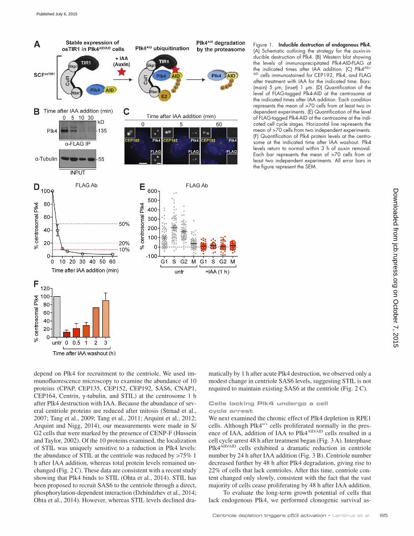
FLAG antibody and determined the abundance of Plk4 by im-munoblot. Addition of the auxin indole-3-acetic acid (IAA) to Plk4AID/AID cells led to rapid Plk4 degradation, with Plk4 fall-ing below the limit of detection within 10 min of IAA addition (Fig. 1 B). The level of Plk4 at the centrosome was measured at various times after IAA addition using antibodies raised to the FLAG tag or to the C terminus of Plk4. Staining with the monoclonal FLAG antibody revealed that the level of Plk4 at the centrosomes of interphase cells declined by >95% within 30 min of IAA addition (Fig. 1, C and D), whereas staining with a polyclonal antibody to the C terminus of Plk4 revealed a >80% reduction during the same time period (Fig. S1 F). Plk4 destruction occurred in all cell cycle phases (Fig. 1 E) and re-quired the presence of the osTIR1 F-box protein (Fig. S1 G). Importantly, the degradation of Plk4 was fully reversible, with the level of Plk4 at the centriole recovering to original levels within 3 h of IAA removal (Fig. 1 F).
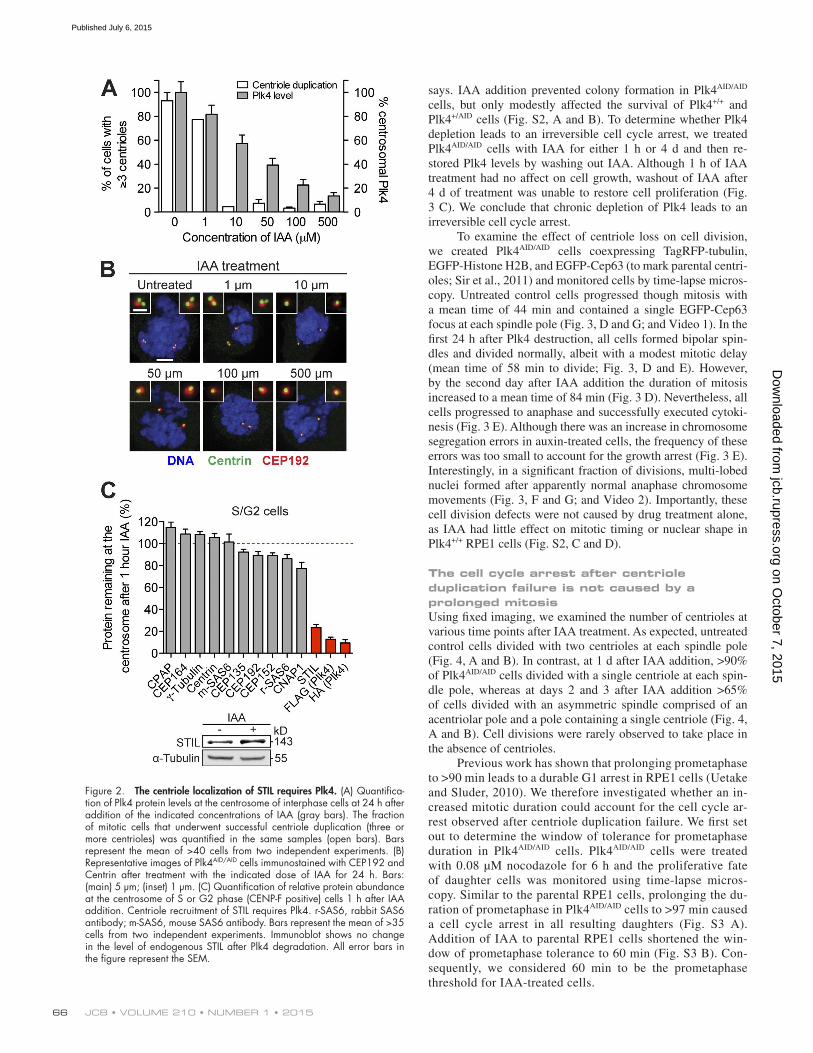
To examine whether treatment with IAA leads to the ex-pected failure of centriole duplication, we assessed centriole number in cells undergoing mitosis one cell cycle (30 h) after IAA addition. In untreated Plk4AID/AID cells, centriole duplica-tion occurred successfully in >90% of cells (0 µM IAA; Fig. 2, A and B). In contrast, IAA addition caused a failure of centriole duplication in >90% of Plk4AID/AID cells (500 µM IAA; Fig. 2, A and B). We conclude that Plk4AID/AID cells offer a new tool to achieve rapid and reversible depletion of endogenous Plk4.
A graded reduction in protein levels reveals a threshold level of Plk4 required for centriole duplicationReduced expression of Plk4 has been associated with tumor-igenesis, but it remains unknown how much Plk4 protein is required for centriole duplication (Ko et al., 2005; Pellegrino et al., 2010; Liu et al., 2012). Because Plk4 autoregulates its own stability, generating a progressive reduction in Plk4 protein levels is challenging, as a reduced abundance of Plk4 mRNA may not lead to a comparable down-regulation of Plk4 protein (Holland et al., 2012a). We therefore set out to use auxin-induc-ible destruction to generate a graded reduction in Plk4 levels. We used quantitative microscopy with directly labeled Plk4 primary antibodies to determine the abundance of Plk4 at the centriole in cells treated with various concentrations of IAA. Exposing Plk4AID/AID cells to increasing concentrations of IAA resulted in gradual reduction in the average level of Plk4 at the centriole (Fig. 2 A). We examined whether centriole duplication occurred successfully with different levels of Plk4 protein. In cells treated with 1 µM IAA, the average level of Plk4 at the centrosome was 81% of that in untreated cells (Fig. 2, A and B). Under these conditions, centrioles duplicated in >75% of cells. However, at 10 µM IAA, whereas centrosomal Plk4 levels were maintained at 57% of that in control cells, <10% of cells underwent centriole duplication (Fig. 2, A and B). We conclude that Plk4 levels are finely balanced and that a less than twofold reduction in the level of Plk4 at the centrosome leads to a fail-ure of centriole duplication.
The centriole localization of STIL is uniquely sensitive to the levels of Plk4RNA interference of Plk4 leads to slow protein depletion, mak-ing it challenging to distinguish between direct and indirect ef-fects of Plk4 removal. The ability to rapidly remove Plk4 from cells provided an opportunity to identify proteins that directly
on October 7, 2015
jcb.rupress.orgD
ownloaded from
Published July 6, 2015
Centriole depletion triggers p53 activation • lambrus et al. 65
depend on Plk4 for recruitment to the centriole. We used im-munofluorescence microscopy to examine the abundance of 10 proteins (CPAP, CEP135, CEP152, CEP192, SAS6, CNAP1, CEP164, Centrin, γ-tubulin, and STIL) at the centrosome 1 h after Plk4 destruction with IAA. Because the abundance of sev-eral centriole proteins are reduced after mitosis (Strnad et al., 2007; Tang et al., 2009; Tang et al., 2011; Arquint et al., 2012; Arquint and Nigg, 2014), our measurements were made in S/G2 cells that were marked by the presence of CENP-F (Hussein and Taylor, 2002). Of the 10 proteins examined, the localization of STIL was uniquely sensitive to a reduction in Plk4 levels: the abundance of STIL at the centriole was reduced by >75% 1 h after IAA addition, whereas total protein levels remained un-changed (Fig. 2 C). These data are consistent with a recent study showing that Plk4 binds to STIL (Ohta et al., 2014). STIL has been proposed to recruit SAS6 to the centriole through a direct, phosphorylation-dependent interaction (Dzhindzhev et al., 2014; Ohta et al., 2014). However, whereas STIL levels declined dra-
matically by 1 h after acute Plk4 destruction, we observed only a modest change in centriole SAS6 levels, suggesting STIL is not required to maintain existing SAS6 at the centriole (Fig. 2 C).
Cells lacking Plk4 undergo a cell cycle arrestWe next examined the chronic effect of Plk4 depletion in RPE1 cells. Although Plk4+/+ cells proliferated normally in the pres-ence of IAA, addition of IAA to Plk4AID/AID cells resulted in a cell cycle arrest 48 h after treatment began (Fig. 3 A). Interphase Plk4AID/AID cells exhibited a dramatic reduction in centriole number by 24 h after IAA addition (Fig. 3 B). Centriole number decreased further by 48 h after Plk4 degradation, giving rise to 22% of cells that lack centrioles. After this time, centriole con-tent changed only slowly, consistent with the fact that the vast majority of cells cease proliferating by 48 h after IAA addition.
To evaluate the long-term growth potential of cells that lack endogenous Plk4, we performed clonogenic survival as-
Figure 1. Inducible destruction of endogenous Plk4. (A) Schematic outlining the strategy for the auxin-in-ducible destruction of Plk4. (B) Western blot showing the levels of immunoprecipitated Plk4-AID-FLAG at the indicated times after IAA addition. (C) Plk4AID/
AID cells immunostained for CEP192, Plk4, and FLAG after treatment with IAA for the indicated time. Bars: (main) 5 µm; (inset) 1 µm. (D) Quantification of the level of FLAG-tagged Plk4-AID at the centrosome at the indicated times after IAA addition. Each condition represents the mean of >70 cells from at least two in-dependent experiments. (E) Quantification of the level of FLAG-tagged Plk4-AID at the centrosome at the indi-cated cell cycle stages. Horizontal line represents the mean of >70 cells from two independent experiments. (F) Quantification of Plk4 protein levels at the centro-some at the indicated time after IAA washout. Plk4 levels return to normal within 3 h of auxin removal. Each bar represents the mean of >70 cells from at least two independent experiments. All error bars in the figure represent the SEM.
on October 7, 2015
jcb.rupress.orgD
ownloaded from
Published July 6, 2015
JCB • Volume 210 • NumBer 1 • 201566
says. IAA addition prevented colony formation in Plk4AID/AID cells, but only modestly affected the survival of Plk4+/+ and Plk4+/AID cells (Fig. S2, A and B). To determine whether Plk4 depletion leads to an irreversible cell cycle arrest, we treated Plk4AID/AID cells with IAA for either 1 h or 4 d and then re-stored Plk4 levels by washing out IAA. Although 1 h of IAA treatment had no affect on cell growth, washout of IAA after 4 d of treatment was unable to restore cell proliferation (Fig. 3 C). We conclude that chronic depletion of Plk4 leads to an irreversible cell cycle arrest.
To examine the effect of centriole loss on cell division, we created Plk4AID/AID cells coexpressing TagRFP-tubulin, EGFP-Histone H2B, and EGFP-Cep63 (to mark parental centri-oles; Sir et al., 2011) and monitored cells by time-lapse micros-copy. Untreated control cells progressed though mitosis with a mean time of 44 min and contained a single EGFP-Cep63 focus at each spindle pole (Fig. 3, D and G; and Video 1). In the first 24 h after Plk4 destruction, all cells formed bipolar spin-dles and divided normally, albeit with a modest mitotic delay (mean time of 58 min to divide; Fig. 3, D and E). However, by the second day after IAA addition the duration of mitosis increased to a mean time of 84 min (Fig. 3 D). Nevertheless, all cells progressed to anaphase and successfully executed cytoki-nesis (Fig. 3 E). Although there was an increase in chromosome segregation errors in auxin-treated cells, the frequency of these errors was too small to account for the growth arrest (Fig. 3 E). Interestingly, in a significant fraction of divisions, multi-lobed nuclei formed after apparently normal anaphase chromosome movements (Fig. 3, F and G; and Video 2). Importantly, these cell division defects were not caused by drug treatment alone, as IAA had little effect on mitotic timing or nuclear shape in Plk4+/+ RPE1 cells (Fig. S2, C and D).
The cell cycle arrest after centriole duplication failure is not caused by a prolonged mitosisUsing fixed imaging, we examined the number of centrioles at various time points after IAA treatment. As expected, untreated control cells divided with two centrioles at each spindle pole (Fig. 4, A and B). In contrast, at 1 d after IAA addition, >90% of Plk4AID/AID cells divided with a single centriole at each spin-dle pole, whereas at days 2 and 3 after IAA addition >65% of cells divided with an asymmetric spindle comprised of an acentriolar pole and a pole containing a single centriole (Fig. 4, A and B). Cell divisions were rarely observed to take place in the absence of centrioles.
Previous work has shown that prolonging prometaphase to >90 min leads to a durable G1 arrest in RPE1 cells (Uetake and Sluder, 2010). We therefore investigated whether an in-creased mitotic duration could account for the cell cycle ar-rest observed after centriole duplication failure. We first set out to determine the window of tolerance for prometaphase duration in Plk4AID/AID cells. Plk4AID/AID cells were treated with 0.08 µM nocodazole for 6 h and the proliferative fate of daughter cells was monitored using time-lapse micros-copy. Similar to the parental RPE1 cells, prolonging the du-ration of prometaphase in Plk4AID/AID cells to >97 min caused a cell cycle arrest in all resulting daughters (Fig. S3 A). Addition of IAA to parental RPE1 cells shortened the win-dow of prometaphase tolerance to 60 min (Fig. S3 B). Con-sequently, we considered 60 min to be the prometaphase threshold for IAA-treated cells.
Figure 2. The centriole localization of STIL requires Plk4. (A) Quantifica-tion of Plk4 protein levels at the centrosome of interphase cells at 24 h after addition of the indicated concentrations of IAA (gray bars). The fraction of mitotic cells that underwent successful centriole duplication (three or more centrioles) was quantified in the same samples (open bars). Bars represent the mean of >40 cells from two independent experiments. (B) Representative images of Plk4AID/AID cells immunostained with CEP192 and Centrin after treatment with the indicated dose of IAA for 24 h. Bars: (main) 5 µm; (inset) 1 µm. (C) Quantification of relative protein abundance at the centrosome of S or G2 phase (CENP-F positive) cells 1 h after IAA addition. Centriole recruitment of STIL requires Plk4. r-SAS6, rabbit SAS6 antibody; m-SAS6, mouse SAS6 antibody. Bars represent the mean of >35 cells from two independent experiments. Immunoblot shows no change in the level of endogenous STIL after Plk4 degradation. All error bars in the figure represent the SEM.
on October 7, 2015
jcb.rupress.orgD
ownloaded from
Published July 6, 2015
Centriole depletion triggers p53 activation • lambrus et al. 67
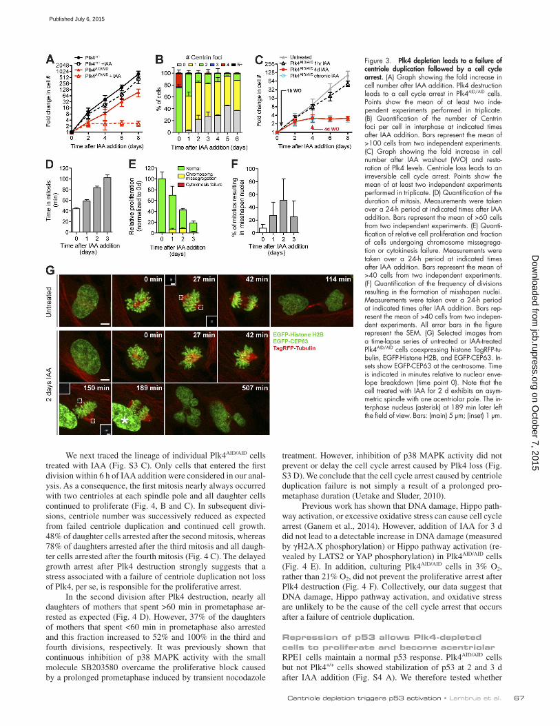
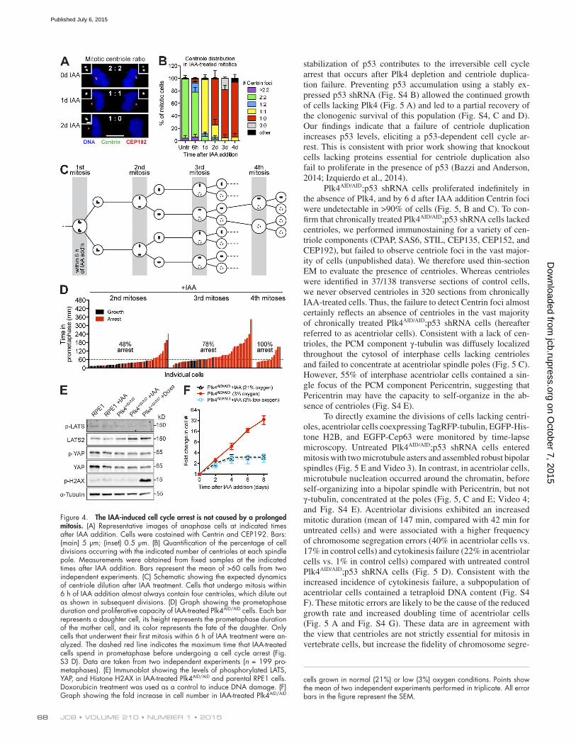
We next traced the lineage of individual Plk4AID/AID cells treated with IAA (Fig. S3 C). Only cells that entered the first division within 6 h of IAA addition were considered in our anal-ysis. As a consequence, the first mitosis nearly always occurred with two centrioles at each spindle pole and all daughter cells continued to proliferate (Fig. 4, B and C). In subsequent divi-sions, centriole number was successively reduced as expected from failed centriole duplication and continued cell growth. 48% of daughter cells arrested after the second mitosis, whereas 78% of daughters arrested after the third mitosis and all daugh-ter cells arrested after the fourth mitosis (Fig. 4 C). The delayed growth arrest after Plk4 destruction strongly suggests that a stress associated with a failure of centriole duplication not loss of Plk4, per se, is responsible for the proliferative arrest.
In the second division after Plk4 destruction, nearly all daughters of mothers that spent >60 min in prometaphase ar-rested as expected (Fig. 4 D). However, 37% of the daughters of mothers that spent <60 min in prometaphase also arrested and this fraction increased to 52% and 100% in the third and fourth divisions, respectively. It was previously shown that continuous inhibition of p38 MAPK activity with the small molecule SB203580 overcame the proliferative block caused by a prolonged prometaphase induced by transient nocodazole
treatment. However, inhibition of p38 MAPK activity did not prevent or delay the cell cycle arrest caused by Plk4 loss (Fig. S3 D). We conclude that the cell cycle arrest caused by centriole duplication failure is not simply a result of a prolonged pro-metaphase duration (Uetake and Sluder, 2010).
Previous work has shown that DNA damage, Hippo path-way activation, or excessive oxidative stress can cause cell cycle arrest (Ganem et al., 2014). However, addition of IAA for 3 d did not lead to a detectable increase in DNA damage (measured by γH2A.X phosphorylation) or Hippo pathway activation (re-vealed by LATS2 or YAP phosphorylation) in Plk4AID/AID cells (Fig. 4 E). In addition, culturing Plk4AID/AID cells in 3% O2, rather than 21% O2, did not prevent the proliferative arrest after Plk4 destruction (Fig. 4 F). Collectively, our data suggest that DNA damage, Hippo pathway activation, and oxidative stress are unlikely to be the cause of the cell cycle arrest that occurs after a failure of centriole duplication.
Repression of p53 allows Plk4-depleted cells to proliferate and become acentriolarRPE1 cells maintain a normal p53 response. Plk4AID/AID cells but not Plk4+/+ cells showed stabilization of p53 at 2 and 3 d after IAA addition (Fig. S4 A). We therefore tested whether
Figure 3. Plk4 depletion leads to a failure of centriole duplication followed by a cell cycle arrest. (A) Graph showing the fold increase in cell number after IAA addition. Plk4 destruction leads to a cell cycle arrest in Plk4AID/AID cells. Points show the mean of at least two inde-pendent experiments performed in triplicate. (B) Quantification of the number of Centrin foci per cell in interphase at indicated times after IAA addition. Bars represent the mean of >100 cells from two independent experiments. (C) Graph showing the fold increase in cell number after IAA washout (WO) and resto-ration of Plk4 levels. Centriole loss leads to an irreversible cell cycle arrest. Points show the mean of at least two independent experiments performed in triplicate. (D) Quantification of the duration of mitosis. Measurements were taken over a 24-h period at indicated times after IAA addition. Bars represent the mean of >60 cells from two independent experiments. (E) Quanti-fication of relative cell proliferation and fraction of cells undergoing chromosome missegrega-tion or cytokinesis failure. Measurements were taken over a 24-h period at indicated times after IAA addition. Bars represent the mean of >40 cells from two independent experiments. (F) Quantification of the frequency of divisions resulting in the formation of misshapen nuclei. Measurements were taken over a 24-h period at indicated times after IAA addition. Bars rep-resent the mean of >40 cells from two indepen-dent experiments. All error bars in the figure represent the SEM. (G) Selected images from a time-lapse series of untreated or IAA-treated Plk4AID/AID cells coexpressing histone TagRFP-tu-bulin, EGFP-Histone H2B, and EGFP-CEP63. In-sets show EGFP-CEP63 at the centrosome. Time is indicated in minutes relative to nuclear enve-lope breakdown (time point 0). Note that the cell treated with IAA for 2 d exhibits an asym-metric spindle with one acentriolar pole. The in-terphase nucleus (asterisk) at 189 min later left the field of view. Bars: (main) 5 µm; (inset) 1 µm.
on October 7, 2015
jcb.rupress.orgD
ownloaded from
Published July 6, 2015
JCB • Volume 210 • NumBer 1 • 201568
stabilization of p53 contributes to the irreversible cell cycle arrest that occurs after Plk4 depletion and centriole duplica-tion failure. Preventing p53 accumulation using a stably ex-pressed p53 shRNA (Fig. S4 B) allowed the continued growth of cells lacking Plk4 (Fig. 5 A) and led to a partial recovery of the clonogenic survival of this population (Fig. S4, C and D). Our findings indicate that a failure of centriole duplication increases p53 levels, eliciting a p53-dependent cell cycle ar-rest. This is consistent with prior work showing that knockout cells lacking proteins essential for centriole duplication also fail to proliferate in the presence of p53 (Bazzi and Anderson, 2014; Izquierdo et al., 2014).
Plk4AID/AID;p53 shRNA cells proliferated indefinitely in the absence of Plk4, and by 6 d after IAA addition Centrin foci were undetectable in >90% of cells (Fig. 5, B and C). To con-firm that chronically treated Plk4AID/AID;p53 shRNA cells lacked centrioles, we performed immunostaining for a variety of cen-triole components (CPAP, SAS6, STIL, CEP135, CEP152, and CEP192), but failed to observe centriole foci in the vast major-ity of cells (unpublished data). We therefore used thin-section EM to evaluate the presence of centrioles. Whereas centrioles were identified in 37/138 transverse sections of control cells, we never observed centrioles in 320 sections from chronically IAA-treated cells. Thus, the failure to detect Centrin foci almost certainly reflects an absence of centrioles in the vast majority of chronically treated Plk4AID/AID;p53 shRNA cells (hereafter referred to as acentriolar cells). Consistent with a lack of cen-trioles, the PCM component γ-tubulin was diffusely localized throughout the cytosol of interphase cells lacking centrioles and failed to concentrate at acentriolar spindle poles (Fig. 5 C). However, 55% of interphase acentriolar cells contained a sin-gle focus of the PCM component Pericentrin, suggesting that Pericentrin may have the capacity to self-organize in the ab-sence of centrioles (Fig. S4 E).
To directly examine the divisions of cells lacking centri-oles, acentriolar cells coexpressing TagRFP-tubulin, EGFP-His-tone H2B, and EGFP-Cep63 were monitored by time-lapse microscopy. Untreated Plk4AID/AID;p53 shRNA cells entered mitosis with two microtubule asters and assembled robust bipolar spindles (Fig. 5 E and Video 3). In contrast, in acentriolar cells, microtubule nucleation occurred around the chromatin, before self-organizing into a bipolar spindle with Pericentrin, but not γ-tubulin, concentrated at the poles (Fig. 5, C and E; Video 4; and Fig. S4 E). Acentriolar divisions exhibited an increased mitotic duration (mean of 147 min, compared with 42 min for untreated cells) and were associated with a higher frequency of chromosome segregation errors (40% in acentriolar cells vs. 17% in control cells) and cytokinesis failure (22% in acentriolar cells vs. 1% in control cells) compared with untreated control Plk4AID/AID;p53 shRNA cells (Fig. 5 D). Consistent with the increased incidence of cytokinesis failure, a subpopulation of acentriolar cells contained a tetraploid DNA content (Fig. S4 F). These mitotic errors are likely to be the cause of the reduced growth rate and increased doubling time of acentriolar cells (Fig. 5 A and Fig. S4 G). These data are in agreement with the view that centrioles are not strictly essential for mitosis in vertebrate cells, but increase the fidelity of chromosome segre-
Figure 4. The IAA-induced cell cycle arrest is not caused by a prolonged mitosis. (A) Representative images of anaphase cells at indicated times after IAA addition. Cells were costained with Centrin and CEP192. Bars: (main) 5 µm; (inset) 0.5 µm. (B) Quantification of the percentage of cell divisions occurring with the indicated number of centrioles at each spindle pole. Measurements were obtained from fixed samples at the indicated times after IAA addition. Bars represent the mean of >60 cells from two independent experiments. (C) Schematic showing the expected dynamics of centriole dilution after IAA treatment. Cells that undergo mitosis within 6 h of IAA addition almost always contain four centrioles, which dilute out as shown in subsequent divisions. (D) Graph showing the prometaphase duration and proliferative capacity of IAA-treated Plk4AID/AID cells. Each bar represents a daughter cell, its height represents the prometaphase duration of the mother cell, and its color represents the fate of the daughter. Only cells that underwent their first mitosis within 6 h of IAA treatment were an-alyzed. The dashed red line indicates the maximum time that IAA-treated cells spend in prometaphase before undergoing a cell cycle arrest (Fig. S3 D). Data are taken from two independent experiments (n = 199 pro-metaphases). (E) Immunoblot showing the levels of phosphorylated LATS, YAP, and Histone H2AX in IAA-treated Plk4AID/AID and parental RPE1 cells. Doxorubicin treatment was used as a control to induce DNA damage. (F) Graph showing the fold increase in cell number in IAA-treated Plk4AID/AID
cells grown in normal (21%) or low (3%) oxygen conditions. Points show the mean of two independent experiments performed in triplicate. All error bars in the figure represent the SEM.
on October 7, 2015
jcb.rupress.orgD
ownloaded from
Published July 6, 2015

Centriole depletion triggers p53 activation • lambrus et al. 69
gation and cytokinesis (Khodjakov and Rieder, 2001; Debec et al., 2010; Sir et al., 2013).
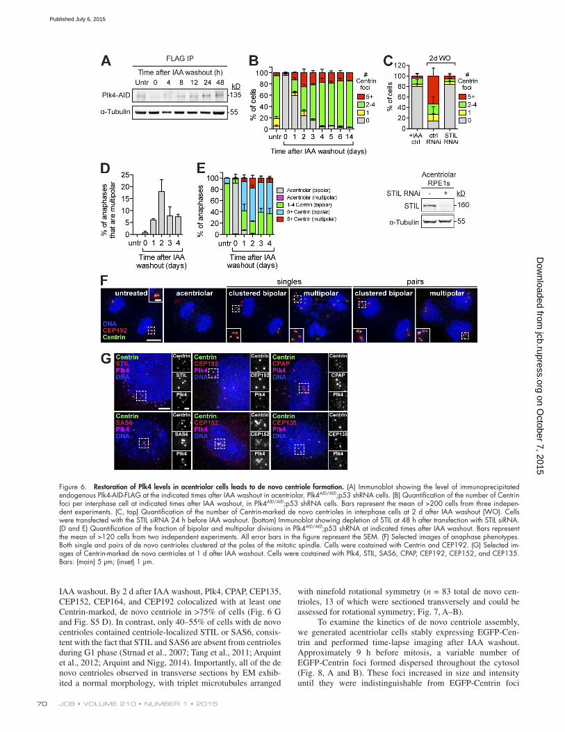
Restoration of endogenous Plk4 levels in acentriolar cells results in de novo centriole formationIn vertebrate somatic cells, de novo centriole assembly is initi-ated after the eradication of the existing centrioles by laser ab-lation or microsurgery (Khodjakov et al., 2002; La Terra et al., 2005; Uetake et al., 2007). In this case a variable number of de novo centrioles are spontaneously generated. We therefore ex-amined the effect of restoring endogenous Plk4 levels in acen-triolar cells. Endogenous Plk4 levels returned to normal within 12 h of IAA washout (Fig. 6 A) and promoted the penetrant formation of de novo centrioles: by 2 d after IAA washout, de novo centriole assembly occurred in 74% of cells and increased to >95% of cells by 4 d after IAA removal (Fig. 6 B and Fig. S4 H). Knockdown of the cartwheel component STIL prevented de novo centriole assembly, suggesting that, similar to canoni-cal centriole duplication, de novo centrioles assemble through a cartwheel-dependent mechanism (Fig. 6 C). Collectively, these
data show that Plk4 levels are rate limiting for both canonical and de novo centriole assembly.
De novo centriole assembly occurred stochastically throughout the cytosol of cells, resulting in the formation of a variable number of centrioles (Fig. 6 B and Fig. S4 H). Whereas cells with one to four de novo centrioles always divided nor-mally, cells with supernumerary de novo centrioles underwent frequent (6–18%) multipolar divisions (Fig. 6, D–F). Because the progeny of multipolar divisions are invariably inviable (Ganem et al., 2009), aberrant mitotic divisions are likely to limit the proliferative potential of cells with supernumerary de novo centrioles. The decreased growth potential of cells with supernumerary centrioles may explain why the relative fraction of cells with a normal centriole number gradually increases in abundance after IAA washout (Fig. 6 B).
De novo centriole assembly required cell cycle pro-gression, as arresting cells in G1 phase with mimosine or the CDK2/4 inhibitor PD0332991 greatly reduced the frequency of de novo centriole formation by 2 d after IAA washout (Fig. S5, A–C; Watson et al., 1991; Fry et al., 2004). To examine the composition of de novo centrioles we performed immunostain-ing for several centriole components at 1, 2, 3, and 4 d after
Figure 5. Depletion of p53 allows contin-ued growth in the absence of Plk4. (A) Graph showing the fold increase in cell number after IAA addition. Points show the mean of at least two independent experiments performed in triplicate. (B) Quantification of the number of Centrin foci per cell in interphase at indicated times after IAA addition. Bars represent the mean of >150 cells from two independent experiments. (C) Selected images of untreated control or acentriolar Plk4AID/AID;p53 shRNA cells immunostained with α-tubulin, γ-tubulin, and Centrin. Bars: (main) 5 µm; (inset) 1 µm. (D) Quantification of the duration of mitosis, the frequency of divisions resulting in the for-mation of misshapen nuclei, and the frequency of cytokinesis failure and chromosome misseg-regation in acentriolar Plk4AID/AID;p53 shRNA cells. Bars represent the mean of >100 cells from two independent experiments. All error bars in the figure represent the SEM. (E) Se-lected images from a time-lapse series of un-treated control or acentriolar Plk4AID/AID;p53 shRNA cells coexpressing histone TagRFP-tubu-lin, EGFP-Histone H2B, and EGFP-CEP63. In-sets show EGFP-CEP63 at the centrosome. The acentriolar cell spends longer in mitosis and fails cytokinesis. Time is indicated in minutes relative to nuclear envelope breakdown (time point 0). Bars: (main) 5 µm; (inset) 1 µm.
on October 7, 2015
jcb.rupress.orgD
ownloaded from
Published July 6, 2015
JCB • Volume 210 • NumBer 1 • 201570
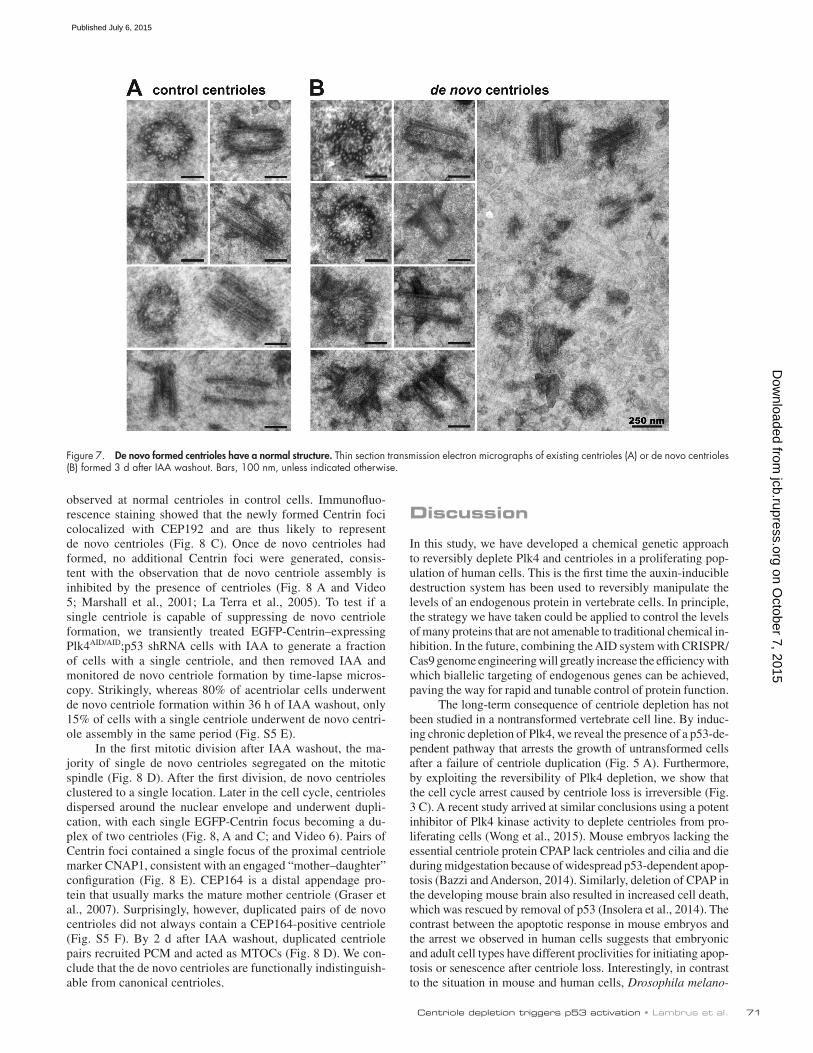
IAA washout. By 2 d after IAA washout, Plk4, CPAP, CEP135, CEP152, CEP164, and CEP192 colocalized with at least one Centrin-marked, de novo centriole in >75% of cells (Fig. 6 G and Fig. S5 D). In contrast, only 40–55% of cells with de novo centrioles contained centriole-localized STIL or SAS6, consis-tent with the fact that STIL and SAS6 are absent from centrioles during G1 phase (Strnad et al., 2007; Tang et al., 2011; Arquint et al., 2012; Arquint and Nigg, 2014). Importantly, all of the de novo centrioles observed in transverse sections by EM exhib-ited a normal morphology, with triplet microtubules arranged
with ninefold rotational symmetry (n = 83 total de novo cen-trioles, 13 of which were sectioned transversely and could be assessed for rotational symmetry; Fig. 7, A–B).
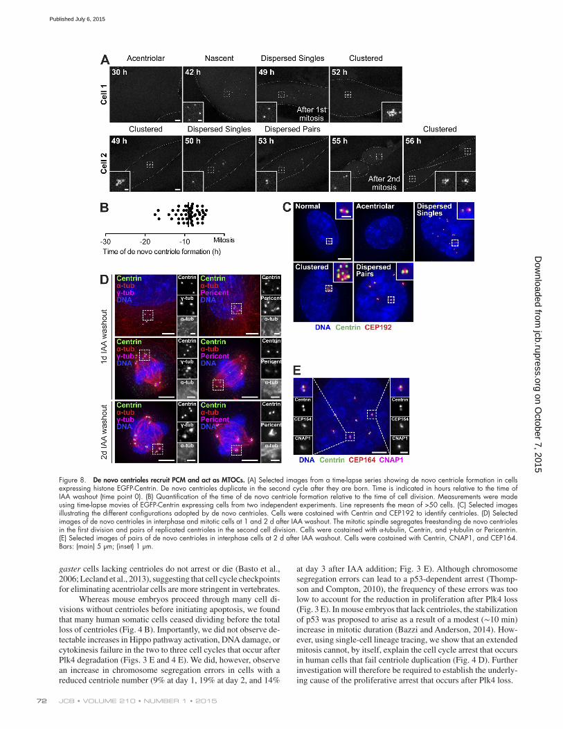
To examine the kinetics of de novo centriole assembly, we generated acentriolar cells stably expressing EGFP-Cen-trin and performed time-lapse imaging after IAA washout. Approximately 9 h before mitosis, a variable number of EGFP-Centrin foci formed dispersed throughout the cytosol (Fig. 8, A and B). These foci increased in size and intensity until they were indistinguishable from EGFP-Centrin foci
Figure 6. Restoration of Plk4 levels in acentriolar cells leads to de novo centriole formation. (A) Immunoblot showing the level of immunoprecipitated endogenous Plk4-AID-FLAG at the indicated times after IAA washout in acentriolar, Plk4AID/AID;p53 shRNA cells. (B) Quantification of the number of Centrin foci per interphase cell at indicated times after IAA washout, in Plk4AID/AID;p53 shRNA cells. Bars represent the mean of >200 cells from three indepen-dent experiments. (C, top) Quantification of the number of Centrin-marked de novo centrioles in interphase cells at 2 d after IAA washout (WO). Cells were transfected with the STIL siRNA 24 h before IAA washout. (bottom) Immunoblot showing depletion of STIL at 48 h after transfection with STIL siRNA. (D and E) Quantification of the fraction of bipolar and multipolar divisions in Plk4AID/AID;p53 shRNA at indicated times after IAA washout. Bars represent the mean of >120 cells from two independent experiments. All error bars in the figure represent the SEM. (F) Selected images of anaphase phenotypes. Both single and pairs of de novo centrioles clustered at the poles of the mitotic spindle. Cells were costained with Centrin and CEP192. (G) Selected im-ages of Centrin-marked de novo centrioles at 1 d after IAA washout. Cells were costained with Plk4, STIL, SAS6, CPAP, CEP192, CEP152, and CEP135. Bars: (main) 5 µm; (inset) 1 µm.
on October 7, 2015
jcb.rupress.orgD
ownloaded from
Published July 6, 2015
Centriole depletion triggers p53 activation • lambrus et al. 71
observed at normal centrioles in control cells. Immunofluo-rescence staining showed that the newly formed Centrin foci colocalized with CEP192 and are thus likely to represent de novo centrioles (Fig. 8 C). Once de novo centrioles had formed, no additional Centrin foci were generated, consis-tent with the observation that de novo centriole assembly is inhibited by the presence of centrioles (Fig. 8 A and Video 5; Marshall et al., 2001; La Terra et al., 2005). To test if a single centriole is capable of suppressing de novo centriole formation, we transiently treated EGFP-Centrin–expressing Plk4AID/AID;p53 shRNA cells with IAA to generate a fraction of cells with a single centriole, and then removed IAA and monitored de novo centriole formation by time-lapse micros-copy. Strikingly, whereas 80% of acentriolar cells underwent de novo centriole formation within 36 h of IAA washout, only 15% of cells with a single centriole underwent de novo centri-ole assembly in the same period (Fig. S5 E).
In the first mitotic division after IAA washout, the ma-jority of single de novo centrioles segregated on the mitotic spindle (Fig. 8 D). After the first division, de novo centrioles clustered to a single location. Later in the cell cycle, centrioles dispersed around the nuclear envelope and underwent dupli-cation, with each single EGFP-Centrin focus becoming a du-plex of two centrioles (Fig. 8, A and C; and Video 6). Pairs of Centrin foci contained a single focus of the proximal centriole marker CNAP1, consistent with an engaged “mother–daughter” configuration (Fig. 8 E). CEP164 is a distal appendage pro-tein that usually marks the mature mother centriole (Graser et al., 2007). Surprisingly, however, duplicated pairs of de novo centrioles did not always contain a CEP164-positive centriole (Fig. S5 F). By 2 d after IAA washout, duplicated centriole pairs recruited PCM and acted as MTOCs (Fig. 8 D). We con-clude that the de novo centrioles are functionally indistinguish-able from canonical centrioles.
Discussion
In this study, we have developed a chemical genetic approach to reversibly deplete Plk4 and centrioles in a proliferating pop-ulation of human cells. This is the first time the auxin-inducible destruction system has been used to reversibly manipulate the levels of an endogenous protein in vertebrate cells. In principle, the strategy we have taken could be applied to control the levels of many proteins that are not amenable to traditional chemical in-hibition. In the future, combining the AID system with CRISPR/Cas9 genome engineering will greatly increase the efficiency with which biallelic targeting of endogenous genes can be achieved, paving the way for rapid and tunable control of protein function.
The long-term consequence of centriole depletion has not been studied in a nontransformed vertebrate cell line. By induc-ing chronic depletion of Plk4, we reveal the presence of a p53-de-pendent pathway that arrests the growth of untransformed cells after a failure of centriole duplication (Fig. 5 A). Furthermore, by exploiting the reversibility of Plk4 depletion, we show that the cell cycle arrest caused by centriole loss is irreversible (Fig. 3 C). A recent study arrived at similar conclusions using a potent inhibitor of Plk4 kinase activity to deplete centrioles from pro-liferating cells (Wong et al., 2015). Mouse embryos lacking the essential centriole protein CPAP lack centrioles and cilia and die during midgestation because of widespread p53-dependent apop-tosis (Bazzi and Anderson, 2014). Similarly, deletion of CPAP in the developing mouse brain also resulted in increased cell death, which was rescued by removal of p53 (Insolera et al., 2014). The contrast between the apoptotic response in mouse embryos and the arrest we observed in human cells suggests that embryonic and adult cell types have different proclivities for initiating apop-tosis or senescence after centriole loss. Interestingly, in contrast to the situation in mouse and human cells, Drosophila melano-
Figure 7. De novo formed centrioles have a normal structure. Thin section transmission electron micrographs of existing centrioles (A) or de novo centrioles (B) formed 3 d after IAA washout. Bars, 100 nm, unless indicated otherwise.
on October 7, 2015
jcb.rupress.orgD
ownloaded from
Published July 6, 2015
JCB • Volume 210 • NumBer 1 • 201572
gaster cells lacking centrioles do not arrest or die (Basto et al., 2006; Lecland et al., 2013), suggesting that cell cycle checkpoints for eliminating acentriolar cells are more stringent in vertebrates.
Whereas mouse embryos proceed through many cell di-visions without centrioles before initiating apoptosis, we found that many human somatic cells ceased dividing before the total loss of centrioles (Fig. 4 B). Importantly, we did not observe de-tectable increases in Hippo pathway activation, DNA damage, or cytokinesis failure in the two to three cell cycles that occur after Plk4 degradation (Figs. 3 E and 4 E). We did, however, observe an increase in chromosome segregation errors in cells with a reduced centriole number (9% at day 1, 19% at day 2, and 14%
at day 3 after IAA addition; Fig. 3 E). Although chromosome segregation errors can lead to a p53-dependent arrest (Thomp-son and Compton, 2010), the frequency of these errors was too low to account for the reduction in proliferation after Plk4 loss (Fig. 3 E). In mouse embryos that lack centrioles, the stabilization of p53 was proposed to arise as a result of a modest (∼10 min) increase in mitotic duration (Bazzi and Anderson, 2014). How-ever, using single-cell lineage tracing, we show that an extended mitosis cannot, by itself, explain the cell cycle arrest that occurs in human cells that fail centriole duplication (Fig. 4 D). Further investigation will therefore be required to establish the underly-ing cause of the proliferative arrest that occurs after Plk4 loss.
Figure 8. De novo centrioles recruit PCM and act as MTOCs. (A) Selected images from a time-lapse series showing de novo centriole formation in cells expressing histone EGFP-Centrin. De novo centrioles duplicate in the second cycle after they are born. Time is indicated in hours relative to the time of IAA washout (time point 0). (B) Quantification of the time of de novo centriole formation relative to the time of cell division. Measurements were made using time-lapse movies of EGFP-Centrin expressing cells from two independent experiments. Line represents the mean of >50 cells. (C) Selected images illustrating the different configurations adopted by de novo centrioles. Cells were costained with Centrin and CEP192 to identify centrioles. (D) Selected images of de novo centrioles in interphase and mitotic cells at 1 and 2 d after IAA washout. The mitotic spindle segregates freestanding de novo centrioles in the first division and pairs of replicated centrioles in the second cell division. Cells were costained with α-tubulin, Centrin, and γ-tubulin or Pericentrin. (E) Selected images of pairs of de novo centrioles in interphase cells at 2 d after IAA washout. Cells were costained with Centrin, CNAP1, and CEP164. Bars: (main) 5 µm; (inset) 1 µm.
on October 7, 2015
jcb.rupress.orgD
ownloaded from
Published July 6, 2015
Centriole depletion triggers p53 activation • lambrus et al. 73
Removal of p53 allowed human cells to proliferate indef-initely despite the absence of centrioles (Fig. 5 A). As centro-somes increase the speed of spindle assembly, these acentriolar cells formed bipolar spindles at a considerably slower rate than cells containing centrioles. In addition, acentriolar cells dis-played chromosomal instability and frequent cytokinesis failure (Fig. 5 D). Similar mitotic errors have been reported in acen-triolar D. melanogaser cells and transformed vertebrate cell lines, highlighting the important role of centrioles in ensuring the fidelity of cell division in adult cells (Khodjakov and Rieder, 2001; Basto et al., 2006; Debec et al., 2010; Sir et al., 2013). Surprisingly, however, acentriolar mouse embryos showed no detectable increase in chromosome segregation errors (Bazzi and Anderson, 2014). The first five rounds of cell division in the mouse embryo are naturally acentriolar (Szollosi et al., 1972) and it is therefore possible that mouse embryonic divisions are less dependent on centrioles for faithful chromosome segregation.
Overexpression of Plk4 did not promote de novo centriole formation in acentriolar mouse zygotes (Coelho et al., 2013), but was capable of inducing de novo centriole formation in unfertilized fly eggs (Peel et al., 2007; Rodrigues-Martins et al., 2007) and activated Xenopus laevis oocytes (Eckerdt et al., 2011). Here we have shown that restoring endogenous levels of Plk4 induces de novo centriole formation in acentriolar human somatic cells. We speculate that de novo centrioles are created from Plk4 foci that form stochastically in the cytosol of acentri-olar cells (Fig. S5 G). Once these foci surpass a threshold level of Plk4 activity, cartwheel assembly would be initiated, leading to centriole formation. Consistent with this idea, we were able to observe recruitment of the cartwheel components STIL and SAS6 to Plk4 foci as an early event occurring before incorpora-tion of Centrin into de novo centrioles (Fig. S5 G).
De novo centrioles assembled very efficiently (within two to three cell cycles) after restoration of Plk4 levels (Fig. 6 B). This is in accordance with previous work examining the kinet-ics of de novo centriole formation in somatic human cells (La Terra et al., 2005; Uetake et al., 2007) and green algae (Mar-shall et al., 2001). Procentrioles formed in S phase are normally modified during mitosis to activate their ability to recruit PCM and function as MTOCs in the next cell cycle (Wang et al., 2011). In our experiments, cells that lack centrioles undergo a prolonged prometaphase, providing increased time for newly created de novo centrioles to be modified and converted into MTOCs. Consistently, we observed that in the first cell cycle the vast majority of de novo centrioles recruited small amounts of PCM in mitosis and were capable of segregating themselves on the mitotic spindle (Fig. 8 D). De novo centrioles duplicated in the second cell cycle after their creation, consistent with a previous study demonstrating that newly formed centrioles are modified in mitosis to license their duplication (Fig. 8, A and D; Wang et al., 2011).
A single centriole was shown to suppress de novo cen-triole assembly in the vast majority of cells, supporting the conclusion that existing centrioles inhibit the de novo assembly pathway (Fig. S5 E; Marshall et al., 2001; La Terra et al., 2005). Like canonical centriole assembly, de novo centriole formation requires Plk4 activity (Wong et al., 2015). An intriguing possi-bility, therefore, is that existing centrioles sequester an activator of Plk4 and thereby act to inhibit the cytosolic pool of Plk4 and suppress de novo centriole formation. The ability to induce timed de novo centriole biogenesis in populations of cycling acentriolar cells offers an excellent tool to dissect the molecular
events involved in centriole formation and how existing centri-oles antagonize the de novo pathway.
Materials and methods
Antibody productionA C-terminal Plk4 fragment (aa 510–970) was cloned into a pET-23b bacterial expression vector (EMD Millipore) containing a C-termi-nal 6xHis tag. Recombinant protein was purified from Escherichia coli using Ni-NTA beads (QIAGEN) and used for immunization (ProSci). A STIL C-terminal peptide VGTFLDVKRLRQLPKLF (aa 1,271–1,287) was synthesized and conjugated to KLH for immu-nization. Rabbit immune sera were affinity purified using standard procedures. Affinity purified antibodies were directly conjugated to DyLight 550 and DyLight 650 fluorophores (Thermo Fisher Scientific) for use in immunofluorescence.
Cell culturehTERT RPE-1 cells were maintained at 37°C in a 5% CO2 atmosphere with 21% oxygen. Cells were grown in DMEM/F12 medium containing 10% fetal bovine serum (Sigma-Aldrich), 0.348% sodium bicarbonate, 100 U/ml penicillin, 100 U/ml streptomycin, and 2 mM l-glutamine. Nocodazole (Sigma-Aldrich) was dissolved in DMSO and used at a final concentration of 3.3 µM; Mimosine (Sigma-Aldrich) was dis-solved in diluted HCl and used at a final concentration of 250 µM; PD0332991 (Sigma-Aldrich) was dissolved in DMSO and used at a final concentration of 1 µM; SB203580 (Sigma-Aldrich) was dissolved in DMSO and used at a final concentration of 10 µM; Doxorubicin (Sigma-Aldrich) was dissolved in water and used at a final concentra-tion of 200 ng/ml; and IAA (Sigma-Aldrich) was dissolved in water and used at 500 µM unless otherwise stated.
Gene targeting and stable cell linesGene targeting was performed using adeno-associated virus (AAV; Ber-dougo et al., 2009; Holland et al., 2012b). To generate the Plk4 target-ing construct we cloned an AID degron followed by a 3xFLAG epitope tag, a translational stop codon, the Plk4 3′ UTR, and 150 bp of adjacent genomic sequence into the pBluescript derivative pNY to create the AID-3xFLAG-3′UTR;loxP-NeoR-loxP vector. 5′ and 3′ homology arms were PCR amplified from genomic RPE1 DNA and cloned on either side of the central AID-3xFLAG-3′UTR;loxP-NeoR-loxP cassette. The entire insert was transferred into the pAAV vector backbone and fully sequenced to verify its integrity. hTERT-RPE1 cells were transduced with infectious AID-3xFLAG-3′UTR;loxP-NeoR-loxP–containing AAV particles. Plk4AID/+ cells were isolated by selection in G418 and resistant clones were screened by PCR using primers binding in NeoR and a ge-nomic sequence adjacent to the targeting construct. A second targeting construct was created containing a HA tag in place of the FLAG epitope tag and a BlastR in place of NeoR. An additional round of gene targeting was performed in Plk4AID/+ cells using AID-HA-3′UTR;loxP-BlastR-loxP AAV particles. The intergenic position of the NeoR and BlastR cas-settes reduces the likelihood that their presence will disrupt the function of the Plk4 gene. This facilitates the isolation of homozygous targeted al-leles by allowing concurrent selection for both markers. Plk4AID/AID cells were isolated by coselection in G418 and blasticidin and resistant clones were screened by PCR using primers that bind in BlastR and a genomic sequence adjacent to the targeting construct. Two independent Plk4AID/
AID clones were created and the targeted Plk4-AID-3xFLAG and Plk4-AID-3xHA alleles were sequenced to verify their integrity. osTIR1-9xMyc was cloned into the pBabe backbone under the control of the gag promoter and introduced into Plk4AID/AID cells using retroviral delivery.
on October 7, 2015
jcb.rupress.orgD
ownloaded from
Published July 6, 2015
JCB • Volume 210 • NumBer 1 • 201574
Stable integrants were selected in 5 µg/ml puromycin and single clones were isolated by limiting dilution. EGFP-tagged Cep63 was cloned into the FUGW backbone under the control of the ubiquitin promoter, whereas EGFP-tagged histone H2B and TagRFP-tubulin were cloned into the FUGW backbone under the control of the CMV promoter. All three constructs were introduced into Plk4AID/AID cells using lentiviral de-livery and single clones were isolated by limiting dilution. A p53 shRNA was introduced into cells using lentiviral delivery and single clones were isolated using fluorescence-activated cell sorting (Tiscornia et al., 2003).
Antibody techniquesTo purify Plk4-AID-3xFLAG, cells were lysed in lysis buffer (10 mM Tris, pH 7.5, 0.1% Triton X-100, 100 mM NaCl, 1 mM EDTA, 1 mM EGTA, 50 mM NaF, 20 mM, β-glycerophosphate, 0.1 mM DTT, 200 nM microcystin, 1 mM PMSF, and 1 µM LPC) and sonicated and soluble extracts were prepared. The supernatant was incubated with anti-FLAG M2 magnetic beads (Sigma-Aldrich). Beads were washed five times in lysis buffer and immunopurified protein was analyzed by immunoblot.
For immunoblot analysis, protein samples were separated by SDS-PAGE, transferred onto nitrocellulose membranes with a Trans-Blot Turbo Transfer System (Bio-Rad Laboratories), and then probed with the following antibodies: DM1A (mouse anti–α-tubulin, 1:5,000; Sigma-Al-drich), Plk4 (rabbit, 1:3,200; this study), FLAG M2 (mouse, 1:1,000; Sigma-Aldrich), STIL (rabbit, 1:2,500; Bethyl Laboratories, Inc.), Yap (rabbit, 1:1,000, Cell Signaling Technology), p-Yap Ser127 (rabbit, 1:1,000; Cell Signaling Technology), LATS2 (rabbit, 1:1,000; Cell Sig-naling Technology), p-LATS S1077 (rabbit, 1:500; Yu et al., 2010; a gift of D. Pan, Johns Hopkins School of Medicine, Baltimore, MD), and p-Histone H2A.X (Ser139; rabbit, 1:1,000; Cell Signaling Technology).
For immunofluorescence, cells were grown on 18-mm glass cov-erslips and fixed in 100% ice-cold methanol for 10 min. Cells were blocked in 2.5% FBS, 200 mM glycine, and 0.1% Triton X-100 in PBS for 1 h. Antibody incubations were conducted in the blocking solution for 1 h. DNA was detected using DAPI and cells were mounted in Pro-long Antifade (Invitrogen). Staining was performed with the following primary antibodies: GTU-88 (mouse anti–γ-tubulin, 1:250; Abcam), Centrin (mouse, 1:1,000; EMD Millipore), CNAP1 (guinea pig, raised against the CNAP1 peptide sequence SPTQQDGRGQKNSDAKC, 1:1,000, a gift from O. Stemmann, University of Bayreuth, Bayreuth, Germany), CEP152 (rabbit, 1:5,000; Bethyl Laboratories, Inc.), Plk4-650 (directly labeled rabbit, 1:1,000; this study), STIL-550 (directly labeled rabbit, 1:1,000; this study), CEP135 (rabbit, raised against CEP135 aa 695–838, 1:1,000; a gift from A. Hyman, Max Planck In-stitute for Molecular Cell Biology and Genetics, Dresden, Germany), CEP192-Cy3 (directly labeled rabbit, raised against CEP192 aa 1–211, 1:1,000; a gift from K. Oegema, Ludwig Institute for Cancer Research, La Jolla, CA), SAS6-Cy3 (directly labeled rabbit, raised against SAS6 aa 501–657, 1:1,000, a gift from K. Oegema), SAS6 (mouse, 1:1,000; Santa Cruz Biotechnology, Inc.), CPAP-Cy3 (directly labeled rab-bit, 1:1,000; a gift from K. Oegema), CENP-F (sheep, raised against CENP-F aa 1,363–1,640, 1:1,000; a gift from S. Taylor, University of Manchester, Manchester, UK), HA (rat anti-HA High Affinity, 1:200; Roche), and FLAG (mouse anti-FLAG M2, 1:1,000; Sigma-Aldrich). Secondary donkey antibodies were conjugated to Alexa Fluor 488, 555, or 650 (Life Technologies). To mark S phase, cells were pulsed with EdU before fixation in 100% ice-cold methanol at −20°C for 10 min. Cells were washed three times with 0.1% Triton X-100 in PBS and stained using a Click-It EdU Alexa Fluor 555 imaging kit (Life Tech-nologies) according to the manufacturer’s recommendations.
For the cell cycle analysis of Plk4 levels shown in Fig. 1 E, cells were pulsed with EdU for 1 h before fixation in 100% ice-cold methanol at −20°C for 10 min. Cells were washed three times with
0.1% Triton X-100, stained using a Click-It EdU Alexa Fluor 555 imaging kit (Life Technologies), and blocked in 2.5% FBS, 200 mM glycine, and 0.1% Triton X-100 in PBS for 1 h. Immunofluorescence microscopy was performed using the following antibodies: CENP-F (sheep, raised against CENP-F aa 1,363–1,640, 1:1,000; a gift from S. Taylor), GTU-88 (mouse anti–γ-tubulin, 1:250; Abcam), and Plk4-650 (directly labeled rabbit, 1:1,000; this study). G1 phase cells were classified as CENP-F and EdU negative, S phase cells were classified as EdU positive, and G2 phase cells were classified as CENP-F posi-tive and EdU negative. The γ-tubulin staining was used to define the position of the centrosome.
Immunofluorescence images were collected using a Deltavision Elite system (GE Healthcare) controlling a camera (pco.edge 5.5; Sci-entific CMOS). Acquisition parameters were controlled by SoftWoRx suite (GE Healthcare). Images were collected at room temperature (25°C) using a 40× 1.35 NA, 60× 1.42 NA, or 100× 1.4 NA oil objec-tive (Olympus) at 0.2-µm z-sections and subsequently deconvolved in SoftWoRx suite. Images were acquired using immersion oil (n = 1.516; Applied Precision). For quantitation of signal intensity at the centro-some, deconvolved 2D maximum intensity projections were saved as 16-bit TIFF images. Signal intensity was determined using ImageJ by drawing a circular region of interest (ROI) around the centriole (ROI S). A larger concentric circle (ROI L) was drawn around ROI S. ROI S and L were transferred to the channel of interest and the signal in ROI S was calculated using the formula IS − [(IL − IS/AL − AS) × AS], where A is area and I is integrated pixel intensity.
Live-cell microscopyCells were seeded into four-chamber, 35-mm glass bottom culture dishes (Greiner) and maintained at 37°C in an environmental control station. Images were collected using a Deltavision Elite system con-trolling a camera (pco.edge 5.5.). Images were acquired with a 40× 1.35 NA oil lens using immersion oil (n = 1.526). Every 5 min, 7 × 3-µm z-sections were acquired for EGFP and RFP and maximum inten-sity projection was created using SoftWoRx. Alternatively, cells were imaged using a 40× 1.4 NA Plan Apochromat oil-immersion objective (Carl Zeiss) on a confocal microscope (LSM 780; Carl Zeiss) equipped with a solid-state 488- and 514-nm laser and a spectral GaAsP detector. Images were acquired using immersion oil (n = 1.518; Carl Zeiss). Ac-quisition parameters, shutters, and focus were controlled by ZEN black software (Carl Zeiss). 15 × 1.6-µm z-sections were acquired simultane-ously for EGFP and/or RFP at 5- or 10-min time intervals. Maximum intensity projections were created using ZEN black. Movies were as-sembled and analyzed using FIJI.
Lineage tracingFor nocodazole treatment, coverslips were assembled into observation chambers with medium containing 0.08 µM nocodazole and fields of cells were continuously followed by video time-lapse microscopy at 37°C for 6 h. After 6 h, the field of view was marked with a dia-mond scribe; the bottom of the observation chamber was removed and washed out with fresh medium several times before being reassembled with fresh medium as previously described (Uetake and Sluder, 2012). The previously marked fields were continuously followed for at least 96 h. Lineage tracing of individual cells was performed as previously described (Uetake et al., 2007). For IAA and SB203580 treatment, cells were exposed to the drug for the duration of imaging. Images were collected using a microscope (DMRXE; Leica) equipped with phase-contrast optics and a 10× 0.3 NA objective (Leica). Images were captured with an Orca ER (Hamamatsu Photonics) camera using HCT software (Hamamatsu Photonics) and exported as AVI movies to be viewed with QuickTime (Apple).
on October 7, 2015
jcb.rupress.orgD
ownloaded from
Published July 6, 2015
Centriole depletion triggers p53 activation • lambrus et al. 75
EMCells were grown on 35-mm Nunclon Delta–treated culture dishes (Thermo Fisher Scientific) and fixed (2.5% glutaraldehyde, 3 mM MgCl2, and 0.1 M sodium cacodylate, pH 7.2) for 1 h at room tempera-ture. Cells were rinsed with 0.1 M sodium cacodylate and post-fixed with 0.5% OsO4 for 1 h on ice. After a dH2O rinse, plates were stained with 0.1% tannic acid, rinsed twice with dH2O, and stained en bloc with 2% aqueous uranyl acetate. Samples were dehydrated in a graded series of ethanol, embedded in Eponate 12 resin (Ted Pella), and polymerized at 60°C overnight. Ultrathin 60-nm sections were cut, mounted, and stained with 1% tannic acid, followed by 2% uranyl acetate and lead citrate. Images were acquired using a transmission electron microscope (7600; Hitachi) equipped with a charge coupled device 2,080 × 2,048-pixel camera (Advanced Microscopy Techniques).
Cell biologyTo prepare cells for flow cytometry, cell pellets were fixed in cold 70% EtOH for 24 h, washed once in PBS, and resuspended in PBS sup-plemented with 0.5 mg/ml RNase A and 50 mg/ml propidium iodide. Samples were incubated at room temperature for 30 min and analyzed on a flow cytometer (FACSCalibur; BD). For RNA interference, 2 × 10⁵ cells were seeded in a 6-well plate and duplexed siRNAs were introduced using RNAiMax (Life Technologies). siRNA directed against STIL (5′-GCUCCAAACAGUUUCUGCUGGAAU-3′) was purchased from GE Healthcare.
Online supplemental materialFig. S1 shows the strategy for the generation of Plk4AID/AID cells and that Plk4 destruction occurs rapidly in an osTIR1-dependent manner. Fig. S2 shows that Plk4 destruction dramatically reduces clonogenic survival and that treatment of parental RPE1 cells with IAA does not alter mitotic timing. Fig. S3 shows that Plk4AID/AID cells have a window of prometaphase tolerance of 97 min, that IAA treatment reduces the window of hTERT RPE1 cells to 60 min, and that inhibition of p38 MAPK activity with SB203580 does not prevent the cell cycle arrest that occurs as a result of centriole duplication failure. Fig. S4 shows that p53 is stabilized after Plk4 destruction, that depletion of p53 par-tially rescues the clonogenic survival of cells lacking Plk4, that mitotic spindles form in the absence of centrioles, and that the incidence of polyploidy increases in acentriolar cells. Fig. S5 shows that de novo centrioles do not form in G1-arrested cells, that the composition of de novo centrioles is similar at 1–4 d after their assembly, and that a single existing centriole is capable of suppressing de novo centriole forma-tion. Video 1 shows a normal cell division in an untreated Plk4AID/AID cell. Video 2 shows an IAA-treated Plk4AID/AID cell dividing with an asymmetric spindle. Video 3 shows a normal cell division in an un-treated Plk4AID/AID;p53 shRNA cell. Video 4 shows a Plk4AID/AID;p53 shRNA cell dividing without centrioles. Video 5 shows de novo centri-ole formation after recovery of Plk4 levels. Video 6 shows duplication of de novo formed centrioles. Online supplemental material is available at http://www.jcb.org/cgi/content/full/jcb.201502089/DC1.
Acknowledgments
The authors would like to thank Stephen Taylor for providing the sheep CENP-F antibody, Anthony Hyman for providing the CEP135 anti-body, Duojia Pan for providing the p-LATS S1077 antibody, Olaf Stemmann for providing the CNAP1 antibody, and Karen Oegema for providing the CEP192, CPAP, and SAS6 antibodies. We thank Bar-bara Smith and the Johns Hopkins School of Medicine Microscope Facility for help with EM analysis. We thank the Johns Hopkins Center for Sensory Biology/Hearing and Balance imaging center for use of
their confocal microscope. We thank Michelle Levine and Randall Reed for comments on the manuscript.
This work was supported by the Leukemia Research Foundation, a W.W. Smith Charitable Trust Research Grant, a March of Dimes Basil O’Conner Scholar Award, a Pew Scholar Award, a Kimmel Scholar Award, and research grants GM 114119 (to A.J. Holland) and GM 30758 (to G. Sluder) from the National Institutes of Health.
The authors declare no competing financial interests.
Submitted: 24 February 2015Accepted: 1 June 2015
ReferencesArquint, C., and E.A. Nigg. 2014. STIL microcephaly mutations interfere with
APC/C-mediated degradation and cause centriole amplification. Curr. Biol. 24:351–360. http://dx.doi.org/10.1016/j.cub.2013.12.016
Arquint, C., K.F. Sonnen, Y.D. Stierhof, and E.A. Nigg. 2012. Cell-cycle-regulated expression of STIL controls centriole number in human cells. J. Cell Sci. 125:1342–1352. http://dx.doi.org/10.1242/jcs.099887
Basto, R., J. Lau, T. Vinogradova, A. Gardiol, C.G. Woods, A. Khodjakov, and J.W. Raff. 2006. Flies without centrioles. Cell. 125:1375–1386. http://dx.doi.org/10.1016/j.cell.2006.05.025
Bazzi, H., and K.V. Anderson. 2014. Acentriolar mitosis activates a p53-depen-dent apoptosis pathway in the mouse embryo. Proc. Natl. Acad. Sci. USA. 111:E1491–E1500. http://dx.doi.org/10.1073/pnas.1400568111
Berdougo, E., M.E. Terret, and P.V. Jallepalli. 2009. Functional dissection of mi-totic regulators through gene targeting in human somatic cells. Methods Mol. Biol. 545:21–37.
Bettencourt-Dias, M., A. Rodrigues-Martins, L. Carpenter, M. Riparbelli, L. Lehmann, M.K. Gatt, N. Carmo, F. Balloux, G. Callaini, and D.M. Glover. 2005. SAK/PLK4 is required for centriole duplication and fla-gella development. Curr. Biol. 15:2199–2207. http://dx.doi.org/10.1016/j.cub.2005.11.042
Brownlee, C.W., J.E. Klebba, D.W. Buster, and G.C. Rogers. 2011. The Protein Phosphatase 2A regulatory subunit Twins stabilizes Plk4 to induce cen-triole amplification. J. Cell Biol. 195:231–243. http://dx.doi.org/10.1083/jcb.201107086
Coelho, P.A., L. Bury, B. Sharif, M.G. Riparbelli, J. Fu, G. Callaini, D.M. Glover, and M. Zernicka-Goetz. 2013. Spindle formation in the mouse embryo requires Plk4 in the absence of centrioles. Dev. Cell. 27:586–597. http://dx.doi.org/10.1016/j.devcel.2013.09.029
Cunha-Ferreira, I., A. Rodrigues-Martins, I. Bento, M. Riparbelli, W. Zhang, E. Laue, G. Callaini, D.M. Glover, and M. Bettencourt-Dias. 2009. The SCF/Slimb ubiquitin ligase limits centrosome amplification through deg-radation of SAK/PLK4. Curr. Biol. 19:43–49. http://dx.doi.org/10.1016/j.cub.2008.11.037
Cunha-Ferreira, I., I. Bento, A. Pimenta-Marques, S.C. Jana, M. Lince-Faria, P. Duarte, J. Borrego-Pinto, S. Gilberto, T. Amado, D. Brito, et al. 2013. Regulation of autophosphorylation controls PLK4 self-destruction and centriole number. Curr. Biol. 23:2245–2254. http://dx.doi.org/10.1016/j.cub.2013.09.037
Debec, A., W. Sullivan, and M. Bettencourt-Dias. 2010. Centrioles: active play-ers or passengers during mitosis? Cell. Mol. Life Sci. 67:2173–2194. http://dx.doi.org/10.1007/s00018-010-0323-9
Dzhindzhev, N.S., G. Tzolovsky, Z. Lipinszki, S. Schneider, R. Lattao, J. Fu, J. Debski, M. Dadlez, and D.M. Glover. 2014. Plk4 phosphorylates Ana2 to trigger Sas6 recruitment and procentriole formation. Curr. Biol. 24:2526–2532. http://dx.doi.org/10.1016/j.cub.2014.08.061
Eckerdt, F., T.M. Yamamoto, A.L. Lewellyn, and J.L. Maller. 2011. Identification of a polo-like kinase 4-dependent pathway for de novo centriole forma-tion. Curr. Biol. 21:428–432. http://dx.doi.org/10.1016/j.cub.2011.01.072
Fry, D.W., P.J. Harvey, P.R. Keller, W.L. Elliott, M. Meade, E. Trachet, M. Albassam, X. Zheng, W.R. Leopold, N.K. Pryer, and P.L. Toogood. 2004. Specific inhibition of cyclin-dependent kinase 4/6 by PD 0332991 and associated antitumor activity in human tumor xenografts. Mol. Cancer Ther. 3:1427–1438.
Ganem, N.J., S.A. Godinho, and D. Pellman. 2009. A mechanism linking extra centrosomes to chromosomal instability. Nature. 460:278–282. http://dx.doi.org/10.1038/nature08136
Ganem, N.J., H. Cornils, S.Y. Chiu, K.P. O’Rourke, J. Arnaud, D. Yimlamai, M. Théry, F.D. Camargo, and D. Pellman. 2014. Cytokinesis failure triggers
on October 7, 2015
jcb.rupress.orgD
ownloaded from
Published July 6, 2015
JCB • Volume 210 • NumBer 1 • 201576
hippo tumor suppressor pathway activation. Cell. 158:833–848. http://dx.doi.org/10.1016/j.cell.2014.06.029
Gönczy, P. 2012. Towards a molecular architecture of centriole assembly. Nat. Rev. Mol. Cell Biol. 13:425–435. http://dx.doi.org/10.1038/nrm3373
Graser, S., Y.D. Stierhof, S.B. Lavoie, O.S. Gassner, S. Lamla, M. Le Clech, and E.A. Nigg. 2007. Cep164, a novel centriole appendage protein required for primary cilium formation. J. Cell Biol. 179:321–330. http://dx.doi.org/10.1083/jcb.200707181
Guderian, G., J. Westendorf, A. Uldschmid, and E.A. Nigg. 2010. Plk4 trans-autophosphorylation regulates centriole number by controlling βTrCP-mediated degradation. J. Cell Sci. 123:2163–2169. http://dx.doi.org/10.1242/jcs.068502
Habedanck, R., Y.D. Stierhof, C.J. Wilkinson, and E.A. Nigg. 2005. The Polo ki-nase Plk4 functions in centriole duplication. Nat. Cell Biol. 7:1140–1146. http://dx.doi.org/10.1038/ncb1320
Holland, A.J., and D.W. Cleveland. 2014. Polo-like kinase 4 inhibition: a strategy for cancer therapy? Cancer Cell. 26:151–153. http://dx.doi.org/10.1016/j.ccr.2014.07.017
Holland, A.J., W. Lan, S. Niessen, H. Hoover, and D.W. Cleveland. 2010. Polo-like kinase 4 kinase activity limits centrosome overduplication by au-toregulating its own stability. J. Cell Biol. 188:191–198. http://dx.doi.org/10.1083/jcb.200911102
Holland, A.J., D. Fachinetti, S. Da Cruz, Q. Zhu, B. Vitre, M. Lince-Faria, D. Chen, N. Parish, I.M. Verma, M. Bettencourt-Dias, and D.W. Cleveland. 2012a. Polo-like kinase 4 controls centriole duplication but does not di-rectly regulate cytokinesis. Mol. Biol. Cell. 23:1838–1845. http://dx.doi.org/10.1091/mbc.E11-12-1043
Holland, A.J., D. Fachinetti, Q. Zhu, M. Bauer, I.M. Verma, E.A. Nigg, and D.W. Cleveland. 2012b. The autoregulated instability of Polo-like kinase 4 lim-its centrosome duplication to once per cell cycle. Genes Dev. 26:2684–2689. http://dx.doi.org/10.1101/gad.207027.112
Hussein, D., and S.S. Taylor. 2002. Farnesylation of Cenp-F is required for G2/M progression and degradation after mitosis. J. Cell Sci. 115:3403–3414.
Insolera, R., H. Bazzi, W. Shao, K.V. Anderson, and S.H. Shi. 2014. Cortical neurogenesis in the absence of centrioles. Nat. Neurosci. 17:1528–1535. http://dx.doi.org/10.1038/nn.3831
Izquierdo, D., W.J. Wang, K. Uryu, and M.F. Tsou. 2014. Stabilization of cart-wheel-less centrioles for duplication requires CEP295-mediated centri-ole-to-centrosome conversion. Cell Reports. 8:957–965. http://dx.doi.org/10.1016/j.celrep.2014.07.022
Khodjakov, A., and C.L. Rieder. 2001. Centrosomes enhance the fidelity of cyto-kinesis in vertebrates and are required for cell cycle progression. J. Cell Biol. 153:237–242. http://dx.doi.org/10.1083/jcb.153.1.237
Khodjakov, A., C.L. Rieder, G. Sluder, G. Cassels, O. Sibon, and C.L. Wang. 2002. De novo formation of centrosomes in vertebrate cells arrested during S phase. J. Cell Biol. 158:1171–1181. http://dx.doi.org/10.1083/jcb.200205102
Kitagawa, D., I. Vakonakis, N. Olieric, M. Hilbert, D. Keller, V. Olieric, M. Bortfeld, M.C. Erat, I. Flückiger, P. Gönczy, and M.O. Steinmetz. 2011. Structural basis of the 9-fold symmetry of centrioles. Cell. 144:364–375. http://dx.doi.org/10.1016/j.cell.2011.01.008
Klebba, J.E., D.W. Buster, A.L. Nguyen, S. Swatkoski, M. Gucek, N.M. Rusan, and G.C. Rogers. 2013. Polo-like kinase 4 autodestructs by generating its Slimb-binding phosphodegron. Curr. Biol. 23:2255–2261. http://dx.doi.org/10.1016/j.cub.2013.09.019
Ko, M.A., C.O. Rosario, J.W. Hudson, S. Kulkarni, A. Pollett, J.W. Dennis, and C.J. Swallow. 2005. Plk4 haploinsufficiency causes mitotic infidelity and carcinogenesis. Nat. Genet. 37:883–888. http://dx.doi.org/10.1038/ng1605
La Terra, S., C.N. English, P. Hergert, B.F. McEwen, G. Sluder, and A. Khodjakov. 2005. The de novo centriole assembly pathway in HeLa cells: cell cycle progression and centriole assembly/maturation. J. Cell Biol. 168:713–722. http://dx.doi.org/10.1083/jcb.200411126
Lecland, N., A. Debec, A. Delmas, S. Moutinho-Pereira, N. Malmanche, A. Bouissou, C. Dupré, A. Jourdan, B. Raynaud-Messina, H. Maiato, and A. Guichet. 2013. Establishment and mitotic characterization of new Drosophila acentriolar cell lines from DSas-4 mutant. Biol. Open. 2:314–323. http://dx.doi.org/10.1242/bio.20133327
Liu, L., C.Z. Zhang, M. Cai, J. Fu, G.G. Chen, and J. Yun. 2012. Downregulation of polo-like kinase 4 in hepatocellular carcinoma associates with poor prognosis. PLoS ONE. 7:e41293. http://dx.doi.org/10.1371/journal.pone.0041293
Marshall, W.F., Y. Vucica, and J.L. Rosenbaum. 2001. Kinetics and regulation of de novo centriole assembly. Implications for the mechanism of cen-triole duplication. Curr. Biol. 11:308–317. http://dx.doi.org/10.1016/S0960-9822(01)00094-X
Mason, J.M., D.C. Lin, X. Wei, Y. Che, Y. Yao, R. Kiarash, D.W. Cescon, G.C. Fletcher, D.E. Awrey, M.R. Bray, et al. 2014. Functional charac-terization of CFI-400945, a Polo-like kinase 4 inhibitor, as a potential anticancer agent. Cancer Cell. 26:163–176. http://dx.doi.org/10.1016/j.ccr.2014.05.006
Miki-Noumura, T. 1977. Studies on the de novo formation of centrioles: aster formation in the activated eggs of sea urchin. J. Cell Sci. 24:203–216.
Nigg, E.A., and J.W. Raff. 2009. Centrioles, centrosomes, and cilia in health and disease. Cell. 139:663–678. http://dx.doi.org/10.1016/j.cell.2009.10.036
Nishimura, K., T. Fukagawa, H. Takisawa, T. Kakimoto, and M. Kanemaki. 2009. An auxin-based degron system for the rapid depletion of proteins in nonplant cells. Nat. Methods. 6:917–922. http://dx.doi.org/10.1038/nmeth.1401
Ohta, M., T. Ashikawa, Y. Nozaki, H. Kozuka-Hata, H. Goto, M. Inagaki, M. Oyama, and D. Kitagawa. 2014. Direct interaction of Plk4 with STIL ensures formation of a single procentriole per parental centriole. Nat. Commun. 5:5267. http://dx.doi.org/10.1038/ncomms6267
Palazzo, R.E., E. Vaisberg, R.W. Cole, and C.L. Rieder. 1992. Centriole dupli-cation in lysates of Spisula solidissima oocytes. Science. 256:219–221. http://dx.doi.org/10.1126/science.1566068
Peel, N., N.R. Stevens, R. Basto, and J.W. Raff. 2007. Overexpressing centri-ole-replication proteins in vivo induces centriole overduplication and de novo formation. Curr. Biol. 17:834–843. http://dx.doi.org/10.1016/j.cub.2007.04.036
Pellegrino, R., D.F. Calvisi, S. Ladu, V. Ehemann, T. Staniscia, M. Evert, F. Dombrowski, P. Schirmacher, and T. Longerich. 2010. Oncogenic and tumor suppressive roles of polo-like kinases in human hepatocellular car-cinoma. Hepatology. 51:857–868.
Rodrigues-Martins, A., M. Riparbelli, G. Callaini, D.M. Glover, and M. Bettencourt-Dias. 2007. Revisiting the role of the mother centriole in centriole biogenesis. Science. 316:1046–1050. http://dx.doi.org/10.1126/science.1142950
Rogers, G.C., N.M. Rusan, D.M. Roberts, M. Peifer, and S.L. Rogers. 2009. The SCFSlimb ubiquitin ligase regulates Plk4/Sak levels to block centri-ole reduplication. J. Cell Biol. 184:225–239. http://dx.doi.org/10.1083/jcb.200808049
Silkworth, W.T., I.K. Nardi, L.M. Scholl, and D. Cimini. 2009. Multipolar spin-dle pole coalescence is a major source of kinetochore mis-attachment and chromosome mis-segregation in cancer cells. PLoS ONE. 4:e6564. http://dx.doi.org/10.1371/journal.pone.0006564
Sir, J.H., A.R. Barr, A.K. Nicholas, O.P. Carvalho, M. Khurshid, A. Sossick, S. Reichelt, C. D’Santos, C.G. Woods, and F. Gergely. 2011. A primary mi-crocephaly protein complex forms a ring around parental centrioles. Nat. Genet. 43:1147–1153. http://dx.doi.org/10.1038/ng.971
Sir, J.H., M. Pütz, O. Daly, C.G. Morrison, M. Dunning, J.V. Kilmartin, and F. Gergely. 2013. Loss of centrioles causes chromosomal instability in vertebrate somatic cells. J. Cell Biol. 203:747–756. http://dx.doi.org/10.1083/jcb.201309038
Strnad, P., S. Leidel, T. Vinogradova, U. Euteneuer, A. Khodjakov, and P. Gönczy. 2007. Regulated HsSAS-6 levels ensure formation of a single procentriole per centriole during the centrosome duplication cycle. Dev. Cell. 13:203–213. http://dx.doi.org/10.1016/j.devcel.2007.07.004
Suh, M.R., J.W. Han, Y.R. No, and J. Lee. 2002. Transient concentration of a γ-tubulin-related protein with a pericentrin-related protein in the forma-tion of basal bodies and flagella during the differentiation of Naegleria gruberi. Cell Motil. Cytoskeleton. 52:66–81. http://dx.doi.org/10.1002/cm.10033
Szollosi, D., P. Calarco, and R.P. Donahue. 1972. Absence of centrioles in the first and second meiotic spindles of mouse oocytes. J. Cell Sci. 11:521–541.
Szöllosi, D., and J.P. Ozil. 1991. De novo formation of centrioles in partheno-genetically activated, diploidized rabbit embryos. Biol. Cell. 72:61–66. http://dx.doi.org/10.1016/0248-4900(91)90079-3
Tang, C.J., R.H. Fu, K.S. Wu, W.B. Hsu, and T.K. Tang. 2009. CPAP is a cell-cycle regulated protein that controls centriole length. Nat. Cell Biol. 11:825–831. http://dx.doi.org/10.1038/ncb1889
Tang, C.J.C., S.Y. Lin, W.B. Hsu, Y.N. Lin, C.T. Wu, Y.C. Lin, C.W. Chang, K.S. Wu, and T.K. Tang. 2011. The human microcephaly protein STIL interacts with CPAP and is required for procentriole formation. EMBO J. 30:4790–4804. http://dx.doi.org/10.1038/emboj.2011.378
Thompson, S.L., and D.A. Compton. 2010. Proliferation of aneuploid human cells is limited by a p53-dependent mechanism. J. Cell Biol. 188:369–381. http://dx.doi.org/10.1083/jcb.200905057
Tiscornia, G., O. Singer, M. Ikawa, and I.M. Verma. 2003. A general method for gene knockdown in mice by using lentiviral vectors expressing small in-terfering RNA. Proc. Natl. Acad. Sci. USA. 100:1844–1848. http://dx.doi.org/10.1073/pnas.0437912100
on October 7, 2015
jcb.rupress.orgD
ownloaded from
Published July 6, 2015
Centriole depletion triggers p53 activation • lambrus et al. 77
Tsou, M.F., and T. Stearns. 2006. Controlling centrosome number: licenses and blocks. Curr. Opin. Cell Biol. 18:74–78. http://dx.doi.org/10.1016/j.ceb.2005.12.008
Uetake, Y., and G. Sluder. 2010. Prolonged prometaphase blocks daughter cell proliferation despite normal completion of mitosis. Curr. Biol. 20:1666–1671. http://dx.doi.org/10.1016/j.cub.2010.08.018
Uetake, Y., and G. Sluder. 2012. Biomedical optical phase microscopy and na-noscopy. In Practical Methodology for Long-Term Recordings of Live Human Cells. Academic Press, Waltham, MA. 43–52.
Uetake, Y., J. Loncarek, J.J. Nordberg, C.N. English, S. La Terra, A. Khodjakov, and G. Sluder. 2007. Cell cycle progression and de novo centriole assem-bly after centrosomal removal in untransformed human cells. J. Cell Biol. 176:173–182. http://dx.doi.org/10.1083/jcb.200607073
van Breugel, M., M. Hirono, A. Andreeva, H.A. Yanagisawa, S. Yamaguchi, Y. Nakazawa, N. Morgner, M. Petrovich, I.O. Ebong, C.V. Robinson, et al. 2011. Structures of SAS-6 suggest its organization in centrioles. Science. 331:1196–1199. http://dx.doi.org/10.1126/science.1199325
van Breugel, M., R. Wilcken, S.H. McLaughlin, T.J. Rutherford, and C.M. Johnson. 2014. Structure of the SAS-6 cartwheel hub from Leishmania major. eLife. 3:e01812. http://dx.doi.org/10.7554/eLife.01812
Wang, W.J., R.K. Soni, K. Uryu, and M.F. Tsou. 2011. The conversion of centri-oles to centrosomes: essential coupling of duplication with segregation. J. Cell Biol. 193:727–739. http://dx.doi.org/10.1083/jcb.201101109
Watson, P.A., H.H. Hanauske-Abel, A. Flint, and M. Lalande. 1991. Mimosine reversibly arrests cell cycle progression at the G1–S phase border. Cytometry. 12:242–246. http://dx.doi.org/10.1002/cyto.990120306
Wong, Y.L., J.V. Anzola, R.L. Davis, M. Yoon, A. Motamedi, A. Kroll, C.P. Seo, J.E. Hsia, S.K. Kim, J.W. Mitchell, et al. 2015. Reversible centriole de-pletion with an inhibitor of Polo-like kinase 4. Science. 348:1155–1160. http://dx.doi.org/10.1126/science.aaa5111
Yu, J., Y. Zheng, J. Dong, S. Klusza, W.M. Deng, and D. Pan. 2010. Kibra func-tions as a tumor suppressor protein that regulates Hippo signaling in con-junction with Merlin and Expanded. Dev. Cell. 18:288–299. http://dx.doi.org/10.1016/j.devcel.2009.12.012
on October 7, 2015
jcb.rupress.orgD
ownloaded from
Published July 6, 2015