p05 pediatric elbow
TRANSCRIPT

Fractures and Dislocations about the Elbow in the
Pediatric Patient
Kevin Shea, MD

Elbow Fractures in Children• Very common injuries (approximately 65% of
pediatric trauma)• Radiographic assessment is very difficult for non-
orthopaedists, because of the complexity and variability of the physeal anatomy and development
• A thorough physical examination is essential, because neurovascular injuries can occur before and after reduction
• Compartment syndromes are rare with elbow trauma, but can occur

Elbow Fractures in Children:Physical Examination
• In most cases, children will not move the elbow if a fracture is present, although this may not be the case for non-displaced fractures.
• Swelling about the elbow is a constant feature, except for non-displaced fracture. Swelling may not develop in the first 12 to 24 hours.
• Complete vascular exam in necessary, especially in supracondylar fractures. The doppler device may be necessary to document vascular status
• Neurologic exam is essential, as nerve injuries are common. In most cases, full recovery can be expected

Elbow Fractures in Children:Physical Examination
• Neuro-motor exam may be limited by the childs ability to cooperate because of age, pain, or fear.
• Thumb extension– EPL (radial – PIN branch)
• Thumb flexion – FPL (median – AIN branch)
• Cross fingers - Adductors (ulnar)

Elbow Fractures in Children:Physical Examination
• Always palpate the arm and forearm for signs of compartment syndrome.
• Thorough documentation of all findings is important. A simple record of “neurovascular status is intact” is unacceptable.
• Individual assessment and recording of motor, sensory, and vascular function is essential

Elbow Fractures in Children:Radiographs
• AP and Lateral views are important initial views. In fracture situations, these views may be less than ideal, because it can be difficult to position the injured extremity.
• Oblique views may be necessary for evaluation, especially for the evaluation of suspected lateral condyle fractures.
• Comparision views frequently obtained by primary care or ER physicians, although these are rarely used by orthopaedists.

Elbow Fractures in Children:Radiograph Anatomy/Landmarks
• Bauman’s angle is formed by a line perpendicular to the axis of the humerus, and a line that goes through the physis of the capitellum.
• There is a wide range of normal for this value, and it can vary with rotation of the radiograph.
• In this case, the medial impaction and varus position reduces Bauman’s angle.

Elbow Fractures in Children:Radiograph Anatomy/Landmarks
• Anterior Humeral Line: This is drawn along the anterior humeral cortex. It should pass through the middle of the capitellum.

Elbow Fractures in Children:Radiograph Anatomy/Landmarks
• The capitellum is angulated anteriorly about 30 degrees.
• The appearance of the distal humerus is similar to a hockey stick.
30

Elbow Fractures in Children:Radiograph Anatomy/Landmarks
• The physis of the capitellum is usually wider posteriorly, compared to the anterior portion of the physis
Wider

Elbow Fractures in Children:Radiograph Anatomy/Landmarks
• Radiocapitellar line – should intersect the capitellum
• Make it a habit to evaluate this line on every pediatric elbow film

Supracondylar Humerus Fractures
• Most common fracture around the elbow in children (60 percent of elbow fractures)
• 95 percent are extension type injuries, which produces posterior displacement of the distal fragment
• Occurs from a fall on an outstretched hand• Ligamentous laxity and hyperextension of the elbow
are important mechanical factors• May be associated with a distal radius or forearm
fracture

Supracondylar Humerus Fractures:Classification
• Gartland (1959)• Type 1 non-displaced
• Type 2 Angulated/displaced fracture with intact posterior cortex
• Type 3 Complete displacement, with no contact between fragments

Type 1: Non-displaced
• Note the non- displaced fracture (Red Arrow)
• Note the posterior fat pad (Yellow Arrows)

Type 2: Angulated/Displaced Fracture with Intact Posterior Cortex

Type 2: Angulated/Displaced Fracture with Intact Posterior Cortex• In many cases, the type 2
fractures will be impacted medially, leading to varus angulation.
• The varus malposition must be considered when reducing these fractures, which may require a valgus moment for realignment.

Type 3: Complete Displacement, with No Contact between Fragments

Supracondylar Humerus Fractures: Associated Injuries
• Nerve injury incidence is high, between 7 and 16 % (radial, median, and ulnar nerve)
• Anterior interosseous nerve injury is most commonly injured nerve
• In many cases, assessment of nerve integrity is limited , because children can not always cooperate with the exam
• Carefully document premanipulation exam, as postmanipulation neurologic deficits can alter decision making

Supracondylar Humerus Fractures: Associated Injuries
• 5% have associated distal radius fracture
• Physical exam of distal forearm
• Radiographs if needed• If displaced pin radius
also

Supracondylar Humerus Fractures: Associated Injuries
• Vascular injuries are rare, but pulses should always be assessed before and after reduction
• In the absence of a radial and/or ulnar pulse, the fingers may still be well-perfused, because of the excellent collateral ciculation about the elbow
• Doppler device can be used for assessment

Supracondylar Humerus Fractures: Associated Injuries
• Type 3 supracondylar fracture, with absent ulnar and radial pulses, but fingers had capillary refill less than 2 seconds. The normal hand has capillary refill less than 0.5 seconds.
• The pink,pulseless extremity

Supracondylar Humerus Fractures - Anatomy
• The medial and lateral columns are connected by a thin wafer of bone, that is approximately 2-3 mm wide in the central portion.
• If the fracture is malreduced, it is inherently unstable. The medial or lateral columns displace easily into varus or valgus

Supracondylar Humerus Fractures: Treatment
• Type 1 Fractures:• In most cases, these can be treated with
immobilization for approximately 3 weeks, at 90 degrees of flexion. If there is significant swelling, do not flex to 90 degrees until the swelling subsides.

Supracondylar Humerus Fractures: Treatment
• Type 2 Fractures: Posterior Angulation• If minimal (anterior humeral line hits part of
capitellum) -immobilization for 3 weeks. Close followup is necessary to monitor for loss of reduction
• Anterior humeral line misses capitellum - reduction may be necessary. The degree of posterior angulation that requires reduction is controversial- check opposite extremity for hyperextension
• If varus/valgus malalignment exists, most authors recommend reduction.

Supracondylar Humerus Fractures: Treatment
• Type 2 Fractures:• Reduction of these fractures is usually not difficult,
although maintaining the reduction usually requires flexion beyond 90 degrees.
• Excessive flexion may not be tolerated because of swelling, and these fractures may require percutaneous pinning to maintain the reduction.
• Most authors suggest that percutaneous pinning is the safest form of treatment for many of these fractures, as the pins maintain the reduction and allow the elbow to be immobilized in a more extended position

Supracondylar Humerus Fractures: Treatment
• Type 3 Fractures:• These fractures have a high risk of neurologic and/or
vascular compromise, and can be associated with a significant amount of swelling.
• Current treatment protocols use percutaneous pin fixation in almost all cases.
• In rare cases, open reduction may be necessary, especially in cases of vascular disruption.

Supracondylar Humerus Fractures: OR Setup
• The monitor should be positioned across from the OR table, to allow easy visualization of the monitor during the reduction and pinning
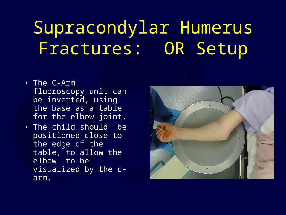
Supracondylar Humerus Fractures: OR Setup
• The C-Arm fluoroscopy unit can be inverted, using the base as a table for the elbow joint.
• The child should be positioned close to the edge of the table, to allow the elbow to be visualized by the c-arm.

Supracondylar Elbow Fractures:Type 2 with Valgus Impaction• During reduction of
medially impacted fractures, valgus force should be applied to address this deformity.

Type 3 Supracondylar Fracture

Type 3 Supracondylar Fracture,Operative Reduction
• Closed reduction with flexion
• AP view with elbow held in flexed position to maintain reduction.

Supracondylar Elbow Fractures:Type 2 with Valgus Impaction
• The elbow may need to be held in a hyperflexed position to maintain the reduction during pinning.
• The pins are placed with the elbow held in this position

Medial Impaction Fracture
Type II fracture with medial impaction – not recognized and varus / extension not reduced

Medial Impaction Fracture
Cubitus varus 2 years later

Lateral Pin Placement
• AP and Lateral views with 2 pins

C-arm Views• Oblique views with the C-arm can be
useful to help verify the reduction

Supracondylar Fracture: Pin Fixation
• Different authors have recommended different pin fixation methods.
• The medial pin can injury the ulnar nerve. Some advocate 2 lateral pins to avoid injuring the median nerve.
• If the lateral pins are placed close together at the fracture site, the pins may not provide much resistance to rotation and further displacement. If 2 lateral pins are used, they should be widely spaced at the fracture site.
• Some recommend one lateral, and one medial pin

Supracondylar Humerus Fractures- Pin Fixation
• Many children have anterior subluxation of the ulnar nerve with hyperflexion of the elbow
• Some recommend place two lateral pins, assess fracture stability
• If unstable then extend elbow to take tension off ulnar nerve and place medial pin

Supracondylar Humerus Fractures
• After the pins have been placed, and a stable reduction obtained, the elbow can be extended to review the AP radiograph. Baumann’s angle can be assessed on these radiographs, although there can be a wide range of normal values for this measurement.
• With the elbow extended, the carrying angle of the elbow should be reviewed, and clinical comparison as well as radiograph comparison can be performed to assure an adequate reduction.

Supracondylar Humerus Fractures
• If pin fixation is used, the pins are usually bent and cut outside the skin.
• The skin is protected from the pins by placing felt pad around the pins.
• The arm is immobilized.• The pins are removed in the clinic 3 to 3 1/2
weeks later.• If adequate callus formation is present, gentle
range of motion exercises are initiated.• In most cases, full recovery of motion can be
expected.

Supracondylar Humerus Fractures: Indications for
Open Reduction• Inadequate reduction
with closed methods• Vascular injury• Open fractures

Supracondylar Humerus Fractures: Complications
• Compartment syndrome• Vascular injury /
compromise• Loss of reduction /
Malunion –cubitus varus• Loss of motion• Pin track infection• Neurovascular injury with
pin placement

Supracondylar Humerus Fractures- Flexion Type
• Rare, only 2%• Distal fracture
fragment anterior,flexed
• Reduce in extension• Pin or immobilize in
extension

Distal Humeral Complete Physeal Separation
• Often in very young children
• May be sign of NAT• Swollen elbow,“muffled
crepitance” on exam• Through area of wider
cross sectional area than SC humerus fx
• Restore alignment, may need pinning
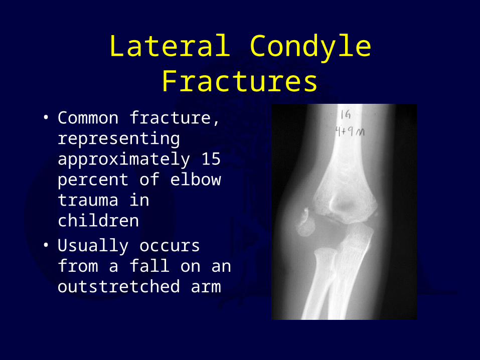
Lateral Condyle Fractures
• Common fracture, representing approximately 15 percent of elbow trauma in children
• Usually occurs from a fall on an outstretched arm

Lateral Condyle Fractures: Jakob Classification
• Type 1: Non-displaced fracture. Fracture line does not cross through the articular surface
• Type 2: Minimally displaced. Fracture extends to the articular surface, but the capitellum is not rotated or significantly displaced
• Type 3: Completely displaced. Fracture extends to the articular surface, and the capitellum is rotated and significantly displaced

Lateral Condyle Elbow Fractures: Treatment
• Type 1: • Oblique radiographs
may be necessary to confirm that this is not displaced. Frequent radiographs in the cast are necessary to ensure that the fracture does not displace in the cast.

Lateral Condyle Fractures: Jakob Classification
• Type 2: • If displaced more than 2 mm on
any radiograph (AP / Lateral / Oblique views), this will probably require reduction and pinning. Closed reduction and percutaneous pinning can be attempted, but the reduction at the articular surface must be anatomic.
• If displaced, and the articular surface is not congruous, ORIF is necessary
Fracture line exiting posterior metaphysis (arrow) typical for lateral condyle fractures

Lateral Condyle Fractures: Jakob Classification
• Type 3: • ORIF is necessary.
• A lateral Kocher approach is used for reduction, and pins are placed to maintain the reduction.
• Careful dissection needed to preserve soft tissue attachments (and thus blood supply) to the lateral condyle fragment

Lateral Condyle ORIF

Lateral Condyle Fractures:Complications
• Non-union: This usually occurs if the patient is not evaluated by a physician, or the fracture displaced despite casting
• This complication is well-described in fractures which were displaced more than 2 mm, and not treated with pin fixation
• Late complication of progressive valgus and ulnar neuropathy reported

Lateral Condyle Fractures - Complications
• AVN can occur after excessive surgical dissection
• Cubitus varus can occur, may be because of malreduction or a result of lateral column overgrowth

Medial Epicondyle Fracture
• Represents 5-10 percent of pediatric elbow fractures
• Occurs with valgus stress to the elbow, which avulses the medial epicondyle
• Frequently associated with an elbow dislocation

Medial Epicondyle Fracture:Classification and Treatment
• Nondisplaced and minimally displaced (less than 5 mm of displacement)-May be treated without fixation, and early motion to avoid stiffness.
• Displaced more than 5 mm. Treatment is controversial, with some recommending operative, and others recommending non-operative treatment. Some have suggested that surgery is indicated in the presence of valgus instability, or in patients who are throwing athletes.
• Only absolute indication is entrapped fragment after dislocation with incongruent elbow joint
• Long term studies – favor nonoperative treatment

Medial Epicondyle Fracture:Elbow Dislocation with Medial Epicondyle Avulsion
Medial EpicondyleAvulsion
After elbow reduction, medial epicondyleavulsion fragment is obvious

Medial Epicondyle Fracture:• Elbow dislocation with medial epicondyle
avulsion, treated with ORIF.

Displaced Medial Epicondyle Fracture
• 12 year old female UE weight bearing athlete
• Treated nonoperatively
• Full motion, no valgus instability at 6 weeks
• Returned to competition at 8 weeks

Olecranon Fractures
• Relatively rare fracture in children (increased incidence in children with OI)
• May be associated with elbow subluxation/ dislocation, or radial head fracture.
• The diagnosis may be difficult in a younger child, as the olecranon does not ossify until 8-9 years.
• In older children, the fracture may occur through the olecranon physis.
• Anatomic reduction is necessary in displaced fractures, restore active elbow extension.

Olecranon Fractures
• Olecranon fracture treated with ORIF in 14 year old, with tension band fixation.

Rare Distal Humeral Fractures• Lateral Epicondyle: rare,
usually represent a small avulsion fracture. Treated with early immobilization.
• T-Condylar fractures: These occur in patients that are almost skeletally mature. Treatment is similar to adult intra-articular elbow fractures.
• Medial Condyle: rare, treated with ORIF if displaced.

Proximal Radial Fractures
• 1% of children’s fractures• 90% involve physis or neck• Normally some angulation of head to radial
shaft (0-15 degrees)• No ligaments attach to head or neck• Much of radial neck extraarticular (no
effusion with fracture)

Proximal Radial Fractures - Types
• Valgus fractures – Salter I or II (intraarticular fractures rare)
• Metaphyseal fractures• Associated with elbow
dislocations or proximal ulna fractures
• Can be completely displaced, rotated

Proximal Radius Fractures- Treatment
• Greater than 30 degrees angulation- manipulate
• Usually can obtain acceptable reduction in fractures with less than 60 degrees initial angulation
• Traction, varus force in supination & extension, flex and pronate
• Ace wrap or Esmarch reduction

Proximal Radius Fractures- Treatment
• Unable to reduce closed
• Percutaneous pin reduction
• Intramedullary pin reduction
• Open reduction via lateral approach

Completely Displaced, Malrotated Radial Neck Fracture
After closed reduction the articular surface (arrow) is facing distally, 180 degrees malrotated

Proximal Radial Fractures - Complications
• Loss of forearm rotation• Radial head overgrowth• Premature physeal closure – valgus• Nonunion of radial neck rare• AVN• Proximal synostosis

Monteggia Lesions: Ulnar Fracture- Radial Head Dislocation
Bado classification
• Type I – anterior radial head dislocation
• Type II – posterior radial head dislocation
• Type III – lateral radial head dislocation
• Type IV – associated fracture of radius

Monteggia Lesions
• Most important is to make the diagnosis initially
• Radiocapitellar line critical
• A commonly missed diagnosis
• Every ulna fracture should have good elbow joint radiographs to avoid missing Monteggia lesion

Monteggia Lesions
• Be wary of plastic deformation of ulna or minimally displaced ulna fracture with radial head dislocation
• On lateral radiograph the ulna should be straight
Note anterior bow of ulnar shaft, and anterior radial head dislocation
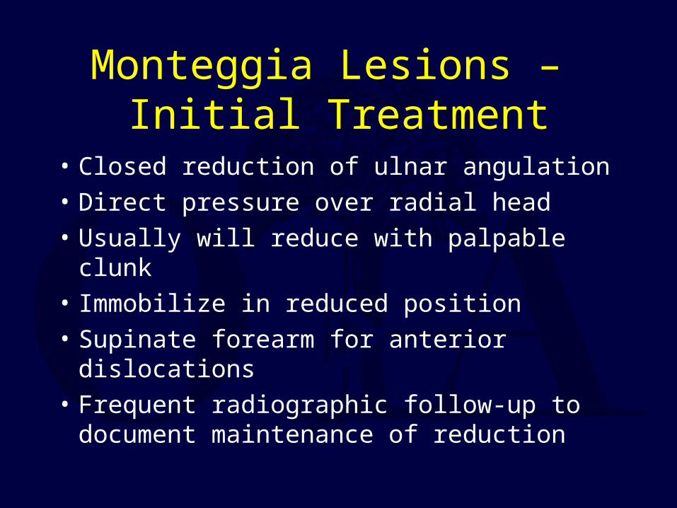
Monteggia Lesions – Initial Treatment
• Closed reduction of ulnar angulation• Direct pressure over radial head• Usually will reduce with palpable clunk• Immobilize in reduced position• Supinate forearm for anterior dislocations• Frequent radiographic follow-up to
document maintenance of reduction

Monteggia Lesions
• If unable to obtain or maintain reduction of radial head
• Operative stabilization of ulnar fracture to correct angulation (oblique fractures may need plate fixation)
• Assess radial head stability- flexion may help for anterior dislocation

Missed Monteggia Lesion
Healed prox ulna fx with anterior bow
Anterior radial head dislocation and heterotopic ossification

Missed Monteggia Lesions- Possible Long Term Sequelae
• Progressive valgus• Proximal radial migration
with disruption of normal forearm and distal radioulnar joint mechanics
• Posterior interosseous nerve traction palsy
• Collateral ligament instability

Missed Monteggia Lesions – Treatment Options
• Annular ligament reconstructions – Bell-Tawse, fascia lata, Peterson
• Ulnar osteotomy • Combination• Transcapitellar pinning
– be wary of possible pin breakage

Missed Monteggia – Ulnar Osteotomy and Radiocapitellar Pin
Return to Pediatrics Index