oxygen therapy learning module cat.1 and 2, 2016 · oxygen therapy learning module for category 1...
TRANSCRIPT

______
N
______
___
Manual Number:
Site:
Oxygen Therapy for Acute Adult Inpatients
Learning Module for
Allied Health Staff (Category 1 and 2)
Allied Health Services February 5, 2016

Oxygen Therapy Learning Module for Category 1 and 2 Staff 2
This document has been reviewed and revised in 2015 by an Allied Health provincial multi-disciplinary group to reflect the needs of all areas of the province. It is intended for the use of adult acute care Allied Health Staff across AHS and is based on previous educational material produced in the Calgary Zone. The original document was developed in 2006 by a group of Calgary Health Region staff including Physical Therapists, Management and Program Facilitators. In 2013 it was reviewed and revised by the Allied Health Educators of the Calgary Zone, AHS.
Copyright © (2015) Alberta Health Services. This material is protected by Canadian and other international copyright laws. All rights reserved. This material may not be copied, published, distributed or reproduced in any way in whole or in part without the express written permission of Alberta Health Services (please contact Health Professions, Strategy and Practice Senior Practice Lead –Physiotherapy June Norris at 780-735-3481/ [email protected]). This material is intended for general information only and is provided on an "as is", "where is" basis. Although reasonable efforts were made to confirm the accuracy of the information, Alberta Health Services does not make any representation or warranty, express, implied or statutory, as to the accuracy, reliability, completeness, applicability or fitness for a particular purpose of such information. This material is not a substitute for the advice of a qualified health professional. Alberta Health Services expressly disclaims all liability for the use of these materials, and for any claims, actions, demands or suits arising from such use. Allied Health Services Februray 5, 2016

Oxygen Therapy Learning Module for Category 1 and 2 Staff 3
Table of Contents Page
Introduction……………………………………………………………………………..………..... 4
Purpose of Education…………………………………………...……………………..…………... 5
Learning Objectives.…………………………………………………………………..…………... 6
Learning Resources…………………………………………………………………..…………… 6
Definitions and Abbreviations…………………………………………………………………….. 7
Instructions for Completion…………………………………………………………..…………... 8
Section One: Respiratory Physiology and Anatomy………………………………..……………. 9
Section Two: Role of Oxygen Therapy in the Acute Care Setting………………………………... 13
Section Three: Activity and Oxygen………………………………………………..…………… 16
Section Four: Physical Assessment and Signs and Symptoms of Respiratory Distress….…….... 22
Section Five: Determination of Level of Risk and Documentation………….…………………... 26
Section Six: Oxygen Equipment………………...…………………………….…………….…….. 32
Section Seven: Oxygen Delivery System……………………………………..…………….……. 41
Section Eight: Pulse Oximetry (SpO2)………………………...………………………….…….. 45
Section Nine: Allied Health/Rehabilitation Services Acute Care Sites Emergency Response…… 49
Appendix A Self Test Answer Key ……………..……………………………………………………...... 52
Appendix B Post Quiz…..………………………………………………………………….....…………… 53
Appendix C Practical Component ……….………………..……………………………….……………... 58
Appendix D Performance Checklist ……………………………………………………………………... 59
Appendix E References …………………………………………………………………..………………. 60
Allied Health Services Februray 5, 2016

Oxygen Therapy Learning Module for Category 1 and 2 Staff 4
Introduction
In compliance with the Oxygen Management - Allied Health Adult Acute Care Inpatients Guideline, this module is designed to provide Allied Health (excluding respiratory therapists) with the necessary knowledge and skills to provide and support safe delivery of oxygen therapy.
Oxygen is an odorless, colorless, tasteless gas constituting one fifth of the earth’s atmosphere and is essential to the life of living organisms. Oxygen is also a medical intervention in the prevention and treatment of hypoxia.
Oxygen therapy is an area of patient care where recognition of its need and efficient administration can have a significant impact on a patient’s well being. Caring safely for patients receiving oxygen therapy is a vital part of Allied Health practice.
A patient not requiring supplemental oxygen therapy will receive 21% oxygen from the air. This amount of oxygen is adequate provided that the airway is not compromised and there are enough oxygen carrying components in the blood. As well, the cardiovascular system must be intact and able to circulate blood to all body tissues. If any of these systems fail, higher concentrations of oxygen must be delivered to the patient’s lungs. This makes it more likely that adequate levels of oxygen will reach all vital body tissues necessary to sustain life.
Oxygen therapy should be provided continuously unless the need has been shown to be associated only with
specific situations (e.g. exercise and sleep). Care should be taken to avoid interruption of oxygen therapy in situations including ambulation or transport for procedures.2
ALWAYS REMEMBER OXYGEN THERAPY IS A MEDICAL INTERVENTION Ensure correct patient, correct flow rate, correct device, and correct connection to an oxygen source. If using a portable system check sufficient O2 reserve is available. Without oxygen we cannot live.
Without supplemental oxygen therapy, many patients cannot survive.
Allied Health Services Februray 5, 2016

Oxygen Therapy Learning Module for Category 1 and 2 Staff 5
Purpose of the Educational Program
This learning module will familiarize the learner with the information necessary to work safely with patients who are receiving oxygen therapy. Participants must successfully complete the theory component of this module by achieving 90% on the qualification quiz. The quiz is open book format. The practical component of the oxygen training includes demonstration of the correct application of oxygen therapy.
This module is intended to promote patient safety by enhancing the knowledge and skills of Allied Health staff who care for adult patients receiving oxygen therapy. Discussion and consultation with nursing and respiratory therapy is recommended whenever necessary. This module is not an educational package to ensure competency of health professionals to treat cardiopulmonary patients with chest physiotherapy techniques, or to complete full chest assessments.
The educational program for oxygen therapy has been standardized. It is comprised of four components:
• Oxygen Therapy Learning Module • Review of Oxygen Management – Allied Health Adult Acute Care Inpatients Guideline • Qualification quiz • Practical component
Who Should Complete This Module
This module is mandatory for the following staff, working in acute care adult inpatient settings.
Category 1 Staff • Occupational Therapists • Physical Therapists • Speech Language Pathologists
Category 2 Staff
• Therapist Assistants
Demonstration and maintenance of competency is done annually by reading the module and completing the quiz. As part of orientation, new staff will also do a practical component under the guidance of a local educator or designate. The practical component may be required yearly at the discretion of Allied Health management.
Students Students of category 1 staff (OT, PT, SLP) and students of category 2 staff (Therapist Assistants) are able to provide oxygen therapy, as set out in the Oxygen Management - Allied Health Adult Acute Care Inpatients Guideline, under the supervision (direct, indirect, or remote) of category 1 staff who have demonstrated oxygen therapy competency. The category 1 supervisor is responsible for determining the competence of the student, with respect to knowledge and skill in providing oxygen therapy and for providing the appropriate amount and type of supervision.11
Allied Health Services Februray 5, 2016

Oxygen Therapy Learning Module for Category 1 and 2 Staff 6
Learning Objectives
On completion of the learning module, the learner will be able to:
1. Differentiate between ventilation, internal respiration, and external respiration. 2. Identify the major muscles of respiration. 3. Identify factors affecting external and internal respiration. 4. Define hypoxemia and hypoxia. 5. Identify the conditions and indications for oxygen therapy. 6. Identify the dangers, problems and contraindications for oxygen. 7. Identify the normal response to physical activity. 8. Identify the communication components needed for safe assignment of patients with oxygen to a
category 2 staff member. 9. Identify the components required for physical assessment/monitoring of patients. 10. Identify the normal adult ranges for vital signs. 11. Identify the signs of respiratory instability/distress and when to obtain assistance. 12. Determine the level of risk for a patient receiving oxygen and differentiate between low, moderate, and
high risk. 13. Identify the key elements for oxygen therapy documentation. 14. Identify the different oxygen tanks. 15. Identify the reserve volume in an oxygen tank and safe duration time for use. 16. Understand how to safely fill a liquid oxygen tank. 17. Identify injury prevention measures when working with oxygen. 18. Differentiate between low flow and high flow oxygen delivery systems. 19. Identify different oxygen delivery devices. 20. Define pulse oximetry. 21. Identify the indications for pulse oximetry. 22. Understand the limitations of pulse oximetry. 23. Identify when to access medical assistance. 24. Differentiate between initiating a Code Blue and calling for medical assistance. 25. Understand the importance of the Goals of Care.
Learning Resources
Resources that will assist the learner in oxygen safety:
Clinical Leaders and Clinical Educators
Books: Chung,F., Reid,W.D., (2004) Cardiopulmonary Physical Therapy SLACK Incorporated New Jersey DeTurk,W.E., Cahalin,L.P., (2004) Cardiovascular and Pulmonary Physical Therapy - An Evidence-Based Approach McGraw-Hill Companies Inc. USA Frownfelter, D.L.,Dean, E., (2006) Cardiovascular and Pulmonary Physical Therapy . Mosby Inc. Kisner,C., Colby,L.A., (1990) Therapeutic Exercise Foundations and Techniques, Davis and Co. Philadelphia PA
Allied Health Services Februray 5, 2016

Oxygen Therapy Learning Module for Category 1 and 2 Staff 7
Definitions and Abbreviations
For the purposes of this learning module:
Acute care adult inpatient settings – include rural and community hospitals, regional hospitals, metropolitan hospitals, and tertiary hospitals.
Adult - 18 years or older.
Artificial Airway - e.g. tracheostomy tube, endotracheal tube, or nasotracheal tube.
Category 1 Staff - professionals who work with adult inpatients requiring oxygen therapy. The following health care professionals are included in this category: Physiotherapists, Occupational Therapists, and Speech-Language Pathologists.11
Category 2 Staff - health care providers who work with adult inpatients requiring oxygen therapy. The following health care providers are included in this category: Therapist Assistants. 11
FiO2 - fraction of inspired oxygen.
Hypoxemia - a decreased oxygen tension (Pa02) in the blood below the normal range.
Hypoxia - an inadequate supply of oxygen to the tissue or cell.
Initiate - place patients/clients on supplemental oxygen, who previously were not on supplemental oxygen.
Most Responsible Health Practitioner – means the health practitioner who has responsibility and accountability for the specific treatment/procedure(s) provided to a patient and who is authorized by Alberta Health Services to perform the duties required to fulfill the delivery of such a treatment/procedure(s), within the scope of his/her practice.11 Oxygen Therapy Risk Assessment - clinical determination of the patient’s/client’s inability to tolerate interruption of therapeutic oxygen administration.
PaCO2 - partial pressure of carbon dioxide in arterial circulation.
Pulse Oximetry – is the measurement of the oxygen saturation of hemoglobin in arterial blood, with 100% as the maximum reading possible. It is a measure of the average amount of oxygen bound to each hemoglobin molecule.
Respiratory Acidosis - a decrease in pH in the blood due to an increase in partial pressure of carbon dioxide in the arterial blood (PaCo2).
Significant Change - a variation in values outside of a predefined range, set by an appropriate regulated health professional (category 1 staff, physician, nurse practitioner, registered nurse, licensed practical nurse, or respiratory therapist). The predefined range will be individual for each patient.
SaO2 - arterial oxygen saturation of hemoglobin.
Allied Health Services Februray 5, 2016
Oxygen Therapy Learning Module for Category 1 and 2 Staff 8
SpO2 - the measurement of functional saturation of oxyhemoglobin. This measurement is obtained non- invasively (i.e. via pulse oximeter). Student - an individual enrolled in an entry-level health care discipline/education program, leading to initial entry-to-practice as a regulated or non-regulated health care provider.11
Titration - the process of gradually adjusting the dose of oxygen until the desired effect is achieved.
Valid Oxygen Order - an order issued by a health professional authorized to provide medication orders within AHS.
Instruction for completion
1. Please do not write in this module. Record your answers to the self-test on a separate piece of paper.
2. Review the oxygen therapy learning objectives identified on page 6. Note: if this is a re-certification, you can skip to #4 and only review the learning objectives if answers are incorrect.
3. Review each section of the learning module and consult additional cited resources as needed.
4. Complete the self tests at the end of each section of the module. Compare answers with the
information provided in the module. Refer to Appendix A for the answer key. Review sections of the module as needed for clarification.
5. Complete the post quiz “Oxygen Therapy Quiz for Allied Health Category 1 and 2” through
MyLearning Link. Print and submit a copy of the completed course page to your clinical educator or designate. Note: The quiz is open book. The pass mark is 90% (30/33 correct answers). Errors will cue you to review the pertinent information and make corrections.
6. New staff are also required to complete the practical component and the performance checklist (page
58 & 59). Submit the checklist to your clinical educator or designate once completed.
Allied Health Services Februray 5, 2016
Oxygen Therapy Learning Module for Category 1 and 2 Staff 9
Section One
Respiratory Physiology and Anatomy
Learning Objectives Upon completion of this section, the learner will be able to:
1. Differentiate between ventilation, internal respiration and external respiration. 2. Identify the major muscles of respiration. 3. Identify factors affecting external and internal respiration.
Respiratory Physiology
Respiration is the exchange of gases between the atmosphere, the blood, and the cells. These are the three processes involved in this exchange:
A. Ventilation – breathing in and out. B. External Respiration – how the lungs exchange oxygen (O2) and eliminate carbon dioxide (CO2). C. Internal Respiration – how the cells exchange oxygen and carbon dioxide.
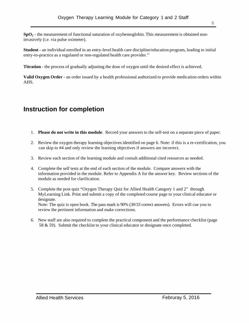
A. Ventilation - Air in / Air out
This is the movement of air in and out of the body during inspiration and expiration. During inspiration air is drawn in through the nose, mouth, and throat, through the larynx and down the trachea. The trachea then branches into right and left primary bronchus, followed by secondary bronchi that enter both the right and left lobes of the lungs. The bronchi continue to divide, becoming smaller and smaller, very much resembling the branches of a tree, ending at the smallest air passage called the terminal bronchiole. It is here, at the terminal bronchiole, that the tiny air sacs called alveoli are located and where the exchange of gases takes place.
B. External Respiration – O2 in and CO2 out
This is the exchange of O2 from the alveoli in the lungs, for CO2 from the blood capillaries which surround the alveoli. The thin membranes and the differences in gas pressures on each side of these membranes cause O2 to enter the bloodstream from the alveoli, and CO2 to enter the alveoli from the bloodstream. The CO2 is removed from the alveoli when the person exhales.
Allied Health Services Februray 5, 2016

Oxygen Therapy Learning Module for Category 1 and 2 Staff 10
Efficiency of external respiration depends on various factors. Some of these are:
• Total surface area available for O2 – CO2 exchange: any disease or injury which decreases or
obstructs the gas exchange surface area of the lung, decreases the efficiency of external respiration. An example of this would be COPD.
• Minute volume of respiration: minute volume is the total volume of air taken in during a minute of
breathing. Slow respiratory rates and/or shallow respirations decrease the minute volume, thereby reducing the efficiency of external respiration. For example sedative drugs can result in a slower respiratory rate, or respirations may be shallow due to pain from abdominal surgery.
C. Internal Respiration
This is the exchange of O2 and CO2 between the blood and the cells. Once oxygen diffuses from the alveoli into the capillaries, it binds with the hemoglobin in red blood cells. The oxygenated blood is transported to the left side of the heart via the pulmonary veins, and then pumped throughout the body via the arterial system to the cells. It is in the capillaries that O2 is released from the blood to supply the cells, and CO2 is picked up for elimination.
Factors affecting internal respiration:
• Amount of O2 in the blood • Medical conditions e.g. anemia • Loss of blood e.g. trauma • Impairment of circulation • Presence of drugs or chemicals that impair cellular O2 exchange
(Reference 6)
Allied Health Services Februray 5, 2016
Oxygen Therapy Learning Module for Category 1 and 2 Staff 11
Respiratory Anatomy
The thorax (chest wall) protects the principle organs of respiration and circulation, as well as the liver and stomach. The posterior thorax is formed by the 12 thoracic vertebrae and the posterior surface of the 12 rib pairs. The anterior thorax is formed by the sternum and the costo-chondral cartilage. The lateral thorax is formed by the ribs.16 The thorax provides the bony sites of attachment for the muscles of ventilation which mechanically enlarge the thorax for inspiration (breathing in) or compress the thorax for expiration (breathing out).
The diaphragm is the principle muscle of respiration and separates the thoracic and abdominal cavities. During inspiration, the diaphragm contracts to force the abdominal contents away from the thorax (abdomen moves out) and elevates the ribs in a bucket handle fashion. The external intercostals and part of the internal intercostals also contract during inspiration. Expiration is achieved through the elastic recoil of the thorax and abdominal wall. During forced deep breathing or labored breathing, a large number of accessory muscles may also contract.
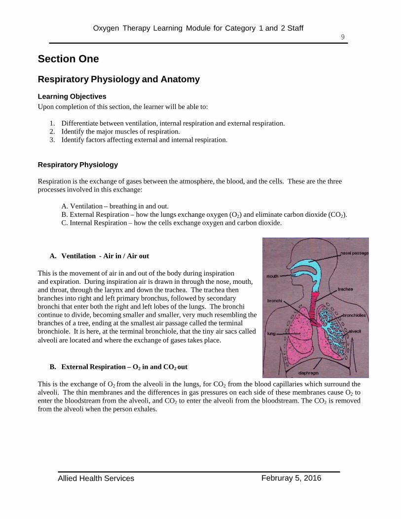
Inspiratory accessory muscles include the scalenes, sternocleidomastoids, upper trapezius, pectoralis major, and erector spinae muscles. Expiratory accessory muscles include the rectus abdominis, transverse abdominis, internal obliques, external obliques and pectoralis major muscles.
Respiratory Muscles (Anterior View)
(Reference 5) Posterior view not shown – the trapezius muscles form a diamond shaped sheet extending from the head down the back and out to both shoulders. The erector spinae is a large muscle extending from the sacrum to the skull.
For further information on the respiratory muscles please refer to DeTurk - Cardiovascular and Pulmonary Physical Therapy – an Evidence Based Approach.13
Allied Health Services Februray 5, 2016

Oxygen Therapy Learning Module for Category 1 and 2 Staff 12
Self Test Section One
1. Respiration is the exchange of gases between the , the and the cells.
A. Atmosphere, Blood
B. Air, Skin
C. Atmosphere, Skin
D. None of the above
2. The principle muscle(s) for respiration is the:
A. Sternocleidomastoid
B. Pectoralis major
C. Diaphragm
D. Abdominal muscles
3. Normal expiration is achieved through:
A. Contraction of the pectoralis major
B. Elastic recoil of the thorax and abdominal wall
C. Contraction of the sternocleidomastoid
D. Relaxation of the pectoralis major
Allied Health Services Februray 5, 2016

Oxygen Therapy Learning Module for Category 1 and 2 Staff 13
SECTION TWO
Role of Oxygen Therapy in the Acute Care Setting
Learning Objectives Upon completion of this section, the learner will be able to:
4. Define hypoxemia and hypoxia. 5. Identify the conditions and indications for oxygen therapy. 6. Identify the dangers, problems and contraindications for oxygen.
Role of Oxygen Therapy in the Acute Care Setting
Hypoxemia Is an inadequate supply of oxygen in the arterial blood. In the clinical setting the pulse oximeter is used to estimate oxyhemoglobin saturation (SpO2). The acceptable SpO2 is usually greater than 90%, but may vary dependent on the patient, and the nature of the condition being treated.12 For example, COPD patients may have an acceptable oxygen saturation of 88%; therefore, always refer to individual patient specific oxygen orders. Examples of medical conditions that may cause acute hypoxemia:
• Asthma • Pneumonia • COPD exacerbation • Pneumothorax • Heart failure • Pulmonary edema • Pleural effusions • Pulmonary emboli
Hypoxemia is important as it can lead to Hypoxia
Hypoxia Is an inadequate supply of oxygen to the tissue or cells. Cellular hypoxia occurs when oxygen transport fails to meet the tissue demand for oxygen. This may be due to a problem with the lungs (e.g. airway obstruction, including: secretions, foreign objects, or tumors,) hypoventilation due to disease, injury and drugs, or blood flow due to the circulatory system. Hypoxia secondary to problems with the blood (anemia) or the circulatory system (e.g. decreased cardiac output) responds poorly to oxygen therapy. 9
Indications for Oxygen in the Acute Care Setting • Hypoxemia. • To decrease the work of breathing. • To decrease myocardial work. This may be done to target a specific organ in order to prevent ischemic
damage and pain.12
• Post operatively, oxygen may be ordered for a specific time frame and/or flow rate rate.
Allied Health Services Februray 5, 2016

Oxygen Therapy Learning Module for Category 1 and 2 Staff 14
Dangers, Problems, and Contraindications for Oxygen Oxygen administration can result in detrimental effects in some cases. These include:
• COPD Patients – Some patients with COPD may become increasingly hypercapnic (elevated
levels of carbon dioxide in the blood) when treated with excessive amounts of oxygen. In these patients, it is recommended to keep their SpO2 in the range of 88% to 92%. If their oxygen flow rates need to be significantly increased, clarification is required from the physician involved.
Note: Prevention of acute hypoxia should be your first priority.
Do not deprive the hypoxic COPD patient of oxygen while awaiting further medical intervention
• Bleomycin Induced Pneumonitis – Oxygen therapy is contraindicated.20 If oxygen needs to be applied, it is at very low levels (i.e. 0.5LPM). In this scenario, clarification of the parameters must be obtained from the Physician.
• Absorption Atelectasis – About 80% of the gas in the alveoli is nitrogen. If high concentrations of
oxygen are provided, the nitrogen is displaced. When the oxygen diffuses across the alveolar-capillary membrane into the blood stream, the nitrogen is no longer present to distend the alveoli, which contributes to their collapse and atelectasis.12
• Oxygen Toxicity – High levels of oxygen provided for more than 24 hours usually results in some
lung damage because of oxygen radical production. Oxygen radical production occurs because of incomplete reduction of oxygen to water. Oxygen radicals are very reactive molecules that can damage membranes, proteins and many cell structures in the lungs.12
The goal of oxygen therapy is:
To achieve an optimal arterial oxygen tension by giving the lowest possible most effective dose of oxygen, while avoiding its toxic effects. For most patients, SpO2 92% to 98% will achieve this goal.9
For COPD patients with hypoventilation, a target SpO2 of 88% to 92% may be ordered to prevent further increases in PaCO2.9
In circumstances where there is not an oxygen order in place, category 1 health care professionals may initiate oxygen based on their assessment of the patient’s condition. Consultation and an oxygen order for further oxygen therapy shall be obtained as soon as possible.11
Allied Health Services Februray 5, 2016

Oxygen Therapy Learning Module for Category 1 and 2 Staff 15
Self Test Section Two
1. Every patient needs to have SpO2 greater than 92%
A. True
B. False
2. Indications for oxygen in the acute care setting include:
A. Hypoxemia Yes No
B. To decrease the work of breathing Yes No
C. To decrease myocardial work Yes No
3. COPD patients may have an acceptable oxygen saturation of 88%
A. True
B. False
Allied Health Services Februray 5, 2016

Oxygen Therapy Learning Module for Category 1 and 2 Staff 16
Section Three
Activity and Oxygen
Learning Objectives Upon completion of this section, the learner will be able to:
7. Identify the normal response to physical activity. 8. Identify the communication components needed for safe assignment of patients with oxygen to a category 2 staff member.
Activity and Oxygen
Endurance is the ability to work for a prolonged period of time and resist fatigue. Endurance is dependent on an individual’s oxygen transport system which works to supply all muscles with the oxygen required to perform tasks. Many factors influence the oxygen transport system. For the purpose of this module the following will be considered:
System Function Normal response to exercise Heart pumps the blood increased heart rate and force of heart beat Lungs takes the O2 to the blood increased respiratory rate and deep breathing Vascular System transports the oxygen increased blood pressure Muscles utilize the oxygen increased use of oxygen delivered
An acute care patient may have pathology or be taking medications that will affect his/her response to exercise. In addition, the patient may be deconditioned.
Systems affected may include: Heart: the use of heart rate limiting drugs, decreased blood circulation, or heart disease. Lungs: limited oxygenation in lungs secondary to conditions like pneumonia, COPD, pleural effusions, pulmonary fibrosis, etc. Muscle: decreased available muscle mass makes the body work harder to do the same task.
The deconditioned body responds quickly to exercise to improve heart, lung and muscle function. To start out, a deconditioned patient may be medicated with supplemental oxygen to decrease the stress on the whole body - especially the heart.
General Considerations16
At no time should a patient be exercising using less oxygen than is used at rest. • A patient should be asked why they need to stop exercising. It may be due to muscle cramping caused by
increased oxygen demands of the muscles, versus shortness of breath. Although cardiopulmonary factors are generally considered the most important, skeletal muscle dysfunction has been increasingly recognized as a key factor that contributes to exercise intolerance. The perception of increased leg effort or discomfort is the main symptom that limits exercise in 40-45% of patients with COPD.3
• Large bodies will expend more energy (may need more rest or oxygen) than small bodies.21
• Recovery after vigorous exercise may take 5-10 minutes or more to return to baseline vitals.
Allied Health Services Februray 5, 2016

Oxygen Therapy Learning Module for Category 1 and 2 Staff 17
Monitoring a deconditioned patient during exertion
System Findings What it means Category 2
(Therapist Assistant)
Action to perform
Category 1 (Therapist) Action to perform
Heart Rate (HR) use cardiac monitor, manual pulse check or pulse oximeter (correlated with manual pulse check).
Increased Normal to a set maximum identified by the therapist.
If maximum HR reached, rest patient and monitor HR for 3 minutes.21 .If still at or near maximum, call any category 1 staff or RN.
Regulated rehabilitation professional will use their clinical judgment and assessment skills.
May require the following interventions:
• Stop treatment, let patient rest, and observe vital signs, once stable document event.
• Change position • Change oxygen level
and/or delivery system • Contact Nursing
/Respiratory Therapist.
• Contact doctor • Activate the emergency
response system at your site – see Section 9.
Decreased Be concerned if lower than set minimum.
Call any category 1 staff or RN.
No change May be on rate control drugs, rate control pacemaker or cardiac transplant patient.
Monitor Sp02 and respiratory rate closely
Lungs (SpO2) use pulse oximeter
Increased Normal after exercise
Decreased Normal within range specified by therapist.
Below specified range, rest patient, do breathing exercises, and if required increase oxygen within specified parameters. If saturation does not return to specified range, then call any category 1 staff or
RN/RT to assess. Respiratory rate (RR) count breaths
Increased Normal with exercise to set maximum identified by the category 1 staff.
Above set maximum stop activity, encourage slow deep breaths and check SpO2. If too fast or distressed call any category 1 staff or RN/RT to assess.
Decreased Normal if taking deeper breaths.
Exercise will affect one or more systems. • Therapists should set restrictions on areas of compromise for the patients. • When in doubt, ask.
Allied Health Services Februray 5, 2016

Oxygen Therapy Learning Module for Category 1 and 2 Staff 18
Oxygen titration and weaning: Titration of oxygen (process of gradually adjusting the dose of oxygen until the desired effect is achieved) can occur when the oxygen order provides instruction on titration/discontinuation. All changes in oxygen flow rate must be documented. The patient must be reassessed 10-30 minutes after the change in FiO2 (fraction of inspired oxygen) or change in flow rate. The new assessment finding(s) must be documented on the chart.
Category 2 staff (Therapist Assistants), who have demonstrated competency, may adjust oxygen and switch between oxygen supply sources for low risk patients (refer to section 5 for risk levels) under the supervision of a category 1 staff (OT,PT,SLP), who have demonstrated oxygen therapy competency. O2 flow rate and Fi02 may be increased during activity, within the guidelines set by the category 1 staff. These guidelines are patient specific and need to be communicated to the therapist assistant (TA).11 The therapist must assess the patient and communicate all the information to the TA before the TA sees the patient.
Example: Mr. X
• Must keep his oxygen saturation (SpO2) greater than 92% during activity. • The O2 may be increased from 3 to a maximum of 5 liters per minute during activity, if required • O2 flow rate should be returned to baseline within 5 minutes after exercise, as long as SpO2 is
maintained within established parameters. • SpO2 should be rechecked at least 10 minutes later. • O2 saturations must be taken prior to activity, during the activity, immediately post activity and/or after
any change in oxygen delivery. Observe patient for changes to breathing pattern and rate.
All category 1 and 2 staff are expected to check the chart prior to treating the patient. For category 2 staff, if the flow rate or FiO2 (fraction of inspired oxygen) is outside the parameters provided by the category 1 staff, they need to clarify flow rate or FiO2 with the category 1 staff.
Expectations of the Category 1 staff for assigning patients with O2 to a Therapist Assistant
1. Determine the patient’s level of risk according to the definitions on page 26 of this module
2. Determine the activities, location of activity, and parameters for treatment. Please note:
• Therapist Assistants are able to independently treat those patients in the low risk categories. • Therapist Assistants may work with high and moderate risk patients on the patient care unit, providing
they don’t adjust the level of O2, or switch between oxygen supply sources during their treatment, and the category 1 staff has deemed the patient medically stable.11
• There may be exceptional circumstances when Therapist Assistants who work exclusively in Pulmonary or Thoracic units and who have received specialized training, may switch oxygen sources for stable high risk patients. The category 2 staff member must receive direction from the category 1 staff assessing the patient. That direction will be based on discussion by the category 1 staff and with approval from the physician or nurse practitioner. Category 1 staff will document the outcome of the discussion with the attending physician or nurse practitioner.11
Allied Health Services Februray 5, 2016

Oxygen Therapy Learning Module for Category 1 and 2 Staff 19
• Therapist Assistants are unable to independently treat patients in the moderate and high risk category
off the patient care unit. They are able, under direct supervision of a category 1 staff, to provide a second set of hands. Again there may be exceptional circumstances when Therapist Assistants who work exclusively in Pulmonary or Thoracic units and who have received specialized training may switch oxygen sources, transport patients or work with them away from the nursing unit for stable moderate risk patients. The category 2 staff member must receive direction from the Category 1 staff assessing the patient. That direction will be based on discussion by the Category 1 staff and with approval from the physician or nurse practitioner Category 1 staff will document the outcome of the discussion with the attending physician or nurse practitioner.11
• Other policies and procedures exist that guide the transportation of patients with respiratory instability or on high flow oxygen away from the care unit. All staff working with these patients must be aware of these policies.
3. Communicate relevant information to the category 2 staff.
This would include the following:
• O2 liters per minute or the FiO2
• Level of risk • SpO2 range • Heart rate range • Respiratory rate range • If flow rate of O2 can be adjusted up or down to maintain specified SpO2
• Max O2 flow rate a patient can be raised to, before the category 1 staff or RN/RT must be called • Indicate if the O2 is not to be changed • Frequency of treatment • Treatment goals (these should be specific and time limited) • Treatment activity (this needs to be specific, with identified ways to progress)
Allied Health Services Februray 5, 2016

Oxygen Therapy Learning Module for Category 1 and 2 Staff 20
An Example of Information for Therapist Assistant Assignment,
which may be utilized at your site with adaptions.
Allied Health Services Februray 5, 2016

Oxygen Therapy Learning Module for Category 1 and 2 Staff 21
Self Test Section Three
1. The normal response of the heart rate to exercise is
A. No change
B. Decreases
C. Increases
2. During exercise your patient’s heart rate increases to maximum (as set by the therapist). What would you do?
A. Call immediately for medical intervention.
B. Continue exercise at the same level.
C. Rest the patient for 3 minutes while monitoring heart rate.
3. During exercise your patient’s respiratory rate increases above the maximum (as set by the therapist). What would you do?
A. Explain to the patient that increased respiratory rate is a normal response to exercise, while encouraging the patient to continue the exercise program at the same intensity. B . Rest the patient, check SpO2 and encourage slow deep breaths. C. Encourage patient to continue the exercise program at the same intensity and document the results in the medical chart.
4. What action would you take if during exercise your patient’s SpO2 decreases below the range set by the physician?
A. Encourage deep breathing while maintaining the current exercise program B. Rest the patient. Encourage deep breaths and increase oxygen flow rate if required, up to prescribed maximum.
C. Change to a less intense exercise and monitor.
5. What should the Therapist Assistant do, when they are assigned a client on oxygen and have not been given all the information needed?
A. Treat the patient and contact the therapist afterwards.
B. Provide no service and contact the therapist for clarification.
Allied Health Services Februray 5, 2016

Oxygen Therapy Learning Module for Category 1 and 2 Staff 22
Section Four Physical Assessment, Monitoring and Signs of Respiratory Distress
Learning Objectives Upon completion of this section, the learner will be able to:
9. Identify the components required for physical assessment/monitoring of patients. 10. Identify the normal adult ranges for vital signs. 11. Identify the signs of respiratory instability/distress and when to obtain assistance.
Physical assessment and monitoring
All staff need to be able to monitor patients and immediately identify values outside the norm (as defined for the patient), trends in values, and signs of respiratory distress. Measurement and observation of these parameters is to be completed pre and post rehabilitation intervention, on all patients receiving oxygen.
• Respiratory Rate (RR): Is usually assessed by observing the movement of the chest wall and /or the
abdomen. It is very important that the patient is unaware that this measure is being taken and that the health professional does not place his or her hand on the patient’s chest wall or abdomen to take it. If the patient becomes aware, he/she may alter his/her RR and an inaccurate measurement will occur. Respiratory rate should increase during exercise.
• Respiratory Pattern:
o Normal breathing – Upper chest (thoracic) or abdominal (diaphragmatic) pattern. Upper chest – the thorax elevates and expands during inspiration and the abdomen remains
relatively motionless. Abdominal – during inspiration the abdomen expands and the thorax remains relatively
motionless.
o Abnormal Breathing Excessive accessory muscle use - excessive upper chest motion with increased use of the
sternocleidomastoid, scalene and other accessory muscles of inspiration. Paradoxical breathing pattern – is the reverse pattern of normal breathing.
• Oxygen flow rate or Fraction of inspired Oxygen (FiO2): refer to the section in manual on oxygen
equipment - page 32. • Oxygen delivery device: refer to the section in manual on oxygen delivery devices - page 41.
• Connection of oxygen delivery device to oxygen source: refer to the section in the manual on oxygen
equipment - page 32. • Saturation of oxygen by pulse oximetry (SpO2): refer to the section in the manual on pulse oximetry -
page 45.
Allied Health Services Februray 5, 2016

Oxygen Therapy Learning Module for Category 1 and 2 Staff 23
• Pulse: In most cases the radial pulse is used. Two or three fingers (not your thumb) are placed just lateral
to the flexor tendons on the radial side of the wrist. Gentle pressure is applied and released until the pulse is palpated and counted for 15 seconds. The value is then multiplied by 4 to determine the beats per minute. If the pulse is irregular, count the pulse for the full minute.
• Blood Pressure (BP): Is not done routinely on all patients.
Normal Adult Ranges Respiratory Rate 12 to 16 breaths per minute
Heart Rate 60 to 100 beats per minute SpO2 95 to 100 % BP approximately 120 systolic; approximately 80 diastolic23
(Reference 5)
Signs of Respiratory Instability/Distress
Respiratory instability or distress occurs when the respiratory system cannot eliminate enough carbon dioxide to prevent respiratory acidosis and/or pick up enough oxygen, resulting in hypoxemia.
There are many causes of respiratory instability/distress. Some examples are lung disorders (e.g. COPD or asthma), mechanical disorders (e.g. spinal cord injury or chest trauma), and depression or overstimulation of the respiratory center, caused by drugs, CVA, metabolic issues or head injury.
During rehabilitation treatment there may be an increased demand for oxygen. If this is not readily available, it may lead to respiratory instability/distress.
Signs of Respiratory Instability/Distress • Oxygen Saturation
• consistently less than the prescribed acceptable range indicated in an order from the most responsible health practitioner or • less than 90%, if no range has been identified, on supplementary oxygen 11
• Respiratory Rate • less than eight or greater than thirty11
• a significant change from baseline respiratory rate as per clinical judgment11
• Color Changes – A bluish color seen around the mouth, on the inside of the lips, or on the fingernails, may occur when a person is not getting as much oxygen as needed. The color of the skin may also appear pale or gray.25
• Evidence of excessive use of accessory muscles of respiration, evidence of forced exhalation or increased work of breathing.11 • Grunting – A grunting sound can be heard each time the person exhales. This grunting is the body’s way to
try to keep air in the lungs so they will stay open.25 • Nasal Flaring – The nostrils spreading open while breathing may indicate that the person has to work
harder to breathe.25
• Accessory Muscle Use – See illustration below. • Paradoxical Breathing Pattern
Allied Health Services Februray 5, 2016

Oxygen Therapy Learning Module for Category 1 and 2 Staff 24
• Retractions (Indrawing) – The chest appears to sink in just below the neck and/or under the breast bone
with each breath in an attempt to bring more air into the lungs.25
• Excessive secretions requiring suction.11
• Sweating – There may be increased sweat on the head, but the skin does not feel warm to touch. More often the skin may feel cold and clammy. This may happen when the breathing rate is very fast.25
• Wheezing – A tight whistling or musical sound heard with each breath. This may indicate that the air passages may be smaller, making it more difficult to breathe.25
A patient who is showing signs of respiratory instability/distress is considered to be breathing inadequately and should be treated accordingly. Treatment consists of providing adequate oxygenation and reversing the respiratory instability. If a category 2 staff encounters a patient experiencing one or more of these symptoms they should immediately stop any activity and notify the nurse, or any category 1 staff. The category 1 staff or RN should do an assessment of the situation and treat accordingly. If needed, they should immediately page Respiratory Therapy or activate the emergency response system at your site – see Section 9.
Under no circumstances should a patient in respiratory distress, who has not been assessed
by a category 1 staff or RN, be sent back to the nursing unit.
Note: A known unstable airway can cause respiratory instability. All of the following can place a patient at greater risk for unstable airway: retropharyngeal abscess, laryngospasm, smoke inhalation & facial burns from a thermal or chemical injury, neck masses, epiglottitis, inflammation by whatever cause, foreign body, laryngitis, asthma and COPD exacerbations, tracheostomy, laryngectomy or tracheal stent, chest injuries, fractured ribs causing hemo & pneumo thoracies.
A patient with a diagnosis of emphysema. Note the generalized muscle wasting, shortness of breath with pursed lip breathing and use of accessory muscles with a forward leaning posture.13
Allied Health Services Februray 5, 2016

Oxygen Therapy Learning Module for Category 1 and 2 Staff 25
Self Test Section Four
1. Match the vital signs with the correct normal adult range.
1. SpO2 % A. 12 to 16 2. BP B. 95 to 100 3. Respiratory Rate per minute C. 60 to 100 4. Heart Rate per minute D. Approximately 120 systolic /
Approximately 80 diastolic
2.Which of the following are signs of respiratory distress?
A. SpO2 consistently less than prescribed range Yes No
B. Respiratory rate outside the patients prescribed range Yes No
C. SpO2 greater than 98% Yes No
D. Patient struggling to clear secretions (i.e. choking) Yes No
E. Excessive use of accessory muscles Yes No
Allied Health Services Februray 5, 2016

Oxygen Therapy Learning Module for Category 1 and 2 Staff 26
Section Five
Determination of Level of Risk and Documentation
Learning Objectives Upon completion of this section, the learner will be able to:
12. Determine the level of risk for a patient receiving oxygen and differentiate between low, moderate, and
high risk. 13. Identify the key elements for oxygen therapy documentation.
Determination of level of risk The oxygen therapy risk assessment is the clinical determination of the patient’s/client’s inability to tolerate interruption of therapeutic oxygen administration. It is important that
• Category 1 staff can determine the level of risk and communicate this to the category 2 staff. • Category 2 staff can differentiate between low, moderate, and high risk.
High Risk:
• Patient/client requires greater than eight liters per minute (8 Lpm) of oxygen (a lower oxygen requirement may be determined by the site or most responsible health practitioner); or
• Patient/client requires greater than 50 per cent concentration of oxygen or • Patient/client exhibits one or more symptoms of respiratory instability (see page 23), or • Patient/client requires transition from a heated high flow oxygen therapy system (the integration of
heated humidification and a precise blend of air and oxygen delivered via an innovative nasal cannula or tracheostomy interface) for transport away from the unit.
Moderate Risk: • Patient/client requires six (6) to eight (8) liters of oxygen per minute • Patient/client requires between 40 and 50 per cent concentration of oxygen • Patient/client is receiving oxygen via an artificial airway (less than 7 days post-operatively after artificial airway insertion).11
Low Risk: • Patient/client receiving supplementary oxygen who do not meet the criteria for high or moderate
risk.
Other policies and procedures exist that guide the transportation of patients with respiratory instability or on high flow oxygen away from the care unit. All staff working with these patients must be aware of these policies.
After demonstrating competency, category 2 staff may adjust oxygen or switch between oxygen supply sources for low risk patient/client under the supervision of a category 1 staff member, who has demonstrated competency in oxygen therapy.
Allied Health Services Februray 5, 2016

Oxygen Therapy Learning Module for Category 1 and 2 Staff 27
Category 2 staff must not adjust or switch between oxygen supply sources for moderate and high risk patients. Category 2 staff may work with moderate and high risk patients on the patient care unit, providing they don’t adjust oxygen or switch between oxygen supply sources during their treatment, and the category 1 staff has deemed the patient medically stable.11
Category 2 staff are unable to independently treat patients in the moderate and high risk category off the patient care unit. They are able, under direct supervision of a category 1 staff, to provide a second set of hands.
There may be exceptional circumstances for Therapist Assistants who work exclusively in Pulmonary or Thoracic units and who received specialized training may: • Switch oxygen sources for stable high risk patients • Switch oxygen sources, transport patients or work with them away from the nursing unit for stable
moderate risk patients In both these exceptional cases above, the category 2 staff member must receive direction from the category 1 staff assessing the patient. This direction will be based on discussion by the category 1 staff and with approval from the physician or nurse practitioner category 1 staff will document the outcome of the discussion with the attending physician or nurse practitioner.11
Documentation for patients/clients receiving oxygen therapy
Assessment and documentation of oxygen administration will include: oxygen flow rate or FiO2, the oxygen delivery device, and the connection of oxygen delivery device to oxygen source. Initiation and all changes to the FiO2 or flow rate shall be documented by all disciplines. The SpO2 (where monitors are available), vital signs and respiratory assessment (including respiratory rate, and abnormal patterns of respirations) are obtained and documented on the progress record as follows, for all patients:
• Before and 10-30 minutes after an FiO2 change or change in O2 flow rate • When the patient’s/client’s condition changes • Prior to and post transport of patients, away from the patient care area, for diagnostic or
therapeutic procedures • After connection/reconnection to a portable oxygen delivery system • Before and after any intervention that may have an impact on oxygenation
Allied Health documentation must occur at least once per rehabilitation visit. This applies whether the patient is treated in the rehabilitation department or on the nursing unit. This may not apply to patients who are in acute care while waiting for placement in an alternate level of care. Documentation must include all necessary content as outlined in the Oxygen Management - Allied Health Adult Acute Care Inpatients Guideline, the AHS charting guidelines and your service area site standards.
Timing of the documentation process is best approached from the perspective of two scenarios –
• The patient’s condition is as expected, and no changes need to be made • The patient’s response is not as anticipated
Where the patient’s condition and response to treatment are within the expected parameters, documentation is completed as soon as possible and no later than the end of the shift. The actual time of treatment should be recorded within the note.
Allied Health Services Februray 5, 2016

Oxygen Therapy Learning Module for Category 1 and 2 Staff 28
Where the patient’s condition and response to treatment are outside of the expected parameters, documentation should occur as soon as possible - ideally immediately. Also other health care providers must be informed as required. Depending on the zone, program and facility, documentation may occur on a flow sheet or a multi-
disciplinary progress note. Documentation may be in electronic or paper format. Progress notes may be in SOAP, DARP, and DPARE
Examples of paper based multi-disciplinary progress notes.
Date/Time
Role/ Discipline
Focus/ Content
Notes (SOAP)
2014/03/24 09:30
SLP
Oxygen
S. Patient received in the rehab department for swallowing assessment. Patient had complaints of being short of breath upon eating, causing coughing occasionally while eating.
Swallow Assessment
O. Respiratory assessment completed and oxygen tank checked for adequate oxygen upon arrival and before transportation back to the nursing unit. Patient arrived at rehab on 2 liters per minute (lpm) oxygen (O2) via nasal prongs on a Grab’nGo tank. SpO2 95%, respiratory rate (RR) 15, heart rate (HR) 93. The Patient SpO2, RR, and HR were maintained during the swallow assessment. Patient observed holding food in cheek to catch breath before initiating swallow.
A. Oropharyngeal dysphagia. P. Continued follow up re: management of modified diet texture.
S. Peach MSc., R.SLP
Date/Time
Role/ Discipline
Focus/ Content
Notes (DARP)
2014/06/22 10:45
Physical Therapy
Oxygen/Mobility
D. Patient arrived in the department on 2 liters per minute (lpm) oxygen (O2) by nasal prongs on a liquid portable tank. SpO2 at rest 94%, heart rate (HR) 100, respiratory rate (RR) 18. Pt had no complaints of breathlessness.
A. Respiratory assessment completed and oxygen tank checked for adequate oxygen upon arrival and before transportation back to the nursing unit. Patient ambulated with a 4 wheeled walker 10 meters X 2 repetitions. Patient requires one person standby assist to ambulate. Exercise program reviewed with patient and repetitions increased.
R. Patient stopped ambulating due to leg fatigue. SpO2 on 2 lpm O2 after exercise 90%, HR 110, RR 20. 5 minutes post exercise SpO2 95%, HR100, RR18.
P. Patient will be seen by Therapy Assistant 5x a week to continue with above ambulation and exercise program. Will be seen by Physical Therapist Monday and Wednesday next week to review and revise exercise and ambulation program. B. Utiful PT MSc.
Allied Health Services Februray 5, 2016

Oxygen Therapy Learning Module for Category 1 and 2 Staff 29
Date/Time Role/
Discipline Focus/ Content Notes (DARP)
2014/06/22 11:30
Occupational Therapist
Oxygen/Kitchen Assessment
D. Patient arrived in the department on 4 liters per minute (lpm), oxygen (O2), via nasal prongs, on a liquid O2 tank. SpO2 90%, heart rate (HR) 95, respiratory rate (RR) 20. Pt had no complaints of breathlessness.
A. Respiratory assessment completed and oxygen tank checked for adequate oxygen upon arrival and before transportation back to the nursing unit. Kitchen assessment initiated, but not completed. O2 maintained on 4 lpm during assessment. O2 saturations checked periodically throughout the testing session.
R. Patient required 4 rest periods during the 45 minute assessment session. Patient needed to rest due to dyspnea and leg fatigue. SpO2 90%, RR 22, HR 110 at each rest period.
P. Complete kitchen assessment tomorrow. S Plint MSc. OT
Date/Time Role/
Discipline Focus/ Content Notes (DARP)
2014/06/22 14:50
SLP
Oxygen
D. Patient received in the department on 3 liters per minute (lpm) oxygen (O2) via nasal prongs on a Grab’nGo tank. Oxygen tank checked for adequate oxygen supply, SpO2 95%, respiratory rate (RR) 15, heart rate (HR) 93. Pt had no complaints of being short of breath.
Communication Assessment
A. Patient seen in Speech Language Pathology (SLP) office to initiate communication assessment. Respiratory assessment completed upon arrival and before transportation back to the nursing unit.
R. Patient needed to rest X 2 during the 30 minute session, due to dyspnea. SpO2 90%, RR 18, HR 93, at each rest session. Returned to baseline within 1 minute. Oxygen tank checked for adequate oxygen before return to unit.
P. Continue with daily communication assessment. S. Peach MSc., R.SLP
Date/Time Role/
Discipline Focus/ Content
Notes (DARP)
2014/06/22 14:50
Therapist Assistant
Mobility
D. Patient received in the department on 2 liters per minute (lpm) oxygen (O2) by nasal prongs via a liquid portable tank. Oxygen tank checked for adequate oxygen supply, SpO2 94%, respiratory rate (RR) 18, heart rate (HR) 80. Pt had no complaints of being short of breath.
A. Patient ambulated with a 4 wheeled walker 10 meters X 2 repetitions. Patient requires 1 person standby assist to ambulate.
R. Patient stopped ambulating due to leg fatigue. SpO2 90% on 2 liters after exercise. HR 85, RR 20. Vitals returned to baseline within 5 mins. Oxygen tank checked for adequate oxygen before return to unit
P. Therapist Assistant will continue to see patient 5x a week. Pt will continue a program of exercises and ambulation as outlined by Physiotherapist F.Ixit Therapy Assistant
Allied Health Services Februray 5, 2016

Oxygen Therapy Learning Module for Category 1 and 2 Staff 30
Below is an example of an electronic flow sheet from the Calgary Zone
Allied Health Services Februray 5, 2016
Oxygen Therapy Learning Module for Category 1 and 2 Staff 31
Self Test Section Five
1. Therapist Assistants may switch between oxygen supply sources for:
A. Low risk patients
B. Moderate risk patients
C. High risk patients
D. Low and moderate risk patients
2. Match the level of risk – low, moderate and high with the amount of oxygen being received.
A. Greater than 8 liters of oxygen Low
B. Less than 6 liters of oxygen Moderate
C. Between 40and 50 per cent concentration of oxygen High
3. Documentation for patient/client receiving oxygen therapy will include the following
A. Oxygen delivery device (e.g. nasal prongs) Yes No
B. Oxygen flow rate or FiO2 Yes No
C. Oxygen source Yes No
D. SpO2 (if monitor is available) Yes No
E. Respiratory rate Yes No
Allied Health Services Februray 5, 2016

Oxygen Therapy Learning Module for Category 1 and 2 Staff 32
Section Six
Oxygen Equipment
Learning Objectives Upon completion of this section, the learner will be able to:
14. Identify the different oxygen tanks. 15. Identify the reserve volume in an oxygen tank and safe duration time for use. 16. Understand how to safely fill a liquid oxygen tank. 17. Identify injury prevention measures when working with oxygen.
Oxygen Equipment
Oxygen Cylinders Oxygen cylinders (e.g. Grab’nGo) are made from steel or aluminum, and hold compressed oxygen at 2000- 2200 psi (pounds per square inch). A pressure regulator reduces the 2000-2200 psi cylinder pressure to about 50 psi. The flow meter controls the rate of oxygen delivery to the patient.
The safe residual pressure for an oxygen cylinder while actively being used is 500 psi. An oxygen tank should never be allowed to run dry and should be changed at 500 psi.
The Grab’nGo systems operate ONLY when positioned at the flow rates marked on the flow-adjusting knob. The flow rates are marked in liters per minute. Refer to page 34 for flow rates.22 If the flow rate your client is on is not available on the Grab’nGo, select the next highest level available. For example, a client is on wall oxygen at 2.5LPM. The only option on the Grab’nGo is 2 or 3; therefore, choose 3 LPM if no other portable system is available. Remember when they go back on wall oxygen at 2.5LPM they need to be monitored to ensure they can tolerate the decrease in oxygen. See Section 3 for details. For high flow rates if the correct setting is not available on the oxygen tank, any change in flow rates should be discussed with the respiratory therapist or attending physician and documented. Some sites have oxygen cylinders that need to have a regulator attached before use, refer to page 35 for details.
Liquid Oxygen Units/Tanks
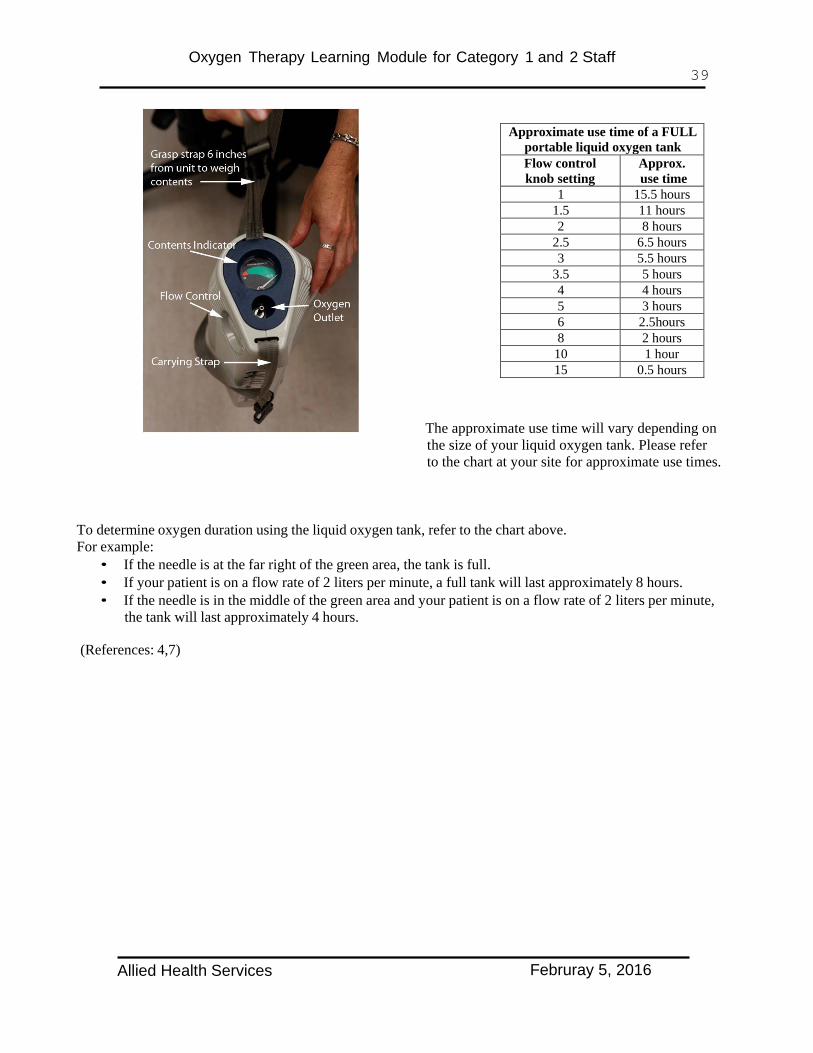
Liquid tanks are measured by weight. To determine the amount of oxygen in a liquid tank, grasp the strap closest to the content indicator, approximately 6 inches above the tank. Lift the unit off the floor. The needle, on the indicator, is spring loaded and will move when the tank is lifted. Repeat 3 times to get an average reading. The needle in the green area indicates the amount of oxygen available. The needle in the red area indicates a new oxygen source is needed. If you find a liquid oxygen tank not working, report it to the nursing unit, remove from circulation and ensure unit is repaired/replaced.
Tanks with Low Capacity Alarm Function If these are available at your site you must recognize the implications of an activated low capacity alarm and act accordingly (refer to page 36 for an example). Replace the portable oxygen cylinder with a cylinder containing more oxygen or change the oxygen delivery system to a wall outlet if avavilable.
Allied Health Services Februray 5, 2016

Oxygen Therapy Learning Module for Category 1 and 2 Staff 33
Portable Oxygen Safety Improper handling of portable oxygen can create a potentially unsafe work area. Improper handling of a portable oxygen cylinder, can convert it into an unguided missile with enormous destructive power. If the oxygen cylinder is punctured, or if a valve breaks off, the results can be lethal. Oxygen cylinders (E-size) should always be transported in a cylinder (tank) holder and never lying on a bed or stretcher. Oxygen cylinders and liquid oxygen canisters are to be secured to prevent tipping and falling. Liquid oxygen tanks must be transported upright. Placing a liquid oxygen tank on its side will lead to leakage of liquid oxygen, which will cause skin burns and corrosion of building materials (like laminate floors or tiles). Liquid oxygen tanks must never be placed on a patient’s lap or beside them on a stretcher. This can result in frostbite to the patient.
Instruct the patient/client and family in the following safety measures as appropriate: 9
• Do not use oxygen in the presence of an open flame. • Do not smoke around oxygen. • Do not wear synthetic fabrics that can build up static electricity. • Do not apply oils and petroleum products to the patient’s/client’s face. • Do not allow the oxygen tubing to become tangled as this may result in kinks cutting off the
oxygen supply. • For Home Care clients, assess the environment to ensure liquid oxygen storage is available in a
well ventilated area, away from direct heat or sunlight and properly grounded. If storage racks are not available lay compressed gas oxygen cylinders flat when storing them.
Points of Emphasis
• Under no circumstances should a patient be sent from or back to a nursing unit with a low/empty portable oxygen source.
• A portable oxygen source should be assessed for the amount of oxygen, before and after treatment, to ensure adequate supply. A patient must always be provided with enough oxygen to last for the treatment time, porter wait time, transportation time, and any delays after returning to the unit, before being returned to the wall supply.
• If there is an inadequate supply, replace with a full tank. • Always test that the system is intact and you can feel flow coming out of the nozzle when
turning the oxygen tank on. • Prior to reattaching to the wall outlet, ensure the proper oxygen flow rate. • Always ensure the correct flow rate is set on the portable oxygen source. If your patient is on
2.5lpm and the tank gauge has settings for 2 lpm or 3 lpm, you would place them on 3lpm for the duration of using the tank. Remember when they go back onto wall Oxygen at 2.5 lpm they need to be monitored to ensure they can tolerate the decrease in oxygen. See Section 3 for details.
• An additional source of oxygen should be available for all high risk patients If a patient arrives in the department or is found on the unit connected to an empty tank, complete a Patient Safety Learning Report through the Reporting and Learning System (RLS) after ensuring that the patient has a new oxygen tank.
To determine oxygen duration using the Grab’nGo oxygen tank, refer to the chart on the next page. For example:
• If the needle is at 1800 psi, and your patient is on 3 liters per minute flow, the tank will last approximately 136 minutes.
• If the needle is at 800 psi, and your patient is on 3 liters per minute flow, the tank will last only 31 minutes.
Allied Health Services Februray 5, 2016

Oxygen Therapy Learning Module for Category 1 and 2 Staff 34
When you first use the Grab’nGo tear off the lower section of the label to indicate the cylinder is in use. Once the tank reaches 500psi tear off the IN USE section of the label to indicate that the tank is now empty.
Allied Health Services Februray 5, 2016

Oxygen Therapy Learning Module for Category 1 and 2 Staff 35
Allied Health Services Februray 5, 2016

Oxygen Therapy Learning Module for Category 1 and 2 Staff 36
Allied Health Services Februray 5, 2016

Oxygen Therapy Learning Module for Category 1 and 2 Staff 37
A Guide to Portable Oxygen Units Filling the portable unit
*In acute care settings it is best practice to use thermal gloves and eye protection while filling portable liquid oxygen units.
• It is very important to connect the portable liquid unit all the way down onto the reservoir and hold it there while filling.
• If the portable sounds like its filling, but the hissing sound slowly fades within a minute, you have not held the portable down all the way. Reconnect the portable and try again.
• If the portable makes a hissing sound and seems like it is leaking after it’s been filled, this is normal. The pressure relief valve is releasing pressure from excess filling.
• If the portable is leaking, it will vent immediately.
1. Clean and dry the fill connectors on both the stationary and portable units to prevent freezing. Hold the portable unit with both hands and position the contoured case over the matching recessed area on top of the stationary reservoir Ensure that the fill connectors are properly engaged. Place one hand on top of the portable, directly over the fill connector and press straight down. The portable will lower a further 10 mm into place.
2. While still holding the portable down in the fill position, open side vent valve. A loud hissing sound will occur. This will continue until the valve is closed. Filling the portable completely should not take longer than two minutes. An irregular hissing sound will occur when the unit is full. You may also note that there is a white cloud of liquid oxygen around the reservoir. Close the valve when this occurs as the unit is full.
3. Disengage the portable from the stationary reservoir by holding the carrying strap above the unit and depressing the release button.
Place portable unit upright on the floor. There will be some ‘steam’ at the bottom of the portable unit – this is normal. To weigh contents: with one hand grasp the strap closest to the contents indicator, about six inches from portable. The needle will move into the green area on the content indicator. Repeat 3 times for an average reading.
Allied Health Services Februray 5, 2016

Oxygen Therapy Learning Module for Category 1 and 2 Staff 38
• The portable unit may make a ‘honk’ sound if knocked over or picked up suddenly. If the ‘honking’
continues after being placed upright, it should be considered faulty and should not be used. If the unit is found lying down and is not “honking,” it should be sent for maintenance.
Trouble shooting the portable oxygen system Problem Cause Solution
Portable unit not filling Unit has become disengaged from the reservoir.
1. Push portable down to secure it onto the reservoir. 2. Make sure the vent valve lever is open.
Unit has been filled but no oxygen coming out
Unit is frozen due to excessive filling.
1. Allow the unit to sit at room temperature until it thaws. 2. If the problem persists once the unit is thawed, it is considered faulty and should not be used.
Portable unit cannot disengage from the reservoir after filling procedure
Unit is frozen onto valve. This can happen after filling numerous portables in a row. Allow 15 minutes between filling portables.
1. Make sure the vent valve is in the closed position and let sit for 15-20 minutes to thaw. 2. Do not attempt to pull off or you will break the lip seal on the portable.
Filling valve on the reservoir is frosted or very wet
Reservoir has just been filled or more than 1 portable has been filled.
1. Wipe excessive moisture with a lint free cloth. 2. Use as required once the fill valve is dry.
Safety
• Keep the unit away from electrical equipment or sources of heat. • Keep flammable materials away from unit. • Always turn unit off when not in use. • Liquid oxygen is extremely cold -300ºF (-184ºC). Liquid oxygen, or parts of the equipment that have been
in contact with liquid oxygen, can cause frostbite. • If the unit is turned over, oxygen will escape. • Liquid oxygen units should be transported in such a manner that they do not come into direct contact with
the patient and must be in an upright position, at all times, to prevent leakage.
Allied Health Services Februray 5, 2016
Oxygen Therapy Learning Module for Category 1 and 2 Staff 39
Approximate use time of a FULL portable liquid oxygen tank Flow control knob setting
Approx. use time
1 15.5 hours 1.5 11 hours 2 8 hours
2.5 6.5 hours 3 5.5 hours
3.5 5 hours 4 4 hours 5 3 hours 6 2.5hours 8 2 hours
10 1 hour 15 0.5 hours
The approximate use time will vary depending on the size of your liquid oxygen tank. Please refer to the chart at your site for approximate use times.
To determine oxygen duration using the liquid oxygen tank, refer to the chart above. For example:
• If the needle is at the far right of the green area, the tank is full. • If your patient is on a flow rate of 2 liters per minute, a full tank will last approximately 8 hours. • If the needle is in the middle of the green area and your patient is on a flow rate of 2 liters per minute,
the tank will last approximately 4 hours.
(References: 4,7)
Allied Health Services Februray 5, 2016

Oxygen Therapy Learning Module for Category 1 and 2 Staff 40
Self Test Section Six
1. An oxygen cylinder should be changed when the gauge reads?
A. 800 psi
B. 500 psi
C. 1000 psi
D. 100 psi
2. Why should oxygen cylinders be handled with care?
A. Costly to repair Yes No
B. Injury to patient/staff may occur Yes No
C. Can become an unguided missile with enormous destructive power Yes No
3. If a portable liquid oxygen tank is set at 2 LPM, what is the approximate use time for a full tank?
A. 8 hrs
B. 6.5 hrs
C. 5.5 hrs
D. 2 hrs
4. Your patient arrives in the treatment area on 4 LPM and the liquid portable tank is half full. Is there sufficient oxygen supply for the patient to receive 30 minutes treatment and 15 minutes travel time?
A. Yes
B. No
5. The oxygen tank has a choice of 2 or 3 liters per minute flow rate, your patient is on 2.5 LPM via nasal prongs, what would you set the oxygen flow rate to?
A. 2 liters per minute.
B. 3 liters per minute
C. 4 liters per minute
Allied Health Services Februray 5, 2016

Oxygen Therapy Learning Module for Category 1 and 2 Staff 41
Section Seven
Oxygen Delivery Devices
Learning Objectives Upon completion of this section, the learner will be able to:
18. Differentiate between low flow and high flow oxygen delivery systems. 19. Identify different oxygen delivery devices.
Allied Health Services Februray 5, 2016

Oxygen Therapy Learning Module for Category 1 and 2 Staff 42
Oxygen Delivery Devices
Low Flow
Provides variable oxygen concentration (fraction of inspired oxygen = FiO2) Concentration cannot be guaranteed. It is influenced by the patient’s rate and depth of breathing.
Flow meter rate
Point of interest
Nasal prongs/cannula
1-6 LPM
Ensure nostrils are not blocked. (Effectiveness is decreased with deformity, secretions, silastic or NG tubes). Tubing color is clear. Flow is adjusted to meet a target SpO2.
High flow nasal prongs/ cannula
1-15 LPM Typically 7-15 LPM
Used for clients who require higher flows to adequately oxygenate, but cannot tolerate face mask for long periods. Tubing color is usually green.
Simple oxygen mask
5-10 LPM Never less than 5 LPM to prevent re-breathing CO2.
Not recommended for long term use or when changing levels of oxygen.
High Concentration Oxygen Mask with Reservoir
Oxygen rate is set to ensure reservoir bag remains at least 2/3rd inflated during inspiration. Generally run at 10 LPM
The reservoir bag provides for extra oxygen when the patient breathes faster or deeper. Ensure the reservoir bag remains partially inflated during inspiration.
Interfaces used with high flow devices (venturi, cold nebulizer and high flow nebulizer) include: a) Aerosol mask with or without tusks (See photo attached to high flow nebulizer description) b) Tracheostomy mask / collar c) Face tent
Can be used with a venturi device for short term application (i.e. patient transport). Other than for the purpose of transport, must be used with a cold nebulizer or high flow nebulizer.
Used for patients who find a mask claustrophobic, have burns to the face or have facial/nasal surgery. Use with a cold nebulizer.
Allied Health Services Februray 5, 2016

Oxygen Therapy Learning Module for Category 1 and 2 Staff 43
High flow
Meets all of the inspiratory demands of the patient and therefore, provides fixed oxygen concentrations. Concentration is precise and constant, regardless of the patient’s breathing pattern.
Oxygen concentration
Flowmeter rate
Point of interest
Venturi device
Available in 24% (blue) 28% (yellow) 31% (white) 35% (green) 40% (pink) 50% (orange)
= 2 LPM = 4 LPM = 6 LPM = 8 LPM = 8 LPM = 12 LPM
The minimum flow rate for a particular oxygen concentration is stamped on the bottom of each venturi adapter.
Cold nebulizer
28%, 30%, 35%, 40%, 50%, 70%, or 100% The dial on the neck of the nebulizer gives the O2 concentration.
10-15 LPM
The nebulizer unit attaches directly to the oxygen flow meter. Adjust the oxygen flow meter so that mist is visible exiting the mask during inspiration. Adjust the venturi opening on top of the nebulizer by turning the collar to the desired oxygen concentration. At settings above 50%, the flow delivered by the device will be diluted by the patients inspiratory efforts lowering the effective FiO2. Do not use for transport.
High flow nebulizers
(shown with tusks)
60%, 65%, 75%, 85% or 96%. The dial on the neck of the nebulizer gives the O2 concentration.
Oxygen flow meter must be open all the way (“flush”).
Despite these high oxygen concentration settings on the dial, the actual delivered oxygen concentration is less. However, it is still greater than with a cold nebulizer. The addition of tusks to an aerosol mask, increases the reservoir size and provides a higher oxygen concentration. Do not use for transport.
High flow oxygen with heated humidity via nasal prongs or trach interface
30% - 100%
10-60 LPM
Patients who may benefit include those with: high oxygen requirements, high inspiratory flow demands, retained secretions, poor compliance with high flow oxygen delivery via an aerosol mask, hypothermic core body temperature. Do not use for transport. This device may be referred to as “Optiflow”
(References 8,9,10,15.)
There may be other oxygen delivery devices used at your site, please contact Nursing or Respiratory Therapy for further details on a specific device.
Allied Health Services Februray 5, 2016

Oxygen Therapy Learning Module for Category 1 and 2 Staff 44
Self Test Section Seven
1. Regular Nasal prongs are used for flow rates up to A. 6 B. 8 C. 4 D. 3
LPM for adults.
2. Match the oxygen delivery device to the most appropriate statement/ photo.
Answer
(A,B,or C)
Device
Statement / Photo
High Concentration Oxygen Mask with Reservoir
A. Should not exceed 6 LPM
Regular Nasal Prongs
B.
High Flow Nasal Prongs
C. Used for clients that require higher flows to adequately oxygenate,
but cannot tolerate a face mask for long periods of time.
Allied Health Services Februray 5, 2016

Oxygen Therapy Learning Module for Category 1 and 2 Staff 45
Section Eight Pulse Oximetry
Learning Objectives Upon completion of this section, the learner will be able to:
20. Define pulse oximetry. 21. Identify the indications for pulse oximetry. 22. Understand the limitations of pulse oximetry.
Pulse Oximetry
Pulse oximetry is a measure of how well oxygenated the hemoglobin in arterial blood is. It is used in conjunction with other assessment tools (see section 4) to help assess a person’s ability to tolerate physical activity such as exercising, feeding, chewing and swallowing.
A pulse oximeter measures a person’s pulse (in beats per minute) and estimates the arterial oxygen saturation of hemoglobin (in percent) through a sensor typically clipped to a finger, but is also effective on the toe, different sensors are available for the earlobe or bridge of the nose.
The sensing device detects changes in oxygen saturation level, by monitoring light signals generated by the oximeter and reflected by the blood through the tissue at the probe site. As the oxygen saturation changes, so does the amount of light absorbed.
A pulse oximetry reading, in and of itself, is not a reliable indicator of oxygenation status and must be done in conjunction with clinical assessment. This includes, but is not limited to, appearance, respiratory rate, depth of ventilation, peripheral circulation, and blood pressure. See Section 4: Physical Assessment and Signs of Respiratory Distress.
What does a pulse oximeter measure:
• The oxygen saturation of hemoglobin in arterial blood, with 100% as the maximum reading possible. It is a measure of the average amount of oxygen bound to each hemoglobin molecule.
• Pulse rate is measured in beats per minute, averaged over 20 seconds.
A pulse oximeter gives no information about the following: • The amount of hemoglobin in the blood. • The amount of oxygen dissolved in the blood. • The cardiac output or blood pressure.
A pulse oximeter also gives no indication of a patient’s ventilation, only of their oxygenation. It can give a false sense of security, if supplemental oxygen is being given. In addition, there may be a delay between the occurrence of a potentially hypoxic event, and a pulse oximeter detecting low oxygen saturation. Pulse oximeters rely on adequate perfusion to function effectively
Pulse oximetry readings correlate closely with the oxygen saturations obtained by arterial blood gases, when the SpO2 (saturation of oxyhemoglobin) level is above 70%. Oxygen saturation values below 70%, obtained by pulse oximetry, are unreliable.
Allied Health Services Februray 5, 2016

Oxygen Therapy Learning Module for Category 1 and 2 Staff 46
Advantages of measuring blood oxygen saturation by pulse oximetry: • Non-invasive • Inexpensive • Easy to tolerate • Allows continuous measurement
Indications
• To obtain baseline data and data trends, during intervention and post intervention • To detect a patient at possible risk of hypoxemia • Symptoms of respiratory distress • Monitoring and adjusting O2 therapy • During invasive procedures • Following anesthesia • During exercise
Limitation
• Pulse oximeters vary in accuracy, especially at saturations < 70%. • Poor perfusion to the extremities makes it difficult for the sensor to lock onto a pulsatile artery. • Ambient light may enter the space between the sensor and the finger, affecting the optics of the
oximeter and give a false low reading. • Skin pigmentation (dark skin) may cause an over estimation of oxygen saturation, at saturations below
80%. • Acrylic nails and nail polish lowers the saturation reading. • Movement that occurs with tremor, shivering or patient transport, may mimic vascular pulsations and
result in an inaccurate reading. • Cold hands or feet may cause a poor signal and lead to an inaccurate reading. • False high readings are obtained from smokers and anyone who is exposed to carbon monoxide. A
falsely high reading may be produced up to 4 hours after smoking a cigarette.19
Clinical Points of interest • Therapists should be aware of the trends in changes of the SpO2 reading and be able to identify and
record factors that cause increases and decreases in the individual patient’s SpO2. • Post treatment it should be noted whether improvements in SpO2 are maintained. • The target SpO2 for individual patients may be different and may depend on the patient’s pathology.
The medical chart should be checked for a specified SpO2 goal or range. • Is the pulse oximeter reading correct? The reading can be correlated with other measures as follows:
palpation of radial pulse, heart rate reading on the monitor, signal quality indicator on the monitor (shown as 1, 2, or 3 bars with increasing quality).
• To optimize infection prevention, the finger probe and device should be cleaned with a disinfecting wipe between patients.
• If the patient has a BP cuff on their arm ensure the pulse oximeter is placed on the arm without the BP Cuff.
Allied Health Services Februray 5, 2016

Oxygen Therapy Learning Module for Category 1 and 2 Staff 47
The pulse oximeter
SpO2 reading
Heart rate reading
Backlight on/off
Probe connection site
Power on/off
Volume up Volume down
Alarm silence
Finger probe permanent (or disposable)
Instructions for operating the functions are usually located on the front or back of the oximeter. For your site follow the manufacturer’s user manual. (References: 1,9,14,17,18,19.)
Clinical Implications - Review
• A clinician can obtain a baseline pulse rate and oxygen saturation level and then compare them when the person is engaged in physical activity (such as exercise, feeding, chewing, and swallowing).
• Generally readings between 95 and 100% are considered normal, however the target SpO2 for individual patients may be different and may depend on the patient’s pathology.The medical chart should be checked for a specified SpO2 goal or range.
• A downward trend from the baseline may be a sign of fatigue, increased work of breathing, or aspiration. During feeding and swallowing, a drop in SpO2 may suggest possible aspiration.24 NB: There is inconsistent evidence that pulse oximetry can reliably determine the occurrence of aspiration events during feeding and swallowing. Clinical decisions should not be based solely on information obtained from this procedure.
• Levels below the target SpO2, or 90% if no range has been identified and on supplementary oxygen may be life threatening.
• Ensure the pulse oximeter is reading correctly.
Allied Health Services Februray 5, 2016

Oxygen Therapy Learning Module for Category 1 and 2 Staff 48
Self Test Section Eight
1. What does pulse oximetry estimate?
A. Arterial oxygenation saturation of hemoglobin Yes No
B. Amount of hemoglobin
Yes
No
C. Pulse rate
Yes
No
D. Blood pressure
Yes
No
2. Below what percentage, is the pulse oximetry reading unreliable?
A. 88%
B. 80%
C. 85%
D. 70%
3. You would know the pulse oximeter was reading correctly when?
A. Correlates with respiratory rate
B. Patient is able to talk normally
C. Correlates with pulse
Allied Health Services Februray 5, 2016

Oxygen Therapy Learning Module for Category 1 and 2 Staff 49
Section Nine
Allied Health/Rehabilitation Services Acute Care Sites Emergency Response
Learning Objectives Upon completion of this section, the learner will be able to:
23. Identify when to access medical assistance. 24. Differentiate between initiating a Code Blue and calling for medical assistance. 25. Understand the importance of the Goals of Care.
Calling for Medical Assistance is required for any of the following:
• Any patient you are seriously worried about • Respiratory rate less than 8 or greater than 30 • Signs of respiratory instability (see section 4) • Acute Change in O2 Sats less than 90 despite O2 greater than 5LPM • Heart rate less than 40 or greater than 140 • BP systolic less than 90 or greater than 200 or acute drop in systolic blood pressure • Sudden decrease in level of consciousness or Glasgow Coma Scale drop greater than 2 points • Seizures
Indications for calling a Code Blue
• Unresponsive, not breathing with or without pulse.
Please refer to your site specific guidelines to learn how to activate the emergency response system in your facility. In cases of acute respiratory distress, category 1 and 2 staff may initiate oxygen therapy (i.e. place patient on supplementary oxygen), or increase flow rate, while awaiting further medical direction. This is contraindicated if the oxygen order specifically states not to increase oxygen under any circumstances.11
Staff working with patients away from the nursing unit, must be aware of the patient’s goals of care designation at all times.
Goals of Care documentation Patients who have discussed and decided upon a Goals of Care designation will have the goals of care documented in the electronic and or paper chart. While on hospital property, the goals of care designation in the chart is the “source of truth”. When a patient is away from the nursing unit, and in an area to which the code team does not respond, during a medical emergency, EMS will need evidence of the patient’s Goals of Care designation to treat appropriately. The green sleeve (where available) is the universally identified container for Goals of Care documentation in the community and becomes the source of truth for Goals of Care designation. EMS will look for this green sleeve when responding to a medical emergency.
Allied Health Services Februray 5, 2016
Oxygen Therapy Learning Module for Category 1 and 2 Staff 50
Facilities with electronic charting A current Goals of Care order must be printed from the electronic charting, and placed in a green sleeve. The green sleeve is then taken on the outing with the patient. On returning the patient to the unit the Allied Health staff member ensures that the print out of the order is destroyed. The patient’s own green sleeve often contains other supporting documentation that is better left with the chart during the Allied Health outing.
Facilities with paper charting A current copy of the Goals of Care paper order (#103547) must be made, and placed in the chart. The green sleeve and original order is then taken on the outing with the patient. On returning the patient to the unit the Allied Health staff member ensures that the original order goes on the chart and the copy of the order is destroyed. The patient’s own green sleeve often contains other supporting documentation that is better left with the chart during the Allied Health outing.
Allied Health Services Februray 5, 2016

Oxygen Therapy Learning Module for Category 1 and 2 Staff 51
Self Test Section Nine 1. You would call a Code Blue when
A. No pulse Yes No
B. Respiratory rate is greater than 26 Yes No
C. No breathing Yes No
Allied Health Services Februray 5, 2016

Oxygen Therapy Learning Module for Category 1 and 2 Staff 52
Appendix A – Self Test Answer Key
Section 1
1. A 2. C 3. B
Section 2
1. B 2. A – Yes, B – Yes, C – Yes 3. A
Section 3
1. C 2. C 3. B 4. B 5. B
Section 4
1. 1 - B, 2 - D, 3 - A, 4 - C 2. A - Yes, B - Yes, C - No, D - Yes, E – Yes
Section 5
1. A 2. Low - B, Moderate - C, High – A 3. A - Yes, B - Yes, C - Yes, D - Yes, E - Yes,
Section 6
1. B 2. A - Yes, B - Yes, C - Yes, 3. A 4. A 5. B
Section 7
1. A 2. 1 - B, 2 - A, 3 - C,
Section 8
1. A – Yes, B – No, C – Yes, D – No 2. D 3. C
Section 9
1. A – Yes, B – No, C – Yes
Allied Health Services Februray 5, 2016

Oxygen Therapy Learning Module for Category 1 and 2 Staff 53
Appendix B – Quiz
1. COPD patients may have an acceptable oxygen saturation of 88%
A. True
B. False
2. Every patient needs to have SpO2 greater than 92%
A. True
B. False
3. What action would you take if during exercise your patient’s heart rate increases to maximum (as set by the therapist)?
A. Continue exercise at the same level
B. Rest the patient for 3 minutes, while monitoring heart rate
C. Call immediately for medical intervention
4. During exercise your patient’s SpO2 decreases below the range set by the physician. What would you do?
A. Rest the patient, encourage deep breaths and increase the flowrate if required, up to prescribed maximum
B. Encourage deep breathing while maintaining the current exercise program
C. Change to a less intense exercise and monitor
Allied Health Services Februray 5, 2016

Oxygen Therapy Learning Module for Category 1 and 2 Staff 54
5. During exercise your patient’s respiratory rate increases above the maximum (as set by the therapist). What would you do?
A. Rest the patient, check SpO2 and encourage slow deep breaths
B. Explain to the patient that increased respiratory rate us a normal response to exercise, while encouraging the patient to continue the exercise program at the same intensity.
C. Encourage patient to continue the exercise program at the same intensity and document the results in the medical chart
6. Match the vital signs with the correct normal adult range.
1. SpO2 % A. 12 to 16 2. BP B. 60 to 100 3. Respiratory Rate per minute C. 95 to 100 4. Heart Rate per minute D. approximately 120 systolic /
approximately 80 diastolic
7. Therapist Assistants may switch between oxygen supply sources for:
A. High risk patients
B. Moderate risk patients
C. Low and moderate risk patients
D. Low patients
8. Match the level of risk – low, moderate and high, with the amount of oxygen being received.
A. Less than 6 liters of oxygen Moderate
B. Greater than 8 liters of oxygen Low
C. Between 40 and 50 per cent concentration of oxygen High
9. Documentation for patient/client receiving oxygen therapy will include the following:
B. Oxygen delivery device (e.g. nasal prongs) Yes No
C. Oxygen flow rate or FiO2 Yes No
D. SpO2, if monitor is available Yes No
Allied Health Services Februray 5, 2016

Oxygen Therapy Learning Module for Category 1 and 2 Staff 55
10. What should the Therapist Assistant do, when they are assigned a client on oxygen and have not been given all the information needed?
A. Treat the patient and contact the therapist afterwards
B. Contact the therapist as soon as possible for clarification and provide no service until further information is received.
11. Signs of respiratory instability that require immediate attention include the following.
A. SpO2 consistently less than prescribed range Yes No
B. SpO2 greater than 98%
Yes
No
C. Respiratory rate less than prescribed range
D. Patient struggling to clear secretions (i.e. choking)
E. Excessive use of accessory muscles
Yes
Yes
Yes
No
No
No
12. Why should oxygen cylinders be handled with care?
A. Costly to repair Yes No
B. Injury to patient/staff may occur Yes No
C. If damaged can become an unguided missile with enormous
destructive power Yes No
Allied Health Services Februray 5, 2016

Oxygen Therapy Learning Module for Category 1 and 2 Staff 56
Approximate use time of a FULL portable liquid oxygen tank
Flow control knob setting Approx. use time 1 15.5 hours
1.5 11 hours 2 8 hours
2.5 6.5 hours 3 5.5 hours
3.5 5 hours 4 4 hours 5 3 hours 6 2.5hours 8 2 hours
10 1 hour 15 0.5 hours
13. If a portable liquid oxygen tank is set at 3 LPM, what is the approximate use time for a full tank?
A. 8 hrs
B. 6.5 hrs
C. 5.5 hrs
D. 2 hrs
14. Your patient arrives in the treatment area on 5 LPM and the liquid portable tank is half full. Is there sufficient oxygen supply for the patient to receive 30 minutes treatment and 15 minutes travel time?
A. Yes
B. No
15. The oxygen tank has a choice of 2 or 3 liters, flow rate, your patient is on 2.5 LPM via nasal prongs, what would you set the oxygen flow rate to?
A. 2 liters per minute.
B. 3 liters per minute
C. 4 liters per minute
Allied Health Services Februray 5, 2016

Oxygen Therapy Learning Module for Category 1 and 2 Staff 57
16. Match the oxygen delivery device to the most appropriate statement/ photo.
Answer
(A,B,or C)
Device
Statement / Photo
Regular Nasal Prongs
A. Used for clients that require higher flows to adequately oxygenate,
but cannot tolerate a face mask for long periods of time.
High Concentration Oxygen Mask with Reservoir
B.
High Flow Nasal Prongs
C. Should not exceed 6 LPM
17. You would know the pulse oximeter was reading correctly when?
A. Correlates with the pulse
B. Patient is able to talk normally
C. Correlates with the respiratory rate
Allied Health Services Februray 5, 2016
Oxygen Therapy Learning Module for Category 1 and 2 Staff 58
Appendix C - Practical Component (New Staff)
Demonstrate competency in the following activities:
1. Filling the liquid portable oxygen system - if available.
2. Moving the O2 delivery system from wall to portable system and back to wall outlet, while providing the correct level of oxygen for liquid oxygen and portable oxygen cylinder.
3. Evaluation of O2 reserve in the liquid oxygen and portable oxygen cylinder.
4. Measurement of respiratory rate.
5. Correct use of pulse oximeter, including correlating the pulse oximeter reading with the heart rate.
Allied Health Services Februray 5, 2016

Oxygen Therapy Learning Module for Category 1 and 2 Staff 59
Appendix D – Performance Checklist (New staff)
Oxygen Therapy
Performance Checklist
Name: Unit/Dept: Allied Health /Rehabilitation
Criteria Completed 1. Complete post test.
2. Fill the portable liquid oxygen system - if available.
3. Move the O2 delivery system from wall to portable system and back to wall outlet. Provide the correct level of oxygen for each device.
4. Evaluate O2 reserve in the liquid oxygen and portable oxygen cylinder.
5. Measure respiratory rate.
6. Measure saturation of oxyhemoglobin via a pulse oximeter and correlate the reading with the heart rate.
Date: Signature of Trainer:
Allied Health Services Februray 5, 2016

Oxygen Therapy Learning Module for Category 1 and 2 Staff 60
Appendix E – References
Guideline References • Oxygen Management - Allied Health Adult Acute Care Inpatients Guideline • AHS Emergency Procedures - Code Blue Policy
(AHS staff, not the public, can access these documents on the internal web Insite > Out Teams/Departments > Emergency/Disaster Management > Emergency Response Plans (Sites/Services)
References
1. AARC Clinical Practice Guideline. (1992) Pulse oximetry. Respiratory care, 37(8) 891-897. 2. AARC Clinical Practice Guideline – Oxygen Therapy for Adults in the Acute Care Facility – 2002
Revision and Update –Respiratory Care 2002; 47(6): 717-720. 3. AACVPR (2007) Guidelines for Pulmonary Rehabilitation Programs. Chest 2007, Volume 131:4-42. 4. Airgas Product and Service Information Manual. 5. Calgary Health Region Cardiorespiratory Rehabilitation Steering Committee. 6. Calgary Heath Region Diagnostic Imaging Department Oxygen Therapy Manual. 7. Calgary Health Region Memo. Dr. Bruce MacLeod. May 20th 2005. Liquid Oxygen Tanks. 8. Calgary Health Region, Nursing Policy and Procedure Manual Oxygen Therapy 0-12. 9. Calgary Health Region, Nursing Professional Development, (November 2005) Oxygen Therapy
Learning Module. 10. Calgary Health Region, Respiratory Services Policy and Procedure Manual Oxygen Therapy Clin-022. 11. Oxygen Management - Allied Health Adult Acute Care Inpatients Guideline. 12. Chung,F., Reid,W.D., (2004) Cardiopulmonary Physical Therapy SLACK Incorporated New Jersey. 13. DeTurk,W.E., Cahalin,L.P., (2004) Cardiovascular and Pulmonary Physical Therapy - An Evidence-
Based Approach McGraw-Hill Companies Inc. USA. 14. Fearnley,S.J.,(1995) Update in Anesthesia. Practical procedures, Issue 5. 15. Fisher and Paykel - Optiflow. Retrieved from the Calgary Zone Respiratory Services Webpage May
2013. 16. Frownfelter,D.L., Dean Elizabeth. Cardiovascular and Pulmonary Physical Therapy. (2006)Mosby
Inc. 17. Grap,Mary Jo., (2002) Pulse Oximetry. Critical Care Nurse, 22 (3),69-76. 18. Hill,E., Stoneham,M.D., (2008) Update in Anesthesia. 24 (2) 156-159. 19. Howell M, The correct use of pulse oximetry in measuring oxygen status. Professional Nurse 2002
Mar; 17 (7):416-418. 20. Ingrassia, TS III, et al (1991) Oxygen-Exacerbated Bleomycin Pulmonary Toxicity Mayo Clin Proc
66:173-178. 21. Kisner,C., Colby,L.A., (1990) Therapeutic Exercise Foundations and Techniques, Davis and Co.
Philadelphia, PA. 22. Praxair Grab N Go Operating Instructions Booklet B20442 ECN-13275 (05/04). 23. Schnell H, Puntillo. K,. Critical Care Nursing Secrets (201) Hanley and Belfus Incorporated. 24. Sherman B, et al (1999) Assessment of dysphagia with the use of pulse oximetry. Dysphagia 14 (3): 152-6
25. UAB Health System. Signs of Respiratory Distress. www.health.uab.edu
Allied Health Services Februray 5, 2016