oxidant damage of lipids and proteins membranes unstable
TRANSCRIPT

Oxidant Damage of the Lipids and Proteinsof the Erythrocyte Membranesin Unstable Hemoglobin DiseaseEVIDENCE FOR THE ROLE OF LIPID PEROXIDATION
THOMASP. FLYNN, DAVID W. ALLEN, GERHARDJ. JOHNSON,and JAMES G. WHITE,Departments of Medicine, St. Paul-Ramsey Medical Center, St. Paul,Minnesota 55101; the Veterans Administration Medical Center and theUniversity of Minnesota, and the Departments of Pediatrics and LaboratoryMedicine and Pathology, the University of Minnesota, Minneapolis,Minnesota 55455
A B S T R A C T Since unstable hemoglobins have beenconsidered a source of reactive oxygen radicals, andoxidative membrane damage a prehemolytic event,we examined the erythrocyte membranes of six pa-tients (three splenectomized) with hemoglobin Kolndisease. In the hydrogen peroxide stress test, the pa-tients' erythrocytes generated more than twice themalonyldialdehyde (a lipid peroxidative product) thancontrol erythrocytes. Fluorescence spectra of lipid ex-tracts of the patients' erythrocytes showed an excita-tion maximum at 400 nm and an emission maximumof 460 nm, characteristic of malonyldialdehyde lipidadducts. Two types of membrane polypeptide aggre-gates were found in the erythrocytes of the splenec-tomized patients. The first, which were dissociable bytreatment with mercaptoethanol, contained disulfide-linked spectrin, band 3 and globin. The second, notdissociable by mercaptoethanol, had an amino acidcomposition similar to that of erythrocyte membranesand spectrin (unlike globin) and like that of aggregatesproduced by the action of malonyldialdehyde on nor-mal erythrocyte membranes. Atomic absorption spec-troscopy of hemoglobin Koln erythrocytes showedno increase in calcium content implying that thesecross-links were not due to calcium-stimulated trans-glutaminase. Using a micropipette technique, we dem-onstrated that erythrocytes containing membrane
This work was presented, in part, at the 23rd AnnualMeeting of the American Society of Hematology, San An-tonio, TX, 5 December 1981, and reported in abstract formin 1981. Blood. 58(Suppl. 1): 41a. (Abstr.)
Received for publication 16 August 1982 and in revisedform 10 January 1983.
aggregates from splenectomized patients were less de-formable while aggregate-free erythrocytes from non-splenectomized patients had normal deformability.Weconclude that the erythrocyte membranes in he-moglobin Koln disease show evidence of lipid perox-idation with production of malonyldialdehyde, andthat the nondissociable membrane aggregates formedin this disease are likely cross-linked by malonyldi-aldehyde. Because the erythrocytes containing mem-brane aggregates from splenectomized patients withunstable hemoglobin disease show decreased mem-brane deformability, we hypothesize that this abnor-mality results in premature erythrocyte destruction invivo.
INTRODUCTION
Approximately 60 human hemoglobin (Hb)' variantshave been called unstable by virtue of their increasedpropensity to denature, precipitate, and form Heinzbodies (1). This type of Hb mutation produces unstableHb disease, manifested by variable degrees of chronichemolysis, Heinz body-containing erythrocytes anddipyrroluria (1). Many of the unstable Hb are char-acterized by an amino acid substitution in the globinchain segment that forms the heme pocket (2). Alter-ations in the physicochemical properties of the hemepocket secondary to these amino acid substitutions may
' Abbreviations used in this paper: G6PD, glucose-6-phos-phate dehydrogenase; Hb, hemoglobin; MDA, malonyldi-aldehyde; ME, mercaptoethanol; non-SPX, nonsplenectom-ized; SDS-PAGE, polyacrylamide gel electrophoresis in so-dium dodecyl sulfate; SPX, splenectomized.
J. Clin. Invest. © The American Society for Clinical Investigation, Inc. * 0021-9738/83/05/1215/09 $1.00Volume 71 May 1983 1215-1223
1215

TABLE IHigh Molecular Weight Membrane Polypeptide Aggregates, Erythrocyte Calcium,
and Glutathione Content in Hb Koln Patients
Glutathione' Calcium Aggregates' Nondissociable aggregates'
&vnol/g Hb pmol/liter RBCI % rnembrane protein
Controls 6.44±0.34 15-50§ 0 0SPX 3.87±0.3011 33.3 1.22±0.18 0.38±0.13Non-SPX 6.05±1.78 34.3 0 0
Results expressed as mean±SD, n 2 3.I Erythrocytes.§ Range observed in multiple control specimens.
Significantly less than controls and non-SPX patients (P < 0.005).
lead to excessive generation of superoxide and othertoxic oxygen radicals that could oxidize erythrocytecomponents (3). Theoretically the formation of toxicoxygen radicals within the erythrocyte could result inoxidative damage to the membrane as well as cyto-plasmic constituents, but apart from the observationthat erythrocyte membrane sulfhydryls may formmixed disulfide linkages with denatured Hb (4), nodirect evidence of oxidant-induced membrane damagehas been presented.
Wehave found disulfide-bonded membrane poly-peptide aggregates in the erythrocytes of patients withchronic hemolytic glucose-6-phosphate dehydrogenase(G6PD) mutants (5, 6), and have shown that such ox-idant-induced erythrocyte membrane injury is asso-ciated with decreased erythrocyte survival in a in vivomodel system (7). Jain and Hochstein (8) demonstratedthat the malonyldialdehyde (MDA) generated by theperoxidation of membrane lipids, in addition to form-ing phospholipid adducts, can also cross-link eryth-rocyte membrane proteins into nondissociable aggre-gates, in contrast to disulfide-linked aggregates. In anattempt to discover evidence of oxidant-induced mem-brane damage in unstable Hb disease, we studied theerythrocytes from several members of a family withHb Koln (9). These studies indicate the presence ofoxidative erythrocyte damage involving both mem-brane lipids and proteins. The nature of the lesionsdemonstrated suggest that they play an etiologic rolein the hemolysis that occurs in unstable Hb disease.
METHODS
Subjects. Six affected members of a family with an un-stable Hb were studied. Three were splenectomized (SPX)(ages 37, 37, 40), three nonsplenectomized (non-SPX) (ages11, 14, 15). Blood samples were drawn into tubes containingsodium heparin or disodium EDTA as were samples fromsimultaneous normal volunteers. Other control samples from
patients with various hemolytic disorders were studied asindicated, when appropriate. One patient's unstable Hb wasidentified as Hb Koln by determination of its primary struc-ture (9, 10).
A
12
34.1
567 A
Hb
B;
4,
C D
IN
I.
E
IiI
IN
..
.*j
.ti
I
I ::
F
8.f
U'
U:
_:a
0
+ME 4IME +MECONTROL NON-SPX SPX..
Hb KOLN HbKOLN
FIGURE 1 SDS-PAGEof erythrocyte membranes of control(A and B), non-SPX Hb Koln patient (C and D) and SPX HbKoln patient (E and F). Gels A, C, and E were run in theabsence of ME, and gels B, D, and F in the presence of ME.High molecular weight polypeptide aggregates are indicatedby the arrow and are shown to be only partially dissociableby ME. Bands are numbered according to Steck (28). Theheavy bands just below band 7 and at Hb on gels E and Fwere shown to be mono- and dimeric globin by simultaneouselectrophoresis with known globin fractions.
1216 T. P. Flynn, D. W. Allen, G. J. Johnson, and J. G. White
Ii

Studies on intact erythrocytes. Complete blood counts,reticulocyte counts, and Heinz body determinations wereperformed by standard methods. Erythrocyte reduced glu-tathione levels were determined by the method of Beutler(11). MDAgeneration from membrane lipids by H202 stresswas analyzed according to Stocks et al. (12). Erythrocytecalcium was measured on three times washed erythrocytesby Dr. John Eaton using atomic absorption spectroscopy (13,14). Erythrocyte micropipette deformability was measuredon cells washed three times in Hanks' buffered saline as be-fore (6).
Studies on erythrocyte membrane proteins. Erythrocytemembranes were prepared in a cold room at 00-40C withmethods described by Dodge et al. (15) and Fairbanks et al.(16). Polyacrylamide gel electrophoresis in sodium dodecylsulfate (SDS-PAGE) was performed by the method of Fair-banks et al. (16). Protein was measured (17) before electro-phoresis or gel filtration chromatography; membrane sam-ples were heated in 1% SDS at 1000C for 2 min with orwithout 1% mercaptoethanol (ME). Polypeptide aggregatesat the origin of the gel were estimated by planimetry interms of the total membrane proteins present on the gels.A correction for the meniscus of the gels was used. Mem-brane polypeptide aggregates were also analyzed and iso-lated by a 2 X 80-cm gel filtration column of Biogel A50m(Bio-Rad Laboratories, Richmond, CA) developed with 0.01Mphosphate buffer pH 7.0 with 0.1% SDSand 0.2% sodiumazide. The same column was used to prepare spectrin.
SDS-PAGEusing gels containing 4% acrylamide was per-formed on patient membranes without the addition of ME.One of each set of duplicate gels was stored at -70'C whilethe other was stained with Coomassie Blue to assess the pres-ence of high molecular weight aggregates at the origin. A2-3-mm slice was then taken from the top of the unstainedgels known to contain aggregates at the origin, immersed inelectrophoresis buffer containing 1% ME, heated to 1000C
,, 0.60
0
oo 0.4
0.2-
for 2 min and allowed to stand at room temperature for 2h. The slice was then placed atop a gel containing 5% ac-rylamide and electrophoresis carried out.
Nondissociable high molecular weight polypeptide aggre-gates were isolated from erythrocyte membranes by gel fil-tration chromatography (as above) in the presence of ME.These fractions were then dialyzed against H20, the proteinhydrolyzed 24 h in 6 N HCI and dried in a rotary still. Afteraddition of jl-alanine as an internal standard, the dried hy-drolysate was dissolved in H20, saturated with sodiumbicarbonate and reacted with {3,5-3H}l-fluoro-2,4 dinitro-benzene (24 Ci/mM, New England Nuclear, Boston, MA)diluted to 0.15 mCi/mM with carrier. The resulting dini-trophenyl amino acids were separated by thin-layer chro-matography using silica gel plates according to Brenner etal. (18) and quantitated by scraping the appropriate dini-trophenyl amino acids located by UV photography and de-termining radioactivity on a scintillation counter. Aminoacid composition was calculated using dinitrophenyl ,B-ala-nine as an internal standard and, for ease of comparison withliterature results, was expressed as moles percent of the totalamino acids known to be present, not just those amino acidsmeasured by this technique.
Studies on erythrocyte membrane lipids. Erythrocytemembrane lipids were extracted in duplicate with isopro-panol/chloroform (3:2) by the method of Rose and Oklander(19). The final extract was filtered through glass wool andthe fluorescence excitation and emission spectra obtained atroorfi temperature on an Aminco-Bowman spectrophoto-fluorometer (American Instrument Co., Inc., Silver Spring,MD), standardized with quinine sulfate (1 ug/ml 0.1 NH2SO4). The slit arrangement for recording fluorescencespectra was slits 3, 4, and 6, set at 3, 1, and 3 mm, respec-tively. Fluorescence of simultaneously prepared isopropa-nol/chloroform blanks was subtracted from erythrocyte ex-tract readings, and corrections were made for variations in
1 3
2 4 6 8
Migration, cm
FIGURE 2 Densitometric scan of polypeptide aggregates treated with ME. Gel top slice con-taining high molecular weight aggregates run without ME (such as from gel E, Fig. 1), wassoaked and heated in ME, placed atop another gel and electrophoresis performed. A densito-metric scan of one such Coomassie blue-stained gel is shown. The spectrin, band 3, and globindissociated from the aggregates by sulfhydryl reduction are indicated. Also note the significantamount of polypeptide remaining at the origin despite sulfhydryl reduction.
Oxidant Membrane Damage in Unstable Hemoglobin Disease 1217

quinine sulfate fluorescence due to fluctuations in xenonlamp intensity. An aliquot of each erythrocyte extract wassubmitted to washing and lipid phosphorus determinationsby the method of Chen et al. (20). Fluorescence was recordedin arbitrary units based on the above instrument settings asthe peak at an excitation wavelength of 400 nmand emissionwavelength of 460 nm. Results were then expressed as flu-orescence units per microgram of lipid phosphorus in theextract.
Standard statistical methods were used throughout, withresults expressed as mean±standard deviation, and the t testwas used to determine the significance of the difference be-tween means (21).
0.5
0.4
EC0a 0.3
CXo0.20
0.1
RESULTS
All six family members studied with Hb Koln had com-pensated hemolysis (Hb 13.4±0.4 g/dl) with persistentreticulocytosis, SPX: 6.0±1.1%; non-SPX: 10.4±0.4%.Heinz bodies were seen in a significant percentage oferythrocytes only in the SPX patients (58±3%) andwere associated with decreased erythrocyte reducedglutathione (Table I). No increase in erythrocyte cal-cium was present in either the SPXor non-SPX patients(Table I).
Spectrin
VO
20 40 60 80
Fraction NumberFIGURE 3 Gel filtration chromatography on a 2 X 80-cm column of Biogel A5Omof 20 mg oferythrocyte membranes from an SPX patient with Hb Koin disease. The membranes were
reduced before analysis (1% SDS, 1% ME, 1000C, 2 min). The column profile plots absorbancyat 280 nm vs. fraction number. Note the presence in the exclusion volume (Vo) of 280 nm
absorbing material that was found not to react with either Coomassie blue stain for protein(Fig. 4) or with the Lowry et al. (17) protein method and may be related to material earlierobserved by Lux and John (29).
1218 T. P. Flynn, D. W. Allen, G. J. Johnson, and J. G. White
A typical analysis by SDS-PAGEof unreduced andreduced erythrocyte membrane protein samples fromcontrol, non-SPX, and SPX Hb Koln patients is shownin Fig. 1. High molecular weight polypeptide aggre-gates are present at the origin of the gels in SPX pa-tients (Fig. 1). These aggregates were not seen in themembranes of normal control (A, B) or in non-SPXpatients (C, D), whether treated with MEor not. Inthe SPX patients (E, F) a portion of the aggregates wasseen to dissociate when the membranes were heatedwith MEbut a significant amount of high molecularweight protein material (hereafter referred to as "non-dissociable aggregates") remained despite such sulfhy-dryl reduction (F).
The components of the high molecular weight ag-gregates solubilized by disulfide bond reduction werefound to be mainly spectrin, band 3, and globin, asshown in the densitometric scan (Fig. 2). Characteristicpeaks for spectrin (bands 1 and 2), band 3, and globinwere identified by simultaneous electrophoresis withknown proteins. These scans also confirmed that a sig-nificant amount of the high molecular weight aggre-gates was not dissociable by sulfhydryl reduction andremained at the origin of the gel (Fig. 2).
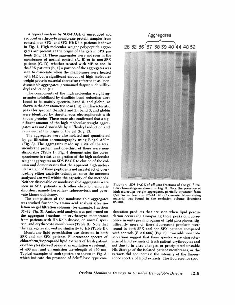
The aggregates were also isolated and quantitatedby gel filtration chromatography using Biogel A50m(Fig. 3). The aggregates made up 1.2% of the totalmembrane protein and one-third of these were non-dissociable (Table I). Fig. 4 demonstrates the corre-spondence in relative migration of the high molecularweight aggregates on SDS-PAGEto elution of the col-umn and demonstrates that the apparent high molec-ular weight of these peptides is not an artef act of over-loading either analytic technique, since the amountsanalyzed are well within the capacity of the methods.Neither dissociable or nondissociable aggregates wereseen in SPX patients with other chronic hemolyticdisorders, namely hereditary spherocytosis and pyru-vate kinase deficiency.
The composition of the nondissociable aggregateswas studied further by amino acid analysis after iso-lation on gel filtration columns (for example, fractions37-43, Fig. 3). Amino acid analysis was performed onthe aggregate fractions of erythrocyte membranesfrom patients with Hb Koln disease, on normal spec-trin, and erythrocyte membranes (Table II). Note thatthe aggregates showed no similarity to Hb (Table II).
Membrane lipid peroxidation was detected in bothSPX and non-SPX patients. Fluorescence spectra ofchloroform/isopropanol lipid extracts of fresh patienterythrocytes showed peaks at an excitation wavelengthof 400 nm, and an emission wavelength of 460 nm.Typical examples of such spectra are shown in Fig. 5,which indicate the presence of Schiff base-type con-
Aggregates
28 32 36 37 38 39 40 44 48 52
......... ,..
_,
FIGURE 4 SDS-PAGEof effluent fractions of the gel filtra-tion chromatogram shown in Fig. 3. Note the presence ofhigh molecular weight aggregates, partially separated fromspectrin in fractions 37-44. No Coomassie blue-stainingmaterial was found in the exclusion volume (fractions28-32).
densation products that are seen when lipid peroxi-dation occurs (8). Comparing these peaks of fluores-cence in units per microgram of lipid phosphorus, sig-nificantly more of these fluorescent products werefound in both SPX and non-SPX patients comparedwith controls (P < 0.005) (Fig. 6). Two additional ob-servations suggest that these spectra were character-istic of lipid extracts of fresh patient erythrocytes andnot due to in vitro changes, or precipitated unstableHb. Storage of the isolated patient membranes, or theextracts did not increase the intensity of the fluores-cence spectra of lipid extracts. The fluorescence spec-
Oxidant Membrane Damage in Unstable Hemoglobin Disease 1219

TABLE IIComparison of the Amino Acid Composition of Nondissociable Aggregates from
Erythrocyte Membranes of Hb Koin Disease with Normal Spectrinand Normal Erythrocyte Membranes
Erythrocyte Hb Koinmembranes aggregates Spectrin
(n = 2) (n = 4) (n = 3) Hb-
Glutamic acid 12.2±0.03 14.9±1.0 19.8±0.8 5.6Aspartic acid 8.1±0.6 9.4±0.2 10.7±1.4 8.7Serine 6.0±0.3 8.3±0.9 3.7±0.1 5.6Threonine 6.2±0.6 5.8±0.4 4.5±0.9 5.6Glycine 6.6±0.1 8.0±0.8 5.6±0.3 7.0Alanine 8.0±0.7 7.9±0.3 11.0±2.1 12.5Lysine and tyrosine 7.7±0.3 7.0±0.3 7.9±0.5 9.7Phenylalanine 4.2±0.3 3.5±0.04 3.2±0.2 5.2Valine 6.9±0.5 6.0±0.3 5.9±0.2 10.8Leucine and isoleucine 17.7±0.8 16.6±1.2 17.7±1.2 12.5
Total of amino acidsanalyzed 83.2 87.3 87.3 83.2
Data represent moles percent±standard deviation.Bunn, H. F., B. G. Forget, and H. M. Ranney. 1977. Human Hemoglobins. W. B.
Saunders, Co., Philadelphia. 15-17.
trum of the lipid extract of membrane-free heat-pre-cipitated Hb Koin was negative. MDAgeneration frommembrane lipids under the stress of hydrogen peroxide
30r
0- 25._
,20-Z0C
C)0* 1000
U. 5
F-_
360 380 400 420 440 460 480 500Wavelength nm
FIGURE 5 Fluorescence spectra of chloroform/isopropanolextracts of fresh erythrocytes. Results are expressed as ar-bitrary fluorescence units per microgram lipid phosphorusin the extract. One fluorescence unit is equivalent to thatproduced by 0.1 ng quinine sulfate/ml 0.1 N H2SO4. Spectraof representative duplicate extracts from normal control(0), SPX (0), and non-SPX (O) unstable Hb patients areshown.
in vitro was also assessed. These results paralleled thefluorescence results, with significantly increased MDAgenerated in both SPX and non-SPX patient cells com-pared with controls (P < 0.005) (Fig. 7).
The results of a typical experiment comparing themicropipette deformability of control erythrocytes,and erythrocytes from two non-SPX siblings and theirSPX mother, all with Hb Koln disease, are shown inFig. 8. In this experiment 15 erythrocytes from eachsubject were subjected to 10-cm H20 negative pressureusing a pipette of 0.9-Atm i.d., and the micrometers oftongue extension measured. As can be seen, the eryth-rocyte membranes from the SPX Hb Koln patient are
30
0L25-r 220-
;15-I-....o co 10
Controls SPX Non-SPXHb Koln Hb KOan
FIGURE 6 Fluorescence in lipid extracts of fresh erythro-cytes. Units are expressed as in Fig. 5. Results are shown asmean and standard deviation of peak fluorescence (excitation400 nm, emission 460 nm). Fluorescence in patient's samplesis significantly greater than in controls (P < 0.005).
1220 T. P. Flynn, D. W. Allen, G. J. Johnson, and J. G. White
a
0
-a--
00
0

.00I0)x0
Ec
0)
*010co
*0c0co
12001100
1000900800700600500400300200100
Control SPX Hb Koln Non-SPXHb Koln
FIGURE 7 MDAgeneration by H202 stress. Erythrocytes were stressed in vitro with exogenousH202 and the resulting MDAassayed. Patient erythrocytes generated significantly more MDAthan controls (P < 0.005). Abbreviations and symbols are as in Fig. 5.
significantly less deformable than the control eryth-rocytes, but those of the non-SPX patients are not sig-nificantly different from controls.
DISCUSSION
The present studies provide evidence that oxidativedamage to both lipids and proteins of the erythrocytemembrane occurs in unstable Hb disease. Depletionof membrane thiols has been previously reported andascribed to reaction with Heinz bodies (4). In the stud-ies reported here, the membrane damage is detectablein freshly obtained erythrocytes not otherwise oxida-tively stressed, and thus likely results from the oxidant-generating capacity of the unstable Hb.
Membrane lipid peroxidation was detected by thepresence of increased fluorescence in lipid extracts.Such lipid extractable fluorescence has been shown byDillard and Tappel (22) to be 10-100 times more sen-sitive than direct MDAassay as a measure of lipidperoxidation. The major species responsible for thisfluorescence appears to be conjugated Schiff base fluo-rophores formed by cross-linking two primary amines
with MDA. In lipid extracts the amino groups wouldobviously be those of aminophospholipids, but cross-links could also form between amino groups of pep-tides. Jain and Hochstein (8) have shown that exposingerythrocytes to MDAin vitro produces both fluores-cent chromolipids and protein cross-links not dissoci-able by sulfhydryl reduction. Our patients' membraneshad significantly increased amounts of these fluores-cent species compared with normals, whether thespleen was intact or had been removed.
In addition to the lipid peroxidation, two types ofhigh molecular weight polypeptide aggregates wereseen in the erythrocyte membranes, but only in SPXfamily members. Two-thirds of these high molecularweight aggregates were dissociable by sulfhydryl re-duction analogous to those in chronic hemolytic G6PDmutants (5, 6). Also, like these G6PD mutants, thepresence of the disulfide-bonded aggregates was as-sociated with decreased levels of erythrocyte reducedglutathione. However, in unstable Hb disease the com-ponents of the disulfide-bonded peptide material weredifferent from that found in G6PD mutants in thatglobin was a principal constituent. These observations
Oxidant Membrane Damage in Unstable Hemoglobin Disease 1221

10
0
Q)
eEZ
2-
0
5 6 7 8 9
pM Tongue Extension
FIGURE 8 Distribution of micropipette deformability inerythrocytes from a control (-.-), two non-SPX Hb Koln pa-tients (-h-) (... A ...), and one SPX patient (-_). The numberof erythrocytes with a given membrane deformability isplotted against that deformability (micrometers tongue ex-tension±0.5 Amat -10 cm H20). The erythrocytes from theSPX patient are significantly less deformable than controlerythrocytes (P < 0.005), but those of the non-SPX patientsare not. Other experiments with slightly different pipettesize and negative pressures gave the same results.
are consistent with the earlier concept of disulfidebinding of Heinz bodies to the membrane (4), andmore recent evidence that Hb is reversibly bound toband 3 (23), to which it can become disulfide-linkedby oxidation (24).
These disulfied-bonded aggregates were accompa-nied by additional high molecular weight polypeptidesthat remained after sulfhydryl reduction and are likelycross-linked by MDA. At neutral pH, MDArapidlypolymerizes to form longer bifunctional aldehydes,with actually increased capacity for cross-linking anddenaturing protein (25). When normal erythrocytemembranes are briefly treated with fresh MDA(26)only spectrin appears to be polymerized, but with in-cubated MDAmore and more membrane polypeptidesbecome included.2 The amino acid composition dem-onstrates that these aggregates cannot be aggregated
2 Allen, D. W. Unpublished observations.
Hb, and the composition resembles aggregated spec-trin or erythrocyte membranes.
It is likely significant that both membrane polypep-tide aggregates and decreased erythrocyte membranedeformability were detected only in cells from SPXpatients. Wehave shown in an in vivo model systemin dogs that erythrocytes with diamide-induced mem-brane polypeptide aggregates and decreased deform-ability are preferentially removed from the circulationand sequestered in the spleen (7). Further, our pa-tients' erythrocyte membranes had sustained damagein the form of nondissociable aggregates. The actionof MDAon erythrocytes, which produces similar ag-gregates, has recently been shown to produce signifi-cant alterations in erythrocyte deformability with allevidence pointing to effects on the cell membrane asthe mechanism (26). Since each of the lesions we de-scribe taken separately can alter erythrocyte deform-ability, their combination is likely of even greater sig-nificance as evidenced by the decreased deformabilityof our SPX patients' erythrocytes.
Removing the spleen in unstable Hb disease makesthe erythrocyte membrane defect and decreased de-formability apparent. The modest decrease in reticu-locytosis that occurs with splenectomy (6 vs. 10% fornon-SPX patients) illustrates the limited value of re-moval of a prime locus of Heinz body pitting (27) and,we suspect, a likely site also for removal of erythro-cytes with cross-linked membrane skeletons (7). De-termination of the relative importance of splenic re-moval of Heinz bodies or stiff membrane skeletons inthe hemolytic process will require further investiga-tion.
ACKNOWLEDGMENTSThe authors are grateful to Jerome Groat, Claude Burgoyne,Linda Leis, Stephen Burris and Barbara Finkel for technicalassistance.
We are indebted to Dr. Virgil Fairbanks of the MayoClinic, Rochester, MN, for permitting us to study these pa-tients, to Dr. John Eaton of the University of Minnesota,Minneapolis, MN, for determining the erythrocyte calciumlevels, and to Dr. Walter Schroeder of the California Instituteof Technology, Pasadena, CA, for identifying the primarystructure of the unstable Hb.
This work was supported in part by the Veterans Admin-istration and by National Institutes of Health grant PO1-HL-16833.
REFERENCES1. White, J. M. 1976. The unstable haemoglobins. Br. Med.
Bull. 32: 219-222.2. Bunn, H. F., B. G. Forget, and H. M. Ranney. 1977.
HumanHemoglobins. W. B. Saunders Co., Philadelphia.282-311.
3. Carrell, R. W., C. C. Winterbourn, and E. A. Rachmi-
1222 T. P. Flynn, D. W. Allen, G. J. Johnson, and J. G. White

lewitz. 1975. Activated oxygen and haemolysis. Br. J.Haematol. 30: 259-264.
4. Jacob, H. S., M. C. Brain, and J. V. Dacie. 1968. Alteredsulfhydryl reactivity of hemoglobin and red blood cellmembranes in congenital Heinz body hemolytic anemia.J. Clin. Invest. 47: 2664-2677.
5. Allen, D. W., G. J. Johnson, S. Cadman, and M. E. Ka-plan. 1978. Membrane polypeptide aggregates in glu-cose-6-phosphate dehydrogenase-deficient and in vitroaged red blood cells. J. Lab. Clin. Med. 91: 321-327.
6. Johnson, G. J., D. W. Allen, S. Cadman, V. F. Fairbanks,J. G. White, B. C. Lampkin, and M. E. Kaplan. 1979.Red cell membrane polypeptide aggregates in glucose-6-phosphate dehydrogenase mutants with chronic he-molytic disease. A clue to the mechanism of hemolysis.N. Engl. J. Med. 301: 522-527.
7. Johnson, G. J., D. W. Allen, T. P. Flynn, B. Finkel, andJ. G. White. 1980. Decreased survival in vivo of diamide-incubated dog erythrocytes. A model of oxidant-inducedhemolysis. J. Clin. Invest. 66: 955-961.
8. Jain, S. K., and P. Hochstein. 1980. Polymerization ofmembrane components in aging red blood cells. Biochem.Biophys. Res. Commun. 92: 247-254.
9. Carrell, R. W., H. Lehmann, and H. E. Hutchinson.1966. Haemoglobin Koln (fl-98 valine - methionine):an unstable protein causing inclusion-body anemia. Na-ture (Lond.). 210: 915-916.
10. Schroeder, W. A., J. B. Shelton, J. R. Schelton, D. Powars,S. Friedman, J. Baker, J. Z. Finkelstein, B. Miller, C. S.Johnson, J. R. Sharpsteen, L. Sieger, and E. Kawaoka.1982. Identification of eleven human hemoglobin vari-ants by high-performance liquid chromatography: ad-ditional data on functional properties and clinicalexpression. Biochem. Genet. 20: 133-152.
11. Beutler, E. 1975. Red Cell Metabolism: A Manual ofBiochemical Methods. Grune & Stratton, Inc., NewYork.112-114.
12. Stocks, J., E. L. Offerman, C. B. Modell, and T. L. Dor-mandy. 1972. The susceptibility to autoxidation of hu-man red cell lipids in health and disease. Br. J. Hae-matol. 23: 713-724.
13. Eaton, J. W., T. D. Skelton, H. S. Swafford, C. E. Koplin,and H. S. Jacob. 1973. Elevated erythrocyte calcium insickle cell disease. Nature (Lond.). 246: 105-106.
14. Eaton, J. W., E. Berger, J. G. White, and H. S. Jacob.1977. Metabolic and morphologic effects of intra-erythrocytic calcium: implications for the pathogenesisof sickle cell disease. In Zinc Metabolism: Current As-pects in Health and Disease. G. J. Brewer and A. S. Pra-sad, editors. Alan R. Liss, Inc., New York. 275-293.
15. Dodge, J. T., C. Mitchell, and D. J. Hanahan. 1963. Thepreparation and chemical characteristics of hemoglobin-free ghosts of human erythrocytes. Arch. Biochem. Bio-phys. 100: 119-130.
16. Fairbanks, G., T. L. Steck, and D. F. H. Wallach. 1971.Electrophoretic analysis of the major polypeptides of thehuman erythrocyte membrane. Biochemistry. 10: 2606-2617.
17. Lowry, 0. H., N. J. Rosebrough, A. L. Farr, and R. J.Randall. 1951. Protein measurement with the Folin phe-nol reagent. J. Biol. Chem. 193: 265-275.
18. Brenner, M., A. Niederwieser, and G. Pataki. 1965.Amino acids and derivatives. In Thin-Layer Chroma-tography. A Laboratory Handbook. E. Stahl, editor. Ac-ademic Press, Inc., New York. 414-427.
19. Rose, H. G., and M. Oklander. 1965. Improved proce-dure for the extraction of lipids from human erythro-cytes. J. Lipid Res. 6: 428-431.
20. Chen, P. S., T. Y. Toribara, and H. Warner. 1956. Mi-crodetermination of phosphorus. Anal. Chem. 28: 1756-1758.
21. Dixon, W. J., and F. J. Massey. 1957. Introduction toStatistical Analysis. McGraw-Hill Book Co., Inc., NewYork. 2nd edition. 119-124.
22. Dillard, C. J., and A. L. Tappel. 1971. Fluorescent prod-ucts of lipid peroxidation of mitochondria and micro-somes. Lipids. 6: 715-721.
23. Shaklai, N., J. Yguerabide, and H. M. Ranney. 1977.Classification and localization of hemoglobin bindingsites on the red blood cell membrane. Biochemistry. 16:5593-5597.
24. Sayare, M., and M. Fikiet. 1981. Cross-linking of he-moglobin to the cytoplasmic surface of human eryth-rocyte membranes. J. Biol. Chem. 256: 13152-13158.
25. Shin, B. C., J. W. Huggins, and K. L. Carraway. 1972.Effects of pH, concentration, and aging on the malon-aldehyde reaction with proteins. Lipids. 7: 229-233.
26. Pfafferott, C., H. J. Meiselman, and P. Hochstein. 1982.The effect of malonyldialdehyde on erythrocyte deform-ability. Blood. 59: 12-15.
27. Rifkind, R. A. 1965. Heinz body anemia, an ultrastruc-tural study. II. Red cell sequestration and destruction.Blood. 26: 433-448.
28. Steck, T. L. 1974. The organization of proteins in thehuman red blood cell membrane. A review. J. Cell Biol.62: 1-19.
29. Lux, S. E., and K. M. John. 1977. Isolation and partialcharacterization of a high molecular weight red cellmembrane protein complex normally removed by thespleen. Blood. 50: 625-641.
Oxidant Membrane Damage in Unstable Hemoglobin Disease 122t3