overview of the various methods of milf iom techniques … · overview of the various methods of...
TRANSCRIPT
Overview of the various methods of MILF surgery
IOM techniques In MILF surgery
Review of current literature dealing with MILF surgeries and IOM
Case studies dealing with MILF surgeries
LIFs
Many different methods for fusing the lumbar spine
Enough acronyms to make your head spin
TLIF PLIF ALIF XLIFDLIF ILIF MILF
Why so many LIFs Patient variability
Surgeon preference
Availability of resources
Pathology
Instrumentation Gimmicks
The goal is to improve the quality of the patients life Decompression of neural structures
Bone fusions of unstable segments
Traditional Lumbar surgery involves exposure of the Lumbar spine Large amounts of Blood loss and many other
complications are involved
Decompression of neural structures and Bone fusions were thought to be only possible with an open exposure
Minimally Invasive Lumbar Surgery Treat pathology with the least amount of soft tissue
disruption
Design procedures that are minimally invasive through smaller controlled surgical corridors
Goals of Minimally Invasive Lumbar Surgery Reduce complications of open procedures
Shorter hospital says and better patient outcomes
Treat pathology completely (without recurrence or reoperation)
History of Minimally Invasive Lumbar Surgery
Courtesy of J H Oppenheimer I DeCastro and D E McDonnell Neurosurg Focus Volume 27 September 2009
Fig 1 Chronological timelines showing technical and procedural milestones in spine surgery using traditional techniques(first column) and MAST (second and third columns)
Making Minimally Invasive Lumbar Fusions better Imaging and Stereotactic Navigation Intraoperative Monitoring
addition of EMG to the lateral approach has contributed to decrease the complication rate from 30 to less than 1 (20)
Improved Lumbar instrumentation technologies
Problems with MILF and IOM
Instrumentation systems are designed without proper IOM considerations
Surgeons and sales representatives are stimulating and using devices incorrectly
Unforeseen challenges and complications develop in practice
Impact On IOM
The O-arm (Medtronic TM)
Portable CT machine Gives the surgeon the
axial view of the vertebral bodiespedicles
Ensures proper mediallateral screw position O-verpriced
O-ver kill
O-my god this takes forever
The O-arm (Medtronic TM)
Has its own navigation system with trays of equipment
Usually concurrent with our IOM
May NOT always Correlate
Don‟t let the rep discount your monitoring
The O-arm (Medtronic TM)
Seen this somewhere beforehellip
Nohellip that‟s not it
The O-arm (Medtronic TM)
Yeahhellip that‟s it
NeuroVision M5 (NuVasive Inc)
Uses Accelerometers and lasers to co-register data from pre-operative scans
NeuroVision M5 (NuVasive Inc)
Attaches to the end of the C-arm
Uses similar presentation method to the O-arm (Medtronic TM)
NeuroVision M5 (NuVasive Inc)
EMG triggered in unison with the navigation
DLIF (TM Medtronic Inc)
XLIF (TM NuVasive Inc)
Oracle (TM Synthes Inc)
Cougar LS (TM Depuy Inc)
DLIF (Direct Lateral Interbody Fusion) (TM Medtronic Inc)
XLIF (Extreme Lateral Interbody Fusion) (TM NuVasive Inc)
DLIF(TM Medtronic Inc) Cougar LS (TM Depuy Inc) Oracle (TM Synthes Inc)
Depend on in-house IOM staff (you)
Communication with sales representatives is absolutely crucial
Monitoring Tools Certified IOM
technologist
(CNIM)
Neuromonitoring Machine
Stimulation Probes
Communication skills
Monitoring Modalities Triggered EMG
Spontaneous EMG
Ptn SSEP
TcMEP (8)
Getting Started Pre-positioning traces are recommended Patient is positioned laterally Bed may be brokenextended
SSEP and sEMG baselines are acquired as soon as possible Optimal dual plane X-ray or O-arm is obtained Pt is prepped and draped
Getting Started Navigation may or may not be used with tEMG
Incision Identifying the psoas Psoas and transverse process
identified with finger sweep Posterolateral incision may be
necessary
Establishing the Surgical Corridor
Lumbar Plexus Motor nerves reside
in the Posterior 13 of the disc space in 95
of the population (13)
Target area of penetration is the anterior 23 of the disc space
Lumbar Plexus The further down
the spine the more prevalent the plexus becomes
Lumbar Plexus The further down
the spine the more prevalent the plexus becomes
Lumbar Plexus The further down
the spine the more prevalent the plexus becomes
Lumbar Plexus The further down
the spine the more prevalent the plexus becomes
Monitoring Through the Psoas Live real-time feedback must be
given to the surgeon during dissection
Dissecting Psoas The Surgical corridor
is established while adhering to alarmalert criteria
If necessary probe re-direction is performed to obtain favorable monitoring signals
Into the disc Space Surgical Corridor is
established
Probe sheath is used as a guide for dilators
NOTE If dilators are not insulated tEMGwill not yield valid thresholds due to current shunting
Retractor Placement Stimulation of the
docking pin-holes is encouraged (DLIF)
Stimulationmapping of the retracted corridor is recommended (posterior portion)
Discectomy and Fusion sEMG and SSEPs are
continually monitored Disc is prepared Trials inserted Graft placed Closure
Suggestions for Alarm Criteria
Monitoring Through the Psoas Steady State (static) Triggered EMG monitoring A pre-determined stimulation threshold is set
Surgeon is informed when a CMAP is elicited
Monitoring Through the Psoas Dynamic Triggered EMG monitoring Stimulation threshold is varied over time to obtain a CMAP
Threshold values are relayed to surgeon in real time
tEMG AlertAlarm criteria
bdquoStudies have shown that thresholds of 5mA or less may indicate direct contact with nerve tissue Experience has suggested that threshold values greater than 10mA indicate a distance that allows for both continued nerve safety and ample working space‟
Courtesy of Nuvasive inc
More tEMG AlertAlarm criteria
Courtesy of Impulse Monitoring Inc
Free Run or Spontaneous EMG (sEMG) sEMG running throughout psoas dissection and
lateral discectomyfusion Nuerotonic discharges could be missed otherwise
Monitoring SSEP Ptn SSEP and Medianulnar SSEP Standard Alarm Criteria 50 Amplitude
10 Latency
AEEGS guideline 12
Other types of Monitoring Femoral SSEP
Cremaster tEMG and sEMG
Monitors the genitofemoral nerve
Working with automated EMG systems
NeuroVision M5 (TM NuVasive Inc)
NuVasive will almost always use their own tEMGand sEMG
Communication about IOM is still vital
NeuroVision continued (TM NuVasive Inc)
If you are concurrently monitoring SSEPshellip You will be asked to turn stimulation off Get traces as soon as you can I have seen Ptn needles create less stim
artifact with neurovision EMG
NeuroVision Triggered EMG Stimulation Technique (TM NuVasive Inc)
Dynamic Triggered EMG monitoring Non-linear algorithm that continually searches for CMAP
thresholds
Complications and problems with lateral Lumbar approach and Automated systems
Are the dissection Tools being used Insulated
The DilatorsJamshidi MUST be insulated (21)
Minimizes current shunting
Isolated electrode at the distal tip acts as the stimulation source
Insulated Jamshidi
Minimizes current shunting
stimulation source
Insulation
Isolated electrode at the distal tip
Insulated Dilators
Minimizes current shunting
Isolated stimulator at the distal tip
stimulation source
Insulation
Anatomic considerations The other 5 of the
population that has an anterior lumbar plexus (13)
Can lead to postoperative deficits at L4-5 (5)
Anatomic considerations Other nerves that are
not directly monitored (10)
The iliohypogastric nerve
The ilioinguinal nerve
Lateral Femoral Cutaneous nerve
Femoral nerve
Positioning Considerations Inexperienced staffsurgeons may position
incorrectly
SSEP changes may be seen
Altering patient position may be difficult
Heavy Patients
Positioning Considerations Aggressive breaking
of the bed may be used especially to access L4-5
This combined with retraction on the lumbar plexus can lead to post operative deficits (5)
Lateral Lumbar Fusions with Automated Monitoring
Monitoring SSEP Medianulnar SSEP positioning
changes case 1
Case 1 53 yo female
100+ kilos
L4-5 Lateral fusion
Right side up
Monitoring SSEP Posterior Tibial SSEP changes case 2
Case 2 69 yo female
L4-5 Lateral fusion
L4-5 Spondylolisthesis
Right side up
Working with automated lateral systems Large gaps between
data acquisition
Inexperience among repstechsurgeon can lead to missed changes
Monitoring SSEP Posterior Tibial SSEP changes case 3
Case 3 54 yo female
L4-5 Lateral fusion
discogenic back pain lumbar back pain
Left side up
Working with automated lateral systems Tech had excellent
communication with the sales rep
Tech had excellent communication with the surgeon
X-STOP (TM Medtronic Inc)
ILIF (TM NuVasive Inc)
Axialif (TM Trans1 Inc)
Facet Fusions
Minimally Invasive Posterior Fixation System
What What In thehellip
What What is AxiaLIF Trans 1 spine
MILF instrumentation company
Only L5-S1 fusions
Sacral approach to the spine
Also known as the Butt screw
httpAxialif
Tosh0 et al
Facet Screws lock adjacent facets
into place
I personally have been asked to test these
Spinal nerve roots could still be impinged
Stabilization is the goal
axialif-360 facet screws
Monitoring concerns Have not had any
changes with these cases
Not even EMG activity Only known
complications have been perforated bowels
Revision is not likely or possible
Sextant (TM Medtronic Inc)
SpheRx (TM NuVasive Inc)
Denali (TM K2M Inc)
Viper 2 (TM Depuy Inc)
Matis (TM Stryker Inc)
Matrix (TM Synthes Inc)
Ballista (TM Biomet Inc)
Minimally Invasive Posterior Fixation System
Minimally Invasive Posterior Fixation Systems
Getting Started Patient is positioned prone
SSEP and EMG baselines are acquired
Optimal X-ray is obtained
Pt is prepped and draped
Monitoring Tools Certified IOM
technologist
(CNIM)
NeuromontoringMachine
Cannulated EMG Monitoring Pedicle Probe(s)
Insulated dilator(s)
Communication skills
Getting Started Navigation may or may not be used with tEMG
Tapping Pedicles Target angles are
identified through X-ray
Surgeon begins to guide probe down the pedicle
Live real-time T-EMG feedback is given to the surgeon during tapping
Tapping Pedicles AlarmAlert Criteria
is followed while going through the pedicle
If necessary probe re-direction is performed to obtain favorable monitoring signals (2)
Courtesy of Impulse Monitoring Inc
Tapping Pedicles The T-Handle is
removed
A Kwire is inserted
Some Surgeons choose to stimulate kwire (3)
Tapping Pedicles Dilators are inserted
over the kwire
Insulated dilator must be used prior to tapping (12)
Important to be flush against the Transverse Process
Tapping Pedicles Stimulate the tap
when it is all the way in the pedicle
This value is thought to be the most correlative to standard open-pedicle screw EMG stimulation case data(2)
ScrewTower Insertion Screws inserted
EMG and SSEPs are continually monitored Can NOT test
Uninsulated Screw Towers
Current shunting can NOT be avoided
Thresholds are inaccurate (21)
Screw and Rod Insertion EMG and SSEPs are
continually monitored Sextant (TM )Towers Mate Rod inserted Final Tightening
Triggered EMG
EMG activity (Triggered) When the surgeon is inserting screws he can not
visualize the medial portion pedicle with C-arm A AP x-rayB Lateral x-rayC Axial (O-arm only)
C
EMG activity (Triggered) There is a risk of nerve root or spinal cord impingement if the
screw is placed too Medial In open PSF cases a threshold of greater than 20 mA of
stimulation without a CMAP response to muscle corresponding to that specific level justifies the medial placement
EMG activity (Triggered) A threshold of less than 10 mA warrants alert
criteria (4)
Under 5 mA warrants ground for screw removal (4)
Triggered EMG (tEMG) Minimally invasive surgeries do not have the
benefit of open air to insulate the path of stimulation
Current shunting must be avoided
Free Run or Spontaneous EMG (sEMG) sEMG when tapping pediclesscrew insertion Nuerotonic discharges could be missed
otherwise
Nerve root baseline Nerve root baselines can not be acquired through Percutaneous
incisions False positives from chronically compressed nerve roots can
not be accounted for
Alarm Criteria Bindal et al (2)
suggested the following tEMG protocol
Greater than 7 mA for the jamshidi needle (A B amp C)
Needle redirection was recommended for lesser thresholds
Greater than 15 mA for the insulated threaded tap (D)
Alarm Criteria Bindal et al (2) conthellip There were no insulated
threaded tap thresholds that tested under 15 mA
In 20 of the 25 patients CT scans were performed
3 lateral breaches were found
No medial breaches
Personal experience with tEMG Alarm Criteria The area of the jamshidi or PAK
needle (medronic inc) is only a few mm The charge density gives very local
information about the integrity of the pedicle wall
Personal experience with tEMG Alarm Criteria The Lowest CMAP thresholds bottom out when the
jamshidi needle is at the junction between the pedicle and the vertebral body
4 mA 7 mA 15mA
Fictional values
Thresholds between 4-7 mA typically correlate to threaded insulated tap values between 15-20 mA
Further research needs to be conducted between systems
Stimulation Duration (pulse width)
Impact On IOM for Percutaneous Pedicle Screw Testing
Idler C Rolfe K amp Gorek J Accuracy of percutaneous lumbar pedicle screw placement using the oblique or ldquoowl‟s-eyerdquo view and novel guidance technology J Neurosurg Spine 13509ndash515 2010
Featuring Nuvasive‟s new Navigation system
NeuroVision M5 (NuVasive Inc)
Five of 326 screws penetrated the pedicle cortex
3 medial and 2 lateral
pedicle breach rate of 153 Medial 092 lateral 061
overall accuracy of 9847
No mention or correlation of tEMG thresholds for the jamshidi or threaded insulated tap
Idler et al 2010
Not good news for IOMers
Wang M Pinero G Mummaneni P Stimulus-evoked electromyography testing of percutaneous pedicle screws for the detection of pedicle breaches J Neurosurg Spine 13600ndash605 2010 A clinical study of 409 screws in 93 patients
Presented at the 2009 Joint Spine Section Meeting
Results Not Good Out of 5 pedicle breaches ALL were false
negatives
There were 35 false positives as well
Results Not Good 5 medial breaches (2 with neurologic complication)
that tested at MORE than 12 mA
No true positives
Results Not Good 35 false positives out of 409 screws (86)
All screws that tested below 12 mA showed NOradiographic evidence of a medial breach
According to this article MIS EMG is a MISS
Discussion How can this be
Anyone have personal experience with this False Positives
False Negatives
Anyone have a surgeonothers mention this article to you
Possible Explanations For the false
positives May be lateral breaches
Lateral are asymptomatic
Lateral breaches can give low thresholds
What does that mean
Other Explanations source of error would be the differences or variability in
metallic conductivity a 12 rate of pedicle violations would be considered
substantially lower than other reports This finding may likely be related to our dependence on the
intensive use of intraoperative fluoroscopy to place percutaneousscrews
Computed tomography scanning can also be a source of error
FAILFAIL
There is IOM being performed without technologists
Most of these cases are also monitored without Professional oversight interpretation
Examples Surgical Instrumentation Companies
NuVasive (Neurovision tm)
Medtronic (NIM tm)
ANS is bdquoSurgeon Controlled‟
ANS do not bdquoperform‟ IOM or interpret it
Without Professional oversight interpretationhellip The surgeon is liable for any monitoring
complications
The hospital also may be liable for any monitoring complications
The instrumentation company is usually exempt
ANS is not usually bdquoSurgeon Controlled‟
Sales representatives participate in most aspects of IOM
IOM duties performed by the Sales Reps include Providing the IOM machine
Providing the IOM electrodes and supplies
Often applying the electrodes
Checking the validityreliability of the data
Relaying IOM changes to the surgeon
Other ANS Pitfalls The machines are being run by unqualified
under-educated personnel
technical errors often go misidentified or unnoticed
ANS are limited in performance (EMG and MEP only)
ANS are designed as an adjunct to concurrent instrumentation
ANS is notoriously expensive (2 to 3 times the cost of conventional IOM)
Why Conventional IOM
Superior certified personnel and equipment
Cost effectiveness
Multimodality capabilities
Position statements from credentialing bodies ASET httpasetorg
ABRET httpwwwabretorg
Neurophysiologists have specific training to obtain optimal recordings
They are available to troubleshoot hardwaresoftware problems
Specific training for optimal needle electrode placement is important to obtain accurate recordings
IOM is the only focus we do NOT sell or market any other type of services or product especially DURING THE CASE
Technologists have comprehensive IOM experience Technical errors such as EMG artifacts are a
common occurrence and must be identified and eliminated immediately
EMG artifacts Familiarity with anesthesia and the requirements
for effective monitoring
The experience to know if the patient is light andor about to start bucking
EMG artifacts Is this real or artifact
EMG artifacts Is this real or artifact
Not possible to tell out of context
SSEP and other types of monitoring is available and concurrently monitored
Vascular and positional changes are monitored
Clear Cogent and Consistent documentation
Similar features are inherent in current monitoring software
Live data feed
can be presented
at surgeon‟s
request
bdquoREAL TIME MONITORING‟ is a false concept Red lightGreen Light threshold based
monitoring is an oversimplification Clear demarcation of artifactresponse is
imperative
Experienced in DlifXlif monitoring
Experience in minimally invasive percutaneousscrew insertion monitoring techniques
Common pitfalls such as current shunting and charge density false positives can be resolved
ACDF = anterior cervical discectomy and fusion AD = aggressive discectomy ADR = artificial disc replacement ALIF = anterior lumbar interbody fusion APLD =automated percutaneous lumbar discectomy BMP = bone morphogenetic protein CAVHS = Central Arkansas Veterans Healthcare System DLIF = direct lateral interbody fusion IDET = intradiscal electrothermy LD = limited discectomy LPD = laser-assisted percutaneous discectomy MAST = minimal access spinal technique MED = microendoscopic discectomy MIS = minimally invasive surgery PEEK = polyetheretherketone PLIF = posterior lumbar interbody fusion rhBMP-2 = recombinant human BMP-2 TLIF =transforaminal interbody fusion XLIF = extreme lateral interbodyfusion
1 Balzer JR Rose RD Welch WC Sclabassi RJ Simultaneous somatosensory evoked potential and electromyographic recordings during lumbosacral decompression and instrumentation Neurosurgery 421318ndash1325 1998
2 Bindal R Ghosh S Intraoperative electromyography monitoring in minimally invasive transforaminal lumbar interbody fusion J Neurosurg Spine 6126ndash132 2007
3 Burak Ozgur Edward Benzel Steven Garfin Minimally Invasive Spine Surgery A Practical Guide to Anatomy and Techniques Springer 2009
4 Calancie B Madsen P Lebwohl N Stimulus-evoked EMG monitoring during transpedicular lumbosacral spine instrumentationInitialclinical results Spine 192780ndash2786 1994
5 Coe J Meyer SC Lumbar Plexus Palsy after XLIFreg An Avoidable Complication Society of Minimally Invasive Spine Surgery Las Vegas CA Oct 10th 2009
6 Gunnarsson T Krassioukov A Sarjeant R Fehlings M Real-Time Continuous Intraoperative Electromyographic and SomatosensoryEvoked Potential Recordings in Spinal Surgery Correlation of Clinical and Electrophysiologic Findings in a Prospective Consecutive Series of 213 Cases Spine 15 March 2004 - Volume 29 - Issue 6 - pp 677-684
7 Idler C Rolfe K amp Gorek J Accuracy of percutaneous lumbar pedicle screw placement using the oblique or ldquoowl‟s-eyerdquo view and novel guidance technology J Neurosurg Spine 13509ndash515 2010
8 Impulse Monitoring Inc Intraoperative Neuromonitoring Protocol for the Synthes Spine Oracle reg Spacer System 2007
9 Knight R Schwaegler P Hanscom D Roh J Direct Lateral Lumbar Interbody Fusion for Degenerative Conditions Early Complication Profile Journal of Spinal Disorders amp Techniques February 2009 - Volume 22 - Issue 1 - pp 34-37
10 Neuromonitroing for Transpsoas Lateral Lumbar Surgery 2006 Dr Jon Block 2006 lthttpwwwIOMstudycomgt
11 Medtronic Sofamor Danek USA Inc DLIFreg Surgical Technique 2009
12 Medtronic Sofamor Danek USA Inc CD Horizonreg Sextantreg II Rod Insertion System Surgical Steps 2009
13 Moro T Kikuchi S Konno S Yaginuma H An anatomic study of the lumbar plexus with respect to retroperitoneal endoscopic surgery Spine 2003 28423-428
14 Nuvasive Inc XLIFreg Surgical Technique 2007
15 O‟Brien RA MD MBA From Synthes sales consultant training presentation Nov 2007
16 Oppenheimer H DeCastro I amp McDonnell D Minimally invasive spine technology and minimally invasive spine surgery a historical review Neurosurg Focus Volume 27 September 2009
17 Takatomo Moro MD Shin-ichi Kikuchi MD PhD Shin-ichi Konno MD PhD Hiroyuki Yaginuma MD PhD An Anatomic Study of the Lumbar Plexus with Respect to Retroperitoneal Endoscopic Surgery SPINE Vol 28 Number 5 pp 423-428
18 Tohmeh A Rodgers W Peterson M Dynamically evoked discrete-threshold electromyography in the extreme lateral interbody fusion approach J Neurosurg Spine 2011 Jan14(1)31-7 Epub 2010 Dec 17
19 Toleikis JR Skelly JP Carlvin AO Toleikis SC Bernard TN Burkus JK et al The usefulness of electrical stimulation for assessing pedicle screw placements J Spinal Disord 13 283ndash289 2000
20 Uribe J Vale F Dakwar E Electromyographic Monitoring and Its Anatomical Implications in Minimally Invasive Spine Surgery Spine 15 December 2010 - Volume 35 - Issue 26S - pp S368-S374
21 Wang M Pinero G Mummaneni P Stimulus-evoked electromyography testing of percutaneous pedicle screws for the detection of pedicle breaches J Neurosurg Spine 13600ndash605 2010
Images courtesy of Axon Inc Biomet Inc Cadwell Inc Depuy Inc IOMstudycom Impulse Monitoring Inc K2M Inc Medtronic Inc NuVasive Inc NuWave Monitoring LLC Stryker Inc
Synthes Inc Tosh0 Trans1 Inc Trufuse Inc x-spine Inc
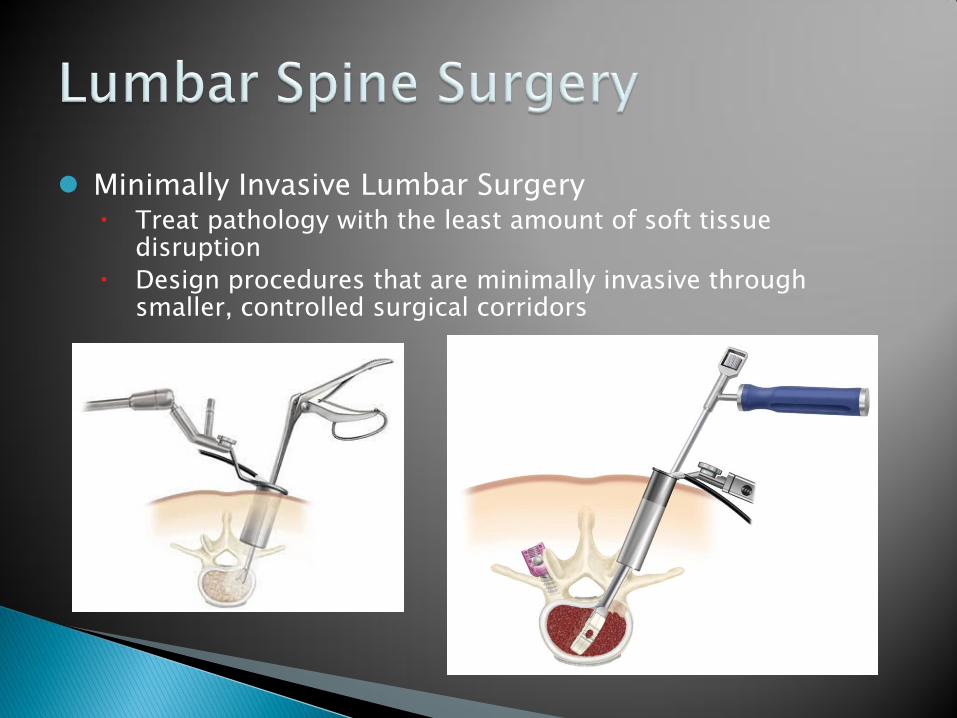

The O-arm (Medtronic TM)
Has its own navigation system with trays of equipment
Usually concurrent with our IOM
May NOT always Correlate
Don‟t let the rep discount your monitoring
The O-arm (Medtronic TM)
Seen this somewhere beforehellip
Nohellip that‟s not it
The O-arm (Medtronic TM)
Yeahhellip that‟s it
NeuroVision M5 (NuVasive Inc)
Uses Accelerometers and lasers to co-register data from pre-operative scans
NeuroVision M5 (NuVasive Inc)
Attaches to the end of the C-arm
Uses similar presentation method to the O-arm (Medtronic TM)
NeuroVision M5 (NuVasive Inc)
EMG triggered in unison with the navigation
DLIF (TM Medtronic Inc)
XLIF (TM NuVasive Inc)
Oracle (TM Synthes Inc)
Cougar LS (TM Depuy Inc)
DLIF (Direct Lateral Interbody Fusion) (TM Medtronic Inc)
XLIF (Extreme Lateral Interbody Fusion) (TM NuVasive Inc)
DLIF(TM Medtronic Inc) Cougar LS (TM Depuy Inc) Oracle (TM Synthes Inc)
Depend on in-house IOM staff (you)
Communication with sales representatives is absolutely crucial
Monitoring Tools Certified IOM
technologist
(CNIM)
Neuromonitoring Machine
Stimulation Probes
Communication skills
Monitoring Modalities Triggered EMG
Spontaneous EMG
Ptn SSEP
TcMEP (8)
Getting Started Pre-positioning traces are recommended Patient is positioned laterally Bed may be brokenextended
SSEP and sEMG baselines are acquired as soon as possible Optimal dual plane X-ray or O-arm is obtained Pt is prepped and draped
Getting Started Navigation may or may not be used with tEMG
Incision Identifying the psoas Psoas and transverse process
identified with finger sweep Posterolateral incision may be
necessary
Establishing the Surgical Corridor
Lumbar Plexus Motor nerves reside
in the Posterior 13 of the disc space in 95
of the population (13)
Target area of penetration is the anterior 23 of the disc space
Lumbar Plexus The further down
the spine the more prevalent the plexus becomes
Lumbar Plexus The further down
the spine the more prevalent the plexus becomes
Lumbar Plexus The further down
the spine the more prevalent the plexus becomes
Lumbar Plexus The further down
the spine the more prevalent the plexus becomes
Monitoring Through the Psoas Live real-time feedback must be
given to the surgeon during dissection
Dissecting Psoas The Surgical corridor
is established while adhering to alarmalert criteria
If necessary probe re-direction is performed to obtain favorable monitoring signals
Into the disc Space Surgical Corridor is
established
Probe sheath is used as a guide for dilators
NOTE If dilators are not insulated tEMGwill not yield valid thresholds due to current shunting
Retractor Placement Stimulation of the
docking pin-holes is encouraged (DLIF)
Stimulationmapping of the retracted corridor is recommended (posterior portion)
Discectomy and Fusion sEMG and SSEPs are
continually monitored Disc is prepared Trials inserted Graft placed Closure
Suggestions for Alarm Criteria
Monitoring Through the Psoas Steady State (static) Triggered EMG monitoring A pre-determined stimulation threshold is set
Surgeon is informed when a CMAP is elicited
Monitoring Through the Psoas Dynamic Triggered EMG monitoring Stimulation threshold is varied over time to obtain a CMAP
Threshold values are relayed to surgeon in real time
tEMG AlertAlarm criteria
bdquoStudies have shown that thresholds of 5mA or less may indicate direct contact with nerve tissue Experience has suggested that threshold values greater than 10mA indicate a distance that allows for both continued nerve safety and ample working space‟
Courtesy of Nuvasive inc
More tEMG AlertAlarm criteria
Courtesy of Impulse Monitoring Inc
Free Run or Spontaneous EMG (sEMG) sEMG running throughout psoas dissection and
lateral discectomyfusion Nuerotonic discharges could be missed otherwise
Monitoring SSEP Ptn SSEP and Medianulnar SSEP Standard Alarm Criteria 50 Amplitude
10 Latency
AEEGS guideline 12
Other types of Monitoring Femoral SSEP
Cremaster tEMG and sEMG
Monitors the genitofemoral nerve
Working with automated EMG systems
NeuroVision M5 (TM NuVasive Inc)
NuVasive will almost always use their own tEMGand sEMG
Communication about IOM is still vital
NeuroVision continued (TM NuVasive Inc)
If you are concurrently monitoring SSEPshellip You will be asked to turn stimulation off Get traces as soon as you can I have seen Ptn needles create less stim
artifact with neurovision EMG
NeuroVision Triggered EMG Stimulation Technique (TM NuVasive Inc)
Dynamic Triggered EMG monitoring Non-linear algorithm that continually searches for CMAP
thresholds
Complications and problems with lateral Lumbar approach and Automated systems
Are the dissection Tools being used Insulated
The DilatorsJamshidi MUST be insulated (21)
Minimizes current shunting
Isolated electrode at the distal tip acts as the stimulation source
Insulated Jamshidi
Minimizes current shunting
stimulation source
Insulation
Isolated electrode at the distal tip
Insulated Dilators
Minimizes current shunting
Isolated stimulator at the distal tip
stimulation source
Insulation
Anatomic considerations The other 5 of the
population that has an anterior lumbar plexus (13)
Can lead to postoperative deficits at L4-5 (5)
Anatomic considerations Other nerves that are
not directly monitored (10)
The iliohypogastric nerve
The ilioinguinal nerve
Lateral Femoral Cutaneous nerve
Femoral nerve
Positioning Considerations Inexperienced staffsurgeons may position
incorrectly
SSEP changes may be seen
Altering patient position may be difficult
Heavy Patients
Positioning Considerations Aggressive breaking
of the bed may be used especially to access L4-5
This combined with retraction on the lumbar plexus can lead to post operative deficits (5)
Lateral Lumbar Fusions with Automated Monitoring
Monitoring SSEP Medianulnar SSEP positioning
changes case 1
Case 1 53 yo female
100+ kilos
L4-5 Lateral fusion
Right side up
Monitoring SSEP Posterior Tibial SSEP changes case 2
Case 2 69 yo female
L4-5 Lateral fusion
L4-5 Spondylolisthesis
Right side up
Working with automated lateral systems Large gaps between
data acquisition
Inexperience among repstechsurgeon can lead to missed changes
Monitoring SSEP Posterior Tibial SSEP changes case 3
Case 3 54 yo female
L4-5 Lateral fusion
discogenic back pain lumbar back pain
Left side up
Working with automated lateral systems Tech had excellent
communication with the sales rep
Tech had excellent communication with the surgeon
X-STOP (TM Medtronic Inc)
ILIF (TM NuVasive Inc)
Axialif (TM Trans1 Inc)
Facet Fusions
Minimally Invasive Posterior Fixation System
What What In thehellip
What What is AxiaLIF Trans 1 spine
MILF instrumentation company
Only L5-S1 fusions
Sacral approach to the spine
Also known as the Butt screw
httpAxialif
Tosh0 et al
Facet Screws lock adjacent facets
into place
I personally have been asked to test these
Spinal nerve roots could still be impinged
Stabilization is the goal
axialif-360 facet screws
Monitoring concerns Have not had any
changes with these cases
Not even EMG activity Only known
complications have been perforated bowels
Revision is not likely or possible
Sextant (TM Medtronic Inc)
SpheRx (TM NuVasive Inc)
Denali (TM K2M Inc)
Viper 2 (TM Depuy Inc)
Matis (TM Stryker Inc)
Matrix (TM Synthes Inc)
Ballista (TM Biomet Inc)
Minimally Invasive Posterior Fixation System
Minimally Invasive Posterior Fixation Systems
Getting Started Patient is positioned prone
SSEP and EMG baselines are acquired
Optimal X-ray is obtained
Pt is prepped and draped
Monitoring Tools Certified IOM
technologist
(CNIM)
NeuromontoringMachine
Cannulated EMG Monitoring Pedicle Probe(s)
Insulated dilator(s)
Communication skills
Getting Started Navigation may or may not be used with tEMG
Tapping Pedicles Target angles are
identified through X-ray
Surgeon begins to guide probe down the pedicle
Live real-time T-EMG feedback is given to the surgeon during tapping
Tapping Pedicles AlarmAlert Criteria
is followed while going through the pedicle
If necessary probe re-direction is performed to obtain favorable monitoring signals (2)
Courtesy of Impulse Monitoring Inc
Tapping Pedicles The T-Handle is
removed
A Kwire is inserted
Some Surgeons choose to stimulate kwire (3)
Tapping Pedicles Dilators are inserted
over the kwire
Insulated dilator must be used prior to tapping (12)
Important to be flush against the Transverse Process
Tapping Pedicles Stimulate the tap
when it is all the way in the pedicle
This value is thought to be the most correlative to standard open-pedicle screw EMG stimulation case data(2)
ScrewTower Insertion Screws inserted
EMG and SSEPs are continually monitored Can NOT test
Uninsulated Screw Towers
Current shunting can NOT be avoided
Thresholds are inaccurate (21)
Screw and Rod Insertion EMG and SSEPs are
continually monitored Sextant (TM )Towers Mate Rod inserted Final Tightening
Triggered EMG
EMG activity (Triggered) When the surgeon is inserting screws he can not
visualize the medial portion pedicle with C-arm A AP x-rayB Lateral x-rayC Axial (O-arm only)
C
EMG activity (Triggered) There is a risk of nerve root or spinal cord impingement if the
screw is placed too Medial In open PSF cases a threshold of greater than 20 mA of
stimulation without a CMAP response to muscle corresponding to that specific level justifies the medial placement
EMG activity (Triggered) A threshold of less than 10 mA warrants alert
criteria (4)
Under 5 mA warrants ground for screw removal (4)
Triggered EMG (tEMG) Minimally invasive surgeries do not have the
benefit of open air to insulate the path of stimulation
Current shunting must be avoided
Free Run or Spontaneous EMG (sEMG) sEMG when tapping pediclesscrew insertion Nuerotonic discharges could be missed
otherwise
Nerve root baseline Nerve root baselines can not be acquired through Percutaneous
incisions False positives from chronically compressed nerve roots can
not be accounted for
Alarm Criteria Bindal et al (2)
suggested the following tEMG protocol
Greater than 7 mA for the jamshidi needle (A B amp C)
Needle redirection was recommended for lesser thresholds
Greater than 15 mA for the insulated threaded tap (D)
Alarm Criteria Bindal et al (2) conthellip There were no insulated
threaded tap thresholds that tested under 15 mA
In 20 of the 25 patients CT scans were performed
3 lateral breaches were found
No medial breaches
Personal experience with tEMG Alarm Criteria The area of the jamshidi or PAK
needle (medronic inc) is only a few mm The charge density gives very local
information about the integrity of the pedicle wall
Personal experience with tEMG Alarm Criteria The Lowest CMAP thresholds bottom out when the
jamshidi needle is at the junction between the pedicle and the vertebral body
4 mA 7 mA 15mA
Fictional values
Thresholds between 4-7 mA typically correlate to threaded insulated tap values between 15-20 mA
Further research needs to be conducted between systems
Stimulation Duration (pulse width)
Impact On IOM for Percutaneous Pedicle Screw Testing
Idler C Rolfe K amp Gorek J Accuracy of percutaneous lumbar pedicle screw placement using the oblique or ldquoowl‟s-eyerdquo view and novel guidance technology J Neurosurg Spine 13509ndash515 2010
Featuring Nuvasive‟s new Navigation system
NeuroVision M5 (NuVasive Inc)
Five of 326 screws penetrated the pedicle cortex
3 medial and 2 lateral
pedicle breach rate of 153 Medial 092 lateral 061
overall accuracy of 9847
No mention or correlation of tEMG thresholds for the jamshidi or threaded insulated tap
Idler et al 2010
Not good news for IOMers
Wang M Pinero G Mummaneni P Stimulus-evoked electromyography testing of percutaneous pedicle screws for the detection of pedicle breaches J Neurosurg Spine 13600ndash605 2010 A clinical study of 409 screws in 93 patients
Presented at the 2009 Joint Spine Section Meeting
Results Not Good Out of 5 pedicle breaches ALL were false
negatives
There were 35 false positives as well
Results Not Good 5 medial breaches (2 with neurologic complication)
that tested at MORE than 12 mA
No true positives
Results Not Good 35 false positives out of 409 screws (86)
All screws that tested below 12 mA showed NOradiographic evidence of a medial breach
According to this article MIS EMG is a MISS
Discussion How can this be
Anyone have personal experience with this False Positives
False Negatives
Anyone have a surgeonothers mention this article to you
Possible Explanations For the false
positives May be lateral breaches
Lateral are asymptomatic
Lateral breaches can give low thresholds
What does that mean
Other Explanations source of error would be the differences or variability in
metallic conductivity a 12 rate of pedicle violations would be considered
substantially lower than other reports This finding may likely be related to our dependence on the
intensive use of intraoperative fluoroscopy to place percutaneousscrews
Computed tomography scanning can also be a source of error
FAILFAIL
There is IOM being performed without technologists
Most of these cases are also monitored without Professional oversight interpretation
Examples Surgical Instrumentation Companies
NuVasive (Neurovision tm)
Medtronic (NIM tm)
ANS is bdquoSurgeon Controlled‟
ANS do not bdquoperform‟ IOM or interpret it
Without Professional oversight interpretationhellip The surgeon is liable for any monitoring
complications
The hospital also may be liable for any monitoring complications
The instrumentation company is usually exempt
ANS is not usually bdquoSurgeon Controlled‟
Sales representatives participate in most aspects of IOM
IOM duties performed by the Sales Reps include Providing the IOM machine
Providing the IOM electrodes and supplies
Often applying the electrodes
Checking the validityreliability of the data
Relaying IOM changes to the surgeon
Other ANS Pitfalls The machines are being run by unqualified
under-educated personnel
technical errors often go misidentified or unnoticed
ANS are limited in performance (EMG and MEP only)
ANS are designed as an adjunct to concurrent instrumentation
ANS is notoriously expensive (2 to 3 times the cost of conventional IOM)
Why Conventional IOM
Superior certified personnel and equipment
Cost effectiveness
Multimodality capabilities
Position statements from credentialing bodies ASET httpasetorg
ABRET httpwwwabretorg
Neurophysiologists have specific training to obtain optimal recordings
They are available to troubleshoot hardwaresoftware problems
Specific training for optimal needle electrode placement is important to obtain accurate recordings
IOM is the only focus we do NOT sell or market any other type of services or product especially DURING THE CASE
Technologists have comprehensive IOM experience Technical errors such as EMG artifacts are a
common occurrence and must be identified and eliminated immediately
EMG artifacts Familiarity with anesthesia and the requirements
for effective monitoring
The experience to know if the patient is light andor about to start bucking
EMG artifacts Is this real or artifact
EMG artifacts Is this real or artifact
Not possible to tell out of context
SSEP and other types of monitoring is available and concurrently monitored
Vascular and positional changes are monitored
Clear Cogent and Consistent documentation
Similar features are inherent in current monitoring software
Live data feed
can be presented
at surgeon‟s
request
bdquoREAL TIME MONITORING‟ is a false concept Red lightGreen Light threshold based
monitoring is an oversimplification Clear demarcation of artifactresponse is
imperative
Experienced in DlifXlif monitoring
Experience in minimally invasive percutaneousscrew insertion monitoring techniques
Common pitfalls such as current shunting and charge density false positives can be resolved
ACDF = anterior cervical discectomy and fusion AD = aggressive discectomy ADR = artificial disc replacement ALIF = anterior lumbar interbody fusion APLD =automated percutaneous lumbar discectomy BMP = bone morphogenetic protein CAVHS = Central Arkansas Veterans Healthcare System DLIF = direct lateral interbody fusion IDET = intradiscal electrothermy LD = limited discectomy LPD = laser-assisted percutaneous discectomy MAST = minimal access spinal technique MED = microendoscopic discectomy MIS = minimally invasive surgery PEEK = polyetheretherketone PLIF = posterior lumbar interbody fusion rhBMP-2 = recombinant human BMP-2 TLIF =transforaminal interbody fusion XLIF = extreme lateral interbodyfusion
1 Balzer JR Rose RD Welch WC Sclabassi RJ Simultaneous somatosensory evoked potential and electromyographic recordings during lumbosacral decompression and instrumentation Neurosurgery 421318ndash1325 1998
2 Bindal R Ghosh S Intraoperative electromyography monitoring in minimally invasive transforaminal lumbar interbody fusion J Neurosurg Spine 6126ndash132 2007
3 Burak Ozgur Edward Benzel Steven Garfin Minimally Invasive Spine Surgery A Practical Guide to Anatomy and Techniques Springer 2009
4 Calancie B Madsen P Lebwohl N Stimulus-evoked EMG monitoring during transpedicular lumbosacral spine instrumentationInitialclinical results Spine 192780ndash2786 1994
5 Coe J Meyer SC Lumbar Plexus Palsy after XLIFreg An Avoidable Complication Society of Minimally Invasive Spine Surgery Las Vegas CA Oct 10th 2009
6 Gunnarsson T Krassioukov A Sarjeant R Fehlings M Real-Time Continuous Intraoperative Electromyographic and SomatosensoryEvoked Potential Recordings in Spinal Surgery Correlation of Clinical and Electrophysiologic Findings in a Prospective Consecutive Series of 213 Cases Spine 15 March 2004 - Volume 29 - Issue 6 - pp 677-684
7 Idler C Rolfe K amp Gorek J Accuracy of percutaneous lumbar pedicle screw placement using the oblique or ldquoowl‟s-eyerdquo view and novel guidance technology J Neurosurg Spine 13509ndash515 2010
8 Impulse Monitoring Inc Intraoperative Neuromonitoring Protocol for the Synthes Spine Oracle reg Spacer System 2007
9 Knight R Schwaegler P Hanscom D Roh J Direct Lateral Lumbar Interbody Fusion for Degenerative Conditions Early Complication Profile Journal of Spinal Disorders amp Techniques February 2009 - Volume 22 - Issue 1 - pp 34-37
10 Neuromonitroing for Transpsoas Lateral Lumbar Surgery 2006 Dr Jon Block 2006 lthttpwwwIOMstudycomgt
11 Medtronic Sofamor Danek USA Inc DLIFreg Surgical Technique 2009
12 Medtronic Sofamor Danek USA Inc CD Horizonreg Sextantreg II Rod Insertion System Surgical Steps 2009
13 Moro T Kikuchi S Konno S Yaginuma H An anatomic study of the lumbar plexus with respect to retroperitoneal endoscopic surgery Spine 2003 28423-428
14 Nuvasive Inc XLIFreg Surgical Technique 2007
15 O‟Brien RA MD MBA From Synthes sales consultant training presentation Nov 2007
16 Oppenheimer H DeCastro I amp McDonnell D Minimally invasive spine technology and minimally invasive spine surgery a historical review Neurosurg Focus Volume 27 September 2009
17 Takatomo Moro MD Shin-ichi Kikuchi MD PhD Shin-ichi Konno MD PhD Hiroyuki Yaginuma MD PhD An Anatomic Study of the Lumbar Plexus with Respect to Retroperitoneal Endoscopic Surgery SPINE Vol 28 Number 5 pp 423-428
18 Tohmeh A Rodgers W Peterson M Dynamically evoked discrete-threshold electromyography in the extreme lateral interbody fusion approach J Neurosurg Spine 2011 Jan14(1)31-7 Epub 2010 Dec 17
19 Toleikis JR Skelly JP Carlvin AO Toleikis SC Bernard TN Burkus JK et al The usefulness of electrical stimulation for assessing pedicle screw placements J Spinal Disord 13 283ndash289 2000
20 Uribe J Vale F Dakwar E Electromyographic Monitoring and Its Anatomical Implications in Minimally Invasive Spine Surgery Spine 15 December 2010 - Volume 35 - Issue 26S - pp S368-S374
21 Wang M Pinero G Mummaneni P Stimulus-evoked electromyography testing of percutaneous pedicle screws for the detection of pedicle breaches J Neurosurg Spine 13600ndash605 2010
Images courtesy of Axon Inc Biomet Inc Cadwell Inc Depuy Inc IOMstudycom Impulse Monitoring Inc K2M Inc Medtronic Inc NuVasive Inc NuWave Monitoring LLC Stryker Inc
Synthes Inc Tosh0 Trans1 Inc Trufuse Inc x-spine Inc
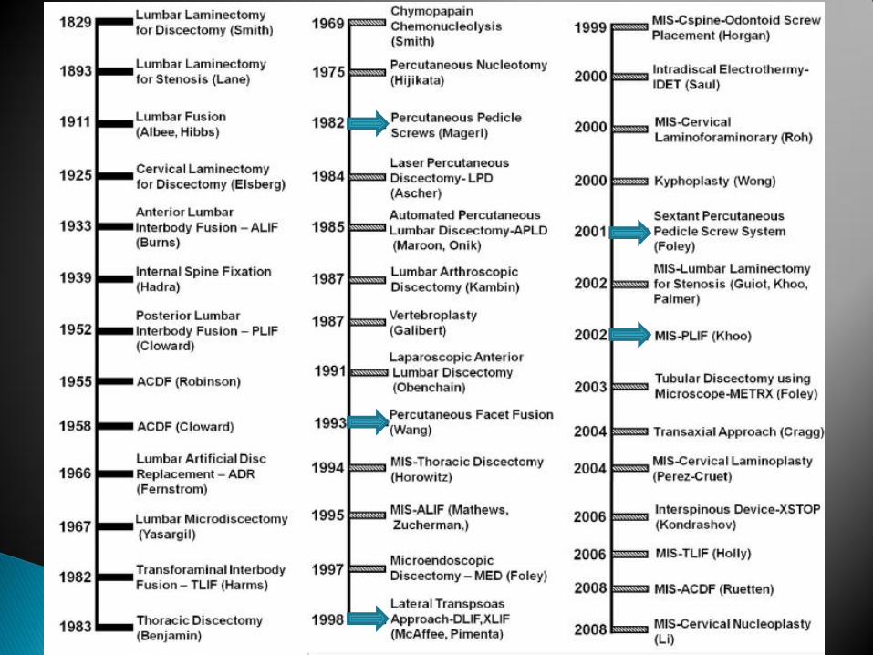
The O-arm (Medtronic TM)
Yeahhellip that‟s it
NeuroVision M5 (NuVasive Inc)
Uses Accelerometers and lasers to co-register data from pre-operative scans
NeuroVision M5 (NuVasive Inc)
Attaches to the end of the C-arm
Uses similar presentation method to the O-arm (Medtronic TM)
NeuroVision M5 (NuVasive Inc)
EMG triggered in unison with the navigation
DLIF (TM Medtronic Inc)
XLIF (TM NuVasive Inc)
Oracle (TM Synthes Inc)
Cougar LS (TM Depuy Inc)
DLIF (Direct Lateral Interbody Fusion) (TM Medtronic Inc)
XLIF (Extreme Lateral Interbody Fusion) (TM NuVasive Inc)
DLIF(TM Medtronic Inc) Cougar LS (TM Depuy Inc) Oracle (TM Synthes Inc)
Depend on in-house IOM staff (you)
Communication with sales representatives is absolutely crucial
Monitoring Tools Certified IOM
technologist
(CNIM)
Neuromonitoring Machine
Stimulation Probes
Communication skills
Monitoring Modalities Triggered EMG
Spontaneous EMG
Ptn SSEP
TcMEP (8)
Getting Started Pre-positioning traces are recommended Patient is positioned laterally Bed may be brokenextended
SSEP and sEMG baselines are acquired as soon as possible Optimal dual plane X-ray or O-arm is obtained Pt is prepped and draped
Getting Started Navigation may or may not be used with tEMG
Incision Identifying the psoas Psoas and transverse process
identified with finger sweep Posterolateral incision may be
necessary
Establishing the Surgical Corridor
Lumbar Plexus Motor nerves reside
in the Posterior 13 of the disc space in 95
of the population (13)
Target area of penetration is the anterior 23 of the disc space
Lumbar Plexus The further down
the spine the more prevalent the plexus becomes
Lumbar Plexus The further down
the spine the more prevalent the plexus becomes
Lumbar Plexus The further down
the spine the more prevalent the plexus becomes
Lumbar Plexus The further down
the spine the more prevalent the plexus becomes
Monitoring Through the Psoas Live real-time feedback must be
given to the surgeon during dissection
Dissecting Psoas The Surgical corridor
is established while adhering to alarmalert criteria
If necessary probe re-direction is performed to obtain favorable monitoring signals
Into the disc Space Surgical Corridor is
established
Probe sheath is used as a guide for dilators
NOTE If dilators are not insulated tEMGwill not yield valid thresholds due to current shunting
Retractor Placement Stimulation of the
docking pin-holes is encouraged (DLIF)
Stimulationmapping of the retracted corridor is recommended (posterior portion)
Discectomy and Fusion sEMG and SSEPs are
continually monitored Disc is prepared Trials inserted Graft placed Closure
Suggestions for Alarm Criteria
Monitoring Through the Psoas Steady State (static) Triggered EMG monitoring A pre-determined stimulation threshold is set
Surgeon is informed when a CMAP is elicited
Monitoring Through the Psoas Dynamic Triggered EMG monitoring Stimulation threshold is varied over time to obtain a CMAP
Threshold values are relayed to surgeon in real time
tEMG AlertAlarm criteria
bdquoStudies have shown that thresholds of 5mA or less may indicate direct contact with nerve tissue Experience has suggested that threshold values greater than 10mA indicate a distance that allows for both continued nerve safety and ample working space‟
Courtesy of Nuvasive inc
More tEMG AlertAlarm criteria
Courtesy of Impulse Monitoring Inc
Free Run or Spontaneous EMG (sEMG) sEMG running throughout psoas dissection and
lateral discectomyfusion Nuerotonic discharges could be missed otherwise
Monitoring SSEP Ptn SSEP and Medianulnar SSEP Standard Alarm Criteria 50 Amplitude
10 Latency
AEEGS guideline 12
Other types of Monitoring Femoral SSEP
Cremaster tEMG and sEMG
Monitors the genitofemoral nerve
Working with automated EMG systems
NeuroVision M5 (TM NuVasive Inc)
NuVasive will almost always use their own tEMGand sEMG
Communication about IOM is still vital
NeuroVision continued (TM NuVasive Inc)
If you are concurrently monitoring SSEPshellip You will be asked to turn stimulation off Get traces as soon as you can I have seen Ptn needles create less stim
artifact with neurovision EMG
NeuroVision Triggered EMG Stimulation Technique (TM NuVasive Inc)
Dynamic Triggered EMG monitoring Non-linear algorithm that continually searches for CMAP
thresholds
Complications and problems with lateral Lumbar approach and Automated systems
Are the dissection Tools being used Insulated
The DilatorsJamshidi MUST be insulated (21)
Minimizes current shunting
Isolated electrode at the distal tip acts as the stimulation source
Insulated Jamshidi
Minimizes current shunting
stimulation source
Insulation
Isolated electrode at the distal tip
Insulated Dilators
Minimizes current shunting
Isolated stimulator at the distal tip
stimulation source
Insulation
Anatomic considerations The other 5 of the
population that has an anterior lumbar plexus (13)
Can lead to postoperative deficits at L4-5 (5)
Anatomic considerations Other nerves that are
not directly monitored (10)
The iliohypogastric nerve
The ilioinguinal nerve
Lateral Femoral Cutaneous nerve
Femoral nerve
Positioning Considerations Inexperienced staffsurgeons may position
incorrectly
SSEP changes may be seen
Altering patient position may be difficult
Heavy Patients
Positioning Considerations Aggressive breaking
of the bed may be used especially to access L4-5
This combined with retraction on the lumbar plexus can lead to post operative deficits (5)
Lateral Lumbar Fusions with Automated Monitoring
Monitoring SSEP Medianulnar SSEP positioning
changes case 1
Case 1 53 yo female
100+ kilos
L4-5 Lateral fusion
Right side up
Monitoring SSEP Posterior Tibial SSEP changes case 2
Case 2 69 yo female
L4-5 Lateral fusion
L4-5 Spondylolisthesis
Right side up
Working with automated lateral systems Large gaps between
data acquisition
Inexperience among repstechsurgeon can lead to missed changes
Monitoring SSEP Posterior Tibial SSEP changes case 3
Case 3 54 yo female
L4-5 Lateral fusion
discogenic back pain lumbar back pain
Left side up
Working with automated lateral systems Tech had excellent
communication with the sales rep
Tech had excellent communication with the surgeon
X-STOP (TM Medtronic Inc)
ILIF (TM NuVasive Inc)
Axialif (TM Trans1 Inc)
Facet Fusions
Minimally Invasive Posterior Fixation System
What What In thehellip
What What is AxiaLIF Trans 1 spine
MILF instrumentation company
Only L5-S1 fusions
Sacral approach to the spine
Also known as the Butt screw
httpAxialif
Tosh0 et al
Facet Screws lock adjacent facets
into place
I personally have been asked to test these
Spinal nerve roots could still be impinged
Stabilization is the goal
axialif-360 facet screws
Monitoring concerns Have not had any
changes with these cases
Not even EMG activity Only known
complications have been perforated bowels
Revision is not likely or possible
Sextant (TM Medtronic Inc)
SpheRx (TM NuVasive Inc)
Denali (TM K2M Inc)
Viper 2 (TM Depuy Inc)
Matis (TM Stryker Inc)
Matrix (TM Synthes Inc)
Ballista (TM Biomet Inc)
Minimally Invasive Posterior Fixation System
Minimally Invasive Posterior Fixation Systems
Getting Started Patient is positioned prone
SSEP and EMG baselines are acquired
Optimal X-ray is obtained
Pt is prepped and draped
Monitoring Tools Certified IOM
technologist
(CNIM)
NeuromontoringMachine
Cannulated EMG Monitoring Pedicle Probe(s)
Insulated dilator(s)
Communication skills
Getting Started Navigation may or may not be used with tEMG
Tapping Pedicles Target angles are
identified through X-ray
Surgeon begins to guide probe down the pedicle
Live real-time T-EMG feedback is given to the surgeon during tapping
Tapping Pedicles AlarmAlert Criteria
is followed while going through the pedicle
If necessary probe re-direction is performed to obtain favorable monitoring signals (2)
Courtesy of Impulse Monitoring Inc
Tapping Pedicles The T-Handle is
removed
A Kwire is inserted
Some Surgeons choose to stimulate kwire (3)
Tapping Pedicles Dilators are inserted
over the kwire
Insulated dilator must be used prior to tapping (12)
Important to be flush against the Transverse Process
Tapping Pedicles Stimulate the tap
when it is all the way in the pedicle
This value is thought to be the most correlative to standard open-pedicle screw EMG stimulation case data(2)
ScrewTower Insertion Screws inserted
EMG and SSEPs are continually monitored Can NOT test
Uninsulated Screw Towers
Current shunting can NOT be avoided
Thresholds are inaccurate (21)
Screw and Rod Insertion EMG and SSEPs are
continually monitored Sextant (TM )Towers Mate Rod inserted Final Tightening
Triggered EMG
EMG activity (Triggered) When the surgeon is inserting screws he can not
visualize the medial portion pedicle with C-arm A AP x-rayB Lateral x-rayC Axial (O-arm only)
C
EMG activity (Triggered) There is a risk of nerve root or spinal cord impingement if the
screw is placed too Medial In open PSF cases a threshold of greater than 20 mA of
stimulation without a CMAP response to muscle corresponding to that specific level justifies the medial placement
EMG activity (Triggered) A threshold of less than 10 mA warrants alert
criteria (4)
Under 5 mA warrants ground for screw removal (4)
Triggered EMG (tEMG) Minimally invasive surgeries do not have the
benefit of open air to insulate the path of stimulation
Current shunting must be avoided
Free Run or Spontaneous EMG (sEMG) sEMG when tapping pediclesscrew insertion Nuerotonic discharges could be missed
otherwise
Nerve root baseline Nerve root baselines can not be acquired through Percutaneous
incisions False positives from chronically compressed nerve roots can
not be accounted for
Alarm Criteria Bindal et al (2)
suggested the following tEMG protocol
Greater than 7 mA for the jamshidi needle (A B amp C)
Needle redirection was recommended for lesser thresholds
Greater than 15 mA for the insulated threaded tap (D)
Alarm Criteria Bindal et al (2) conthellip There were no insulated
threaded tap thresholds that tested under 15 mA
In 20 of the 25 patients CT scans were performed
3 lateral breaches were found
No medial breaches
Personal experience with tEMG Alarm Criteria The area of the jamshidi or PAK
needle (medronic inc) is only a few mm The charge density gives very local
information about the integrity of the pedicle wall
Personal experience with tEMG Alarm Criteria The Lowest CMAP thresholds bottom out when the
jamshidi needle is at the junction between the pedicle and the vertebral body
4 mA 7 mA 15mA
Fictional values
Thresholds between 4-7 mA typically correlate to threaded insulated tap values between 15-20 mA
Further research needs to be conducted between systems
Stimulation Duration (pulse width)
Impact On IOM for Percutaneous Pedicle Screw Testing
Idler C Rolfe K amp Gorek J Accuracy of percutaneous lumbar pedicle screw placement using the oblique or ldquoowl‟s-eyerdquo view and novel guidance technology J Neurosurg Spine 13509ndash515 2010
Featuring Nuvasive‟s new Navigation system
NeuroVision M5 (NuVasive Inc)
Five of 326 screws penetrated the pedicle cortex
3 medial and 2 lateral
pedicle breach rate of 153 Medial 092 lateral 061
overall accuracy of 9847
No mention or correlation of tEMG thresholds for the jamshidi or threaded insulated tap
Idler et al 2010
Not good news for IOMers
Wang M Pinero G Mummaneni P Stimulus-evoked electromyography testing of percutaneous pedicle screws for the detection of pedicle breaches J Neurosurg Spine 13600ndash605 2010 A clinical study of 409 screws in 93 patients
Presented at the 2009 Joint Spine Section Meeting
Results Not Good Out of 5 pedicle breaches ALL were false
negatives
There were 35 false positives as well
Results Not Good 5 medial breaches (2 with neurologic complication)
that tested at MORE than 12 mA
No true positives
Results Not Good 35 false positives out of 409 screws (86)
All screws that tested below 12 mA showed NOradiographic evidence of a medial breach
According to this article MIS EMG is a MISS
Discussion How can this be
Anyone have personal experience with this False Positives
False Negatives
Anyone have a surgeonothers mention this article to you
Possible Explanations For the false
positives May be lateral breaches
Lateral are asymptomatic
Lateral breaches can give low thresholds
What does that mean
Other Explanations source of error would be the differences or variability in
metallic conductivity a 12 rate of pedicle violations would be considered
substantially lower than other reports This finding may likely be related to our dependence on the
intensive use of intraoperative fluoroscopy to place percutaneousscrews
Computed tomography scanning can also be a source of error
FAILFAIL
There is IOM being performed without technologists
Most of these cases are also monitored without Professional oversight interpretation
Examples Surgical Instrumentation Companies
NuVasive (Neurovision tm)
Medtronic (NIM tm)
ANS is bdquoSurgeon Controlled‟
ANS do not bdquoperform‟ IOM or interpret it
Without Professional oversight interpretationhellip The surgeon is liable for any monitoring
complications
The hospital also may be liable for any monitoring complications
The instrumentation company is usually exempt
ANS is not usually bdquoSurgeon Controlled‟
Sales representatives participate in most aspects of IOM
IOM duties performed by the Sales Reps include Providing the IOM machine
Providing the IOM electrodes and supplies
Often applying the electrodes
Checking the validityreliability of the data
Relaying IOM changes to the surgeon
Other ANS Pitfalls The machines are being run by unqualified
under-educated personnel
technical errors often go misidentified or unnoticed
ANS are limited in performance (EMG and MEP only)
ANS are designed as an adjunct to concurrent instrumentation
ANS is notoriously expensive (2 to 3 times the cost of conventional IOM)
Why Conventional IOM
Superior certified personnel and equipment
Cost effectiveness
Multimodality capabilities
Position statements from credentialing bodies ASET httpasetorg
ABRET httpwwwabretorg
Neurophysiologists have specific training to obtain optimal recordings
They are available to troubleshoot hardwaresoftware problems
Specific training for optimal needle electrode placement is important to obtain accurate recordings
IOM is the only focus we do NOT sell or market any other type of services or product especially DURING THE CASE
Technologists have comprehensive IOM experience Technical errors such as EMG artifacts are a
common occurrence and must be identified and eliminated immediately
EMG artifacts Familiarity with anesthesia and the requirements
for effective monitoring
The experience to know if the patient is light andor about to start bucking
EMG artifacts Is this real or artifact
EMG artifacts Is this real or artifact
Not possible to tell out of context
SSEP and other types of monitoring is available and concurrently monitored
Vascular and positional changes are monitored
Clear Cogent and Consistent documentation
Similar features are inherent in current monitoring software
Live data feed
can be presented
at surgeon‟s
request
bdquoREAL TIME MONITORING‟ is a false concept Red lightGreen Light threshold based
monitoring is an oversimplification Clear demarcation of artifactresponse is
imperative
Experienced in DlifXlif monitoring
Experience in minimally invasive percutaneousscrew insertion monitoring techniques
Common pitfalls such as current shunting and charge density false positives can be resolved
ACDF = anterior cervical discectomy and fusion AD = aggressive discectomy ADR = artificial disc replacement ALIF = anterior lumbar interbody fusion APLD =automated percutaneous lumbar discectomy BMP = bone morphogenetic protein CAVHS = Central Arkansas Veterans Healthcare System DLIF = direct lateral interbody fusion IDET = intradiscal electrothermy LD = limited discectomy LPD = laser-assisted percutaneous discectomy MAST = minimal access spinal technique MED = microendoscopic discectomy MIS = minimally invasive surgery PEEK = polyetheretherketone PLIF = posterior lumbar interbody fusion rhBMP-2 = recombinant human BMP-2 TLIF =transforaminal interbody fusion XLIF = extreme lateral interbodyfusion
1 Balzer JR Rose RD Welch WC Sclabassi RJ Simultaneous somatosensory evoked potential and electromyographic recordings during lumbosacral decompression and instrumentation Neurosurgery 421318ndash1325 1998
2 Bindal R Ghosh S Intraoperative electromyography monitoring in minimally invasive transforaminal lumbar interbody fusion J Neurosurg Spine 6126ndash132 2007
3 Burak Ozgur Edward Benzel Steven Garfin Minimally Invasive Spine Surgery A Practical Guide to Anatomy and Techniques Springer 2009
4 Calancie B Madsen P Lebwohl N Stimulus-evoked EMG monitoring during transpedicular lumbosacral spine instrumentationInitialclinical results Spine 192780ndash2786 1994
5 Coe J Meyer SC Lumbar Plexus Palsy after XLIFreg An Avoidable Complication Society of Minimally Invasive Spine Surgery Las Vegas CA Oct 10th 2009
6 Gunnarsson T Krassioukov A Sarjeant R Fehlings M Real-Time Continuous Intraoperative Electromyographic and SomatosensoryEvoked Potential Recordings in Spinal Surgery Correlation of Clinical and Electrophysiologic Findings in a Prospective Consecutive Series of 213 Cases Spine 15 March 2004 - Volume 29 - Issue 6 - pp 677-684
7 Idler C Rolfe K amp Gorek J Accuracy of percutaneous lumbar pedicle screw placement using the oblique or ldquoowl‟s-eyerdquo view and novel guidance technology J Neurosurg Spine 13509ndash515 2010
8 Impulse Monitoring Inc Intraoperative Neuromonitoring Protocol for the Synthes Spine Oracle reg Spacer System 2007
9 Knight R Schwaegler P Hanscom D Roh J Direct Lateral Lumbar Interbody Fusion for Degenerative Conditions Early Complication Profile Journal of Spinal Disorders amp Techniques February 2009 - Volume 22 - Issue 1 - pp 34-37
10 Neuromonitroing for Transpsoas Lateral Lumbar Surgery 2006 Dr Jon Block 2006 lthttpwwwIOMstudycomgt
11 Medtronic Sofamor Danek USA Inc DLIFreg Surgical Technique 2009
12 Medtronic Sofamor Danek USA Inc CD Horizonreg Sextantreg II Rod Insertion System Surgical Steps 2009
13 Moro T Kikuchi S Konno S Yaginuma H An anatomic study of the lumbar plexus with respect to retroperitoneal endoscopic surgery Spine 2003 28423-428
14 Nuvasive Inc XLIFreg Surgical Technique 2007
15 O‟Brien RA MD MBA From Synthes sales consultant training presentation Nov 2007
16 Oppenheimer H DeCastro I amp McDonnell D Minimally invasive spine technology and minimally invasive spine surgery a historical review Neurosurg Focus Volume 27 September 2009
17 Takatomo Moro MD Shin-ichi Kikuchi MD PhD Shin-ichi Konno MD PhD Hiroyuki Yaginuma MD PhD An Anatomic Study of the Lumbar Plexus with Respect to Retroperitoneal Endoscopic Surgery SPINE Vol 28 Number 5 pp 423-428
18 Tohmeh A Rodgers W Peterson M Dynamically evoked discrete-threshold electromyography in the extreme lateral interbody fusion approach J Neurosurg Spine 2011 Jan14(1)31-7 Epub 2010 Dec 17
19 Toleikis JR Skelly JP Carlvin AO Toleikis SC Bernard TN Burkus JK et al The usefulness of electrical stimulation for assessing pedicle screw placements J Spinal Disord 13 283ndash289 2000
20 Uribe J Vale F Dakwar E Electromyographic Monitoring and Its Anatomical Implications in Minimally Invasive Spine Surgery Spine 15 December 2010 - Volume 35 - Issue 26S - pp S368-S374
21 Wang M Pinero G Mummaneni P Stimulus-evoked electromyography testing of percutaneous pedicle screws for the detection of pedicle breaches J Neurosurg Spine 13600ndash605 2010
Images courtesy of Axon Inc Biomet Inc Cadwell Inc Depuy Inc IOMstudycom Impulse Monitoring Inc K2M Inc Medtronic Inc NuVasive Inc NuWave Monitoring LLC Stryker Inc
Synthes Inc Tosh0 Trans1 Inc Trufuse Inc x-spine Inc

NeuroVision M5 (NuVasive Inc)
EMG triggered in unison with the navigation
DLIF (TM Medtronic Inc)
XLIF (TM NuVasive Inc)
Oracle (TM Synthes Inc)
Cougar LS (TM Depuy Inc)
DLIF (Direct Lateral Interbody Fusion) (TM Medtronic Inc)
XLIF (Extreme Lateral Interbody Fusion) (TM NuVasive Inc)
DLIF(TM Medtronic Inc) Cougar LS (TM Depuy Inc) Oracle (TM Synthes Inc)
Depend on in-house IOM staff (you)
Communication with sales representatives is absolutely crucial
Monitoring Tools Certified IOM
technologist
(CNIM)
Neuromonitoring Machine
Stimulation Probes
Communication skills
Monitoring Modalities Triggered EMG
Spontaneous EMG
Ptn SSEP
TcMEP (8)
Getting Started Pre-positioning traces are recommended Patient is positioned laterally Bed may be brokenextended
SSEP and sEMG baselines are acquired as soon as possible Optimal dual plane X-ray or O-arm is obtained Pt is prepped and draped
Getting Started Navigation may or may not be used with tEMG
Incision Identifying the psoas Psoas and transverse process
identified with finger sweep Posterolateral incision may be
necessary
Establishing the Surgical Corridor
Lumbar Plexus Motor nerves reside
in the Posterior 13 of the disc space in 95
of the population (13)
Target area of penetration is the anterior 23 of the disc space
Lumbar Plexus The further down
the spine the more prevalent the plexus becomes
Lumbar Plexus The further down
the spine the more prevalent the plexus becomes
Lumbar Plexus The further down
the spine the more prevalent the plexus becomes
Lumbar Plexus The further down
the spine the more prevalent the plexus becomes
Monitoring Through the Psoas Live real-time feedback must be
given to the surgeon during dissection
Dissecting Psoas The Surgical corridor
is established while adhering to alarmalert criteria
If necessary probe re-direction is performed to obtain favorable monitoring signals
Into the disc Space Surgical Corridor is
established
Probe sheath is used as a guide for dilators
NOTE If dilators are not insulated tEMGwill not yield valid thresholds due to current shunting
Retractor Placement Stimulation of the
docking pin-holes is encouraged (DLIF)
Stimulationmapping of the retracted corridor is recommended (posterior portion)
Discectomy and Fusion sEMG and SSEPs are
continually monitored Disc is prepared Trials inserted Graft placed Closure
Suggestions for Alarm Criteria
Monitoring Through the Psoas Steady State (static) Triggered EMG monitoring A pre-determined stimulation threshold is set
Surgeon is informed when a CMAP is elicited
Monitoring Through the Psoas Dynamic Triggered EMG monitoring Stimulation threshold is varied over time to obtain a CMAP
Threshold values are relayed to surgeon in real time
tEMG AlertAlarm criteria
bdquoStudies have shown that thresholds of 5mA or less may indicate direct contact with nerve tissue Experience has suggested that threshold values greater than 10mA indicate a distance that allows for both continued nerve safety and ample working space‟
Courtesy of Nuvasive inc
More tEMG AlertAlarm criteria
Courtesy of Impulse Monitoring Inc
Free Run or Spontaneous EMG (sEMG) sEMG running throughout psoas dissection and
lateral discectomyfusion Nuerotonic discharges could be missed otherwise
Monitoring SSEP Ptn SSEP and Medianulnar SSEP Standard Alarm Criteria 50 Amplitude
10 Latency
AEEGS guideline 12
Other types of Monitoring Femoral SSEP
Cremaster tEMG and sEMG
Monitors the genitofemoral nerve
Working with automated EMG systems
NeuroVision M5 (TM NuVasive Inc)
NuVasive will almost always use their own tEMGand sEMG
Communication about IOM is still vital
NeuroVision continued (TM NuVasive Inc)
If you are concurrently monitoring SSEPshellip You will be asked to turn stimulation off Get traces as soon as you can I have seen Ptn needles create less stim
artifact with neurovision EMG
NeuroVision Triggered EMG Stimulation Technique (TM NuVasive Inc)
Dynamic Triggered EMG monitoring Non-linear algorithm that continually searches for CMAP
thresholds
Complications and problems with lateral Lumbar approach and Automated systems
Are the dissection Tools being used Insulated
The DilatorsJamshidi MUST be insulated (21)
Minimizes current shunting
Isolated electrode at the distal tip acts as the stimulation source
Insulated Jamshidi
Minimizes current shunting
stimulation source
Insulation
Isolated electrode at the distal tip
Insulated Dilators
Minimizes current shunting
Isolated stimulator at the distal tip
stimulation source
Insulation
Anatomic considerations The other 5 of the
population that has an anterior lumbar plexus (13)
Can lead to postoperative deficits at L4-5 (5)
Anatomic considerations Other nerves that are
not directly monitored (10)
The iliohypogastric nerve
The ilioinguinal nerve
Lateral Femoral Cutaneous nerve
Femoral nerve
Positioning Considerations Inexperienced staffsurgeons may position
incorrectly
SSEP changes may be seen
Altering patient position may be difficult
Heavy Patients
Positioning Considerations Aggressive breaking
of the bed may be used especially to access L4-5
This combined with retraction on the lumbar plexus can lead to post operative deficits (5)
Lateral Lumbar Fusions with Automated Monitoring
Monitoring SSEP Medianulnar SSEP positioning
changes case 1
Case 1 53 yo female
100+ kilos
L4-5 Lateral fusion
Right side up
Monitoring SSEP Posterior Tibial SSEP changes case 2
Case 2 69 yo female
L4-5 Lateral fusion
L4-5 Spondylolisthesis
Right side up
Working with automated lateral systems Large gaps between
data acquisition
Inexperience among repstechsurgeon can lead to missed changes
Monitoring SSEP Posterior Tibial SSEP changes case 3
Case 3 54 yo female
L4-5 Lateral fusion
discogenic back pain lumbar back pain
Left side up
Working with automated lateral systems Tech had excellent
communication with the sales rep
Tech had excellent communication with the surgeon
X-STOP (TM Medtronic Inc)
ILIF (TM NuVasive Inc)
Axialif (TM Trans1 Inc)
Facet Fusions
Minimally Invasive Posterior Fixation System
What What In thehellip
What What is AxiaLIF Trans 1 spine
MILF instrumentation company
Only L5-S1 fusions
Sacral approach to the spine
Also known as the Butt screw
httpAxialif
Tosh0 et al
Facet Screws lock adjacent facets
into place
I personally have been asked to test these
Spinal nerve roots could still be impinged
Stabilization is the goal
axialif-360 facet screws
Monitoring concerns Have not had any
changes with these cases
Not even EMG activity Only known
complications have been perforated bowels
Revision is not likely or possible
Sextant (TM Medtronic Inc)
SpheRx (TM NuVasive Inc)
Denali (TM K2M Inc)
Viper 2 (TM Depuy Inc)
Matis (TM Stryker Inc)
Matrix (TM Synthes Inc)
Ballista (TM Biomet Inc)
Minimally Invasive Posterior Fixation System
Minimally Invasive Posterior Fixation Systems
Getting Started Patient is positioned prone
SSEP and EMG baselines are acquired
Optimal X-ray is obtained
Pt is prepped and draped
Monitoring Tools Certified IOM
technologist
(CNIM)
NeuromontoringMachine
Cannulated EMG Monitoring Pedicle Probe(s)
Insulated dilator(s)
Communication skills
Getting Started Navigation may or may not be used with tEMG
Tapping Pedicles Target angles are
identified through X-ray
Surgeon begins to guide probe down the pedicle
Live real-time T-EMG feedback is given to the surgeon during tapping
Tapping Pedicles AlarmAlert Criteria
is followed while going through the pedicle
If necessary probe re-direction is performed to obtain favorable monitoring signals (2)
Courtesy of Impulse Monitoring Inc
Tapping Pedicles The T-Handle is
removed
A Kwire is inserted
Some Surgeons choose to stimulate kwire (3)
Tapping Pedicles Dilators are inserted
over the kwire
Insulated dilator must be used prior to tapping (12)
Important to be flush against the Transverse Process
Tapping Pedicles Stimulate the tap
when it is all the way in the pedicle
This value is thought to be the most correlative to standard open-pedicle screw EMG stimulation case data(2)
ScrewTower Insertion Screws inserted
EMG and SSEPs are continually monitored Can NOT test
Uninsulated Screw Towers
Current shunting can NOT be avoided
Thresholds are inaccurate (21)
Screw and Rod Insertion EMG and SSEPs are
continually monitored Sextant (TM )Towers Mate Rod inserted Final Tightening
Triggered EMG
EMG activity (Triggered) When the surgeon is inserting screws he can not
visualize the medial portion pedicle with C-arm A AP x-rayB Lateral x-rayC Axial (O-arm only)
C
EMG activity (Triggered) There is a risk of nerve root or spinal cord impingement if the
screw is placed too Medial In open PSF cases a threshold of greater than 20 mA of
stimulation without a CMAP response to muscle corresponding to that specific level justifies the medial placement
EMG activity (Triggered) A threshold of less than 10 mA warrants alert
criteria (4)
Under 5 mA warrants ground for screw removal (4)
Triggered EMG (tEMG) Minimally invasive surgeries do not have the
benefit of open air to insulate the path of stimulation
Current shunting must be avoided
Free Run or Spontaneous EMG (sEMG) sEMG when tapping pediclesscrew insertion Nuerotonic discharges could be missed
otherwise
Nerve root baseline Nerve root baselines can not be acquired through Percutaneous
incisions False positives from chronically compressed nerve roots can
not be accounted for
Alarm Criteria Bindal et al (2)
suggested the following tEMG protocol
Greater than 7 mA for the jamshidi needle (A B amp C)
Needle redirection was recommended for lesser thresholds
Greater than 15 mA for the insulated threaded tap (D)
Alarm Criteria Bindal et al (2) conthellip There were no insulated
threaded tap thresholds that tested under 15 mA
In 20 of the 25 patients CT scans were performed
3 lateral breaches were found
No medial breaches
Personal experience with tEMG Alarm Criteria The area of the jamshidi or PAK
needle (medronic inc) is only a few mm The charge density gives very local
information about the integrity of the pedicle wall
Personal experience with tEMG Alarm Criteria The Lowest CMAP thresholds bottom out when the
jamshidi needle is at the junction between the pedicle and the vertebral body
4 mA 7 mA 15mA
Fictional values
Thresholds between 4-7 mA typically correlate to threaded insulated tap values between 15-20 mA
Further research needs to be conducted between systems
Stimulation Duration (pulse width)
Impact On IOM for Percutaneous Pedicle Screw Testing
Idler C Rolfe K amp Gorek J Accuracy of percutaneous lumbar pedicle screw placement using the oblique or ldquoowl‟s-eyerdquo view and novel guidance technology J Neurosurg Spine 13509ndash515 2010
Featuring Nuvasive‟s new Navigation system
NeuroVision M5 (NuVasive Inc)
Five of 326 screws penetrated the pedicle cortex
3 medial and 2 lateral
pedicle breach rate of 153 Medial 092 lateral 061
overall accuracy of 9847
No mention or correlation of tEMG thresholds for the jamshidi or threaded insulated tap
Idler et al 2010
Not good news for IOMers
Wang M Pinero G Mummaneni P Stimulus-evoked electromyography testing of percutaneous pedicle screws for the detection of pedicle breaches J Neurosurg Spine 13600ndash605 2010 A clinical study of 409 screws in 93 patients
Presented at the 2009 Joint Spine Section Meeting
Results Not Good Out of 5 pedicle breaches ALL were false
negatives
There were 35 false positives as well
Results Not Good 5 medial breaches (2 with neurologic complication)
that tested at MORE than 12 mA
No true positives
Results Not Good 35 false positives out of 409 screws (86)
All screws that tested below 12 mA showed NOradiographic evidence of a medial breach
According to this article MIS EMG is a MISS
Discussion How can this be
Anyone have personal experience with this False Positives
False Negatives
Anyone have a surgeonothers mention this article to you
Possible Explanations For the false
positives May be lateral breaches
Lateral are asymptomatic
Lateral breaches can give low thresholds
What does that mean
Other Explanations source of error would be the differences or variability in
metallic conductivity a 12 rate of pedicle violations would be considered
substantially lower than other reports This finding may likely be related to our dependence on the
intensive use of intraoperative fluoroscopy to place percutaneousscrews
Computed tomography scanning can also be a source of error
FAILFAIL
There is IOM being performed without technologists
Most of these cases are also monitored without Professional oversight interpretation
Examples Surgical Instrumentation Companies
NuVasive (Neurovision tm)
Medtronic (NIM tm)
ANS is bdquoSurgeon Controlled‟
ANS do not bdquoperform‟ IOM or interpret it
Without Professional oversight interpretationhellip The surgeon is liable for any monitoring
complications
The hospital also may be liable for any monitoring complications
The instrumentation company is usually exempt
ANS is not usually bdquoSurgeon Controlled‟
Sales representatives participate in most aspects of IOM
IOM duties performed by the Sales Reps include Providing the IOM machine
Providing the IOM electrodes and supplies
Often applying the electrodes
Checking the validityreliability of the data
Relaying IOM changes to the surgeon
Other ANS Pitfalls The machines are being run by unqualified
under-educated personnel
technical errors often go misidentified or unnoticed
ANS are limited in performance (EMG and MEP only)
ANS are designed as an adjunct to concurrent instrumentation
ANS is notoriously expensive (2 to 3 times the cost of conventional IOM)
Why Conventional IOM
Superior certified personnel and equipment
Cost effectiveness
Multimodality capabilities
Position statements from credentialing bodies ASET httpasetorg
ABRET httpwwwabretorg
Neurophysiologists have specific training to obtain optimal recordings
They are available to troubleshoot hardwaresoftware problems
Specific training for optimal needle electrode placement is important to obtain accurate recordings
IOM is the only focus we do NOT sell or market any other type of services or product especially DURING THE CASE
Technologists have comprehensive IOM experience Technical errors such as EMG artifacts are a
common occurrence and must be identified and eliminated immediately
EMG artifacts Familiarity with anesthesia and the requirements
for effective monitoring
The experience to know if the patient is light andor about to start bucking
EMG artifacts Is this real or artifact
EMG artifacts Is this real or artifact
Not possible to tell out of context
SSEP and other types of monitoring is available and concurrently monitored
Vascular and positional changes are monitored
Clear Cogent and Consistent documentation
Similar features are inherent in current monitoring software
Live data feed
can be presented
at surgeon‟s
request
bdquoREAL TIME MONITORING‟ is a false concept Red lightGreen Light threshold based
monitoring is an oversimplification Clear demarcation of artifactresponse is
imperative
Experienced in DlifXlif monitoring
Experience in minimally invasive percutaneousscrew insertion monitoring techniques
Common pitfalls such as current shunting and charge density false positives can be resolved
ACDF = anterior cervical discectomy and fusion AD = aggressive discectomy ADR = artificial disc replacement ALIF = anterior lumbar interbody fusion APLD =automated percutaneous lumbar discectomy BMP = bone morphogenetic protein CAVHS = Central Arkansas Veterans Healthcare System DLIF = direct lateral interbody fusion IDET = intradiscal electrothermy LD = limited discectomy LPD = laser-assisted percutaneous discectomy MAST = minimal access spinal technique MED = microendoscopic discectomy MIS = minimally invasive surgery PEEK = polyetheretherketone PLIF = posterior lumbar interbody fusion rhBMP-2 = recombinant human BMP-2 TLIF =transforaminal interbody fusion XLIF = extreme lateral interbodyfusion
1 Balzer JR Rose RD Welch WC Sclabassi RJ Simultaneous somatosensory evoked potential and electromyographic recordings during lumbosacral decompression and instrumentation Neurosurgery 421318ndash1325 1998
2 Bindal R Ghosh S Intraoperative electromyography monitoring in minimally invasive transforaminal lumbar interbody fusion J Neurosurg Spine 6126ndash132 2007
3 Burak Ozgur Edward Benzel Steven Garfin Minimally Invasive Spine Surgery A Practical Guide to Anatomy and Techniques Springer 2009
4 Calancie B Madsen P Lebwohl N Stimulus-evoked EMG monitoring during transpedicular lumbosacral spine instrumentationInitialclinical results Spine 192780ndash2786 1994
5 Coe J Meyer SC Lumbar Plexus Palsy after XLIFreg An Avoidable Complication Society of Minimally Invasive Spine Surgery Las Vegas CA Oct 10th 2009
6 Gunnarsson T Krassioukov A Sarjeant R Fehlings M Real-Time Continuous Intraoperative Electromyographic and SomatosensoryEvoked Potential Recordings in Spinal Surgery Correlation of Clinical and Electrophysiologic Findings in a Prospective Consecutive Series of 213 Cases Spine 15 March 2004 - Volume 29 - Issue 6 - pp 677-684
7 Idler C Rolfe K amp Gorek J Accuracy of percutaneous lumbar pedicle screw placement using the oblique or ldquoowl‟s-eyerdquo view and novel guidance technology J Neurosurg Spine 13509ndash515 2010
8 Impulse Monitoring Inc Intraoperative Neuromonitoring Protocol for the Synthes Spine Oracle reg Spacer System 2007
9 Knight R Schwaegler P Hanscom D Roh J Direct Lateral Lumbar Interbody Fusion for Degenerative Conditions Early Complication Profile Journal of Spinal Disorders amp Techniques February 2009 - Volume 22 - Issue 1 - pp 34-37
10 Neuromonitroing for Transpsoas Lateral Lumbar Surgery 2006 Dr Jon Block 2006 lthttpwwwIOMstudycomgt
11 Medtronic Sofamor Danek USA Inc DLIFreg Surgical Technique 2009
12 Medtronic Sofamor Danek USA Inc CD Horizonreg Sextantreg II Rod Insertion System Surgical Steps 2009
13 Moro T Kikuchi S Konno S Yaginuma H An anatomic study of the lumbar plexus with respect to retroperitoneal endoscopic surgery Spine 2003 28423-428
14 Nuvasive Inc XLIFreg Surgical Technique 2007
15 O‟Brien RA MD MBA From Synthes sales consultant training presentation Nov 2007
16 Oppenheimer H DeCastro I amp McDonnell D Minimally invasive spine technology and minimally invasive spine surgery a historical review Neurosurg Focus Volume 27 September 2009
17 Takatomo Moro MD Shin-ichi Kikuchi MD PhD Shin-ichi Konno MD PhD Hiroyuki Yaginuma MD PhD An Anatomic Study of the Lumbar Plexus with Respect to Retroperitoneal Endoscopic Surgery SPINE Vol 28 Number 5 pp 423-428
18 Tohmeh A Rodgers W Peterson M Dynamically evoked discrete-threshold electromyography in the extreme lateral interbody fusion approach J Neurosurg Spine 2011 Jan14(1)31-7 Epub 2010 Dec 17
19 Toleikis JR Skelly JP Carlvin AO Toleikis SC Bernard TN Burkus JK et al The usefulness of electrical stimulation for assessing pedicle screw placements J Spinal Disord 13 283ndash289 2000
20 Uribe J Vale F Dakwar E Electromyographic Monitoring and Its Anatomical Implications in Minimally Invasive Spine Surgery Spine 15 December 2010 - Volume 35 - Issue 26S - pp S368-S374
21 Wang M Pinero G Mummaneni P Stimulus-evoked electromyography testing of percutaneous pedicle screws for the detection of pedicle breaches J Neurosurg Spine 13600ndash605 2010
Images courtesy of Axon Inc Biomet Inc Cadwell Inc Depuy Inc IOMstudycom Impulse Monitoring Inc K2M Inc Medtronic Inc NuVasive Inc NuWave Monitoring LLC Stryker Inc
Synthes Inc Tosh0 Trans1 Inc Trufuse Inc x-spine Inc
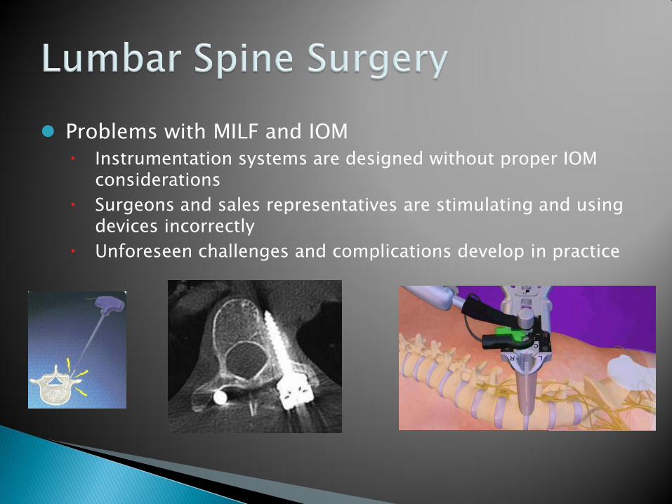

Monitoring Tools Certified IOM
technologist
(CNIM)
Neuromonitoring Machine
Stimulation Probes
Communication skills
Monitoring Modalities Triggered EMG
Spontaneous EMG
Ptn SSEP
TcMEP (8)
Getting Started Pre-positioning traces are recommended Patient is positioned laterally Bed may be brokenextended
SSEP and sEMG baselines are acquired as soon as possible Optimal dual plane X-ray or O-arm is obtained Pt is prepped and draped
Getting Started Navigation may or may not be used with tEMG
Incision Identifying the psoas Psoas and transverse process
identified with finger sweep Posterolateral incision may be
necessary
Establishing the Surgical Corridor
Lumbar Plexus Motor nerves reside
in the Posterior 13 of the disc space in 95
of the population (13)
Target area of penetration is the anterior 23 of the disc space
Lumbar Plexus The further down
the spine the more prevalent the plexus becomes
Lumbar Plexus The further down
the spine the more prevalent the plexus becomes
Lumbar Plexus The further down
the spine the more prevalent the plexus becomes
Lumbar Plexus The further down
the spine the more prevalent the plexus becomes
Monitoring Through the Psoas Live real-time feedback must be
given to the surgeon during dissection
Dissecting Psoas The Surgical corridor
is established while adhering to alarmalert criteria
If necessary probe re-direction is performed to obtain favorable monitoring signals
Into the disc Space Surgical Corridor is
established
Probe sheath is used as a guide for dilators
NOTE If dilators are not insulated tEMGwill not yield valid thresholds due to current shunting
Retractor Placement Stimulation of the
docking pin-holes is encouraged (DLIF)
Stimulationmapping of the retracted corridor is recommended (posterior portion)
Discectomy and Fusion sEMG and SSEPs are
continually monitored Disc is prepared Trials inserted Graft placed Closure
Suggestions for Alarm Criteria
Monitoring Through the Psoas Steady State (static) Triggered EMG monitoring A pre-determined stimulation threshold is set
Surgeon is informed when a CMAP is elicited
Monitoring Through the Psoas Dynamic Triggered EMG monitoring Stimulation threshold is varied over time to obtain a CMAP
Threshold values are relayed to surgeon in real time
tEMG AlertAlarm criteria
bdquoStudies have shown that thresholds of 5mA or less may indicate direct contact with nerve tissue Experience has suggested that threshold values greater than 10mA indicate a distance that allows for both continued nerve safety and ample working space‟
Courtesy of Nuvasive inc
More tEMG AlertAlarm criteria
Courtesy of Impulse Monitoring Inc
Free Run or Spontaneous EMG (sEMG) sEMG running throughout psoas dissection and
lateral discectomyfusion Nuerotonic discharges could be missed otherwise
Monitoring SSEP Ptn SSEP and Medianulnar SSEP Standard Alarm Criteria 50 Amplitude
10 Latency
AEEGS guideline 12
Other types of Monitoring Femoral SSEP
Cremaster tEMG and sEMG
Monitors the genitofemoral nerve
Working with automated EMG systems
NeuroVision M5 (TM NuVasive Inc)
NuVasive will almost always use their own tEMGand sEMG
Communication about IOM is still vital
NeuroVision continued (TM NuVasive Inc)
If you are concurrently monitoring SSEPshellip You will be asked to turn stimulation off Get traces as soon as you can I have seen Ptn needles create less stim
artifact with neurovision EMG
NeuroVision Triggered EMG Stimulation Technique (TM NuVasive Inc)
Dynamic Triggered EMG monitoring Non-linear algorithm that continually searches for CMAP
thresholds
Complications and problems with lateral Lumbar approach and Automated systems
Are the dissection Tools being used Insulated
The DilatorsJamshidi MUST be insulated (21)
Minimizes current shunting
Isolated electrode at the distal tip acts as the stimulation source
Insulated Jamshidi
Minimizes current shunting
stimulation source
Insulation
Isolated electrode at the distal tip
Insulated Dilators
Minimizes current shunting
Isolated stimulator at the distal tip
stimulation source
Insulation
Anatomic considerations The other 5 of the
population that has an anterior lumbar plexus (13)
Can lead to postoperative deficits at L4-5 (5)
Anatomic considerations Other nerves that are
not directly monitored (10)
The iliohypogastric nerve
The ilioinguinal nerve
Lateral Femoral Cutaneous nerve
Femoral nerve
Positioning Considerations Inexperienced staffsurgeons may position
incorrectly
SSEP changes may be seen
Altering patient position may be difficult
Heavy Patients
Positioning Considerations Aggressive breaking
of the bed may be used especially to access L4-5
This combined with retraction on the lumbar plexus can lead to post operative deficits (5)
Lateral Lumbar Fusions with Automated Monitoring
Monitoring SSEP Medianulnar SSEP positioning
changes case 1
Case 1 53 yo female
100+ kilos
L4-5 Lateral fusion
Right side up
Monitoring SSEP Posterior Tibial SSEP changes case 2
Case 2 69 yo female
L4-5 Lateral fusion
L4-5 Spondylolisthesis
Right side up
Working with automated lateral systems Large gaps between
data acquisition
Inexperience among repstechsurgeon can lead to missed changes
Monitoring SSEP Posterior Tibial SSEP changes case 3
Case 3 54 yo female
L4-5 Lateral fusion
discogenic back pain lumbar back pain
Left side up
Working with automated lateral systems Tech had excellent
communication with the sales rep
Tech had excellent communication with the surgeon
X-STOP (TM Medtronic Inc)
ILIF (TM NuVasive Inc)
Axialif (TM Trans1 Inc)
Facet Fusions
Minimally Invasive Posterior Fixation System
What What In thehellip
What What is AxiaLIF Trans 1 spine
MILF instrumentation company
Only L5-S1 fusions
Sacral approach to the spine
Also known as the Butt screw
httpAxialif
Tosh0 et al
Facet Screws lock adjacent facets
into place
I personally have been asked to test these
Spinal nerve roots could still be impinged
Stabilization is the goal
axialif-360 facet screws
Monitoring concerns Have not had any
changes with these cases
Not even EMG activity Only known
complications have been perforated bowels
Revision is not likely or possible
Sextant (TM Medtronic Inc)
SpheRx (TM NuVasive Inc)
Denali (TM K2M Inc)
Viper 2 (TM Depuy Inc)
Matis (TM Stryker Inc)
Matrix (TM Synthes Inc)
Ballista (TM Biomet Inc)
Minimally Invasive Posterior Fixation System
Minimally Invasive Posterior Fixation Systems
Getting Started Patient is positioned prone
SSEP and EMG baselines are acquired
Optimal X-ray is obtained
Pt is prepped and draped
Monitoring Tools Certified IOM
technologist
(CNIM)
NeuromontoringMachine
Cannulated EMG Monitoring Pedicle Probe(s)
Insulated dilator(s)
Communication skills
Getting Started Navigation may or may not be used with tEMG
Tapping Pedicles Target angles are
identified through X-ray
Surgeon begins to guide probe down the pedicle
Live real-time T-EMG feedback is given to the surgeon during tapping
Tapping Pedicles AlarmAlert Criteria
is followed while going through the pedicle
If necessary probe re-direction is performed to obtain favorable monitoring signals (2)
Courtesy of Impulse Monitoring Inc
Tapping Pedicles The T-Handle is
removed
A Kwire is inserted
Some Surgeons choose to stimulate kwire (3)
Tapping Pedicles Dilators are inserted
over the kwire
Insulated dilator must be used prior to tapping (12)
Important to be flush against the Transverse Process
Tapping Pedicles Stimulate the tap
when it is all the way in the pedicle
This value is thought to be the most correlative to standard open-pedicle screw EMG stimulation case data(2)
ScrewTower Insertion Screws inserted
EMG and SSEPs are continually monitored Can NOT test
Uninsulated Screw Towers
Current shunting can NOT be avoided
Thresholds are inaccurate (21)
Screw and Rod Insertion EMG and SSEPs are
continually monitored Sextant (TM )Towers Mate Rod inserted Final Tightening
Triggered EMG
EMG activity (Triggered) When the surgeon is inserting screws he can not
visualize the medial portion pedicle with C-arm A AP x-rayB Lateral x-rayC Axial (O-arm only)
C
EMG activity (Triggered) There is a risk of nerve root or spinal cord impingement if the
screw is placed too Medial In open PSF cases a threshold of greater than 20 mA of
stimulation without a CMAP response to muscle corresponding to that specific level justifies the medial placement
EMG activity (Triggered) A threshold of less than 10 mA warrants alert
criteria (4)
Under 5 mA warrants ground for screw removal (4)
Triggered EMG (tEMG) Minimally invasive surgeries do not have the
benefit of open air to insulate the path of stimulation
Current shunting must be avoided
Free Run or Spontaneous EMG (sEMG) sEMG when tapping pediclesscrew insertion Nuerotonic discharges could be missed
otherwise
Nerve root baseline Nerve root baselines can not be acquired through Percutaneous
incisions False positives from chronically compressed nerve roots can
not be accounted for
Alarm Criteria Bindal et al (2)
suggested the following tEMG protocol
Greater than 7 mA for the jamshidi needle (A B amp C)
Needle redirection was recommended for lesser thresholds
Greater than 15 mA for the insulated threaded tap (D)
Alarm Criteria Bindal et al (2) conthellip There were no insulated
threaded tap thresholds that tested under 15 mA
In 20 of the 25 patients CT scans were performed
3 lateral breaches were found
No medial breaches
Personal experience with tEMG Alarm Criteria The area of the jamshidi or PAK
needle (medronic inc) is only a few mm The charge density gives very local
information about the integrity of the pedicle wall
Personal experience with tEMG Alarm Criteria The Lowest CMAP thresholds bottom out when the
jamshidi needle is at the junction between the pedicle and the vertebral body
4 mA 7 mA 15mA
Fictional values
Thresholds between 4-7 mA typically correlate to threaded insulated tap values between 15-20 mA
Further research needs to be conducted between systems
Stimulation Duration (pulse width)
Impact On IOM for Percutaneous Pedicle Screw Testing
Idler C Rolfe K amp Gorek J Accuracy of percutaneous lumbar pedicle screw placement using the oblique or ldquoowl‟s-eyerdquo view and novel guidance technology J Neurosurg Spine 13509ndash515 2010
Featuring Nuvasive‟s new Navigation system
NeuroVision M5 (NuVasive Inc)
Five of 326 screws penetrated the pedicle cortex
3 medial and 2 lateral
pedicle breach rate of 153 Medial 092 lateral 061
overall accuracy of 9847
No mention or correlation of tEMG thresholds for the jamshidi or threaded insulated tap
Idler et al 2010
Not good news for IOMers
Wang M Pinero G Mummaneni P Stimulus-evoked electromyography testing of percutaneous pedicle screws for the detection of pedicle breaches J Neurosurg Spine 13600ndash605 2010 A clinical study of 409 screws in 93 patients
Presented at the 2009 Joint Spine Section Meeting
Results Not Good Out of 5 pedicle breaches ALL were false
negatives
There were 35 false positives as well
Results Not Good 5 medial breaches (2 with neurologic complication)
that tested at MORE than 12 mA
No true positives
Results Not Good 35 false positives out of 409 screws (86)
All screws that tested below 12 mA showed NOradiographic evidence of a medial breach
According to this article MIS EMG is a MISS
Discussion How can this be
Anyone have personal experience with this False Positives
False Negatives
Anyone have a surgeonothers mention this article to you
Possible Explanations For the false
positives May be lateral breaches
Lateral are asymptomatic
Lateral breaches can give low thresholds
What does that mean
Other Explanations source of error would be the differences or variability in
metallic conductivity a 12 rate of pedicle violations would be considered
substantially lower than other reports This finding may likely be related to our dependence on the
intensive use of intraoperative fluoroscopy to place percutaneousscrews
Computed tomography scanning can also be a source of error
FAILFAIL
There is IOM being performed without technologists
Most of these cases are also monitored without Professional oversight interpretation
Examples Surgical Instrumentation Companies
NuVasive (Neurovision tm)
Medtronic (NIM tm)
ANS is bdquoSurgeon Controlled‟
ANS do not bdquoperform‟ IOM or interpret it
Without Professional oversight interpretationhellip The surgeon is liable for any monitoring
complications
The hospital also may be liable for any monitoring complications
The instrumentation company is usually exempt
ANS is not usually bdquoSurgeon Controlled‟
Sales representatives participate in most aspects of IOM
IOM duties performed by the Sales Reps include Providing the IOM machine
Providing the IOM electrodes and supplies
Often applying the electrodes
Checking the validityreliability of the data
Relaying IOM changes to the surgeon
Other ANS Pitfalls The machines are being run by unqualified
under-educated personnel
technical errors often go misidentified or unnoticed
ANS are limited in performance (EMG and MEP only)
ANS are designed as an adjunct to concurrent instrumentation
ANS is notoriously expensive (2 to 3 times the cost of conventional IOM)
Why Conventional IOM
Superior certified personnel and equipment
Cost effectiveness
Multimodality capabilities
Position statements from credentialing bodies ASET httpasetorg
ABRET httpwwwabretorg
Neurophysiologists have specific training to obtain optimal recordings
They are available to troubleshoot hardwaresoftware problems
Specific training for optimal needle electrode placement is important to obtain accurate recordings
IOM is the only focus we do NOT sell or market any other type of services or product especially DURING THE CASE
Technologists have comprehensive IOM experience Technical errors such as EMG artifacts are a
common occurrence and must be identified and eliminated immediately
EMG artifacts Familiarity with anesthesia and the requirements
for effective monitoring
The experience to know if the patient is light andor about to start bucking
EMG artifacts Is this real or artifact
EMG artifacts Is this real or artifact
Not possible to tell out of context
SSEP and other types of monitoring is available and concurrently monitored
Vascular and positional changes are monitored
Clear Cogent and Consistent documentation
Similar features are inherent in current monitoring software
Live data feed
can be presented
at surgeon‟s
request
bdquoREAL TIME MONITORING‟ is a false concept Red lightGreen Light threshold based
monitoring is an oversimplification Clear demarcation of artifactresponse is
imperative
Experienced in DlifXlif monitoring
Experience in minimally invasive percutaneousscrew insertion monitoring techniques
Common pitfalls such as current shunting and charge density false positives can be resolved
ACDF = anterior cervical discectomy and fusion AD = aggressive discectomy ADR = artificial disc replacement ALIF = anterior lumbar interbody fusion APLD =automated percutaneous lumbar discectomy BMP = bone morphogenetic protein CAVHS = Central Arkansas Veterans Healthcare System DLIF = direct lateral interbody fusion IDET = intradiscal electrothermy LD = limited discectomy LPD = laser-assisted percutaneous discectomy MAST = minimal access spinal technique MED = microendoscopic discectomy MIS = minimally invasive surgery PEEK = polyetheretherketone PLIF = posterior lumbar interbody fusion rhBMP-2 = recombinant human BMP-2 TLIF =transforaminal interbody fusion XLIF = extreme lateral interbodyfusion
1 Balzer JR Rose RD Welch WC Sclabassi RJ Simultaneous somatosensory evoked potential and electromyographic recordings during lumbosacral decompression and instrumentation Neurosurgery 421318ndash1325 1998
2 Bindal R Ghosh S Intraoperative electromyography monitoring in minimally invasive transforaminal lumbar interbody fusion J Neurosurg Spine 6126ndash132 2007
3 Burak Ozgur Edward Benzel Steven Garfin Minimally Invasive Spine Surgery A Practical Guide to Anatomy and Techniques Springer 2009
4 Calancie B Madsen P Lebwohl N Stimulus-evoked EMG monitoring during transpedicular lumbosacral spine instrumentationInitialclinical results Spine 192780ndash2786 1994
5 Coe J Meyer SC Lumbar Plexus Palsy after XLIFreg An Avoidable Complication Society of Minimally Invasive Spine Surgery Las Vegas CA Oct 10th 2009
6 Gunnarsson T Krassioukov A Sarjeant R Fehlings M Real-Time Continuous Intraoperative Electromyographic and SomatosensoryEvoked Potential Recordings in Spinal Surgery Correlation of Clinical and Electrophysiologic Findings in a Prospective Consecutive Series of 213 Cases Spine 15 March 2004 - Volume 29 - Issue 6 - pp 677-684
7 Idler C Rolfe K amp Gorek J Accuracy of percutaneous lumbar pedicle screw placement using the oblique or ldquoowl‟s-eyerdquo view and novel guidance technology J Neurosurg Spine 13509ndash515 2010
8 Impulse Monitoring Inc Intraoperative Neuromonitoring Protocol for the Synthes Spine Oracle reg Spacer System 2007
9 Knight R Schwaegler P Hanscom D Roh J Direct Lateral Lumbar Interbody Fusion for Degenerative Conditions Early Complication Profile Journal of Spinal Disorders amp Techniques February 2009 - Volume 22 - Issue 1 - pp 34-37
10 Neuromonitroing for Transpsoas Lateral Lumbar Surgery 2006 Dr Jon Block 2006 lthttpwwwIOMstudycomgt
11 Medtronic Sofamor Danek USA Inc DLIFreg Surgical Technique 2009
12 Medtronic Sofamor Danek USA Inc CD Horizonreg Sextantreg II Rod Insertion System Surgical Steps 2009
13 Moro T Kikuchi S Konno S Yaginuma H An anatomic study of the lumbar plexus with respect to retroperitoneal endoscopic surgery Spine 2003 28423-428
14 Nuvasive Inc XLIFreg Surgical Technique 2007
15 O‟Brien RA MD MBA From Synthes sales consultant training presentation Nov 2007
16 Oppenheimer H DeCastro I amp McDonnell D Minimally invasive spine technology and minimally invasive spine surgery a historical review Neurosurg Focus Volume 27 September 2009
17 Takatomo Moro MD Shin-ichi Kikuchi MD PhD Shin-ichi Konno MD PhD Hiroyuki Yaginuma MD PhD An Anatomic Study of the Lumbar Plexus with Respect to Retroperitoneal Endoscopic Surgery SPINE Vol 28 Number 5 pp 423-428
18 Tohmeh A Rodgers W Peterson M Dynamically evoked discrete-threshold electromyography in the extreme lateral interbody fusion approach J Neurosurg Spine 2011 Jan14(1)31-7 Epub 2010 Dec 17
19 Toleikis JR Skelly JP Carlvin AO Toleikis SC Bernard TN Burkus JK et al The usefulness of electrical stimulation for assessing pedicle screw placements J Spinal Disord 13 283ndash289 2000
20 Uribe J Vale F Dakwar E Electromyographic Monitoring and Its Anatomical Implications in Minimally Invasive Spine Surgery Spine 15 December 2010 - Volume 35 - Issue 26S - pp S368-S374
21 Wang M Pinero G Mummaneni P Stimulus-evoked electromyography testing of percutaneous pedicle screws for the detection of pedicle breaches J Neurosurg Spine 13600ndash605 2010
Images courtesy of Axon Inc Biomet Inc Cadwell Inc Depuy Inc IOMstudycom Impulse Monitoring Inc K2M Inc Medtronic Inc NuVasive Inc NuWave Monitoring LLC Stryker Inc
Synthes Inc Tosh0 Trans1 Inc Trufuse Inc x-spine Inc

Getting Started Navigation may or may not be used with tEMG
Incision Identifying the psoas Psoas and transverse process
identified with finger sweep Posterolateral incision may be
necessary
Establishing the Surgical Corridor
Lumbar Plexus Motor nerves reside
in the Posterior 13 of the disc space in 95
of the population (13)
Target area of penetration is the anterior 23 of the disc space
Lumbar Plexus The further down
the spine the more prevalent the plexus becomes
Lumbar Plexus The further down
the spine the more prevalent the plexus becomes
Lumbar Plexus The further down
the spine the more prevalent the plexus becomes
Lumbar Plexus The further down
the spine the more prevalent the plexus becomes
Monitoring Through the Psoas Live real-time feedback must be
given to the surgeon during dissection
Dissecting Psoas The Surgical corridor
is established while adhering to alarmalert criteria
If necessary probe re-direction is performed to obtain favorable monitoring signals
Into the disc Space Surgical Corridor is
established
Probe sheath is used as a guide for dilators
NOTE If dilators are not insulated tEMGwill not yield valid thresholds due to current shunting
Retractor Placement Stimulation of the
docking pin-holes is encouraged (DLIF)
Stimulationmapping of the retracted corridor is recommended (posterior portion)
Discectomy and Fusion sEMG and SSEPs are
continually monitored Disc is prepared Trials inserted Graft placed Closure
Suggestions for Alarm Criteria
Monitoring Through the Psoas Steady State (static) Triggered EMG monitoring A pre-determined stimulation threshold is set
Surgeon is informed when a CMAP is elicited
Monitoring Through the Psoas Dynamic Triggered EMG monitoring Stimulation threshold is varied over time to obtain a CMAP
Threshold values are relayed to surgeon in real time
tEMG AlertAlarm criteria
bdquoStudies have shown that thresholds of 5mA or less may indicate direct contact with nerve tissue Experience has suggested that threshold values greater than 10mA indicate a distance that allows for both continued nerve safety and ample working space‟
Courtesy of Nuvasive inc
More tEMG AlertAlarm criteria
Courtesy of Impulse Monitoring Inc
Free Run or Spontaneous EMG (sEMG) sEMG running throughout psoas dissection and
lateral discectomyfusion Nuerotonic discharges could be missed otherwise
Monitoring SSEP Ptn SSEP and Medianulnar SSEP Standard Alarm Criteria 50 Amplitude
10 Latency
AEEGS guideline 12
Other types of Monitoring Femoral SSEP
Cremaster tEMG and sEMG
Monitors the genitofemoral nerve
Working with automated EMG systems
NeuroVision M5 (TM NuVasive Inc)
NuVasive will almost always use their own tEMGand sEMG
Communication about IOM is still vital
NeuroVision continued (TM NuVasive Inc)
If you are concurrently monitoring SSEPshellip You will be asked to turn stimulation off Get traces as soon as you can I have seen Ptn needles create less stim
artifact with neurovision EMG
NeuroVision Triggered EMG Stimulation Technique (TM NuVasive Inc)
Dynamic Triggered EMG monitoring Non-linear algorithm that continually searches for CMAP
thresholds
Complications and problems with lateral Lumbar approach and Automated systems
Are the dissection Tools being used Insulated
The DilatorsJamshidi MUST be insulated (21)
Minimizes current shunting
Isolated electrode at the distal tip acts as the stimulation source
Insulated Jamshidi
Minimizes current shunting
stimulation source
Insulation
Isolated electrode at the distal tip
Insulated Dilators
Minimizes current shunting
Isolated stimulator at the distal tip
stimulation source
Insulation
Anatomic considerations The other 5 of the
population that has an anterior lumbar plexus (13)
Can lead to postoperative deficits at L4-5 (5)
Anatomic considerations Other nerves that are
not directly monitored (10)
The iliohypogastric nerve
The ilioinguinal nerve
Lateral Femoral Cutaneous nerve
Femoral nerve
Positioning Considerations Inexperienced staffsurgeons may position
incorrectly
SSEP changes may be seen
Altering patient position may be difficult
Heavy Patients
Positioning Considerations Aggressive breaking
of the bed may be used especially to access L4-5
This combined with retraction on the lumbar plexus can lead to post operative deficits (5)
Lateral Lumbar Fusions with Automated Monitoring
Monitoring SSEP Medianulnar SSEP positioning
changes case 1
Case 1 53 yo female
100+ kilos
L4-5 Lateral fusion
Right side up
Monitoring SSEP Posterior Tibial SSEP changes case 2
Case 2 69 yo female
L4-5 Lateral fusion
L4-5 Spondylolisthesis
Right side up
Working with automated lateral systems Large gaps between
data acquisition
Inexperience among repstechsurgeon can lead to missed changes
Monitoring SSEP Posterior Tibial SSEP changes case 3
Case 3 54 yo female
L4-5 Lateral fusion
discogenic back pain lumbar back pain
Left side up
Working with automated lateral systems Tech had excellent
communication with the sales rep
Tech had excellent communication with the surgeon
X-STOP (TM Medtronic Inc)
ILIF (TM NuVasive Inc)
Axialif (TM Trans1 Inc)
Facet Fusions
Minimally Invasive Posterior Fixation System
What What In thehellip
What What is AxiaLIF Trans 1 spine
MILF instrumentation company
Only L5-S1 fusions
Sacral approach to the spine
Also known as the Butt screw
httpAxialif
Tosh0 et al
Facet Screws lock adjacent facets
into place
I personally have been asked to test these
Spinal nerve roots could still be impinged
Stabilization is the goal
axialif-360 facet screws
Monitoring concerns Have not had any
changes with these cases
Not even EMG activity Only known
complications have been perforated bowels
Revision is not likely or possible
Sextant (TM Medtronic Inc)
SpheRx (TM NuVasive Inc)
Denali (TM K2M Inc)
Viper 2 (TM Depuy Inc)
Matis (TM Stryker Inc)
Matrix (TM Synthes Inc)
Ballista (TM Biomet Inc)
Minimally Invasive Posterior Fixation System
Minimally Invasive Posterior Fixation Systems
Getting Started Patient is positioned prone
SSEP and EMG baselines are acquired
Optimal X-ray is obtained
Pt is prepped and draped
Monitoring Tools Certified IOM
technologist
(CNIM)
NeuromontoringMachine
Cannulated EMG Monitoring Pedicle Probe(s)
Insulated dilator(s)
Communication skills
Getting Started Navigation may or may not be used with tEMG
Tapping Pedicles Target angles are
identified through X-ray
Surgeon begins to guide probe down the pedicle
Live real-time T-EMG feedback is given to the surgeon during tapping
Tapping Pedicles AlarmAlert Criteria
is followed while going through the pedicle
If necessary probe re-direction is performed to obtain favorable monitoring signals (2)
Courtesy of Impulse Monitoring Inc
Tapping Pedicles The T-Handle is
removed
A Kwire is inserted
Some Surgeons choose to stimulate kwire (3)
Tapping Pedicles Dilators are inserted
over the kwire
Insulated dilator must be used prior to tapping (12)
Important to be flush against the Transverse Process
Tapping Pedicles Stimulate the tap
when it is all the way in the pedicle
This value is thought to be the most correlative to standard open-pedicle screw EMG stimulation case data(2)
ScrewTower Insertion Screws inserted
EMG and SSEPs are continually monitored Can NOT test
Uninsulated Screw Towers
Current shunting can NOT be avoided
Thresholds are inaccurate (21)
Screw and Rod Insertion EMG and SSEPs are
continually monitored Sextant (TM )Towers Mate Rod inserted Final Tightening
Triggered EMG
EMG activity (Triggered) When the surgeon is inserting screws he can not
visualize the medial portion pedicle with C-arm A AP x-rayB Lateral x-rayC Axial (O-arm only)
C
EMG activity (Triggered) There is a risk of nerve root or spinal cord impingement if the
screw is placed too Medial In open PSF cases a threshold of greater than 20 mA of
stimulation without a CMAP response to muscle corresponding to that specific level justifies the medial placement
EMG activity (Triggered) A threshold of less than 10 mA warrants alert
criteria (4)
Under 5 mA warrants ground for screw removal (4)
Triggered EMG (tEMG) Minimally invasive surgeries do not have the
benefit of open air to insulate the path of stimulation
Current shunting must be avoided
Free Run or Spontaneous EMG (sEMG) sEMG when tapping pediclesscrew insertion Nuerotonic discharges could be missed
otherwise
Nerve root baseline Nerve root baselines can not be acquired through Percutaneous
incisions False positives from chronically compressed nerve roots can
not be accounted for
Alarm Criteria Bindal et al (2)
suggested the following tEMG protocol
Greater than 7 mA for the jamshidi needle (A B amp C)
Needle redirection was recommended for lesser thresholds
Greater than 15 mA for the insulated threaded tap (D)
Alarm Criteria Bindal et al (2) conthellip There were no insulated
threaded tap thresholds that tested under 15 mA
In 20 of the 25 patients CT scans were performed
3 lateral breaches were found
No medial breaches
Personal experience with tEMG Alarm Criteria The area of the jamshidi or PAK
needle (medronic inc) is only a few mm The charge density gives very local
information about the integrity of the pedicle wall
Personal experience with tEMG Alarm Criteria The Lowest CMAP thresholds bottom out when the
jamshidi needle is at the junction between the pedicle and the vertebral body
4 mA 7 mA 15mA
Fictional values
Thresholds between 4-7 mA typically correlate to threaded insulated tap values between 15-20 mA
Further research needs to be conducted between systems
Stimulation Duration (pulse width)
Impact On IOM for Percutaneous Pedicle Screw Testing
Idler C Rolfe K amp Gorek J Accuracy of percutaneous lumbar pedicle screw placement using the oblique or ldquoowl‟s-eyerdquo view and novel guidance technology J Neurosurg Spine 13509ndash515 2010
Featuring Nuvasive‟s new Navigation system
NeuroVision M5 (NuVasive Inc)
Five of 326 screws penetrated the pedicle cortex
3 medial and 2 lateral
pedicle breach rate of 153 Medial 092 lateral 061
overall accuracy of 9847
No mention or correlation of tEMG thresholds for the jamshidi or threaded insulated tap
Idler et al 2010
Not good news for IOMers
Wang M Pinero G Mummaneni P Stimulus-evoked electromyography testing of percutaneous pedicle screws for the detection of pedicle breaches J Neurosurg Spine 13600ndash605 2010 A clinical study of 409 screws in 93 patients
Presented at the 2009 Joint Spine Section Meeting
Results Not Good Out of 5 pedicle breaches ALL were false
negatives
There were 35 false positives as well
Results Not Good 5 medial breaches (2 with neurologic complication)
that tested at MORE than 12 mA
No true positives
Results Not Good 35 false positives out of 409 screws (86)
All screws that tested below 12 mA showed NOradiographic evidence of a medial breach
According to this article MIS EMG is a MISS
Discussion How can this be
Anyone have personal experience with this False Positives
False Negatives
Anyone have a surgeonothers mention this article to you
Possible Explanations For the false
positives May be lateral breaches
Lateral are asymptomatic
Lateral breaches can give low thresholds
What does that mean
Other Explanations source of error would be the differences or variability in
metallic conductivity a 12 rate of pedicle violations would be considered
substantially lower than other reports This finding may likely be related to our dependence on the
intensive use of intraoperative fluoroscopy to place percutaneousscrews
Computed tomography scanning can also be a source of error
FAILFAIL
There is IOM being performed without technologists
Most of these cases are also monitored without Professional oversight interpretation
Examples Surgical Instrumentation Companies
NuVasive (Neurovision tm)
Medtronic (NIM tm)
ANS is bdquoSurgeon Controlled‟
ANS do not bdquoperform‟ IOM or interpret it
Without Professional oversight interpretationhellip The surgeon is liable for any monitoring
complications
The hospital also may be liable for any monitoring complications
The instrumentation company is usually exempt
ANS is not usually bdquoSurgeon Controlled‟
Sales representatives participate in most aspects of IOM
IOM duties performed by the Sales Reps include Providing the IOM machine
Providing the IOM electrodes and supplies
Often applying the electrodes
Checking the validityreliability of the data
Relaying IOM changes to the surgeon
Other ANS Pitfalls The machines are being run by unqualified
under-educated personnel
technical errors often go misidentified or unnoticed
ANS are limited in performance (EMG and MEP only)
ANS are designed as an adjunct to concurrent instrumentation
ANS is notoriously expensive (2 to 3 times the cost of conventional IOM)
Why Conventional IOM
Superior certified personnel and equipment
Cost effectiveness
Multimodality capabilities
Position statements from credentialing bodies ASET httpasetorg
ABRET httpwwwabretorg
Neurophysiologists have specific training to obtain optimal recordings
They are available to troubleshoot hardwaresoftware problems
Specific training for optimal needle electrode placement is important to obtain accurate recordings
IOM is the only focus we do NOT sell or market any other type of services or product especially DURING THE CASE
Technologists have comprehensive IOM experience Technical errors such as EMG artifacts are a
common occurrence and must be identified and eliminated immediately
EMG artifacts Familiarity with anesthesia and the requirements
for effective monitoring
The experience to know if the patient is light andor about to start bucking
EMG artifacts Is this real or artifact
EMG artifacts Is this real or artifact
Not possible to tell out of context
SSEP and other types of monitoring is available and concurrently monitored
Vascular and positional changes are monitored
Clear Cogent and Consistent documentation
Similar features are inherent in current monitoring software
Live data feed
can be presented
at surgeon‟s
request
bdquoREAL TIME MONITORING‟ is a false concept Red lightGreen Light threshold based
monitoring is an oversimplification Clear demarcation of artifactresponse is
imperative
Experienced in DlifXlif monitoring
Experience in minimally invasive percutaneousscrew insertion monitoring techniques
Common pitfalls such as current shunting and charge density false positives can be resolved
ACDF = anterior cervical discectomy and fusion AD = aggressive discectomy ADR = artificial disc replacement ALIF = anterior lumbar interbody fusion APLD =automated percutaneous lumbar discectomy BMP = bone morphogenetic protein CAVHS = Central Arkansas Veterans Healthcare System DLIF = direct lateral interbody fusion IDET = intradiscal electrothermy LD = limited discectomy LPD = laser-assisted percutaneous discectomy MAST = minimal access spinal technique MED = microendoscopic discectomy MIS = minimally invasive surgery PEEK = polyetheretherketone PLIF = posterior lumbar interbody fusion rhBMP-2 = recombinant human BMP-2 TLIF =transforaminal interbody fusion XLIF = extreme lateral interbodyfusion
1 Balzer JR Rose RD Welch WC Sclabassi RJ Simultaneous somatosensory evoked potential and electromyographic recordings during lumbosacral decompression and instrumentation Neurosurgery 421318ndash1325 1998
2 Bindal R Ghosh S Intraoperative electromyography monitoring in minimally invasive transforaminal lumbar interbody fusion J Neurosurg Spine 6126ndash132 2007
3 Burak Ozgur Edward Benzel Steven Garfin Minimally Invasive Spine Surgery A Practical Guide to Anatomy and Techniques Springer 2009
4 Calancie B Madsen P Lebwohl N Stimulus-evoked EMG monitoring during transpedicular lumbosacral spine instrumentationInitialclinical results Spine 192780ndash2786 1994
5 Coe J Meyer SC Lumbar Plexus Palsy after XLIFreg An Avoidable Complication Society of Minimally Invasive Spine Surgery Las Vegas CA Oct 10th 2009
6 Gunnarsson T Krassioukov A Sarjeant R Fehlings M Real-Time Continuous Intraoperative Electromyographic and SomatosensoryEvoked Potential Recordings in Spinal Surgery Correlation of Clinical and Electrophysiologic Findings in a Prospective Consecutive Series of 213 Cases Spine 15 March 2004 - Volume 29 - Issue 6 - pp 677-684
7 Idler C Rolfe K amp Gorek J Accuracy of percutaneous lumbar pedicle screw placement using the oblique or ldquoowl‟s-eyerdquo view and novel guidance technology J Neurosurg Spine 13509ndash515 2010
8 Impulse Monitoring Inc Intraoperative Neuromonitoring Protocol for the Synthes Spine Oracle reg Spacer System 2007
9 Knight R Schwaegler P Hanscom D Roh J Direct Lateral Lumbar Interbody Fusion for Degenerative Conditions Early Complication Profile Journal of Spinal Disorders amp Techniques February 2009 - Volume 22 - Issue 1 - pp 34-37
10 Neuromonitroing for Transpsoas Lateral Lumbar Surgery 2006 Dr Jon Block 2006 lthttpwwwIOMstudycomgt
11 Medtronic Sofamor Danek USA Inc DLIFreg Surgical Technique 2009
12 Medtronic Sofamor Danek USA Inc CD Horizonreg Sextantreg II Rod Insertion System Surgical Steps 2009
13 Moro T Kikuchi S Konno S Yaginuma H An anatomic study of the lumbar plexus with respect to retroperitoneal endoscopic surgery Spine 2003 28423-428
14 Nuvasive Inc XLIFreg Surgical Technique 2007
15 O‟Brien RA MD MBA From Synthes sales consultant training presentation Nov 2007
16 Oppenheimer H DeCastro I amp McDonnell D Minimally invasive spine technology and minimally invasive spine surgery a historical review Neurosurg Focus Volume 27 September 2009
17 Takatomo Moro MD Shin-ichi Kikuchi MD PhD Shin-ichi Konno MD PhD Hiroyuki Yaginuma MD PhD An Anatomic Study of the Lumbar Plexus with Respect to Retroperitoneal Endoscopic Surgery SPINE Vol 28 Number 5 pp 423-428
18 Tohmeh A Rodgers W Peterson M Dynamically evoked discrete-threshold electromyography in the extreme lateral interbody fusion approach J Neurosurg Spine 2011 Jan14(1)31-7 Epub 2010 Dec 17
19 Toleikis JR Skelly JP Carlvin AO Toleikis SC Bernard TN Burkus JK et al The usefulness of electrical stimulation for assessing pedicle screw placements J Spinal Disord 13 283ndash289 2000
20 Uribe J Vale F Dakwar E Electromyographic Monitoring and Its Anatomical Implications in Minimally Invasive Spine Surgery Spine 15 December 2010 - Volume 35 - Issue 26S - pp S368-S374
21 Wang M Pinero G Mummaneni P Stimulus-evoked electromyography testing of percutaneous pedicle screws for the detection of pedicle breaches J Neurosurg Spine 13600ndash605 2010
Images courtesy of Axon Inc Biomet Inc Cadwell Inc Depuy Inc IOMstudycom Impulse Monitoring Inc K2M Inc Medtronic Inc NuVasive Inc NuWave Monitoring LLC Stryker Inc
Synthes Inc Tosh0 Trans1 Inc Trufuse Inc x-spine Inc

Lumbar Plexus The further down
the spine the more prevalent the plexus becomes
Lumbar Plexus The further down
the spine the more prevalent the plexus becomes
Lumbar Plexus The further down
the spine the more prevalent the plexus becomes
Lumbar Plexus The further down
the spine the more prevalent the plexus becomes
Monitoring Through the Psoas Live real-time feedback must be
given to the surgeon during dissection
Dissecting Psoas The Surgical corridor
is established while adhering to alarmalert criteria
If necessary probe re-direction is performed to obtain favorable monitoring signals
Into the disc Space Surgical Corridor is
established
Probe sheath is used as a guide for dilators
NOTE If dilators are not insulated tEMGwill not yield valid thresholds due to current shunting
Retractor Placement Stimulation of the
docking pin-holes is encouraged (DLIF)
Stimulationmapping of the retracted corridor is recommended (posterior portion)
Discectomy and Fusion sEMG and SSEPs are
continually monitored Disc is prepared Trials inserted Graft placed Closure
Suggestions for Alarm Criteria
Monitoring Through the Psoas Steady State (static) Triggered EMG monitoring A pre-determined stimulation threshold is set
Surgeon is informed when a CMAP is elicited
Monitoring Through the Psoas Dynamic Triggered EMG monitoring Stimulation threshold is varied over time to obtain a CMAP
Threshold values are relayed to surgeon in real time
tEMG AlertAlarm criteria
bdquoStudies have shown that thresholds of 5mA or less may indicate direct contact with nerve tissue Experience has suggested that threshold values greater than 10mA indicate a distance that allows for both continued nerve safety and ample working space‟
Courtesy of Nuvasive inc
More tEMG AlertAlarm criteria
Courtesy of Impulse Monitoring Inc
Free Run or Spontaneous EMG (sEMG) sEMG running throughout psoas dissection and
lateral discectomyfusion Nuerotonic discharges could be missed otherwise
Monitoring SSEP Ptn SSEP and Medianulnar SSEP Standard Alarm Criteria 50 Amplitude
10 Latency
AEEGS guideline 12
Other types of Monitoring Femoral SSEP
Cremaster tEMG and sEMG
Monitors the genitofemoral nerve
Working with automated EMG systems
NeuroVision M5 (TM NuVasive Inc)
NuVasive will almost always use their own tEMGand sEMG
Communication about IOM is still vital
NeuroVision continued (TM NuVasive Inc)
If you are concurrently monitoring SSEPshellip You will be asked to turn stimulation off Get traces as soon as you can I have seen Ptn needles create less stim
artifact with neurovision EMG
NeuroVision Triggered EMG Stimulation Technique (TM NuVasive Inc)
Dynamic Triggered EMG monitoring Non-linear algorithm that continually searches for CMAP
thresholds
Complications and problems with lateral Lumbar approach and Automated systems
Are the dissection Tools being used Insulated
The DilatorsJamshidi MUST be insulated (21)
Minimizes current shunting
Isolated electrode at the distal tip acts as the stimulation source
Insulated Jamshidi
Minimizes current shunting
stimulation source
Insulation
Isolated electrode at the distal tip
Insulated Dilators
Minimizes current shunting
Isolated stimulator at the distal tip
stimulation source
Insulation
Anatomic considerations The other 5 of the
population that has an anterior lumbar plexus (13)
Can lead to postoperative deficits at L4-5 (5)
Anatomic considerations Other nerves that are
not directly monitored (10)
The iliohypogastric nerve
The ilioinguinal nerve
Lateral Femoral Cutaneous nerve
Femoral nerve
Positioning Considerations Inexperienced staffsurgeons may position
incorrectly
SSEP changes may be seen
Altering patient position may be difficult
Heavy Patients
Positioning Considerations Aggressive breaking
of the bed may be used especially to access L4-5
This combined with retraction on the lumbar plexus can lead to post operative deficits (5)
Lateral Lumbar Fusions with Automated Monitoring
Monitoring SSEP Medianulnar SSEP positioning
changes case 1
Case 1 53 yo female
100+ kilos
L4-5 Lateral fusion
Right side up
Monitoring SSEP Posterior Tibial SSEP changes case 2
Case 2 69 yo female
L4-5 Lateral fusion
L4-5 Spondylolisthesis
Right side up
Working with automated lateral systems Large gaps between
data acquisition
Inexperience among repstechsurgeon can lead to missed changes
Monitoring SSEP Posterior Tibial SSEP changes case 3
Case 3 54 yo female
L4-5 Lateral fusion
discogenic back pain lumbar back pain
Left side up
Working with automated lateral systems Tech had excellent
communication with the sales rep
Tech had excellent communication with the surgeon
X-STOP (TM Medtronic Inc)
ILIF (TM NuVasive Inc)
Axialif (TM Trans1 Inc)
Facet Fusions
Minimally Invasive Posterior Fixation System
What What In thehellip
What What is AxiaLIF Trans 1 spine
MILF instrumentation company
Only L5-S1 fusions
Sacral approach to the spine
Also known as the Butt screw
httpAxialif
Tosh0 et al
Facet Screws lock adjacent facets
into place
I personally have been asked to test these
Spinal nerve roots could still be impinged
Stabilization is the goal
axialif-360 facet screws
Monitoring concerns Have not had any
changes with these cases
Not even EMG activity Only known
complications have been perforated bowels
Revision is not likely or possible
Sextant (TM Medtronic Inc)
SpheRx (TM NuVasive Inc)
Denali (TM K2M Inc)
Viper 2 (TM Depuy Inc)
Matis (TM Stryker Inc)
Matrix (TM Synthes Inc)
Ballista (TM Biomet Inc)
Minimally Invasive Posterior Fixation System
Minimally Invasive Posterior Fixation Systems
Getting Started Patient is positioned prone
SSEP and EMG baselines are acquired
Optimal X-ray is obtained
Pt is prepped and draped
Monitoring Tools Certified IOM
technologist
(CNIM)
NeuromontoringMachine
Cannulated EMG Monitoring Pedicle Probe(s)
Insulated dilator(s)
Communication skills
Getting Started Navigation may or may not be used with tEMG
Tapping Pedicles Target angles are
identified through X-ray
Surgeon begins to guide probe down the pedicle
Live real-time T-EMG feedback is given to the surgeon during tapping
Tapping Pedicles AlarmAlert Criteria
is followed while going through the pedicle
If necessary probe re-direction is performed to obtain favorable monitoring signals (2)
Courtesy of Impulse Monitoring Inc
Tapping Pedicles The T-Handle is
removed
A Kwire is inserted
Some Surgeons choose to stimulate kwire (3)
Tapping Pedicles Dilators are inserted
over the kwire
Insulated dilator must be used prior to tapping (12)
Important to be flush against the Transverse Process
Tapping Pedicles Stimulate the tap
when it is all the way in the pedicle
This value is thought to be the most correlative to standard open-pedicle screw EMG stimulation case data(2)
ScrewTower Insertion Screws inserted
EMG and SSEPs are continually monitored Can NOT test
Uninsulated Screw Towers
Current shunting can NOT be avoided
Thresholds are inaccurate (21)
Screw and Rod Insertion EMG and SSEPs are
continually monitored Sextant (TM )Towers Mate Rod inserted Final Tightening
Triggered EMG
EMG activity (Triggered) When the surgeon is inserting screws he can not
visualize the medial portion pedicle with C-arm A AP x-rayB Lateral x-rayC Axial (O-arm only)
C
EMG activity (Triggered) There is a risk of nerve root or spinal cord impingement if the
screw is placed too Medial In open PSF cases a threshold of greater than 20 mA of
stimulation without a CMAP response to muscle corresponding to that specific level justifies the medial placement
EMG activity (Triggered) A threshold of less than 10 mA warrants alert
criteria (4)
Under 5 mA warrants ground for screw removal (4)
Triggered EMG (tEMG) Minimally invasive surgeries do not have the
benefit of open air to insulate the path of stimulation
Current shunting must be avoided
Free Run or Spontaneous EMG (sEMG) sEMG when tapping pediclesscrew insertion Nuerotonic discharges could be missed
otherwise
Nerve root baseline Nerve root baselines can not be acquired through Percutaneous
incisions False positives from chronically compressed nerve roots can
not be accounted for
Alarm Criteria Bindal et al (2)
suggested the following tEMG protocol
Greater than 7 mA for the jamshidi needle (A B amp C)
Needle redirection was recommended for lesser thresholds
Greater than 15 mA for the insulated threaded tap (D)
Alarm Criteria Bindal et al (2) conthellip There were no insulated
threaded tap thresholds that tested under 15 mA
In 20 of the 25 patients CT scans were performed
3 lateral breaches were found
No medial breaches
Personal experience with tEMG Alarm Criteria The area of the jamshidi or PAK
needle (medronic inc) is only a few mm The charge density gives very local
information about the integrity of the pedicle wall
Personal experience with tEMG Alarm Criteria The Lowest CMAP thresholds bottom out when the
jamshidi needle is at the junction between the pedicle and the vertebral body
4 mA 7 mA 15mA
Fictional values
Thresholds between 4-7 mA typically correlate to threaded insulated tap values between 15-20 mA
Further research needs to be conducted between systems
Stimulation Duration (pulse width)
Impact On IOM for Percutaneous Pedicle Screw Testing
Idler C Rolfe K amp Gorek J Accuracy of percutaneous lumbar pedicle screw placement using the oblique or ldquoowl‟s-eyerdquo view and novel guidance technology J Neurosurg Spine 13509ndash515 2010
Featuring Nuvasive‟s new Navigation system
NeuroVision M5 (NuVasive Inc)
Five of 326 screws penetrated the pedicle cortex
3 medial and 2 lateral
pedicle breach rate of 153 Medial 092 lateral 061
overall accuracy of 9847
No mention or correlation of tEMG thresholds for the jamshidi or threaded insulated tap
Idler et al 2010
Not good news for IOMers
Wang M Pinero G Mummaneni P Stimulus-evoked electromyography testing of percutaneous pedicle screws for the detection of pedicle breaches J Neurosurg Spine 13600ndash605 2010 A clinical study of 409 screws in 93 patients
Presented at the 2009 Joint Spine Section Meeting
Results Not Good Out of 5 pedicle breaches ALL were false
negatives
There were 35 false positives as well
Results Not Good 5 medial breaches (2 with neurologic complication)
that tested at MORE than 12 mA
No true positives
Results Not Good 35 false positives out of 409 screws (86)
All screws that tested below 12 mA showed NOradiographic evidence of a medial breach
According to this article MIS EMG is a MISS
Discussion How can this be
Anyone have personal experience with this False Positives
False Negatives
Anyone have a surgeonothers mention this article to you
Possible Explanations For the false
positives May be lateral breaches
Lateral are asymptomatic
Lateral breaches can give low thresholds
What does that mean
Other Explanations source of error would be the differences or variability in
metallic conductivity a 12 rate of pedicle violations would be considered
substantially lower than other reports This finding may likely be related to our dependence on the
intensive use of intraoperative fluoroscopy to place percutaneousscrews
Computed tomography scanning can also be a source of error
FAILFAIL
There is IOM being performed without technologists
Most of these cases are also monitored without Professional oversight interpretation
Examples Surgical Instrumentation Companies
NuVasive (Neurovision tm)
Medtronic (NIM tm)
ANS is bdquoSurgeon Controlled‟
ANS do not bdquoperform‟ IOM or interpret it
Without Professional oversight interpretationhellip The surgeon is liable for any monitoring
complications
The hospital also may be liable for any monitoring complications
The instrumentation company is usually exempt
ANS is not usually bdquoSurgeon Controlled‟
Sales representatives participate in most aspects of IOM
IOM duties performed by the Sales Reps include Providing the IOM machine
Providing the IOM electrodes and supplies
Often applying the electrodes
Checking the validityreliability of the data
Relaying IOM changes to the surgeon
Other ANS Pitfalls The machines are being run by unqualified
under-educated personnel
technical errors often go misidentified or unnoticed
ANS are limited in performance (EMG and MEP only)
ANS are designed as an adjunct to concurrent instrumentation
ANS is notoriously expensive (2 to 3 times the cost of conventional IOM)
Why Conventional IOM
Superior certified personnel and equipment
Cost effectiveness
Multimodality capabilities
Position statements from credentialing bodies ASET httpasetorg
ABRET httpwwwabretorg
Neurophysiologists have specific training to obtain optimal recordings
They are available to troubleshoot hardwaresoftware problems
Specific training for optimal needle electrode placement is important to obtain accurate recordings
IOM is the only focus we do NOT sell or market any other type of services or product especially DURING THE CASE
Technologists have comprehensive IOM experience Technical errors such as EMG artifacts are a
common occurrence and must be identified and eliminated immediately
EMG artifacts Familiarity with anesthesia and the requirements
for effective monitoring
The experience to know if the patient is light andor about to start bucking
EMG artifacts Is this real or artifact
EMG artifacts Is this real or artifact
Not possible to tell out of context
SSEP and other types of monitoring is available and concurrently monitored
Vascular and positional changes are monitored
Clear Cogent and Consistent documentation
Similar features are inherent in current monitoring software
Live data feed
can be presented
at surgeon‟s
request
bdquoREAL TIME MONITORING‟ is a false concept Red lightGreen Light threshold based
monitoring is an oversimplification Clear demarcation of artifactresponse is
imperative
Experienced in DlifXlif monitoring
Experience in minimally invasive percutaneousscrew insertion monitoring techniques
Common pitfalls such as current shunting and charge density false positives can be resolved
ACDF = anterior cervical discectomy and fusion AD = aggressive discectomy ADR = artificial disc replacement ALIF = anterior lumbar interbody fusion APLD =automated percutaneous lumbar discectomy BMP = bone morphogenetic protein CAVHS = Central Arkansas Veterans Healthcare System DLIF = direct lateral interbody fusion IDET = intradiscal electrothermy LD = limited discectomy LPD = laser-assisted percutaneous discectomy MAST = minimal access spinal technique MED = microendoscopic discectomy MIS = minimally invasive surgery PEEK = polyetheretherketone PLIF = posterior lumbar interbody fusion rhBMP-2 = recombinant human BMP-2 TLIF =transforaminal interbody fusion XLIF = extreme lateral interbodyfusion
1 Balzer JR Rose RD Welch WC Sclabassi RJ Simultaneous somatosensory evoked potential and electromyographic recordings during lumbosacral decompression and instrumentation Neurosurgery 421318ndash1325 1998
2 Bindal R Ghosh S Intraoperative electromyography monitoring in minimally invasive transforaminal lumbar interbody fusion J Neurosurg Spine 6126ndash132 2007
3 Burak Ozgur Edward Benzel Steven Garfin Minimally Invasive Spine Surgery A Practical Guide to Anatomy and Techniques Springer 2009
4 Calancie B Madsen P Lebwohl N Stimulus-evoked EMG monitoring during transpedicular lumbosacral spine instrumentationInitialclinical results Spine 192780ndash2786 1994
5 Coe J Meyer SC Lumbar Plexus Palsy after XLIFreg An Avoidable Complication Society of Minimally Invasive Spine Surgery Las Vegas CA Oct 10th 2009
6 Gunnarsson T Krassioukov A Sarjeant R Fehlings M Real-Time Continuous Intraoperative Electromyographic and SomatosensoryEvoked Potential Recordings in Spinal Surgery Correlation of Clinical and Electrophysiologic Findings in a Prospective Consecutive Series of 213 Cases Spine 15 March 2004 - Volume 29 - Issue 6 - pp 677-684
7 Idler C Rolfe K amp Gorek J Accuracy of percutaneous lumbar pedicle screw placement using the oblique or ldquoowl‟s-eyerdquo view and novel guidance technology J Neurosurg Spine 13509ndash515 2010
8 Impulse Monitoring Inc Intraoperative Neuromonitoring Protocol for the Synthes Spine Oracle reg Spacer System 2007
9 Knight R Schwaegler P Hanscom D Roh J Direct Lateral Lumbar Interbody Fusion for Degenerative Conditions Early Complication Profile Journal of Spinal Disorders amp Techniques February 2009 - Volume 22 - Issue 1 - pp 34-37
10 Neuromonitroing for Transpsoas Lateral Lumbar Surgery 2006 Dr Jon Block 2006 lthttpwwwIOMstudycomgt
11 Medtronic Sofamor Danek USA Inc DLIFreg Surgical Technique 2009
12 Medtronic Sofamor Danek USA Inc CD Horizonreg Sextantreg II Rod Insertion System Surgical Steps 2009
13 Moro T Kikuchi S Konno S Yaginuma H An anatomic study of the lumbar plexus with respect to retroperitoneal endoscopic surgery Spine 2003 28423-428
14 Nuvasive Inc XLIFreg Surgical Technique 2007
15 O‟Brien RA MD MBA From Synthes sales consultant training presentation Nov 2007
16 Oppenheimer H DeCastro I amp McDonnell D Minimally invasive spine technology and minimally invasive spine surgery a historical review Neurosurg Focus Volume 27 September 2009
17 Takatomo Moro MD Shin-ichi Kikuchi MD PhD Shin-ichi Konno MD PhD Hiroyuki Yaginuma MD PhD An Anatomic Study of the Lumbar Plexus with Respect to Retroperitoneal Endoscopic Surgery SPINE Vol 28 Number 5 pp 423-428
18 Tohmeh A Rodgers W Peterson M Dynamically evoked discrete-threshold electromyography in the extreme lateral interbody fusion approach J Neurosurg Spine 2011 Jan14(1)31-7 Epub 2010 Dec 17
19 Toleikis JR Skelly JP Carlvin AO Toleikis SC Bernard TN Burkus JK et al The usefulness of electrical stimulation for assessing pedicle screw placements J Spinal Disord 13 283ndash289 2000
20 Uribe J Vale F Dakwar E Electromyographic Monitoring and Its Anatomical Implications in Minimally Invasive Spine Surgery Spine 15 December 2010 - Volume 35 - Issue 26S - pp S368-S374
21 Wang M Pinero G Mummaneni P Stimulus-evoked electromyography testing of percutaneous pedicle screws for the detection of pedicle breaches J Neurosurg Spine 13600ndash605 2010
Images courtesy of Axon Inc Biomet Inc Cadwell Inc Depuy Inc IOMstudycom Impulse Monitoring Inc K2M Inc Medtronic Inc NuVasive Inc NuWave Monitoring LLC Stryker Inc
Synthes Inc Tosh0 Trans1 Inc Trufuse Inc x-spine Inc

Lumbar Plexus The further down
the spine the more prevalent the plexus becomes
Lumbar Plexus The further down
the spine the more prevalent the plexus becomes
Lumbar Plexus The further down
the spine the more prevalent the plexus becomes
Monitoring Through the Psoas Live real-time feedback must be
given to the surgeon during dissection
Dissecting Psoas The Surgical corridor
is established while adhering to alarmalert criteria
If necessary probe re-direction is performed to obtain favorable monitoring signals
Into the disc Space Surgical Corridor is
established
Probe sheath is used as a guide for dilators
NOTE If dilators are not insulated tEMGwill not yield valid thresholds due to current shunting
Retractor Placement Stimulation of the
docking pin-holes is encouraged (DLIF)
Stimulationmapping of the retracted corridor is recommended (posterior portion)
Discectomy and Fusion sEMG and SSEPs are
continually monitored Disc is prepared Trials inserted Graft placed Closure
Suggestions for Alarm Criteria
Monitoring Through the Psoas Steady State (static) Triggered EMG monitoring A pre-determined stimulation threshold is set
Surgeon is informed when a CMAP is elicited
Monitoring Through the Psoas Dynamic Triggered EMG monitoring Stimulation threshold is varied over time to obtain a CMAP
Threshold values are relayed to surgeon in real time
tEMG AlertAlarm criteria
bdquoStudies have shown that thresholds of 5mA or less may indicate direct contact with nerve tissue Experience has suggested that threshold values greater than 10mA indicate a distance that allows for both continued nerve safety and ample working space‟
Courtesy of Nuvasive inc
More tEMG AlertAlarm criteria
Courtesy of Impulse Monitoring Inc
Free Run or Spontaneous EMG (sEMG) sEMG running throughout psoas dissection and
lateral discectomyfusion Nuerotonic discharges could be missed otherwise
Monitoring SSEP Ptn SSEP and Medianulnar SSEP Standard Alarm Criteria 50 Amplitude
10 Latency
AEEGS guideline 12
Other types of Monitoring Femoral SSEP
Cremaster tEMG and sEMG
Monitors the genitofemoral nerve
Working with automated EMG systems
NeuroVision M5 (TM NuVasive Inc)
NuVasive will almost always use their own tEMGand sEMG
Communication about IOM is still vital
NeuroVision continued (TM NuVasive Inc)
If you are concurrently monitoring SSEPshellip You will be asked to turn stimulation off Get traces as soon as you can I have seen Ptn needles create less stim
artifact with neurovision EMG
NeuroVision Triggered EMG Stimulation Technique (TM NuVasive Inc)
Dynamic Triggered EMG monitoring Non-linear algorithm that continually searches for CMAP
thresholds
Complications and problems with lateral Lumbar approach and Automated systems
Are the dissection Tools being used Insulated
The DilatorsJamshidi MUST be insulated (21)
Minimizes current shunting
Isolated electrode at the distal tip acts as the stimulation source
Insulated Jamshidi
Minimizes current shunting
stimulation source
Insulation
Isolated electrode at the distal tip
Insulated Dilators
Minimizes current shunting
Isolated stimulator at the distal tip
stimulation source
Insulation
Anatomic considerations The other 5 of the
population that has an anterior lumbar plexus (13)
Can lead to postoperative deficits at L4-5 (5)
Anatomic considerations Other nerves that are
not directly monitored (10)
The iliohypogastric nerve
The ilioinguinal nerve
Lateral Femoral Cutaneous nerve
Femoral nerve
Positioning Considerations Inexperienced staffsurgeons may position
incorrectly
SSEP changes may be seen
Altering patient position may be difficult
Heavy Patients
Positioning Considerations Aggressive breaking
of the bed may be used especially to access L4-5
This combined with retraction on the lumbar plexus can lead to post operative deficits (5)
Lateral Lumbar Fusions with Automated Monitoring
Monitoring SSEP Medianulnar SSEP positioning
changes case 1
Case 1 53 yo female
100+ kilos
L4-5 Lateral fusion
Right side up
Monitoring SSEP Posterior Tibial SSEP changes case 2
Case 2 69 yo female
L4-5 Lateral fusion
L4-5 Spondylolisthesis
Right side up
Working with automated lateral systems Large gaps between
data acquisition
Inexperience among repstechsurgeon can lead to missed changes
Monitoring SSEP Posterior Tibial SSEP changes case 3
Case 3 54 yo female
L4-5 Lateral fusion
discogenic back pain lumbar back pain
Left side up
Working with automated lateral systems Tech had excellent
communication with the sales rep
Tech had excellent communication with the surgeon
X-STOP (TM Medtronic Inc)
ILIF (TM NuVasive Inc)
Axialif (TM Trans1 Inc)
Facet Fusions
Minimally Invasive Posterior Fixation System
What What In thehellip
What What is AxiaLIF Trans 1 spine
MILF instrumentation company
Only L5-S1 fusions
Sacral approach to the spine
Also known as the Butt screw
httpAxialif
Tosh0 et al
Facet Screws lock adjacent facets
into place
I personally have been asked to test these
Spinal nerve roots could still be impinged
Stabilization is the goal
axialif-360 facet screws
Monitoring concerns Have not had any
changes with these cases
Not even EMG activity Only known
complications have been perforated bowels
Revision is not likely or possible
Sextant (TM Medtronic Inc)
SpheRx (TM NuVasive Inc)
Denali (TM K2M Inc)
Viper 2 (TM Depuy Inc)
Matis (TM Stryker Inc)
Matrix (TM Synthes Inc)
Ballista (TM Biomet Inc)
Minimally Invasive Posterior Fixation System
Minimally Invasive Posterior Fixation Systems
Getting Started Patient is positioned prone
SSEP and EMG baselines are acquired
Optimal X-ray is obtained
Pt is prepped and draped
Monitoring Tools Certified IOM
technologist
(CNIM)
NeuromontoringMachine
Cannulated EMG Monitoring Pedicle Probe(s)
Insulated dilator(s)
Communication skills
Getting Started Navigation may or may not be used with tEMG
Tapping Pedicles Target angles are
identified through X-ray
Surgeon begins to guide probe down the pedicle
Live real-time T-EMG feedback is given to the surgeon during tapping
Tapping Pedicles AlarmAlert Criteria
is followed while going through the pedicle
If necessary probe re-direction is performed to obtain favorable monitoring signals (2)
Courtesy of Impulse Monitoring Inc
Tapping Pedicles The T-Handle is
removed
A Kwire is inserted
Some Surgeons choose to stimulate kwire (3)
Tapping Pedicles Dilators are inserted
over the kwire
Insulated dilator must be used prior to tapping (12)
Important to be flush against the Transverse Process
Tapping Pedicles Stimulate the tap
when it is all the way in the pedicle
This value is thought to be the most correlative to standard open-pedicle screw EMG stimulation case data(2)
ScrewTower Insertion Screws inserted
EMG and SSEPs are continually monitored Can NOT test
Uninsulated Screw Towers
Current shunting can NOT be avoided
Thresholds are inaccurate (21)
Screw and Rod Insertion EMG and SSEPs are
continually monitored Sextant (TM )Towers Mate Rod inserted Final Tightening
Triggered EMG
EMG activity (Triggered) When the surgeon is inserting screws he can not
visualize the medial portion pedicle with C-arm A AP x-rayB Lateral x-rayC Axial (O-arm only)
C
EMG activity (Triggered) There is a risk of nerve root or spinal cord impingement if the
screw is placed too Medial In open PSF cases a threshold of greater than 20 mA of
stimulation without a CMAP response to muscle corresponding to that specific level justifies the medial placement
EMG activity (Triggered) A threshold of less than 10 mA warrants alert
criteria (4)
Under 5 mA warrants ground for screw removal (4)
Triggered EMG (tEMG) Minimally invasive surgeries do not have the
benefit of open air to insulate the path of stimulation
Current shunting must be avoided
Free Run or Spontaneous EMG (sEMG) sEMG when tapping pediclesscrew insertion Nuerotonic discharges could be missed
otherwise
Nerve root baseline Nerve root baselines can not be acquired through Percutaneous
incisions False positives from chronically compressed nerve roots can
not be accounted for
Alarm Criteria Bindal et al (2)
suggested the following tEMG protocol
Greater than 7 mA for the jamshidi needle (A B amp C)
Needle redirection was recommended for lesser thresholds
Greater than 15 mA for the insulated threaded tap (D)
Alarm Criteria Bindal et al (2) conthellip There were no insulated
threaded tap thresholds that tested under 15 mA
In 20 of the 25 patients CT scans were performed
3 lateral breaches were found
No medial breaches
Personal experience with tEMG Alarm Criteria The area of the jamshidi or PAK
needle (medronic inc) is only a few mm The charge density gives very local
information about the integrity of the pedicle wall
Personal experience with tEMG Alarm Criteria The Lowest CMAP thresholds bottom out when the
jamshidi needle is at the junction between the pedicle and the vertebral body
4 mA 7 mA 15mA
Fictional values
Thresholds between 4-7 mA typically correlate to threaded insulated tap values between 15-20 mA
Further research needs to be conducted between systems
Stimulation Duration (pulse width)
Impact On IOM for Percutaneous Pedicle Screw Testing
Idler C Rolfe K amp Gorek J Accuracy of percutaneous lumbar pedicle screw placement using the oblique or ldquoowl‟s-eyerdquo view and novel guidance technology J Neurosurg Spine 13509ndash515 2010
Featuring Nuvasive‟s new Navigation system
NeuroVision M5 (NuVasive Inc)
Five of 326 screws penetrated the pedicle cortex
3 medial and 2 lateral
pedicle breach rate of 153 Medial 092 lateral 061
overall accuracy of 9847
No mention or correlation of tEMG thresholds for the jamshidi or threaded insulated tap
Idler et al 2010
Not good news for IOMers
Wang M Pinero G Mummaneni P Stimulus-evoked electromyography testing of percutaneous pedicle screws for the detection of pedicle breaches J Neurosurg Spine 13600ndash605 2010 A clinical study of 409 screws in 93 patients
Presented at the 2009 Joint Spine Section Meeting
Results Not Good Out of 5 pedicle breaches ALL were false
negatives
There were 35 false positives as well
Results Not Good 5 medial breaches (2 with neurologic complication)
that tested at MORE than 12 mA
No true positives
Results Not Good 35 false positives out of 409 screws (86)
All screws that tested below 12 mA showed NOradiographic evidence of a medial breach
According to this article MIS EMG is a MISS
Discussion How can this be
Anyone have personal experience with this False Positives
False Negatives
Anyone have a surgeonothers mention this article to you
Possible Explanations For the false
positives May be lateral breaches
Lateral are asymptomatic
Lateral breaches can give low thresholds
What does that mean
Other Explanations source of error would be the differences or variability in
metallic conductivity a 12 rate of pedicle violations would be considered
substantially lower than other reports This finding may likely be related to our dependence on the
intensive use of intraoperative fluoroscopy to place percutaneousscrews
Computed tomography scanning can also be a source of error
FAILFAIL
There is IOM being performed without technologists
Most of these cases are also monitored without Professional oversight interpretation
Examples Surgical Instrumentation Companies
NuVasive (Neurovision tm)
Medtronic (NIM tm)
ANS is bdquoSurgeon Controlled‟
ANS do not bdquoperform‟ IOM or interpret it
Without Professional oversight interpretationhellip The surgeon is liable for any monitoring
complications
The hospital also may be liable for any monitoring complications
The instrumentation company is usually exempt
ANS is not usually bdquoSurgeon Controlled‟
Sales representatives participate in most aspects of IOM
IOM duties performed by the Sales Reps include Providing the IOM machine
Providing the IOM electrodes and supplies
Often applying the electrodes
Checking the validityreliability of the data
Relaying IOM changes to the surgeon
Other ANS Pitfalls The machines are being run by unqualified
under-educated personnel
technical errors often go misidentified or unnoticed
ANS are limited in performance (EMG and MEP only)
ANS are designed as an adjunct to concurrent instrumentation
ANS is notoriously expensive (2 to 3 times the cost of conventional IOM)
Why Conventional IOM
Superior certified personnel and equipment
Cost effectiveness
Multimodality capabilities
Position statements from credentialing bodies ASET httpasetorg
ABRET httpwwwabretorg
Neurophysiologists have specific training to obtain optimal recordings
They are available to troubleshoot hardwaresoftware problems
Specific training for optimal needle electrode placement is important to obtain accurate recordings
IOM is the only focus we do NOT sell or market any other type of services or product especially DURING THE CASE
Technologists have comprehensive IOM experience Technical errors such as EMG artifacts are a
common occurrence and must be identified and eliminated immediately
EMG artifacts Familiarity with anesthesia and the requirements
for effective monitoring
The experience to know if the patient is light andor about to start bucking
EMG artifacts Is this real or artifact
EMG artifacts Is this real or artifact
Not possible to tell out of context
SSEP and other types of monitoring is available and concurrently monitored
Vascular and positional changes are monitored
Clear Cogent and Consistent documentation
Similar features are inherent in current monitoring software
Live data feed
can be presented
at surgeon‟s
request
bdquoREAL TIME MONITORING‟ is a false concept Red lightGreen Light threshold based
monitoring is an oversimplification Clear demarcation of artifactresponse is
imperative
Experienced in DlifXlif monitoring
Experience in minimally invasive percutaneousscrew insertion monitoring techniques
Common pitfalls such as current shunting and charge density false positives can be resolved
ACDF = anterior cervical discectomy and fusion AD = aggressive discectomy ADR = artificial disc replacement ALIF = anterior lumbar interbody fusion APLD =automated percutaneous lumbar discectomy BMP = bone morphogenetic protein CAVHS = Central Arkansas Veterans Healthcare System DLIF = direct lateral interbody fusion IDET = intradiscal electrothermy LD = limited discectomy LPD = laser-assisted percutaneous discectomy MAST = minimal access spinal technique MED = microendoscopic discectomy MIS = minimally invasive surgery PEEK = polyetheretherketone PLIF = posterior lumbar interbody fusion rhBMP-2 = recombinant human BMP-2 TLIF =transforaminal interbody fusion XLIF = extreme lateral interbodyfusion
1 Balzer JR Rose RD Welch WC Sclabassi RJ Simultaneous somatosensory evoked potential and electromyographic recordings during lumbosacral decompression and instrumentation Neurosurgery 421318ndash1325 1998
2 Bindal R Ghosh S Intraoperative electromyography monitoring in minimally invasive transforaminal lumbar interbody fusion J Neurosurg Spine 6126ndash132 2007
3 Burak Ozgur Edward Benzel Steven Garfin Minimally Invasive Spine Surgery A Practical Guide to Anatomy and Techniques Springer 2009
4 Calancie B Madsen P Lebwohl N Stimulus-evoked EMG monitoring during transpedicular lumbosacral spine instrumentationInitialclinical results Spine 192780ndash2786 1994
5 Coe J Meyer SC Lumbar Plexus Palsy after XLIFreg An Avoidable Complication Society of Minimally Invasive Spine Surgery Las Vegas CA Oct 10th 2009
6 Gunnarsson T Krassioukov A Sarjeant R Fehlings M Real-Time Continuous Intraoperative Electromyographic and SomatosensoryEvoked Potential Recordings in Spinal Surgery Correlation of Clinical and Electrophysiologic Findings in a Prospective Consecutive Series of 213 Cases Spine 15 March 2004 - Volume 29 - Issue 6 - pp 677-684
7 Idler C Rolfe K amp Gorek J Accuracy of percutaneous lumbar pedicle screw placement using the oblique or ldquoowl‟s-eyerdquo view and novel guidance technology J Neurosurg Spine 13509ndash515 2010
8 Impulse Monitoring Inc Intraoperative Neuromonitoring Protocol for the Synthes Spine Oracle reg Spacer System 2007
9 Knight R Schwaegler P Hanscom D Roh J Direct Lateral Lumbar Interbody Fusion for Degenerative Conditions Early Complication Profile Journal of Spinal Disorders amp Techniques February 2009 - Volume 22 - Issue 1 - pp 34-37
10 Neuromonitroing for Transpsoas Lateral Lumbar Surgery 2006 Dr Jon Block 2006 lthttpwwwIOMstudycomgt
11 Medtronic Sofamor Danek USA Inc DLIFreg Surgical Technique 2009
12 Medtronic Sofamor Danek USA Inc CD Horizonreg Sextantreg II Rod Insertion System Surgical Steps 2009
13 Moro T Kikuchi S Konno S Yaginuma H An anatomic study of the lumbar plexus with respect to retroperitoneal endoscopic surgery Spine 2003 28423-428
14 Nuvasive Inc XLIFreg Surgical Technique 2007
15 O‟Brien RA MD MBA From Synthes sales consultant training presentation Nov 2007
16 Oppenheimer H DeCastro I amp McDonnell D Minimally invasive spine technology and minimally invasive spine surgery a historical review Neurosurg Focus Volume 27 September 2009
17 Takatomo Moro MD Shin-ichi Kikuchi MD PhD Shin-ichi Konno MD PhD Hiroyuki Yaginuma MD PhD An Anatomic Study of the Lumbar Plexus with Respect to Retroperitoneal Endoscopic Surgery SPINE Vol 28 Number 5 pp 423-428
18 Tohmeh A Rodgers W Peterson M Dynamically evoked discrete-threshold electromyography in the extreme lateral interbody fusion approach J Neurosurg Spine 2011 Jan14(1)31-7 Epub 2010 Dec 17
19 Toleikis JR Skelly JP Carlvin AO Toleikis SC Bernard TN Burkus JK et al The usefulness of electrical stimulation for assessing pedicle screw placements J Spinal Disord 13 283ndash289 2000
20 Uribe J Vale F Dakwar E Electromyographic Monitoring and Its Anatomical Implications in Minimally Invasive Spine Surgery Spine 15 December 2010 - Volume 35 - Issue 26S - pp S368-S374
21 Wang M Pinero G Mummaneni P Stimulus-evoked electromyography testing of percutaneous pedicle screws for the detection of pedicle breaches J Neurosurg Spine 13600ndash605 2010
Images courtesy of Axon Inc Biomet Inc Cadwell Inc Depuy Inc IOMstudycom Impulse Monitoring Inc K2M Inc Medtronic Inc NuVasive Inc NuWave Monitoring LLC Stryker Inc
Synthes Inc Tosh0 Trans1 Inc Trufuse Inc x-spine Inc

Lumbar Plexus The further down
the spine the more prevalent the plexus becomes
Lumbar Plexus The further down
the spine the more prevalent the plexus becomes
Monitoring Through the Psoas Live real-time feedback must be
given to the surgeon during dissection
Dissecting Psoas The Surgical corridor
is established while adhering to alarmalert criteria
If necessary probe re-direction is performed to obtain favorable monitoring signals
Into the disc Space Surgical Corridor is
established
Probe sheath is used as a guide for dilators
NOTE If dilators are not insulated tEMGwill not yield valid thresholds due to current shunting
Retractor Placement Stimulation of the
docking pin-holes is encouraged (DLIF)
Stimulationmapping of the retracted corridor is recommended (posterior portion)
Discectomy and Fusion sEMG and SSEPs are
continually monitored Disc is prepared Trials inserted Graft placed Closure
Suggestions for Alarm Criteria
Monitoring Through the Psoas Steady State (static) Triggered EMG monitoring A pre-determined stimulation threshold is set
Surgeon is informed when a CMAP is elicited
Monitoring Through the Psoas Dynamic Triggered EMG monitoring Stimulation threshold is varied over time to obtain a CMAP
Threshold values are relayed to surgeon in real time
tEMG AlertAlarm criteria
bdquoStudies have shown that thresholds of 5mA or less may indicate direct contact with nerve tissue Experience has suggested that threshold values greater than 10mA indicate a distance that allows for both continued nerve safety and ample working space‟
Courtesy of Nuvasive inc
More tEMG AlertAlarm criteria
Courtesy of Impulse Monitoring Inc
Free Run or Spontaneous EMG (sEMG) sEMG running throughout psoas dissection and
lateral discectomyfusion Nuerotonic discharges could be missed otherwise
Monitoring SSEP Ptn SSEP and Medianulnar SSEP Standard Alarm Criteria 50 Amplitude
10 Latency
AEEGS guideline 12
Other types of Monitoring Femoral SSEP
Cremaster tEMG and sEMG
Monitors the genitofemoral nerve
Working with automated EMG systems
NeuroVision M5 (TM NuVasive Inc)
NuVasive will almost always use their own tEMGand sEMG
Communication about IOM is still vital
NeuroVision continued (TM NuVasive Inc)
If you are concurrently monitoring SSEPshellip You will be asked to turn stimulation off Get traces as soon as you can I have seen Ptn needles create less stim
artifact with neurovision EMG
NeuroVision Triggered EMG Stimulation Technique (TM NuVasive Inc)
Dynamic Triggered EMG monitoring Non-linear algorithm that continually searches for CMAP
thresholds
Complications and problems with lateral Lumbar approach and Automated systems
Are the dissection Tools being used Insulated
The DilatorsJamshidi MUST be insulated (21)
Minimizes current shunting
Isolated electrode at the distal tip acts as the stimulation source
Insulated Jamshidi
Minimizes current shunting
stimulation source
Insulation
Isolated electrode at the distal tip
Insulated Dilators
Minimizes current shunting
Isolated stimulator at the distal tip
stimulation source
Insulation
Anatomic considerations The other 5 of the
population that has an anterior lumbar plexus (13)
Can lead to postoperative deficits at L4-5 (5)
Anatomic considerations Other nerves that are
not directly monitored (10)
The iliohypogastric nerve
The ilioinguinal nerve
Lateral Femoral Cutaneous nerve
Femoral nerve
Positioning Considerations Inexperienced staffsurgeons may position
incorrectly
SSEP changes may be seen
Altering patient position may be difficult
Heavy Patients
Positioning Considerations Aggressive breaking
of the bed may be used especially to access L4-5
This combined with retraction on the lumbar plexus can lead to post operative deficits (5)
Lateral Lumbar Fusions with Automated Monitoring
Monitoring SSEP Medianulnar SSEP positioning
changes case 1
Case 1 53 yo female
100+ kilos
L4-5 Lateral fusion
Right side up
Monitoring SSEP Posterior Tibial SSEP changes case 2
Case 2 69 yo female
L4-5 Lateral fusion
L4-5 Spondylolisthesis
Right side up
Working with automated lateral systems Large gaps between
data acquisition
Inexperience among repstechsurgeon can lead to missed changes
Monitoring SSEP Posterior Tibial SSEP changes case 3
Case 3 54 yo female
L4-5 Lateral fusion
discogenic back pain lumbar back pain
Left side up
Working with automated lateral systems Tech had excellent
communication with the sales rep
Tech had excellent communication with the surgeon
X-STOP (TM Medtronic Inc)
ILIF (TM NuVasive Inc)
Axialif (TM Trans1 Inc)
Facet Fusions
Minimally Invasive Posterior Fixation System
What What In thehellip
What What is AxiaLIF Trans 1 spine
MILF instrumentation company
Only L5-S1 fusions
Sacral approach to the spine
Also known as the Butt screw
httpAxialif
Tosh0 et al
Facet Screws lock adjacent facets
into place
I personally have been asked to test these
Spinal nerve roots could still be impinged
Stabilization is the goal
axialif-360 facet screws
Monitoring concerns Have not had any
changes with these cases
Not even EMG activity Only known
complications have been perforated bowels
Revision is not likely or possible
Sextant (TM Medtronic Inc)
SpheRx (TM NuVasive Inc)
Denali (TM K2M Inc)
Viper 2 (TM Depuy Inc)
Matis (TM Stryker Inc)
Matrix (TM Synthes Inc)
Ballista (TM Biomet Inc)
Minimally Invasive Posterior Fixation System
Minimally Invasive Posterior Fixation Systems
Getting Started Patient is positioned prone
SSEP and EMG baselines are acquired
Optimal X-ray is obtained
Pt is prepped and draped
Monitoring Tools Certified IOM
technologist
(CNIM)
NeuromontoringMachine
Cannulated EMG Monitoring Pedicle Probe(s)
Insulated dilator(s)
Communication skills
Getting Started Navigation may or may not be used with tEMG
Tapping Pedicles Target angles are
identified through X-ray
Surgeon begins to guide probe down the pedicle
Live real-time T-EMG feedback is given to the surgeon during tapping
Tapping Pedicles AlarmAlert Criteria
is followed while going through the pedicle
If necessary probe re-direction is performed to obtain favorable monitoring signals (2)
Courtesy of Impulse Monitoring Inc
Tapping Pedicles The T-Handle is
removed
A Kwire is inserted
Some Surgeons choose to stimulate kwire (3)
Tapping Pedicles Dilators are inserted
over the kwire
Insulated dilator must be used prior to tapping (12)
Important to be flush against the Transverse Process
Tapping Pedicles Stimulate the tap
when it is all the way in the pedicle
This value is thought to be the most correlative to standard open-pedicle screw EMG stimulation case data(2)
ScrewTower Insertion Screws inserted
EMG and SSEPs are continually monitored Can NOT test
Uninsulated Screw Towers
Current shunting can NOT be avoided
Thresholds are inaccurate (21)
Screw and Rod Insertion EMG and SSEPs are
continually monitored Sextant (TM )Towers Mate Rod inserted Final Tightening
Triggered EMG
EMG activity (Triggered) When the surgeon is inserting screws he can not
visualize the medial portion pedicle with C-arm A AP x-rayB Lateral x-rayC Axial (O-arm only)
C
EMG activity (Triggered) There is a risk of nerve root or spinal cord impingement if the
screw is placed too Medial In open PSF cases a threshold of greater than 20 mA of
stimulation without a CMAP response to muscle corresponding to that specific level justifies the medial placement
EMG activity (Triggered) A threshold of less than 10 mA warrants alert
criteria (4)
Under 5 mA warrants ground for screw removal (4)
Triggered EMG (tEMG) Minimally invasive surgeries do not have the
benefit of open air to insulate the path of stimulation
Current shunting must be avoided
Free Run or Spontaneous EMG (sEMG) sEMG when tapping pediclesscrew insertion Nuerotonic discharges could be missed
otherwise
Nerve root baseline Nerve root baselines can not be acquired through Percutaneous
incisions False positives from chronically compressed nerve roots can
not be accounted for
Alarm Criteria Bindal et al (2)
suggested the following tEMG protocol
Greater than 7 mA for the jamshidi needle (A B amp C)
Needle redirection was recommended for lesser thresholds
Greater than 15 mA for the insulated threaded tap (D)
Alarm Criteria Bindal et al (2) conthellip There were no insulated
threaded tap thresholds that tested under 15 mA
In 20 of the 25 patients CT scans were performed
3 lateral breaches were found
No medial breaches
Personal experience with tEMG Alarm Criteria The area of the jamshidi or PAK
needle (medronic inc) is only a few mm The charge density gives very local
information about the integrity of the pedicle wall
Personal experience with tEMG Alarm Criteria The Lowest CMAP thresholds bottom out when the
jamshidi needle is at the junction between the pedicle and the vertebral body
4 mA 7 mA 15mA
Fictional values
Thresholds between 4-7 mA typically correlate to threaded insulated tap values between 15-20 mA
Further research needs to be conducted between systems
Stimulation Duration (pulse width)
Impact On IOM for Percutaneous Pedicle Screw Testing
Idler C Rolfe K amp Gorek J Accuracy of percutaneous lumbar pedicle screw placement using the oblique or ldquoowl‟s-eyerdquo view and novel guidance technology J Neurosurg Spine 13509ndash515 2010
Featuring Nuvasive‟s new Navigation system
NeuroVision M5 (NuVasive Inc)
Five of 326 screws penetrated the pedicle cortex
3 medial and 2 lateral
pedicle breach rate of 153 Medial 092 lateral 061
overall accuracy of 9847
No mention or correlation of tEMG thresholds for the jamshidi or threaded insulated tap
Idler et al 2010
Not good news for IOMers
Wang M Pinero G Mummaneni P Stimulus-evoked electromyography testing of percutaneous pedicle screws for the detection of pedicle breaches J Neurosurg Spine 13600ndash605 2010 A clinical study of 409 screws in 93 patients
Presented at the 2009 Joint Spine Section Meeting
Results Not Good Out of 5 pedicle breaches ALL were false
negatives
There were 35 false positives as well
Results Not Good 5 medial breaches (2 with neurologic complication)
that tested at MORE than 12 mA
No true positives
Results Not Good 35 false positives out of 409 screws (86)
All screws that tested below 12 mA showed NOradiographic evidence of a medial breach
According to this article MIS EMG is a MISS
Discussion How can this be
Anyone have personal experience with this False Positives
False Negatives
Anyone have a surgeonothers mention this article to you
Possible Explanations For the false
positives May be lateral breaches
Lateral are asymptomatic
Lateral breaches can give low thresholds
What does that mean
Other Explanations source of error would be the differences or variability in
metallic conductivity a 12 rate of pedicle violations would be considered
substantially lower than other reports This finding may likely be related to our dependence on the
intensive use of intraoperative fluoroscopy to place percutaneousscrews
Computed tomography scanning can also be a source of error
FAILFAIL
There is IOM being performed without technologists
Most of these cases are also monitored without Professional oversight interpretation
Examples Surgical Instrumentation Companies
NuVasive (Neurovision tm)
Medtronic (NIM tm)
ANS is bdquoSurgeon Controlled‟
ANS do not bdquoperform‟ IOM or interpret it
Without Professional oversight interpretationhellip The surgeon is liable for any monitoring
complications
The hospital also may be liable for any monitoring complications
The instrumentation company is usually exempt
ANS is not usually bdquoSurgeon Controlled‟
Sales representatives participate in most aspects of IOM
IOM duties performed by the Sales Reps include Providing the IOM machine
Providing the IOM electrodes and supplies
Often applying the electrodes
Checking the validityreliability of the data
Relaying IOM changes to the surgeon
Other ANS Pitfalls The machines are being run by unqualified
under-educated personnel
technical errors often go misidentified or unnoticed
ANS are limited in performance (EMG and MEP only)
ANS are designed as an adjunct to concurrent instrumentation
ANS is notoriously expensive (2 to 3 times the cost of conventional IOM)
Why Conventional IOM
Superior certified personnel and equipment
Cost effectiveness
Multimodality capabilities
Position statements from credentialing bodies ASET httpasetorg
ABRET httpwwwabretorg
Neurophysiologists have specific training to obtain optimal recordings
They are available to troubleshoot hardwaresoftware problems
Specific training for optimal needle electrode placement is important to obtain accurate recordings
IOM is the only focus we do NOT sell or market any other type of services or product especially DURING THE CASE
Technologists have comprehensive IOM experience Technical errors such as EMG artifacts are a
common occurrence and must be identified and eliminated immediately
EMG artifacts Familiarity with anesthesia and the requirements
for effective monitoring
The experience to know if the patient is light andor about to start bucking
EMG artifacts Is this real or artifact
EMG artifacts Is this real or artifact
Not possible to tell out of context
SSEP and other types of monitoring is available and concurrently monitored
Vascular and positional changes are monitored
Clear Cogent and Consistent documentation
Similar features are inherent in current monitoring software
Live data feed
can be presented
at surgeon‟s
request
bdquoREAL TIME MONITORING‟ is a false concept Red lightGreen Light threshold based
monitoring is an oversimplification Clear demarcation of artifactresponse is
imperative
Experienced in DlifXlif monitoring
Experience in minimally invasive percutaneousscrew insertion monitoring techniques
Common pitfalls such as current shunting and charge density false positives can be resolved
ACDF = anterior cervical discectomy and fusion AD = aggressive discectomy ADR = artificial disc replacement ALIF = anterior lumbar interbody fusion APLD =automated percutaneous lumbar discectomy BMP = bone morphogenetic protein CAVHS = Central Arkansas Veterans Healthcare System DLIF = direct lateral interbody fusion IDET = intradiscal electrothermy LD = limited discectomy LPD = laser-assisted percutaneous discectomy MAST = minimal access spinal technique MED = microendoscopic discectomy MIS = minimally invasive surgery PEEK = polyetheretherketone PLIF = posterior lumbar interbody fusion rhBMP-2 = recombinant human BMP-2 TLIF =transforaminal interbody fusion XLIF = extreme lateral interbodyfusion
1 Balzer JR Rose RD Welch WC Sclabassi RJ Simultaneous somatosensory evoked potential and electromyographic recordings during lumbosacral decompression and instrumentation Neurosurgery 421318ndash1325 1998
2 Bindal R Ghosh S Intraoperative electromyography monitoring in minimally invasive transforaminal lumbar interbody fusion J Neurosurg Spine 6126ndash132 2007
3 Burak Ozgur Edward Benzel Steven Garfin Minimally Invasive Spine Surgery A Practical Guide to Anatomy and Techniques Springer 2009
4 Calancie B Madsen P Lebwohl N Stimulus-evoked EMG monitoring during transpedicular lumbosacral spine instrumentationInitialclinical results Spine 192780ndash2786 1994
5 Coe J Meyer SC Lumbar Plexus Palsy after XLIFreg An Avoidable Complication Society of Minimally Invasive Spine Surgery Las Vegas CA Oct 10th 2009
6 Gunnarsson T Krassioukov A Sarjeant R Fehlings M Real-Time Continuous Intraoperative Electromyographic and SomatosensoryEvoked Potential Recordings in Spinal Surgery Correlation of Clinical and Electrophysiologic Findings in a Prospective Consecutive Series of 213 Cases Spine 15 March 2004 - Volume 29 - Issue 6 - pp 677-684
7 Idler C Rolfe K amp Gorek J Accuracy of percutaneous lumbar pedicle screw placement using the oblique or ldquoowl‟s-eyerdquo view and novel guidance technology J Neurosurg Spine 13509ndash515 2010
8 Impulse Monitoring Inc Intraoperative Neuromonitoring Protocol for the Synthes Spine Oracle reg Spacer System 2007
9 Knight R Schwaegler P Hanscom D Roh J Direct Lateral Lumbar Interbody Fusion for Degenerative Conditions Early Complication Profile Journal of Spinal Disorders amp Techniques February 2009 - Volume 22 - Issue 1 - pp 34-37
10 Neuromonitroing for Transpsoas Lateral Lumbar Surgery 2006 Dr Jon Block 2006 lthttpwwwIOMstudycomgt
11 Medtronic Sofamor Danek USA Inc DLIFreg Surgical Technique 2009
12 Medtronic Sofamor Danek USA Inc CD Horizonreg Sextantreg II Rod Insertion System Surgical Steps 2009
13 Moro T Kikuchi S Konno S Yaginuma H An anatomic study of the lumbar plexus with respect to retroperitoneal endoscopic surgery Spine 2003 28423-428
14 Nuvasive Inc XLIFreg Surgical Technique 2007
15 O‟Brien RA MD MBA From Synthes sales consultant training presentation Nov 2007
16 Oppenheimer H DeCastro I amp McDonnell D Minimally invasive spine technology and minimally invasive spine surgery a historical review Neurosurg Focus Volume 27 September 2009
17 Takatomo Moro MD Shin-ichi Kikuchi MD PhD Shin-ichi Konno MD PhD Hiroyuki Yaginuma MD PhD An Anatomic Study of the Lumbar Plexus with Respect to Retroperitoneal Endoscopic Surgery SPINE Vol 28 Number 5 pp 423-428
18 Tohmeh A Rodgers W Peterson M Dynamically evoked discrete-threshold electromyography in the extreme lateral interbody fusion approach J Neurosurg Spine 2011 Jan14(1)31-7 Epub 2010 Dec 17
19 Toleikis JR Skelly JP Carlvin AO Toleikis SC Bernard TN Burkus JK et al The usefulness of electrical stimulation for assessing pedicle screw placements J Spinal Disord 13 283ndash289 2000
20 Uribe J Vale F Dakwar E Electromyographic Monitoring and Its Anatomical Implications in Minimally Invasive Spine Surgery Spine 15 December 2010 - Volume 35 - Issue 26S - pp S368-S374
21 Wang M Pinero G Mummaneni P Stimulus-evoked electromyography testing of percutaneous pedicle screws for the detection of pedicle breaches J Neurosurg Spine 13600ndash605 2010
Images courtesy of Axon Inc Biomet Inc Cadwell Inc Depuy Inc IOMstudycom Impulse Monitoring Inc K2M Inc Medtronic Inc NuVasive Inc NuWave Monitoring LLC Stryker Inc
Synthes Inc Tosh0 Trans1 Inc Trufuse Inc x-spine Inc

Lumbar Plexus The further down
the spine the more prevalent the plexus becomes
Monitoring Through the Psoas Live real-time feedback must be
given to the surgeon during dissection
Dissecting Psoas The Surgical corridor
is established while adhering to alarmalert criteria
If necessary probe re-direction is performed to obtain favorable monitoring signals
Into the disc Space Surgical Corridor is
established
Probe sheath is used as a guide for dilators
NOTE If dilators are not insulated tEMGwill not yield valid thresholds due to current shunting
Retractor Placement Stimulation of the
docking pin-holes is encouraged (DLIF)
Stimulationmapping of the retracted corridor is recommended (posterior portion)
Discectomy and Fusion sEMG and SSEPs are
continually monitored Disc is prepared Trials inserted Graft placed Closure
Suggestions for Alarm Criteria
Monitoring Through the Psoas Steady State (static) Triggered EMG monitoring A pre-determined stimulation threshold is set
Surgeon is informed when a CMAP is elicited
Monitoring Through the Psoas Dynamic Triggered EMG monitoring Stimulation threshold is varied over time to obtain a CMAP
Threshold values are relayed to surgeon in real time
tEMG AlertAlarm criteria
bdquoStudies have shown that thresholds of 5mA or less may indicate direct contact with nerve tissue Experience has suggested that threshold values greater than 10mA indicate a distance that allows for both continued nerve safety and ample working space‟
Courtesy of Nuvasive inc
More tEMG AlertAlarm criteria
Courtesy of Impulse Monitoring Inc
Free Run or Spontaneous EMG (sEMG) sEMG running throughout psoas dissection and
lateral discectomyfusion Nuerotonic discharges could be missed otherwise
Monitoring SSEP Ptn SSEP and Medianulnar SSEP Standard Alarm Criteria 50 Amplitude
10 Latency
AEEGS guideline 12
Other types of Monitoring Femoral SSEP
Cremaster tEMG and sEMG
Monitors the genitofemoral nerve
Working with automated EMG systems
NeuroVision M5 (TM NuVasive Inc)
NuVasive will almost always use their own tEMGand sEMG
Communication about IOM is still vital
NeuroVision continued (TM NuVasive Inc)
If you are concurrently monitoring SSEPshellip You will be asked to turn stimulation off Get traces as soon as you can I have seen Ptn needles create less stim
artifact with neurovision EMG
NeuroVision Triggered EMG Stimulation Technique (TM NuVasive Inc)
Dynamic Triggered EMG monitoring Non-linear algorithm that continually searches for CMAP
thresholds
Complications and problems with lateral Lumbar approach and Automated systems
Are the dissection Tools being used Insulated
The DilatorsJamshidi MUST be insulated (21)
Minimizes current shunting
Isolated electrode at the distal tip acts as the stimulation source
Insulated Jamshidi
Minimizes current shunting
stimulation source
Insulation
Isolated electrode at the distal tip
Insulated Dilators
Minimizes current shunting
Isolated stimulator at the distal tip
stimulation source
Insulation
Anatomic considerations The other 5 of the
population that has an anterior lumbar plexus (13)
Can lead to postoperative deficits at L4-5 (5)
Anatomic considerations Other nerves that are
not directly monitored (10)
The iliohypogastric nerve
The ilioinguinal nerve
Lateral Femoral Cutaneous nerve
Femoral nerve
Positioning Considerations Inexperienced staffsurgeons may position
incorrectly
SSEP changes may be seen
Altering patient position may be difficult
Heavy Patients
Positioning Considerations Aggressive breaking
of the bed may be used especially to access L4-5
This combined with retraction on the lumbar plexus can lead to post operative deficits (5)
Lateral Lumbar Fusions with Automated Monitoring
Monitoring SSEP Medianulnar SSEP positioning
changes case 1
Case 1 53 yo female
100+ kilos
L4-5 Lateral fusion
Right side up
Monitoring SSEP Posterior Tibial SSEP changes case 2
Case 2 69 yo female
L4-5 Lateral fusion
L4-5 Spondylolisthesis
Right side up
Working with automated lateral systems Large gaps between
data acquisition
Inexperience among repstechsurgeon can lead to missed changes
Monitoring SSEP Posterior Tibial SSEP changes case 3
Case 3 54 yo female
L4-5 Lateral fusion
discogenic back pain lumbar back pain
Left side up
Working with automated lateral systems Tech had excellent
communication with the sales rep
Tech had excellent communication with the surgeon
X-STOP (TM Medtronic Inc)
ILIF (TM NuVasive Inc)
Axialif (TM Trans1 Inc)
Facet Fusions
Minimally Invasive Posterior Fixation System
What What In thehellip
What What is AxiaLIF Trans 1 spine
MILF instrumentation company
Only L5-S1 fusions
Sacral approach to the spine
Also known as the Butt screw
httpAxialif
Tosh0 et al
Facet Screws lock adjacent facets
into place
I personally have been asked to test these
Spinal nerve roots could still be impinged
Stabilization is the goal
axialif-360 facet screws
Monitoring concerns Have not had any
changes with these cases
Not even EMG activity Only known
complications have been perforated bowels
Revision is not likely or possible
Sextant (TM Medtronic Inc)
SpheRx (TM NuVasive Inc)
Denali (TM K2M Inc)
Viper 2 (TM Depuy Inc)
Matis (TM Stryker Inc)
Matrix (TM Synthes Inc)
Ballista (TM Biomet Inc)
Minimally Invasive Posterior Fixation System
Minimally Invasive Posterior Fixation Systems
Getting Started Patient is positioned prone
SSEP and EMG baselines are acquired
Optimal X-ray is obtained
Pt is prepped and draped
Monitoring Tools Certified IOM
technologist
(CNIM)
NeuromontoringMachine
Cannulated EMG Monitoring Pedicle Probe(s)
Insulated dilator(s)
Communication skills
Getting Started Navigation may or may not be used with tEMG
Tapping Pedicles Target angles are
identified through X-ray
Surgeon begins to guide probe down the pedicle
Live real-time T-EMG feedback is given to the surgeon during tapping
Tapping Pedicles AlarmAlert Criteria
is followed while going through the pedicle
If necessary probe re-direction is performed to obtain favorable monitoring signals (2)
Courtesy of Impulse Monitoring Inc
Tapping Pedicles The T-Handle is
removed
A Kwire is inserted
Some Surgeons choose to stimulate kwire (3)
Tapping Pedicles Dilators are inserted
over the kwire
Insulated dilator must be used prior to tapping (12)
Important to be flush against the Transverse Process
Tapping Pedicles Stimulate the tap
when it is all the way in the pedicle
This value is thought to be the most correlative to standard open-pedicle screw EMG stimulation case data(2)
ScrewTower Insertion Screws inserted
EMG and SSEPs are continually monitored Can NOT test
Uninsulated Screw Towers
Current shunting can NOT be avoided
Thresholds are inaccurate (21)
Screw and Rod Insertion EMG and SSEPs are
continually monitored Sextant (TM )Towers Mate Rod inserted Final Tightening
Triggered EMG
EMG activity (Triggered) When the surgeon is inserting screws he can not
visualize the medial portion pedicle with C-arm A AP x-rayB Lateral x-rayC Axial (O-arm only)
C
EMG activity (Triggered) There is a risk of nerve root or spinal cord impingement if the
screw is placed too Medial In open PSF cases a threshold of greater than 20 mA of
stimulation without a CMAP response to muscle corresponding to that specific level justifies the medial placement
EMG activity (Triggered) A threshold of less than 10 mA warrants alert
criteria (4)
Under 5 mA warrants ground for screw removal (4)
Triggered EMG (tEMG) Minimally invasive surgeries do not have the
benefit of open air to insulate the path of stimulation
Current shunting must be avoided
Free Run or Spontaneous EMG (sEMG) sEMG when tapping pediclesscrew insertion Nuerotonic discharges could be missed
otherwise
Nerve root baseline Nerve root baselines can not be acquired through Percutaneous
incisions False positives from chronically compressed nerve roots can
not be accounted for
Alarm Criteria Bindal et al (2)
suggested the following tEMG protocol
Greater than 7 mA for the jamshidi needle (A B amp C)
Needle redirection was recommended for lesser thresholds
Greater than 15 mA for the insulated threaded tap (D)
Alarm Criteria Bindal et al (2) conthellip There were no insulated
threaded tap thresholds that tested under 15 mA
In 20 of the 25 patients CT scans were performed
3 lateral breaches were found
No medial breaches
Personal experience with tEMG Alarm Criteria The area of the jamshidi or PAK
needle (medronic inc) is only a few mm The charge density gives very local
information about the integrity of the pedicle wall
Personal experience with tEMG Alarm Criteria The Lowest CMAP thresholds bottom out when the
jamshidi needle is at the junction between the pedicle and the vertebral body
4 mA 7 mA 15mA
Fictional values
Thresholds between 4-7 mA typically correlate to threaded insulated tap values between 15-20 mA
Further research needs to be conducted between systems
Stimulation Duration (pulse width)
Impact On IOM for Percutaneous Pedicle Screw Testing
Idler C Rolfe K amp Gorek J Accuracy of percutaneous lumbar pedicle screw placement using the oblique or ldquoowl‟s-eyerdquo view and novel guidance technology J Neurosurg Spine 13509ndash515 2010
Featuring Nuvasive‟s new Navigation system
NeuroVision M5 (NuVasive Inc)
Five of 326 screws penetrated the pedicle cortex
3 medial and 2 lateral
pedicle breach rate of 153 Medial 092 lateral 061
overall accuracy of 9847
No mention or correlation of tEMG thresholds for the jamshidi or threaded insulated tap
Idler et al 2010
Not good news for IOMers
Wang M Pinero G Mummaneni P Stimulus-evoked electromyography testing of percutaneous pedicle screws for the detection of pedicle breaches J Neurosurg Spine 13600ndash605 2010 A clinical study of 409 screws in 93 patients
Presented at the 2009 Joint Spine Section Meeting
Results Not Good Out of 5 pedicle breaches ALL were false
negatives
There were 35 false positives as well
Results Not Good 5 medial breaches (2 with neurologic complication)
that tested at MORE than 12 mA
No true positives
Results Not Good 35 false positives out of 409 screws (86)
All screws that tested below 12 mA showed NOradiographic evidence of a medial breach
According to this article MIS EMG is a MISS
Discussion How can this be
Anyone have personal experience with this False Positives
False Negatives
Anyone have a surgeonothers mention this article to you
Possible Explanations For the false
positives May be lateral breaches
Lateral are asymptomatic
Lateral breaches can give low thresholds
What does that mean
Other Explanations source of error would be the differences or variability in
metallic conductivity a 12 rate of pedicle violations would be considered
substantially lower than other reports This finding may likely be related to our dependence on the
intensive use of intraoperative fluoroscopy to place percutaneousscrews
Computed tomography scanning can also be a source of error
FAILFAIL
There is IOM being performed without technologists
Most of these cases are also monitored without Professional oversight interpretation
Examples Surgical Instrumentation Companies
NuVasive (Neurovision tm)
Medtronic (NIM tm)
ANS is bdquoSurgeon Controlled‟
ANS do not bdquoperform‟ IOM or interpret it
Without Professional oversight interpretationhellip The surgeon is liable for any monitoring
complications
The hospital also may be liable for any monitoring complications
The instrumentation company is usually exempt
ANS is not usually bdquoSurgeon Controlled‟
Sales representatives participate in most aspects of IOM
IOM duties performed by the Sales Reps include Providing the IOM machine
Providing the IOM electrodes and supplies
Often applying the electrodes
Checking the validityreliability of the data
Relaying IOM changes to the surgeon
Other ANS Pitfalls The machines are being run by unqualified
under-educated personnel
technical errors often go misidentified or unnoticed
ANS are limited in performance (EMG and MEP only)
ANS are designed as an adjunct to concurrent instrumentation
ANS is notoriously expensive (2 to 3 times the cost of conventional IOM)
Why Conventional IOM
Superior certified personnel and equipment
Cost effectiveness
Multimodality capabilities
Position statements from credentialing bodies ASET httpasetorg
ABRET httpwwwabretorg
Neurophysiologists have specific training to obtain optimal recordings
They are available to troubleshoot hardwaresoftware problems
Specific training for optimal needle electrode placement is important to obtain accurate recordings
IOM is the only focus we do NOT sell or market any other type of services or product especially DURING THE CASE
Technologists have comprehensive IOM experience Technical errors such as EMG artifacts are a
common occurrence and must be identified and eliminated immediately
EMG artifacts Familiarity with anesthesia and the requirements
for effective monitoring
The experience to know if the patient is light andor about to start bucking
EMG artifacts Is this real or artifact
EMG artifacts Is this real or artifact
Not possible to tell out of context
SSEP and other types of monitoring is available and concurrently monitored
Vascular and positional changes are monitored
Clear Cogent and Consistent documentation
Similar features are inherent in current monitoring software
Live data feed
can be presented
at surgeon‟s
request
bdquoREAL TIME MONITORING‟ is a false concept Red lightGreen Light threshold based
monitoring is an oversimplification Clear demarcation of artifactresponse is
imperative
Experienced in DlifXlif monitoring
Experience in minimally invasive percutaneousscrew insertion monitoring techniques
Common pitfalls such as current shunting and charge density false positives can be resolved
ACDF = anterior cervical discectomy and fusion AD = aggressive discectomy ADR = artificial disc replacement ALIF = anterior lumbar interbody fusion APLD =automated percutaneous lumbar discectomy BMP = bone morphogenetic protein CAVHS = Central Arkansas Veterans Healthcare System DLIF = direct lateral interbody fusion IDET = intradiscal electrothermy LD = limited discectomy LPD = laser-assisted percutaneous discectomy MAST = minimal access spinal technique MED = microendoscopic discectomy MIS = minimally invasive surgery PEEK = polyetheretherketone PLIF = posterior lumbar interbody fusion rhBMP-2 = recombinant human BMP-2 TLIF =transforaminal interbody fusion XLIF = extreme lateral interbodyfusion
1 Balzer JR Rose RD Welch WC Sclabassi RJ Simultaneous somatosensory evoked potential and electromyographic recordings during lumbosacral decompression and instrumentation Neurosurgery 421318ndash1325 1998
2 Bindal R Ghosh S Intraoperative electromyography monitoring in minimally invasive transforaminal lumbar interbody fusion J Neurosurg Spine 6126ndash132 2007
3 Burak Ozgur Edward Benzel Steven Garfin Minimally Invasive Spine Surgery A Practical Guide to Anatomy and Techniques Springer 2009
4 Calancie B Madsen P Lebwohl N Stimulus-evoked EMG monitoring during transpedicular lumbosacral spine instrumentationInitialclinical results Spine 192780ndash2786 1994
5 Coe J Meyer SC Lumbar Plexus Palsy after XLIFreg An Avoidable Complication Society of Minimally Invasive Spine Surgery Las Vegas CA Oct 10th 2009
6 Gunnarsson T Krassioukov A Sarjeant R Fehlings M Real-Time Continuous Intraoperative Electromyographic and SomatosensoryEvoked Potential Recordings in Spinal Surgery Correlation of Clinical and Electrophysiologic Findings in a Prospective Consecutive Series of 213 Cases Spine 15 March 2004 - Volume 29 - Issue 6 - pp 677-684
7 Idler C Rolfe K amp Gorek J Accuracy of percutaneous lumbar pedicle screw placement using the oblique or ldquoowl‟s-eyerdquo view and novel guidance technology J Neurosurg Spine 13509ndash515 2010
8 Impulse Monitoring Inc Intraoperative Neuromonitoring Protocol for the Synthes Spine Oracle reg Spacer System 2007
9 Knight R Schwaegler P Hanscom D Roh J Direct Lateral Lumbar Interbody Fusion for Degenerative Conditions Early Complication Profile Journal of Spinal Disorders amp Techniques February 2009 - Volume 22 - Issue 1 - pp 34-37
10 Neuromonitroing for Transpsoas Lateral Lumbar Surgery 2006 Dr Jon Block 2006 lthttpwwwIOMstudycomgt
11 Medtronic Sofamor Danek USA Inc DLIFreg Surgical Technique 2009
12 Medtronic Sofamor Danek USA Inc CD Horizonreg Sextantreg II Rod Insertion System Surgical Steps 2009
13 Moro T Kikuchi S Konno S Yaginuma H An anatomic study of the lumbar plexus with respect to retroperitoneal endoscopic surgery Spine 2003 28423-428
14 Nuvasive Inc XLIFreg Surgical Technique 2007
15 O‟Brien RA MD MBA From Synthes sales consultant training presentation Nov 2007
16 Oppenheimer H DeCastro I amp McDonnell D Minimally invasive spine technology and minimally invasive spine surgery a historical review Neurosurg Focus Volume 27 September 2009
17 Takatomo Moro MD Shin-ichi Kikuchi MD PhD Shin-ichi Konno MD PhD Hiroyuki Yaginuma MD PhD An Anatomic Study of the Lumbar Plexus with Respect to Retroperitoneal Endoscopic Surgery SPINE Vol 28 Number 5 pp 423-428
18 Tohmeh A Rodgers W Peterson M Dynamically evoked discrete-threshold electromyography in the extreme lateral interbody fusion approach J Neurosurg Spine 2011 Jan14(1)31-7 Epub 2010 Dec 17
19 Toleikis JR Skelly JP Carlvin AO Toleikis SC Bernard TN Burkus JK et al The usefulness of electrical stimulation for assessing pedicle screw placements J Spinal Disord 13 283ndash289 2000
20 Uribe J Vale F Dakwar E Electromyographic Monitoring and Its Anatomical Implications in Minimally Invasive Spine Surgery Spine 15 December 2010 - Volume 35 - Issue 26S - pp S368-S374
21 Wang M Pinero G Mummaneni P Stimulus-evoked electromyography testing of percutaneous pedicle screws for the detection of pedicle breaches J Neurosurg Spine 13600ndash605 2010
Images courtesy of Axon Inc Biomet Inc Cadwell Inc Depuy Inc IOMstudycom Impulse Monitoring Inc K2M Inc Medtronic Inc NuVasive Inc NuWave Monitoring LLC Stryker Inc
Synthes Inc Tosh0 Trans1 Inc Trufuse Inc x-spine Inc
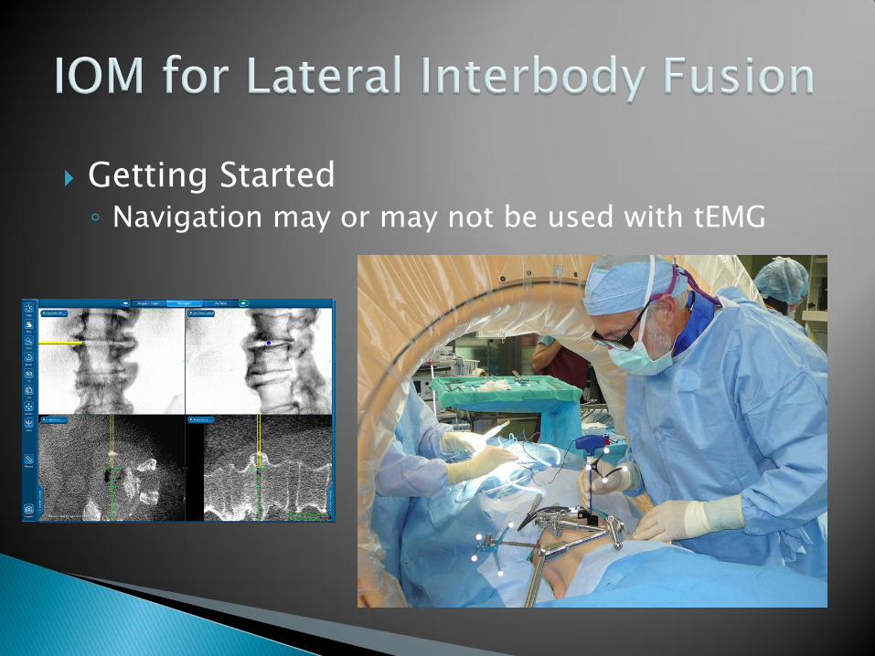
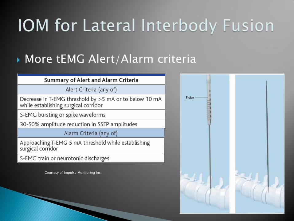

Minimally Invasive Posterior Fixation System
What What In thehellip
What What is AxiaLIF Trans 1 spine
MILF instrumentation company
Only L5-S1 fusions
Sacral approach to the spine
Also known as the Butt screw
httpAxialif
Tosh0 et al
Facet Screws lock adjacent facets
into place
I personally have been asked to test these
Spinal nerve roots could still be impinged
Stabilization is the goal
axialif-360 facet screws
Monitoring concerns Have not had any
changes with these cases
Not even EMG activity Only known
complications have been perforated bowels
Revision is not likely or possible
Sextant (TM Medtronic Inc)
SpheRx (TM NuVasive Inc)
Denali (TM K2M Inc)
Viper 2 (TM Depuy Inc)
Matis (TM Stryker Inc)
Matrix (TM Synthes Inc)
Ballista (TM Biomet Inc)
Minimally Invasive Posterior Fixation System
Minimally Invasive Posterior Fixation Systems
Getting Started Patient is positioned prone
SSEP and EMG baselines are acquired
Optimal X-ray is obtained
Pt is prepped and draped
Monitoring Tools Certified IOM
technologist
(CNIM)
NeuromontoringMachine
Cannulated EMG Monitoring Pedicle Probe(s)
Insulated dilator(s)
Communication skills
Getting Started Navigation may or may not be used with tEMG
Tapping Pedicles Target angles are
identified through X-ray
Surgeon begins to guide probe down the pedicle
Live real-time T-EMG feedback is given to the surgeon during tapping
Tapping Pedicles AlarmAlert Criteria
is followed while going through the pedicle
If necessary probe re-direction is performed to obtain favorable monitoring signals (2)
Courtesy of Impulse Monitoring Inc
Tapping Pedicles The T-Handle is
removed
A Kwire is inserted
Some Surgeons choose to stimulate kwire (3)
Tapping Pedicles Dilators are inserted
over the kwire
Insulated dilator must be used prior to tapping (12)
Important to be flush against the Transverse Process
Tapping Pedicles Stimulate the tap
when it is all the way in the pedicle
This value is thought to be the most correlative to standard open-pedicle screw EMG stimulation case data(2)
ScrewTower Insertion Screws inserted
EMG and SSEPs are continually monitored Can NOT test
Uninsulated Screw Towers
Current shunting can NOT be avoided
Thresholds are inaccurate (21)
Screw and Rod Insertion EMG and SSEPs are
continually monitored Sextant (TM )Towers Mate Rod inserted Final Tightening
Triggered EMG
EMG activity (Triggered) When the surgeon is inserting screws he can not
visualize the medial portion pedicle with C-arm A AP x-rayB Lateral x-rayC Axial (O-arm only)
C
EMG activity (Triggered) There is a risk of nerve root or spinal cord impingement if the
screw is placed too Medial In open PSF cases a threshold of greater than 20 mA of
stimulation without a CMAP response to muscle corresponding to that specific level justifies the medial placement
EMG activity (Triggered) A threshold of less than 10 mA warrants alert
criteria (4)
Under 5 mA warrants ground for screw removal (4)
Triggered EMG (tEMG) Minimally invasive surgeries do not have the
benefit of open air to insulate the path of stimulation
Current shunting must be avoided
Free Run or Spontaneous EMG (sEMG) sEMG when tapping pediclesscrew insertion Nuerotonic discharges could be missed
otherwise
Nerve root baseline Nerve root baselines can not be acquired through Percutaneous
incisions False positives from chronically compressed nerve roots can
not be accounted for
Alarm Criteria Bindal et al (2)
suggested the following tEMG protocol
Greater than 7 mA for the jamshidi needle (A B amp C)
Needle redirection was recommended for lesser thresholds
Greater than 15 mA for the insulated threaded tap (D)
Alarm Criteria Bindal et al (2) conthellip There were no insulated
threaded tap thresholds that tested under 15 mA
In 20 of the 25 patients CT scans were performed
3 lateral breaches were found
No medial breaches
Personal experience with tEMG Alarm Criteria The area of the jamshidi or PAK
needle (medronic inc) is only a few mm The charge density gives very local
information about the integrity of the pedicle wall
Personal experience with tEMG Alarm Criteria The Lowest CMAP thresholds bottom out when the
jamshidi needle is at the junction between the pedicle and the vertebral body
4 mA 7 mA 15mA
Fictional values
Thresholds between 4-7 mA typically correlate to threaded insulated tap values between 15-20 mA
Further research needs to be conducted between systems
Stimulation Duration (pulse width)
Impact On IOM for Percutaneous Pedicle Screw Testing
Idler C Rolfe K amp Gorek J Accuracy of percutaneous lumbar pedicle screw placement using the oblique or ldquoowl‟s-eyerdquo view and novel guidance technology J Neurosurg Spine 13509ndash515 2010
Featuring Nuvasive‟s new Navigation system
NeuroVision M5 (NuVasive Inc)
Five of 326 screws penetrated the pedicle cortex
3 medial and 2 lateral
pedicle breach rate of 153 Medial 092 lateral 061
overall accuracy of 9847
No mention or correlation of tEMG thresholds for the jamshidi or threaded insulated tap
Idler et al 2010
Not good news for IOMers
Wang M Pinero G Mummaneni P Stimulus-evoked electromyography testing of percutaneous pedicle screws for the detection of pedicle breaches J Neurosurg Spine 13600ndash605 2010 A clinical study of 409 screws in 93 patients
Presented at the 2009 Joint Spine Section Meeting
Results Not Good Out of 5 pedicle breaches ALL were false
negatives
There were 35 false positives as well
Results Not Good 5 medial breaches (2 with neurologic complication)
that tested at MORE than 12 mA
No true positives
Results Not Good 35 false positives out of 409 screws (86)
All screws that tested below 12 mA showed NOradiographic evidence of a medial breach
According to this article MIS EMG is a MISS
Discussion How can this be
Anyone have personal experience with this False Positives
False Negatives
Anyone have a surgeonothers mention this article to you
Possible Explanations For the false
positives May be lateral breaches
Lateral are asymptomatic
Lateral breaches can give low thresholds
What does that mean
Other Explanations source of error would be the differences or variability in
metallic conductivity a 12 rate of pedicle violations would be considered
substantially lower than other reports This finding may likely be related to our dependence on the
intensive use of intraoperative fluoroscopy to place percutaneousscrews
Computed tomography scanning can also be a source of error
FAILFAIL
There is IOM being performed without technologists
Most of these cases are also monitored without Professional oversight interpretation
Examples Surgical Instrumentation Companies
NuVasive (Neurovision tm)
Medtronic (NIM tm)
ANS is bdquoSurgeon Controlled‟
ANS do not bdquoperform‟ IOM or interpret it
Without Professional oversight interpretationhellip The surgeon is liable for any monitoring
complications
The hospital also may be liable for any monitoring complications
The instrumentation company is usually exempt
ANS is not usually bdquoSurgeon Controlled‟
Sales representatives participate in most aspects of IOM
IOM duties performed by the Sales Reps include Providing the IOM machine
Providing the IOM electrodes and supplies
Often applying the electrodes
Checking the validityreliability of the data
Relaying IOM changes to the surgeon
Other ANS Pitfalls The machines are being run by unqualified
under-educated personnel
technical errors often go misidentified or unnoticed
ANS are limited in performance (EMG and MEP only)
ANS are designed as an adjunct to concurrent instrumentation
ANS is notoriously expensive (2 to 3 times the cost of conventional IOM)
Why Conventional IOM
Superior certified personnel and equipment
Cost effectiveness
Multimodality capabilities
Position statements from credentialing bodies ASET httpasetorg
ABRET httpwwwabretorg
Neurophysiologists have specific training to obtain optimal recordings
They are available to troubleshoot hardwaresoftware problems
Specific training for optimal needle electrode placement is important to obtain accurate recordings
IOM is the only focus we do NOT sell or market any other type of services or product especially DURING THE CASE
Technologists have comprehensive IOM experience Technical errors such as EMG artifacts are a
common occurrence and must be identified and eliminated immediately
EMG artifacts Familiarity with anesthesia and the requirements
for effective monitoring
The experience to know if the patient is light andor about to start bucking
EMG artifacts Is this real or artifact
EMG artifacts Is this real or artifact
Not possible to tell out of context
SSEP and other types of monitoring is available and concurrently monitored
Vascular and positional changes are monitored
Clear Cogent and Consistent documentation
Similar features are inherent in current monitoring software
Live data feed
can be presented
at surgeon‟s
request
bdquoREAL TIME MONITORING‟ is a false concept Red lightGreen Light threshold based
monitoring is an oversimplification Clear demarcation of artifactresponse is
imperative
Experienced in DlifXlif monitoring
Experience in minimally invasive percutaneousscrew insertion monitoring techniques
Common pitfalls such as current shunting and charge density false positives can be resolved
ACDF = anterior cervical discectomy and fusion AD = aggressive discectomy ADR = artificial disc replacement ALIF = anterior lumbar interbody fusion APLD =automated percutaneous lumbar discectomy BMP = bone morphogenetic protein CAVHS = Central Arkansas Veterans Healthcare System DLIF = direct lateral interbody fusion IDET = intradiscal electrothermy LD = limited discectomy LPD = laser-assisted percutaneous discectomy MAST = minimal access spinal technique MED = microendoscopic discectomy MIS = minimally invasive surgery PEEK = polyetheretherketone PLIF = posterior lumbar interbody fusion rhBMP-2 = recombinant human BMP-2 TLIF =transforaminal interbody fusion XLIF = extreme lateral interbodyfusion
1 Balzer JR Rose RD Welch WC Sclabassi RJ Simultaneous somatosensory evoked potential and electromyographic recordings during lumbosacral decompression and instrumentation Neurosurgery 421318ndash1325 1998
2 Bindal R Ghosh S Intraoperative electromyography monitoring in minimally invasive transforaminal lumbar interbody fusion J Neurosurg Spine 6126ndash132 2007
3 Burak Ozgur Edward Benzel Steven Garfin Minimally Invasive Spine Surgery A Practical Guide to Anatomy and Techniques Springer 2009
4 Calancie B Madsen P Lebwohl N Stimulus-evoked EMG monitoring during transpedicular lumbosacral spine instrumentationInitialclinical results Spine 192780ndash2786 1994
5 Coe J Meyer SC Lumbar Plexus Palsy after XLIFreg An Avoidable Complication Society of Minimally Invasive Spine Surgery Las Vegas CA Oct 10th 2009
6 Gunnarsson T Krassioukov A Sarjeant R Fehlings M Real-Time Continuous Intraoperative Electromyographic and SomatosensoryEvoked Potential Recordings in Spinal Surgery Correlation of Clinical and Electrophysiologic Findings in a Prospective Consecutive Series of 213 Cases Spine 15 March 2004 - Volume 29 - Issue 6 - pp 677-684
7 Idler C Rolfe K amp Gorek J Accuracy of percutaneous lumbar pedicle screw placement using the oblique or ldquoowl‟s-eyerdquo view and novel guidance technology J Neurosurg Spine 13509ndash515 2010
8 Impulse Monitoring Inc Intraoperative Neuromonitoring Protocol for the Synthes Spine Oracle reg Spacer System 2007
9 Knight R Schwaegler P Hanscom D Roh J Direct Lateral Lumbar Interbody Fusion for Degenerative Conditions Early Complication Profile Journal of Spinal Disorders amp Techniques February 2009 - Volume 22 - Issue 1 - pp 34-37
10 Neuromonitroing for Transpsoas Lateral Lumbar Surgery 2006 Dr Jon Block 2006 lthttpwwwIOMstudycomgt
11 Medtronic Sofamor Danek USA Inc DLIFreg Surgical Technique 2009
12 Medtronic Sofamor Danek USA Inc CD Horizonreg Sextantreg II Rod Insertion System Surgical Steps 2009
13 Moro T Kikuchi S Konno S Yaginuma H An anatomic study of the lumbar plexus with respect to retroperitoneal endoscopic surgery Spine 2003 28423-428
14 Nuvasive Inc XLIFreg Surgical Technique 2007
15 O‟Brien RA MD MBA From Synthes sales consultant training presentation Nov 2007
16 Oppenheimer H DeCastro I amp McDonnell D Minimally invasive spine technology and minimally invasive spine surgery a historical review Neurosurg Focus Volume 27 September 2009
17 Takatomo Moro MD Shin-ichi Kikuchi MD PhD Shin-ichi Konno MD PhD Hiroyuki Yaginuma MD PhD An Anatomic Study of the Lumbar Plexus with Respect to Retroperitoneal Endoscopic Surgery SPINE Vol 28 Number 5 pp 423-428
18 Tohmeh A Rodgers W Peterson M Dynamically evoked discrete-threshold electromyography in the extreme lateral interbody fusion approach J Neurosurg Spine 2011 Jan14(1)31-7 Epub 2010 Dec 17
19 Toleikis JR Skelly JP Carlvin AO Toleikis SC Bernard TN Burkus JK et al The usefulness of electrical stimulation for assessing pedicle screw placements J Spinal Disord 13 283ndash289 2000
20 Uribe J Vale F Dakwar E Electromyographic Monitoring and Its Anatomical Implications in Minimally Invasive Spine Surgery Spine 15 December 2010 - Volume 35 - Issue 26S - pp S368-S374
21 Wang M Pinero G Mummaneni P Stimulus-evoked electromyography testing of percutaneous pedicle screws for the detection of pedicle breaches J Neurosurg Spine 13600ndash605 2010
Images courtesy of Axon Inc Biomet Inc Cadwell Inc Depuy Inc IOMstudycom Impulse Monitoring Inc K2M Inc Medtronic Inc NuVasive Inc NuWave Monitoring LLC Stryker Inc
Synthes Inc Tosh0 Trans1 Inc Trufuse Inc x-spine Inc
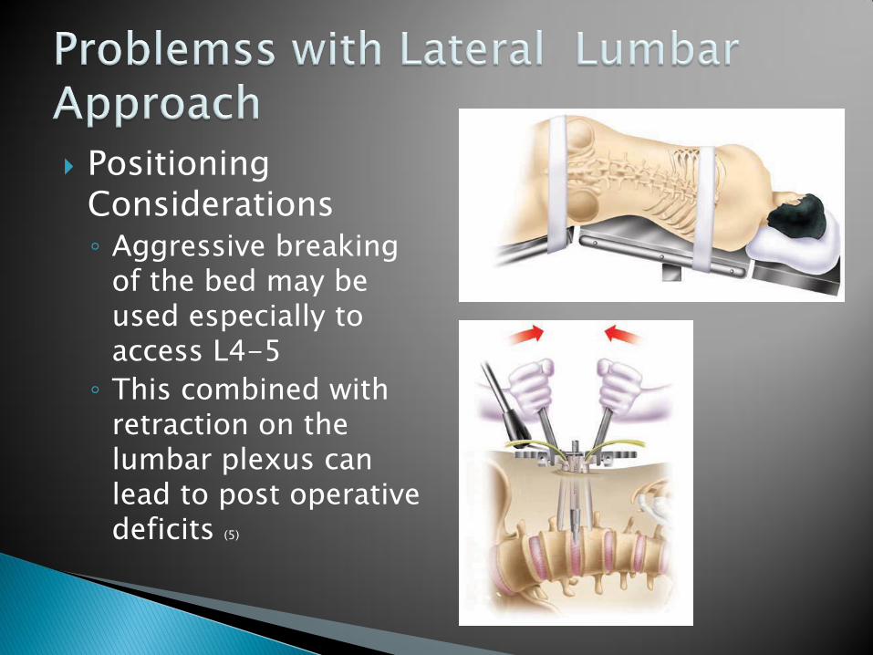
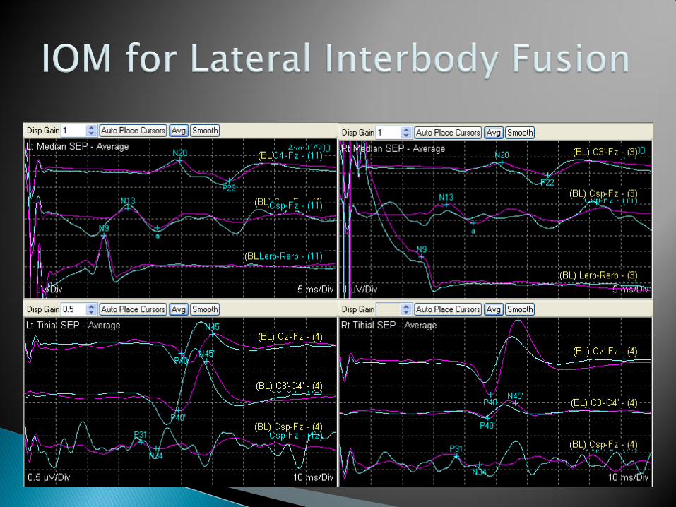
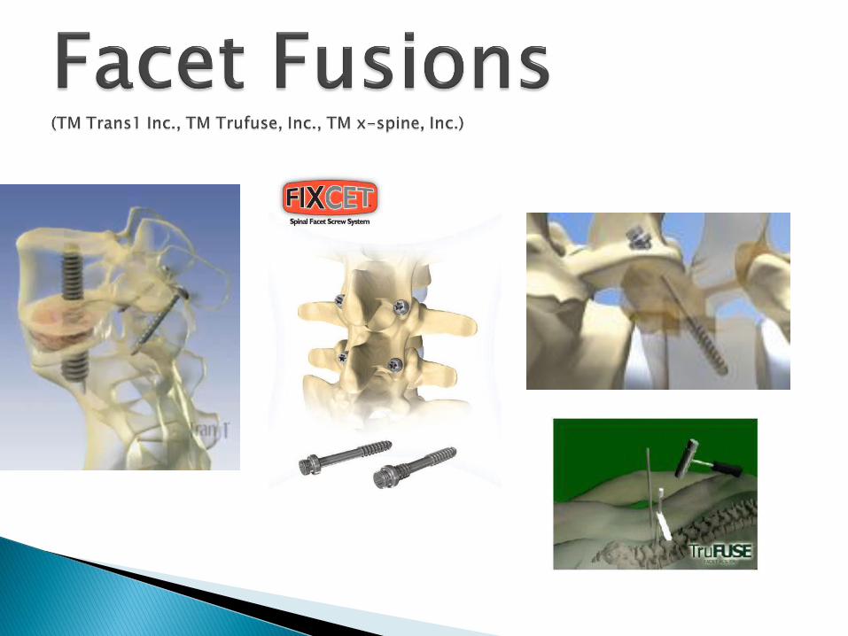

EMG artifacts Is this real or artifact
EMG artifacts Is this real or artifact
Not possible to tell out of context
SSEP and other types of monitoring is available and concurrently monitored
Vascular and positional changes are monitored
Clear Cogent and Consistent documentation
Similar features are inherent in current monitoring software
Live data feed
can be presented
at surgeon‟s
request
bdquoREAL TIME MONITORING‟ is a false concept Red lightGreen Light threshold based
monitoring is an oversimplification Clear demarcation of artifactresponse is
imperative
Experienced in DlifXlif monitoring
Experience in minimally invasive percutaneousscrew insertion monitoring techniques
Common pitfalls such as current shunting and charge density false positives can be resolved
ACDF = anterior cervical discectomy and fusion AD = aggressive discectomy ADR = artificial disc replacement ALIF = anterior lumbar interbody fusion APLD =automated percutaneous lumbar discectomy BMP = bone morphogenetic protein CAVHS = Central Arkansas Veterans Healthcare System DLIF = direct lateral interbody fusion IDET = intradiscal electrothermy LD = limited discectomy LPD = laser-assisted percutaneous discectomy MAST = minimal access spinal technique MED = microendoscopic discectomy MIS = minimally invasive surgery PEEK = polyetheretherketone PLIF = posterior lumbar interbody fusion rhBMP-2 = recombinant human BMP-2 TLIF =transforaminal interbody fusion XLIF = extreme lateral interbodyfusion
1 Balzer JR Rose RD Welch WC Sclabassi RJ Simultaneous somatosensory evoked potential and electromyographic recordings during lumbosacral decompression and instrumentation Neurosurgery 421318ndash1325 1998
2 Bindal R Ghosh S Intraoperative electromyography monitoring in minimally invasive transforaminal lumbar interbody fusion J Neurosurg Spine 6126ndash132 2007
3 Burak Ozgur Edward Benzel Steven Garfin Minimally Invasive Spine Surgery A Practical Guide to Anatomy and Techniques Springer 2009
4 Calancie B Madsen P Lebwohl N Stimulus-evoked EMG monitoring during transpedicular lumbosacral spine instrumentationInitialclinical results Spine 192780ndash2786 1994
5 Coe J Meyer SC Lumbar Plexus Palsy after XLIFreg An Avoidable Complication Society of Minimally Invasive Spine Surgery Las Vegas CA Oct 10th 2009
6 Gunnarsson T Krassioukov A Sarjeant R Fehlings M Real-Time Continuous Intraoperative Electromyographic and SomatosensoryEvoked Potential Recordings in Spinal Surgery Correlation of Clinical and Electrophysiologic Findings in a Prospective Consecutive Series of 213 Cases Spine 15 March 2004 - Volume 29 - Issue 6 - pp 677-684
7 Idler C Rolfe K amp Gorek J Accuracy of percutaneous lumbar pedicle screw placement using the oblique or ldquoowl‟s-eyerdquo view and novel guidance technology J Neurosurg Spine 13509ndash515 2010
8 Impulse Monitoring Inc Intraoperative Neuromonitoring Protocol for the Synthes Spine Oracle reg Spacer System 2007
9 Knight R Schwaegler P Hanscom D Roh J Direct Lateral Lumbar Interbody Fusion for Degenerative Conditions Early Complication Profile Journal of Spinal Disorders amp Techniques February 2009 - Volume 22 - Issue 1 - pp 34-37
10 Neuromonitroing for Transpsoas Lateral Lumbar Surgery 2006 Dr Jon Block 2006 lthttpwwwIOMstudycomgt
11 Medtronic Sofamor Danek USA Inc DLIFreg Surgical Technique 2009
12 Medtronic Sofamor Danek USA Inc CD Horizonreg Sextantreg II Rod Insertion System Surgical Steps 2009
13 Moro T Kikuchi S Konno S Yaginuma H An anatomic study of the lumbar plexus with respect to retroperitoneal endoscopic surgery Spine 2003 28423-428
14 Nuvasive Inc XLIFreg Surgical Technique 2007
15 O‟Brien RA MD MBA From Synthes sales consultant training presentation Nov 2007
16 Oppenheimer H DeCastro I amp McDonnell D Minimally invasive spine technology and minimally invasive spine surgery a historical review Neurosurg Focus Volume 27 September 2009
17 Takatomo Moro MD Shin-ichi Kikuchi MD PhD Shin-ichi Konno MD PhD Hiroyuki Yaginuma MD PhD An Anatomic Study of the Lumbar Plexus with Respect to Retroperitoneal Endoscopic Surgery SPINE Vol 28 Number 5 pp 423-428
18 Tohmeh A Rodgers W Peterson M Dynamically evoked discrete-threshold electromyography in the extreme lateral interbody fusion approach J Neurosurg Spine 2011 Jan14(1)31-7 Epub 2010 Dec 17
19 Toleikis JR Skelly JP Carlvin AO Toleikis SC Bernard TN Burkus JK et al The usefulness of electrical stimulation for assessing pedicle screw placements J Spinal Disord 13 283ndash289 2000
20 Uribe J Vale F Dakwar E Electromyographic Monitoring and Its Anatomical Implications in Minimally Invasive Spine Surgery Spine 15 December 2010 - Volume 35 - Issue 26S - pp S368-S374
21 Wang M Pinero G Mummaneni P Stimulus-evoked electromyography testing of percutaneous pedicle screws for the detection of pedicle breaches J Neurosurg Spine 13600ndash605 2010
Images courtesy of Axon Inc Biomet Inc Cadwell Inc Depuy Inc IOMstudycom Impulse Monitoring Inc K2M Inc Medtronic Inc NuVasive Inc NuWave Monitoring LLC Stryker Inc
Synthes Inc Tosh0 Trans1 Inc Trufuse Inc x-spine Inc

EMG artifacts Is this real or artifact
Not possible to tell out of context
SSEP and other types of monitoring is available and concurrently monitored
Vascular and positional changes are monitored
Clear Cogent and Consistent documentation
Similar features are inherent in current monitoring software
Live data feed
can be presented
at surgeon‟s
request
bdquoREAL TIME MONITORING‟ is a false concept Red lightGreen Light threshold based
monitoring is an oversimplification Clear demarcation of artifactresponse is
imperative
Experienced in DlifXlif monitoring
Experience in minimally invasive percutaneousscrew insertion monitoring techniques
Common pitfalls such as current shunting and charge density false positives can be resolved
ACDF = anterior cervical discectomy and fusion AD = aggressive discectomy ADR = artificial disc replacement ALIF = anterior lumbar interbody fusion APLD =automated percutaneous lumbar discectomy BMP = bone morphogenetic protein CAVHS = Central Arkansas Veterans Healthcare System DLIF = direct lateral interbody fusion IDET = intradiscal electrothermy LD = limited discectomy LPD = laser-assisted percutaneous discectomy MAST = minimal access spinal technique MED = microendoscopic discectomy MIS = minimally invasive surgery PEEK = polyetheretherketone PLIF = posterior lumbar interbody fusion rhBMP-2 = recombinant human BMP-2 TLIF =transforaminal interbody fusion XLIF = extreme lateral interbodyfusion
1 Balzer JR Rose RD Welch WC Sclabassi RJ Simultaneous somatosensory evoked potential and electromyographic recordings during lumbosacral decompression and instrumentation Neurosurgery 421318ndash1325 1998
2 Bindal R Ghosh S Intraoperative electromyography monitoring in minimally invasive transforaminal lumbar interbody fusion J Neurosurg Spine 6126ndash132 2007
3 Burak Ozgur Edward Benzel Steven Garfin Minimally Invasive Spine Surgery A Practical Guide to Anatomy and Techniques Springer 2009
4 Calancie B Madsen P Lebwohl N Stimulus-evoked EMG monitoring during transpedicular lumbosacral spine instrumentationInitialclinical results Spine 192780ndash2786 1994
5 Coe J Meyer SC Lumbar Plexus Palsy after XLIFreg An Avoidable Complication Society of Minimally Invasive Spine Surgery Las Vegas CA Oct 10th 2009
6 Gunnarsson T Krassioukov A Sarjeant R Fehlings M Real-Time Continuous Intraoperative Electromyographic and SomatosensoryEvoked Potential Recordings in Spinal Surgery Correlation of Clinical and Electrophysiologic Findings in a Prospective Consecutive Series of 213 Cases Spine 15 March 2004 - Volume 29 - Issue 6 - pp 677-684
7 Idler C Rolfe K amp Gorek J Accuracy of percutaneous lumbar pedicle screw placement using the oblique or ldquoowl‟s-eyerdquo view and novel guidance technology J Neurosurg Spine 13509ndash515 2010
8 Impulse Monitoring Inc Intraoperative Neuromonitoring Protocol for the Synthes Spine Oracle reg Spacer System 2007
9 Knight R Schwaegler P Hanscom D Roh J Direct Lateral Lumbar Interbody Fusion for Degenerative Conditions Early Complication Profile Journal of Spinal Disorders amp Techniques February 2009 - Volume 22 - Issue 1 - pp 34-37
10 Neuromonitroing for Transpsoas Lateral Lumbar Surgery 2006 Dr Jon Block 2006 lthttpwwwIOMstudycomgt
11 Medtronic Sofamor Danek USA Inc DLIFreg Surgical Technique 2009
12 Medtronic Sofamor Danek USA Inc CD Horizonreg Sextantreg II Rod Insertion System Surgical Steps 2009
13 Moro T Kikuchi S Konno S Yaginuma H An anatomic study of the lumbar plexus with respect to retroperitoneal endoscopic surgery Spine 2003 28423-428
14 Nuvasive Inc XLIFreg Surgical Technique 2007
15 O‟Brien RA MD MBA From Synthes sales consultant training presentation Nov 2007
16 Oppenheimer H DeCastro I amp McDonnell D Minimally invasive spine technology and minimally invasive spine surgery a historical review Neurosurg Focus Volume 27 September 2009
17 Takatomo Moro MD Shin-ichi Kikuchi MD PhD Shin-ichi Konno MD PhD Hiroyuki Yaginuma MD PhD An Anatomic Study of the Lumbar Plexus with Respect to Retroperitoneal Endoscopic Surgery SPINE Vol 28 Number 5 pp 423-428
18 Tohmeh A Rodgers W Peterson M Dynamically evoked discrete-threshold electromyography in the extreme lateral interbody fusion approach J Neurosurg Spine 2011 Jan14(1)31-7 Epub 2010 Dec 17
19 Toleikis JR Skelly JP Carlvin AO Toleikis SC Bernard TN Burkus JK et al The usefulness of electrical stimulation for assessing pedicle screw placements J Spinal Disord 13 283ndash289 2000
20 Uribe J Vale F Dakwar E Electromyographic Monitoring and Its Anatomical Implications in Minimally Invasive Spine Surgery Spine 15 December 2010 - Volume 35 - Issue 26S - pp S368-S374
21 Wang M Pinero G Mummaneni P Stimulus-evoked electromyography testing of percutaneous pedicle screws for the detection of pedicle breaches J Neurosurg Spine 13600ndash605 2010
Images courtesy of Axon Inc Biomet Inc Cadwell Inc Depuy Inc IOMstudycom Impulse Monitoring Inc K2M Inc Medtronic Inc NuVasive Inc NuWave Monitoring LLC Stryker Inc
Synthes Inc Tosh0 Trans1 Inc Trufuse Inc x-spine Inc
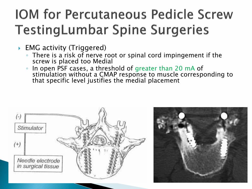

Images courtesy of Axon Inc Biomet Inc Cadwell Inc Depuy Inc IOMstudycom Impulse Monitoring Inc K2M Inc Medtronic Inc NuVasive Inc NuWave Monitoring LLC Stryker Inc
Synthes Inc Tosh0 Trans1 Inc Trufuse Inc x-spine Inc