overexpression or ectopic expression of muc2 is the common property of mucinous carcinomas of the...
TRANSCRIPT

, . 182: 385–391 (1997)
OVEREXPRESSION OR ECTOPIC EXPRESSION OFMUC2 IS THE COMMON PROPERTY OF MUCINOUSCARCINOMAS OF THE COLON, PANCREAS, BREAST,
AND OVARY
. 1*, . 1, . -2**, . 3, . . 1, . 4, . 5, . 6,.-. 2, . 2 . . 1
1Abteilung Gastroenterologie, Universitätsklinikum Benjamin Franklin der Freien Universität Berlin, Hindenburgdamm 30,12200 Berlin, Germany
2Institut für Pathologie, Universitätsklinikum Benjamin Franklin der Freien Universität Berlin, Hindenburgdamm 30,12200 Berlin, Germany
3Abteilung Chirurgie in Universitätsklinikum Benjamin Franklin der Freien Universität Berlin, Hindenburgdamm 30, 12200 Berlin,Germany
4Institut für Pathologie, Heinrich Heine Universität, Postfach 101007, 40001 Düsseldorf, Germany5Pathologisches Institut der Friedrich-Alexander-Universität Erlangen-Nürnberg, Krankenhausstraße 8, 91054 Erlangen, Germany
6Institut für Pathologie am Auguste-Victoria-Krankenhaus, Rubensstraße 125, 12157 Berlin, Germany
SUMMARY
Mucinous carcinomas of the colorectum have been reported to overexpress the intestinal mucin MUC2. The purpose of this study wasto determine whether this alteration is shared by mucinous tumours of the ovary, breast, and pancreas. A total of 40 breast carcinomas(22 of mucinous and 18 of ductal invasive type), 39 ovarian adenocarcinomas (16 mucinous, 23 serous), 47 colorectal carcinomas (25mucinous and 22 non-mucinous), and 41 pancreatic adenocarcinomas (14 mucinous, 27 non-mucinous) were investigated byimmunohistochemistry with the anti-MUC2 monoclonal antibody 4F1 and the expression pattern was ranked. MUC2 mucin is expressedin the normal colonic epithelium; in the normal epithelium of the breast, ovary, and pancreas, it was not detectable by immunohisto-chemistry or by reverse transcriptase-polymerase chain reaction (RT-PCR). In agreement with previous reports, the colonic mucinouscarcinomas differed significantly from the non-mucinous carcinomas by strong MUC2 expression. In all mucinous carcinomas ofthe ovary, breast, and pancreas, de novo expression of the MUC2 gene was observed, which differentiated mucinous and non-mucinous carcinomas of these tissues (P<0·001). The overexpression or ectopic expression of the MUC2 gene exhibited by mucinouscarcinomas of four organs indicates a common genetic lesion associated with the mucinous tumour phenotype. ? 1997 by John Wiley& Sons, Ltd.
J. Pathol. 182: 385–391, 1997.No. of Figures: 4. No. of Tables: 0. No. of References: 31.
KEY WORDS—mucinous carcinoma; colon; ovary; pancreas; breast; MUC2 expression
INTRODUCTION
Mucinous tumours represent a subgroup of carcino-mas exhibiting large amounts of mucus, grossly visibleduring microscopic examination. This morphologicaldefinition applies with some modifications to about10–20 per cent of colonic, 5 per cent of breast, 3 per centof ovarian, and 1 per cent of pancreatic carcinomas. Thecolonic mucinous carcinomas are most precisely definedin this group: according to the WHO definition, at least50 per cent of the microscopically evaluated area in thesetumours must be filled with mucus.1
The genetic alterations of colonic mucinous carcino-mas have been analysed in greatest detail: these tumoursare characterized by frequent and strong expression ofthe MUC2 gene2,3 and a higher frequency of Ki-rasmutations than the non-mucinous carcinomas.4 By con-trast, the frequency of DCC gene loss5 and of mutationsin the p53 gene is lower in mucinous than in non-mucinous colonic carcinomas.6,7 Along with the lowfrequency of mutations in the p53 suppressor gene, alower frequency of expression of the p53 protein has alsobeen observed in mucinous than in non-mucinouscolonic carcinomas.8,9 Both latter findings were inter-preted as indications that the majority of mucinoustumours develop independently of alterations in the p53gene. The analysis of the peculiarities of mucinoustumours, clearly differentiating their genotype from thatof non-mucinous colonic carcinomas, has led to thehypothesis that there is a second, less common pathwayof carcinogenesis, leading to the emergence of themucinous phenotype.5,10 A ‘mucinous pathway of
*Correspondence to: Dr C. Hanski, Klinikum BenjaminFranklin, Hindenburgdamm 30, 12200 Berlin, Germany.
**Present address: Institute für Pathologie, Ludwig-Albert-Universität, 79104 Freiburg, Germany.
Contract grant sponsors: Berliner Krebsgesellschaft; DeutscheKrebshilfe, Proj. No. 10-1025-Ha I; Deutsche Forschungsgemein-schaft, Proj. No. Ha 1520/5-3; Maria-Sonnenfeld Stiftung; FritzBender Stiftung.
CCC 0022–3417/97/080385–07 $17.50 Received 23 August 1996? 1997 by John Wiley & Sons, Ltd. Revised 26 November 1996
Accepted 5 February 1997
carcinogenesis’10 would imply the existence of geneticlesion(s) common to the mucinous tumours of otherorgans. Indeed, the recent observations made on smallgroups of non-colonic mucinous tumours seemed tosupport this hypothesis: overexpression and mutation ofp53 protein in mucinous carcinomas of the breast,11,12ovary,13,14 and pancreas15,16 were found to be lessfrequent than in the non-mucinous tumours of thesetissues. In mucinous ovarian carcinomas, as in thecolonic carcinomas, the frequency of Ki-ras mutations ishigher than in the non-mucinous carcinomas.17,18The objective of the present work was to compare
systematically the expression of the protein core of theMUC2 mucin in mucinous and non-mucinous tumoursof the colon, ovary, breast, and pancreas under stand-ardized conditions, in order to test how far the reportedpeculiarities of the colonic mucinous carcinomas areshared by mucinous tumours of other tissues. Theproperties not shared by the tumours of other organswould appear to be unrelated to the mucinousphenotype in general, but rather to be characteristiconly for the malignant transformation of the colonicepithelium.The results obtained indicate that the frequent and
strong expression of the MUC2 gene is common to themucinous tumours of the four organs. This result sup-ports the concept of a second genetic pathway leading tothe development of the mucinous phenotype.
MATERIALS AND METHODS
Tissue samples and histological typing
Formalin-fixed, paraffin-embedded tissue sampleswere obtained from the Institute of Pathology of theKlinikum Benjamin Franklin Berlin, the Institute ofPathology of the Auguste-Viktoria-Krankenhaus Berlin,the Institute of Pathology of the Heinrich-HeineUniversity Duesseldorf, and the Institute of Pathologyof the University Erlangen, Germany. A total of 40breast carcinomas (22 of mucinous and 18 of ductalinvasive type), 39 ovarian adenocarcinomas (16mucinous, 23 serous), 47 colorectal carcinomas (25mucinous and 22 non-mucinous), and 41 pancreaticadenocarcinomas (14 mucinous, 27 non-mucinous) wereinvestigated. Histological typing was performed andmucinous carcinomas were selected according to theinternational histological classification of tumours bythe WHO. Mucinous carcinomas of the breast containedlarge amounts of extracellular epithelial mucus, whichwas visible grossly and recognizable microscopicallyaround and within tumour cells.19 The epithelial elementof mucinous ovarian carcinomas included a prominentcomponent of mucin-filled cells.20 Carcinomas of thepancreas and colon were included in the mucinous grouponly if more than half of their volume consisted ofmucin lakes.1 This corresponded to the WHO definitionof mucinous carcinomas of the colon. In the case ofmucinous pancreatic adenocarcinomas, this criterionselected a subpopulation of tumours with a particularlypronounced mucinous phenotype.
Tumour stages and grades of differentiation
Except for the non-mucinous breast carcinomas andmucinous ovarian carcinomas, which were of the pT1 topT3 stage, all tumours were of the pT1 to pT4 stage.Tumour grades were from G1 to G3 (well to poorlydifferentiated).
Immunohistochemical staining
Three-micrometre thick sections were stained with themonoclonal anti-MUC2 antibody 4F1 (IgM, kindlyprovided by Dr Devine) using the ABC method andperoxidase detection, as described previously.9 It recog-nized a peptide epitope in the VNTR region of the coreprotein.21 In short, after deparaffinization with xyleneand hydration, endogenous peroxidase was blocked with0·6 per cent hydrogen peroxide in absolute methanol. Toenhance accessibility of the 4F1 epitope, mucins werepartly deglycosylated by treatment with 100 m sodiumperiodate for 3 h at room temperature. To preventnon-specific antibody binding, sections were incubatedin 20 per cent (v/v) fetal calf serum in PBS for 30 min atroom temperature prior to application of the 4F1 anti-body. The antibody 4F1 [final concentration 3 ìg/ml inPBS with 5 per cent (w/v) bovine serum albumin] wasapplied for 1 h at room temperature and detected withbiotin-conjugated goat anti-mouse immunoglobulin[final concentration 5·2 ìg/ml in PBS with 20 per cent(v/v) fetal calf serum]. After application of the ABCcomplex (Dako, Hamburg, Germany) for 30 min, per-oxidase activity was detected with 3,3*-diaminobenzidine(Sigma, St. Louis, MO, U.S.A.) as substrate and thesections were counterstained with haematoxylin, dehy-drated in graded alcohols and xylene, and embedded.Sections in which the first antibody was replaced by PBSserved as negative controls. One section known to givestrong staining was included in each run as a reference.
Evaluation of staining and statistical analysis
Evaluation of all sections was carried out by at leasttwo independent observers. The percentage of stainedcells was scored as 0, <25 per cent of stained cells, <50per cent, <75 per cent, or 75 per cent or more stainedcells. Intensity of staining was scored as 0 (no staining),1 (yellow), 2 (light brown), or 3 (dark brown).The data were plotted on a scatter diagram which for
description was divided into three fields comprisingpoints representing weak, moderate, and strong expres-sion (Fig. 3, inset). The intensity and percentage datawere ranked according to the Mann–Whitney U-test.
Detection of MUC2-mRNA by RT-PCR
Detection was carried out as described previously.3 Inshort, total RNA was extracted from normal humanovarian or mammary tissue obtained from mastectomyor ovariectomy specimens with guanidinium isothio-cyanate22 and treated with 200 U of RNAse-free DNAse(Boehringer, Mannheim, Germany) for 2 h at 37)C in50 m Tris buffer, pH 6·5, containing 10 m MgCl2,
386 C. HANSKI ET AL.
? 1997 by John Wiley & Sons, Ltd. , . 182: 385–391 (1997)
20 U of RNAse inhibitor (Boehringer, Mannheim,Germany), and 10 m DTT (Gibco BRL, Gaithersburg,MD, U.S.A.). After digestion, the DNAse wasdenatured at 100)C for 5 min. 2·5 ìg of total RNA wasreverse-transcribed as described by Murphy et al.23 in a50 ìl volume containing 2·5 ìl of the original hexa-nucleotide solution as primer and 500 U of MMLVreverse transcriptase (MMLV RT, Superscript, Gibco)according to the supplier’s recommendations (Gibco).Alternatively, cDNA was synthesized by using oligo-dTprimers from Pharmacia Biotech (Uppsala, Sweden) andMMLV reverse transcriptase. The reverse transcriptasewas denatured at 100)C for 10 min and a cDNAequivalent of 100 ng of RNA was amplified in a reactionvolume of 25 ìl containing 1#GeneAmp PCR buffer(Perkin Elmer, Cetus, Norwalk, CT, U.S.A.); 0·5 U ofAmpliTaq DNA polymerase (Perkin Elmer); 250 ì ofdATP, dGTP, dCTP, and dTTP; and 50 ng of eachprimer. Prior to amplification, the sample was heated to92)C for 5 min. Amplification was performed for 19–40cycles at 92)C, 1 min; 60)C, 1 min; 72)C, 1 min; and 72)Cfor 7 min after the last cycle in a Biomed Thermocycler60. A negative control consisting of a 25 ìl reaction mixwithout cDNA was included in each amplification ofRNA. Contamination by genomic DNA was excludedby PCR amplification of DNAse-digested RNA. Theprimers used to amplify the human MUC2 mRNAsequence were forward: CCATTCTCAACGACAACCCCTACTACCCC (nucleotides 5341–5370) and reverse:TCCAATGGGAACATCAGGATACATGGTGGC(nucleotides 5500–5529), both from the non-repetitiveregion of the MUC2 gene.25 The size of the amplifiedproduct was 189 bp. A HinfI digest of the amplificationproduct resulted in two fragments of the expected size(64 and 125 bp). Rat pancreatic tissue was obtainedfrom three Wistar rats and mRNA was isolated asdescribed above. For MUC2 gene detection in rat,cDNA primers were forward GTCTCCTACAATGGCCTGTC and reverse ACAGAAGCAGCCTTCCACCA. They amplified a MUC2 region (nucleotides13 819–14 418) homologous in the human and therat cDNA and yielded an amplimer of 600 bp. Thequality of the cDNA was tested by amplifyingthe human pyruvate dehydrogenase gene (PDH) withthe primers forward GGTATGGATGAGGAGCTGGA and reverse CTTCCACAGCCCTCGACTAA(amplimer of 103 bp) or the rat â-actin gene with theprimers forward GTGGGCCGCTCTAGGCACCAand reverse CGGT TGGCCTTAGGGTTCAGGGGGG from Stratagene (Heidelberg, Germany), yieldingan amplimer of 245 bp.
RESULTS
Expression of MUC2 mRNA in normal tissuesThe soluble intestinal mucin MUC2 is abundantly
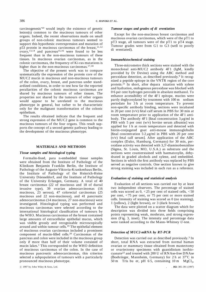
present in normal colonic mucosal tissue.2 In contrast, inthe normal human breast and ovary, MUC2 was notdetected at the protein level by immunohistochemistry(not shown) or at the mRNA level by means of RT-PCR(Fig. 1). MUC2 was also not detected immuno-
histochemically on sections of normal human pancreas(not shown). Since fresh normal human pancreatic tissuewas not available, the total cellular RNA wasextracted from the rat pancreatic tissue and used in theRT-PCR procedure with primers amplifying a sequencecompletely homologous in humans and rats. NoMUC2 mRNA was detected in the pancreatic mRNA(Fig. 1).
Expression of the MUC2 core protein in carcinomas
The analysis of MUC2 expression comprised thenumber of positive cells and the staining intensity; theplot area was divided into three fields defining weak,moderate, and strong expression (Fig. 3, inset).Staining of the non-mucinous colonic carcinomas was
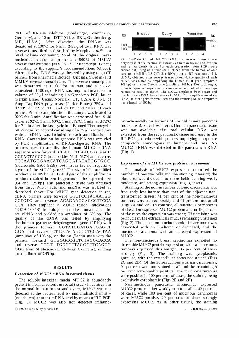
frequently less intense than that of the adjacent non-transformed tissues: 41 per cent of the non-mucinoustumours were stained weakly and 41 per cent not at all(Figs 2A and 2B). In contrast, all mucinous carcinomasof the colon expressed MUC2 protein and in 48 per centof the cases the expression was strong. The staining wasperinuclear, the extracellular mucus remaining unstained(Fig. 2). Thus, the non-mucinous colonic carcinoma wasassociated with an unaltered or decreased, and themucinous carcinoma with an increased expression ofMUC2.3The non-mucinous breast carcinomas exhibited no
detectable MUC2 protein expression, while all mucinoustumours expressed this antigen, 36 per cent of themstrongly (Fig. 3). The staining was cytoplasmic,granular, with the extracellular areas not stained (Figs2C and 2D). Of the non-mucinous ovarian carcinomas,91 per cent were not stained at all and the remaining 9per cent were weakly positive. The mucinous tumourswere positive in 100 per cent of cases, the staining beingexclusively cytoplasmic (Figs 2E and 2F).Non-mucinous pancreatic carcinomas expressed
MUC2 protein either weakly or not at all in 43 per centof cases, while 100 per cent of mucinous carcinomaswere MUC2-positive, 29 per cent of them stronglyexpressing MUC2. As in other tissues, the staining
Fig. 1—Detection of MUC2-mRNA by reverse transcriptase-polymerase chain reaction in extracts of human breast and ovarianand rat pancreatic tissue. For each preparation, four PCRs werecarried out, using as a template 1, cDNA from the human coloniccarcinoma cell line LS174T; 2, mRNA prior to RT reaction; and 3,cDNA, obtained after reverse transcription; 4, the quality of eachcDNA was tested by amplifying the human PDH gene (amplimer103 bp) or the rat â-actin gene (amplimer 245 bp). For each organ,three independent experiments were carried out, of which one rep-resentative result is shown. The MUC2 amplimer from breast andovarian tissue DNA has a length of 189 bp. For amplification of ratDNA, different primers were used and the resulting MUC2 amplimerhas a length of 600 bp
387PHENOTYPE AND GENOTYPE OF MUCINOUS CARCINOMAS
? 1997 by John Wiley & Sons, Ltd. , . 182: 385–391 (1997)
was exclusively cytoplasmic, with no membrane norextracellular mucin stained (Figs 2G and 2H).The ranking of the staining intensity or the percentage
of stained cells in each group indicated that there was asignificant difference in MUC2 expression between themucinous and the non-mucinous carcinomas (P<0·001in all cases) (Fig. 3).
DISCUSSION
The genetic basis of the occurrence of the mucinousphenotype is not clear. Previous data on the genotype ofcolonic mucinous carcinomas suggested that theydevelop along a pathway different from that describedfor non-mucinous tumours.10 The MUC2 gene, whose
Fig. 2—Immunohistochemical detection of the MUC2 protein core with the 4F1 antibody in mucinous (A, C,E, G) and non-mucinous (B, D, F, H) carcinomas of the colon (A, B), breast (C, D), ovary (E, F), andpancreas (G, H). The non-transformed colonic tissue in the vicinity of the non-mucinous tumour is expressingMUC2 (B). Staining of the cytosolic but not of the extracellular mucin is visible in all mucinous tumours.Bar=50 ìm
388 C. HANSKI ET AL.
? 1997 by John Wiley & Sons, Ltd. , . 182: 385–391 (1997)

pattern of expression differentiates the mucinous and thenon-mucinous phenotype of colonic carcinoma, wasinvestigated in the present work. To permit clear-cutconclusions, only well-characterized cases were included.The present data show that MUC2 protein expressionalso differentiates mucinous from non-mucinous carci-nomas of the ovary, breast, and pancreas.MUC2 protein was detected only as an intracellular,
cytoplasmic antigen, indicating that the glycosylation ofthe repetitive region in the secreted molecule preventedbinding of the antibody. The detected increase of expres-sion was not due to altered glycosylation of the maturemolecule in the mucinous carcinomas; in the colonicmucinous tumours a several-fold increase of MUC2expression compared with the normal tissue has alsobeen shown at the mRNA level.2Breast mucinous carcinomas always expressed
MUC2, while in the ductal invasive non-mucinouscarcinomas no MUC2 was detected. The completedichotomy between these two types of breast tumourwas also noted by Yonezawa et al.25 The possibility thatsome ductal tumours do express MUC2 is, however,indicated by the data of Walsh et al.,27 who detectedMUC2 in 20 per cent.
In 100 per cent of ovarian mucinous carcinomas and9 per cent of serous tumours, MUC2 was detected. Theexpression of the intestinal mucin MUC2 in the ovariantumours supports the notion that the development ofmalignant mucinous tumours of the ovary is associatedwith intestinal-type differentiation.28Pancreatic carcinomas of the mucinous type differed
significantly from the non-mucinous tumours in thefrequency and intensity of MUC2 protein expression.Among the non-mucinous pancreatic carcinomas, about60 per cent did, however, express MUC2, making theseparation of the two groups less pronounced than inother organs.In summary, in all four organs the development of
mucinous tumours was associated with frequent andstrong expression of MUC2. In the colon, where expres-sion of MUC2 is a normal phenotypic property, thedevelopment of mucinous carcinoma is accompanied byan enhancement of MUC2 expression. In ovarian, pan-creatic, or breast tissue, where MUC2 is normallyabsent, the development of a mucinous carcinoma isassociated with activation of a silent gene and de novoexpression of MUC2. This event occurs only rarely innon-mucinous carcinomas.
Fig. 3—Ranking of expression of MUC2 core protein in carcinomas of the colon, breast, ovary, andpancreas. The groups differed significantly (P<0·001) in the intensity of staining as well as in the numberof stained cells. Inset: assignment of areas defining weak, moderate, and strong expression
389PHENOTYPE AND GENOTYPE OF MUCINOUS CARCINOMAS
? 1997 by John Wiley & Sons, Ltd. , . 182: 385–391 (1997)
The data on MUC2 expression are of particularinterest in view of the fact that the prognosis of patientswith mucinous carcinomas of the breast, the ovary, andthe pancreas (but not of the colon) is significantly betterthan that of patients with non-mucinous tumours of thesame organs.29 The question of whether the favour-able prognosis is related to the ectopic expression ofMUC2 in these tumours and to possible induction ofan anti-MUC2 immune response warrants furtherinvestigation.Another distinct property differentiating the two
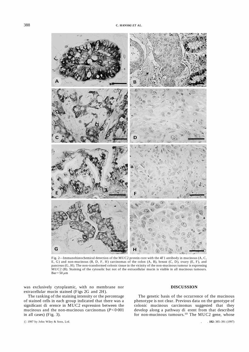
phenotypes of colorectal,6,7 breast,12 ovarian,13,30,31and pancreatic15 tumours is the frequency of p53 genemutations, which is significantly lower in the mucinousthan in the non-mucinous tumours. Collectively, theseresults, in combination with the data on the frequency ofmutations in the Ki-ras gene in carcinomas of the colonand the ovary4,17,18 and the DCC gene in coloniccancers,5 support the hypothesis that mucinouscarcinomas and non-mucinous carcinomas develop as aresult of different sets of lesions. The common propertyof the mucinous carcinomas of all four organs is thefrequent and high expression of the MUC2 gene and therelative rarity of mutations in the p53 gene (Fig. 4).Ectopic MUC2 expression implies a common lesion,
possibly affecting the transcription or the translationprocess. The relative rarity of a p53 lesion suggests thatin mucinous tumours a mechanism alternative to p53mutation may be operative. These genetic propertiescommon to mucinous carcinomas of different originssupport the concept of a ‘mucinous phenotype-related’step(s) in carcinogenesis.10
ACKNOWLEDGEMENTS
This work was supported by the BerlinerKrebsgesellschaft; the Deutsche Krebshilfe, Proj. No.10-1025-Ha I; the Deutsche Forschungsgemeinschaft,Proj. No. Ha 1520/5-3; the Maria-Sonnenfeld Stiftung;and the Fritz Bender Stiftung. We thank Dr P. Devine,Department of Obstetrics and Gynaecology, Universityof Queensland, Australia, for providing the 4F1 anti-body and Dr B. Mann for statistical evaluation of thedata.
REFERENCES
1. Jass JR, Sobin LH. Histological Typing of Intestinal Tumours. 2nd edn.Berlin: Springer-Verlag, 1990.
2. Ho SB, Niehans GA, Lyftogt C, et al. Heterogeneity of mucin geneexpression in normal and neoplastic tissues. Cancer Res 1993; 53: 641–651.
3. Blank M, Klussmann E, Krüger-Krasagakes S, et al. Expression of MUC2-mucin in colorectal adenomas and carcinomas of different histologicaltypes. Int J Cancer 1994; 59: 301–306.
4. Laurent-Puig P, Olschwang S, Delattre O, et al. Association of Ki-rasmutation with differentiation and tumour-formation pathways in colorectalcarcinoma. Int J Cancer 1991; 49: 220–223.
5. Hedrick L, Cho KR, Fearon ER, Wu TC, Kinzler KW, Vogelstein B. TheDCC gene product in cellular differentiation and colorectal tumourigenesis.Genes Dev 1994; 8: 1174–1183.
6. Costa A, Marasca R, Valentinis B, et al. p53 gene point mutations inrelation to p53 nuclear protein accumulation in colorectal cancers. J Pathol1995; 176: 45–53.
7. Hanski C, Tiecke F, Hummel M, et al. Low frequency of p53 gene mutationand protein expression in mucinous colorectal carcinomas. Cancer Lett1996; 103: 163–170.
8. Campo E, de la Calle-Martin O, Miguel R, et al. Loss of heterozygosity ofp53 gene and p53 protein expression in human colorectal carcinomas.Cancer Res 1991; 51: 4436–4442.
9. Hanski C, Bornhoeft G, Shimoda T, et al. Expression of p53 protein ininvasive colorectal carcinomas of different histological type. Cancer 1992;70: 2772–2777.
Fig. 4—Expression of MUC2 and the frequency of mutations in Ki-ras, DCC, and p53 genesdifferentiate the mucinous and the non-mucinous pathway of carcinogenesis. The percentage oftumours of either phenotype in each organ is given above the lines. The height of the lines illustratesthe relative level of MUC2 gene expression; ‘high’ and ‘low’ refer to the relative frequency ofmutations in the respective gene. Note: MUC2 is overexpressed in colonic adenomas but theexpression falls below the normal level in the non-mucinous carcinomas3
390 C. HANSKI ET AL.
? 1997 by John Wiley & Sons, Ltd. , . 182: 385–391 (1997)
10. Hanski C. Is mucinous carcinoma of the colorectum a distinct geneticentity? A review. Br J Cancer 1995; 72: 1350–1356.
11. Domagala W, Harezga B, Szadowska A, Markiewski M, Weber K, OsbornM. Nuclear p53 protein accumulates preferentially in medullary and high-grade ductal but rarely in lobular breast carcinomas. Am J Pathol 1993; 142:669–674.
12. Marchetti A, Buttitta F, Pellegrini S, et al. p53 mutations and histologicaltype of invasive breast carcinoma. Cancer Res 1993; 53: 4665–4669.
13. Milner BJ, Allan LA, Eccles DM, et al. p53 mutation is a common geneticevent in ovarian carcinoma. Cancer Res 1993; 53: 2128–2132.
14. Renninson J, Baker BW, McGown AT, et al. Immunohistochemicaldetection of mutant p53 protein in epithelial ovarian cancer using poly-clonal antibody CM1: correlation with histopathology and clinical features.Br J Cancer 1994; 69: 609–612.
15. Hoshi T, Imai M, Ogawa K. Frequent K-ras mutations and absence of p53mutations in mucin-producing tumours of the pancreas. J Surg Oncol 1994;55: 84–91.
16. Zhang SY, Ruggeri B, Agarwal P, et al. Immunohistochemical analysis ofp53 expression in human pancreatic carcinomas. Arch Pathol Lab Med1994; 118: 150–154.
17. Enomoto T, Weghorst CM, Inoue M, Tanizawa O, Rice JM. K-rasactivation occurs frequently in mucinous adenocarcinomas and rarely inother common epithelial tumours of the human ovary. Am J Pathol 1991;139: 777–785.
18. Ichikawa Y, Nishida M, Suzuki H, et al. Mutation of K-ras protooncogeneis associated with histological subtypes in human mucinous ovariantumours. Cancer Res 1994; 54: 33–35.
19. Scarf RW. Histological Typing of Breast Tumours. Geneva: WHO, 1981.20. Serov SF, Scully RE, Sobin LH. Histological Typing of Ovarian Tumours.
Geneva: WHO, 1973.
21. Devine PL, McGuckin MA, Birrell GW, et al. Monoclonal antibodiesreacting with the MUC2 mucin core protein. Br J Cancer 1993; 67:1182–1188.
22. Chomczynski P, Sacchi N. Single step method of RNA isolation by acidguanidinium thiocyanate–phenol–chloroform extraction. Anal Biochem1987; 162: 156–159.
23. Murphy LD, Herzog CE, Rudick JB, Tito Fojo AT, Bates SE. Use of thepolymerase chain reaction in the quantitation of the mdr-1 gene expression.Biochemistry 1990; 29: 10351–10356.
24. Gum RJ, Hicks JW, Toribara NW, Siddiki B, Kim YS. Molecular cloningof human intestinal mucin (MUC2) cDNA. J Biol Chem 1994; 269:2440–2446.
25. Yonezawa S, Nomoto M, Matsukita S, et al. Expression of MUC2 geneproduct in mucinous carcinoma of the breast: comparison with invasiveductal carcinoma. Acta Histochem Cytochem 1995; 28: 239–246.
26. Scarpa A, Capelli P, Mukai K, et al. Pancreatic adenocarcinomas frequentlyshow p53 gene mutations. Am J Pathol 1993; 142: 1534–1543.
27. Walsh MD, McGuckin MA, Devine PL, Hohn BG, Wright RG. Expressionof MUC2 epithelial mucin in breast carcinoma. J Clin Pathol 1993; 46:922–925.
28. Fenoglio CM, Ferenczy A, Richart RM. Mucinous tumours of the ovary:ultrastructural studies of mucinous cystadenomas with histogeneticconsiderations. Cancer 1975; 36: 1709–1722.
29. Hermanek P, Gospodarowicz MK, Hensen DE, Hutter RVP, Sobin LH(eds). Prognostic Factors in Cancer. Berlin: UICC, Springer-Verlag, 1995.
30. Kappes S, Milde-Langosch K, Kressin P, et al. p53 mutations in ovariantumours, detected by temperature-gradient gel electrophoresis, directsequencing and immunohistochemistry. Int J Cancer 1995; 64: 52–59.
31. Kim JW, Cho YH, Kwon DJ, et al. Aberrations of the p53 tumoursuppressor gene in human epithelial ovarian carcinoma. Gynecol Oncol1995; 57: 199–204.
391PHENOTYPE AND GENOTYPE OF MUCINOUS CARCINOMAS
? 1997 by John Wiley & Sons, Ltd. , . 182: 385–391 (1997)