outpatient physical therapy management of a patient
TRANSCRIPT

University of North DakotaUND Scholarly Commons
Physical Therapy Scholarly Projects Department of Physical Therapy
2019
Outpatient Physical Therapy Management of aPatient Diagnosed with Myotonic MuscularDystrophy Type 2Ashley JobeUniversity of North Dakota
Follow this and additional works at: https://commons.und.edu/pt-grad
Part of the Physical Therapy Commons
This Scholarly Project is brought to you for free and open access by the Department of Physical Therapy at UND Scholarly Commons. It has beenaccepted for inclusion in Physical Therapy Scholarly Projects by an authorized administrator of UND Scholarly Commons. For more information,please contact [email protected].
Recommended CitationJobe, Ashley, "Outpatient Physical Therapy Management of a Patient Diagnosed with Myotonic Muscular Dystrophy Type 2" (2019).Physical Therapy Scholarly Projects. 659.https://commons.und.edu/pt-grad/659

OUTPATIENT PHYSICAL THERAPY MANAGEMENT OF A PATIENT DIAGNOSED WITH
MYOTONIC MUSCULAR DYSTROPHY TYPE 2
A Scholarly Project Submitted to the Graduate Faculty of the
Department of Physical Therapy
School of Medicine
University of North Dakota
by
Ashley Jobe
In Partial Fulfillment ofthe Requirements for the Degree of
Doctor of Physical Therapy
Grand Forks, North Dakota
May, 2019

This Scholarly Project, submitted by Ashley Jobe in partial fulfillment of the requirements for the Degree of Doctor of Physical Therapy from the University of North Dakota, has been read by the Advisor and Chairperson of Physical Therapy under whom the work has been done and is hereby approved.
~d..('()~ (Graduate School Advisorv;::}-
.. li.

Title
Department
Degree
PERMISSION
Outpatient Physical Therapy Management of a Patient Diagnosed with Myotonic Muscular Dystrophy Type 2
Physical Therapy
Doctor of Physical Therapy
In presenting this Scholarly Project in partial fulfillment of the requirements for a graduate degree from the University of North Dakota, I agree that the Department of Physical Therapy shall make it freely available for inspection. I further agree that permission for extensive copying for scholarly purposes may be granted by the professor who supervised my work or, in her absence, by the Chairperson of the department. It is understood that any copying or publication or other use of this Scholarly Project or part thereof for financial gain shall not be allowed without my written permission. It is also understood that due recognition shall be given to me and the University of North Dakota in any scholarly use which may be made of any material in this Scholarly Project.
Date /0 / /0 ( f g
Jll

TABLE OF CONTENTS
LIST OF TABLES ................................................................................................................................................ ;... v
ABSTRACT ....................................................... .. ..................................................................................................... vi
CHAPTERS
I. INTRODUCTION AND BACKGROUND INFORMATION............. ......................................... 1
II. CASE DESCRIPTION........................................................................................................................ 7
Ill. EXAMINATION AND EVALUATION ........................................................................................ 11
IIV. DIAGNOSIS AND PROGNOSIS .................................................................................................... 19
V. INTERVENTIONS .............................................................................................................................. 21
VI. OUTCOMES .................. ................................. ........................................... .......................................... 25
VII. DISCUSSION .................................... .......... ............................................. .......................................... 27
VIII. REFLECTION ............................ ...................................................................................................... 29
IV. CONCLUSION .............................. ........... ............................................................................................ 31
REFERENCES ................................................ .................. ........................................................................ ............... 32
iv

LIST OF TABLES
1. Table 1: Initial and Discharge Examination Scores ................................................................ 14
2. Table 2: Pinch Dynamometry Normative Data for Females Age 60-65 ................................ 17
3. Table 3: Psychometric Properties ofTests and Measures ....................................... ............ 18
v

ABSTRACT
Background and Purpose. This article describes the 3-week outpatient Physical Therapy
management of a 63-year-old female with a diagnosis of Myotonic Muscular Dystrophy
(MMD) Type 2. MMD is an inherited disorder, which progressively impacts muscular,
respiratory, cardiac, nervous, and endocrine systems. MMD Type 2 is characterized by
muscle weakness, muscle pain, and myotonia. The patient in this case study presented to
physical therapy with upper and lower extremity weakness and balance impairment. The
purpose of this article is to describe the initial physical therapy evaluation process for a
patient with MMD, as well as the interventions used for this patient and the patient's
response to treatment. Research involving the physical therapy management of a patient
with MMD Type 2 is scarce, and this article intends to add to the limited available
information. Description. The treatment of this patient involved a generalized
strengthening program for the upper and lower extremities, aerobic conditioning, balance
training, and soft tissue massage for tissue restrictions. Outcomes. Following physical
therapy intervention, the patiettt improved in functional outcome scores and balance
assessment scores. The patient also demonstrated increased tolerance to aerobic exercise.
Grip and pinch strength dynamometry were recorded revealing increased strength.
Variable results were compiled from the manual muscle testing strength assessment, with
a general trend of increased strength following the 3-week period of therapy. Discussion.
The rationale for treatment was based on addressing the patient's presenting symptoms
and goals. Information gathered from a thorough literature review was used to guide
treatment. Treatment was altered or progressed based on the patient's response.
vi
CHAPTER I
INTRODUCTION AND BACKGROUND INFORMATION
Genetics
The origin of Myotonic Muscular Dystrophy (MMD) is genetic. MMD results from an
abnormal repetition within a gene's DNA sequence. The mutated gene is unstable, and
produces aberrant messenger-RNA that clumps inside the cell and interferes with the
production of many proteins.1 These changes impede normal cell function, and lead to the
signs and symptoms of MMD. Due to the etiology of MMD, the disease is further classified as
a spliceopathy.2 MMD is an autosomal dominant disorder. So, when a mutated gene is
present it will be dominant over the paired normal gene. If an individual inherits a mutated
gene, they will develop the disease. The likelihood of a parent with the disease passing the
mutated gene on to their children is 50%. As the disease is passed on through generations,
the signs and symptoms become more severe and begin earlier in life due to a phenomenon
called anticipation. 3
Two different forms of MMD have been identified. Genetic variations between the
two subtypes can help with differential diagnosis. MMD Type 1 is classified by genetic
mutations in the DMPK gene.1 MMD Type 2 results from mutations in the CNBP gene. The
exact functions of these genes are still unclear. However, the protein produced by the
DMPK gene appears to play a role in cell communication and correct cell functioning within
1

the heart, brain and skeletal muscles. The protein produced by CNBP is found primarily in
cardiac and skeletal muscles, where it is involved in protein synthesis and possibly the
regulation of other genes. Additional genetic differences have also been found. For instance,
the repeat expansion observed in MMD Type 2 is much larger than MMD Type 1. 2 However,
the size of the repeat does not correlate with the disease severity or age of onset for
patients with MMD Type 2, unlike patients with MMD Type 1. Physical examination,
electromyography, and muscle biopsies are used to diagnose MMD. Genetic testing is the
only definitive test, though, and is the best for determining the subtype of MMD.3
Prevalence
MMD is estimated to affect at least 1 in 8,000 people worldwide .I The prevalence of
MMD varies by subtype and geographic location. MMD Type 1 is the most common
inherited myopathy that presents in adulthood.4 MMD Type 1 is typically more common
than MMD Type 2, with the exception subpopulations in Germany and Finland where the
two subtypes are equally common. The prevalence of MMD is 3-15/100,000 people in
Europe. It is extremely rare in some areas (0.9/100,000 people in Taiwan) and extremely
common in others (189/100,000 in Saguenay-Lac-Saint-Jean, Canada). Since most research
has been done outside of the United States of America, the exact prevalence of MMD in the
United States is unknown at this time. There are currently no statistics for the prevalence
of MMD Type 2 within the general population.
2

Presentation
MMD progressively impacts muscular, respiratory, cardiac, nervous, and endocrine
systems. The most notable signs and symptoms of MMD include muscle atrophy, muscle
weakness, myotonia, and muscle pain. These impairments are many times accompanied by
balance deficits and difficulties with gait. The multisystem effects of MMD manifest in a
wide variety of health impairments, such as cataracts, cardiac conduction deficits, and
diabetes mellitus. It is important to note that these comorbidities are in fact related to the
patient's diagnosis of MMD.
Not only are there differences in genetic characteristics, there are also differences in
the patients' physical presentation when comparing the subtypes of MMD. This knowledge
is very useful for clinicians. Patients with MMD Type 1 typically develop symptoms of the
disease earlier than those with MMD Type 2. MMD Type 1 can present at birth, but most
often appears in the patients' 20s or 30s. 3 MMD Type 2 typically presents in the patients'
40s. Although many oftheir symptoms are similar, MMD Type 1 tends to be more severe.
MMD Type 1 generally impacts more distal muscles including those of the legs, hands, neck,
and face. Patients with MMD Type 2 typically experience involvement of more proximal
muscles including those of the neck, shoulders, elbows, and hips. Prominent muscle pain,
stiffness, and fatigue are more commonly experienced by patients with MMD Type 2 in
comparison to MMD Type 1.5 These are general trends that can be helpful in coming to a
diagnosis and for treating a patient with MMD, but it is important to note that symptoms
vary largely from person to person so these criteria may not apply to all patients.
3

Research by Dalton, Ranum, and Day describes the clinical presentation of patients
with MMD Type 2. The most prevalent findings are myotonia (90%) and proximal muscle
weakness (82%).5 Patients with MMD Type 2 also commonly report muscle pain, stiffness,
fatigue, and excessive daytime sleepiness. Other clinical features include defects in cardiac
conduction (19%), cataracts (36-78%, increasing with age), and insulin insensitivity (25-
75%, increasing with age). Diabetes mellitus may be more common in MMD Type 2
patients in comparison with MMD Type 1 patients. Cognitive manifestations of MMD Type
2 include difficulties with organization, concentration, and word finding. When performing
a review of systems and history for a patient, these are important factors to address.
Current Literature
The majority of literature and research reviewed in preparation for this case
focused on the patient's primary diagnosis of MMD Type 2. Pandya and Eichinger described
the critical pieces of an initial evaluation for a patient with MMD. 6 Systems review, manual
muscle testing, dynamometry, functional ability assessment, as well as pain and fatigue
assessments provide important data for monitoring a patient's disease progression. The
article also addresses physical therapy management of patients with MMD. General range
of motion is emphasized to maintain joint function and decrease occurrence of muscular
imbalances. Strengthening exercises at a moderate intensity may help to avoid muscular
weakness from disuse. High intensity exercise is not advised, as this can overstress the
patient leading to an increase in symptoms and worsening of the disease progression.
Cardiovascular exercise at a low to moderate intensity is safe for patients with MMD and
4
may benefit the patient. Since MMD can affect the heart, clearance from the patient's
physician is recommended before beginning aerobic exercise.
A study by the United States Department of Health and Human Services suggests
that individuals with chronic conditions or disabilities, such as MMD, would benefit from 2
hours and 30 minutes of moderate intensity exercise every week. 7 This exercise program
should include full body strengthening 2-3 times a week and aerobic conditioning lasting at
least 10 minutes each session. According to Ted Abresch, the director ofthe Research and
Training Center for Neuromuscular Disease at the University of California at Davis, adults
with slowly progressing muscular disorders (such as MMD) show improvements from
moderate intensity exercise. 8 Walking studies have been particularly effective in
decreasing the patients' weight and increasing their quality of life assessment scores.
When selecting modalities, it has been shown that patients with MMD typically have
positive results with therapeutic heat. 6 Cold is less therapeutic, as it increases the
likelihood of myotonic muscle contractions. Adaptive equipment and assistive devices or
orthotics may be helpful to improve the patient's independence and safety. Assessing the
patient's need for such devices is an important part of physical therapy management for
MMD.
When searching for further articles about MMD Type 2, it became apparent that
research is severely lacking. Physical therapy intervention has been proven effective for
other myopathies, but there is limited information available that is specific to MMD Type 2.
The purpose of this article is to describe the initial physical therapy evaluation process for
a patient with MMD Type 2, as well as the interventions used for this patient and the
5

patient's response to treatment. This article aims to add information to the limited supply
that is available, and to increase awareness about the need for research concerning MMD
Type 2.
6

CHAPTER II
CASE DESCRIPTION
History
The patient is a 63-year-old female. She was at a check-up for her sleep apnea when
a nurse practitioner specializing in pulmonology referred her to physical therapy. The
practitioner identified upper and lower extremity weakness and difficulty with standing
during the check-up, which led to the referral. The patient presented to physical therapy
the following week with bilateral upper and lower extremity weakness, decreased muscle
tone of her hands, bilateral grip myotonia, and impaired balance. She was formally
diagnosed with MMD Type 2 approximately five years ago by a neurologist who used
diagnostic EMG studies.
The patient recalled symptoms of grip myotonia beginning when she was around
30-years-old. She reported a history of muscle weakness starting when she was
approximately 40-years-old, which gradually worsened. The patient began using a cane at
age 58. The patient reported a suspected family history of MMD, as she recalled relatives
with similar presentations and health conditions. The patient was of European descent, and
may be of heritage with increased prevalence, although this had not been confirmed. The
patient's past medical history included (but is not limited to), diabetes mellitus,
hypertension, hyperlipidemia, silent aspiration, pharyngeal dysphagia, decreased
7

peristalsis, diaphragm weakness, mixed restrictive and obstructive lung disease,
obstructive sleep apnea, mild aortic insufficiency, bradycardia, cardiac pacemaker
(insertion in 2002 and replacement in 2009), bilateral cataract removal (2008),
cholecystectomy (1992), breast cancer with mastectomy (2008), hysterectomy (1992),
degenerative joint disease, osteopenia, osteoporosis, ligamentous injury to the left knee,
and MMD Type 2. The patient exhibited systemic involvement and had been seen by
specialists for each of her diagnoses. She maintained regular check-ups and treatment by a
multifaceted care team involving cardiologists, neurologists, general practitioners,
pulmonologists, endocrinologists, and orthopedic specialists. Upon review of systems, the
patient reported no changes or new complaints in these areas. The patient also had a
history of falls, which resulted in traumatic coccydynia in 2010, a fractured left radius in
2012, and a distal left femur fracture in 2013. Following each fracture, she was seen in
physical therapy for rehabilitation.
The patient's symptoms associated with MMD Type 2 were worsened by prolonged
activity. Following a day of increased activity, the patient experienced high levels of fatigue.
She reconciled the fatigue with sleep, which would help her feel better. By increasing the
amount of sleep she received over the duration of a few days, she would return to her
baseline level of function.
The patient lived in a rural area of the Midwest. She lived independently with her
husband in their home. Her home had a ramp to the entrance, and approximately 5 stairs
inside that she used on a regular basis. She had family in the area, and often cared for her
teenage grandson when he was done with school. Due to her health conditions, the patient
8

had retired from waitressing. Her leisure activities were limited, and she did not make
many community outings. She typically did not go into the community without her
husband. She reported that driving to her physical therapy sessions was the first time she
had traveled on her own in the last 5 years. The patient had limited resources in regards to
her healthcare. There were no area programs for seniors to exercise, she was not a member
of a support group or care organization related to her diagnosis, and she had to travel for
her healthcare.
The patient's chief complaint was her decreased balance, as she reported multiple
falls. Her main goals for physical therapy included improving her balance, increasing her
muscle strength and endurance so that household chores were less challenging, improving
her walking tolerance for community ambulation, and relief of her muscle pain.
Clinical Impression
The initial clinical impression following the subjective interview was that the
patient would benefit from skilled physical therapy intervention. Observations of her sit-to
stand transfers and gait prior to formal examination, along with her reports of decreased
balance and difficulties with household chores indicated that the patient might benefit from
physical therapy. Skilled physical therapy would work to improve her balance and decrease
her risk of falls. She would also benefit from an exercise program that had been adjusted to
her personal needs, which would counteract her symptoms by increasing both her aerobic
and muscular endurance. Manual therapy techniques, such as soft tissue massage, would
help relieve her pain.
9

The goal of physical therapy was to increase her upper and lower extremity
strength, allowing her increased functional capabilities at home. The purpose of continuing
the strengthening program would be to maintain her functional strength for as long as
possible. The goal of balance training was to decrease the patient's likelihood of falling and
to increase her mobility. All of the proposed goals coincide with the reported goals of the
patient.
Examination Plan
The plan for examination was to document her current functional status.
Observations of her bed mobility, sit-to-stand transfers, and gait would assist in assessing
her transferring abilities and mobility level. Pinch strength, grip strength, and full body
manual muscle testing would gauge her muscular strength and identify areas of weakness.
Balance tests, such as the Berg Balance Scale and the Timed Up and Go Assessment, would
be used to determine her risk of falls and areas of difficulty. Range of motion
measurements would assess ifthe patient had full and pain-free motion of all body parts.
10

CHAPTER III
EXAMlNATION AND EVALUATION
Observation
When walking into the clinic, the patient used a cane in her right hand. Proper gait
sequence was used, although she leaned heavily on her cane. The patient navigated non
complicated environments well, but with increasing complexity she demonstrated
decreased tolerance. For example, in crowded areas or on uneven surfaces she self-selected
a slower gait speed and reported feelings of unsteadiness and weakness. For these reasons,
the patient used her cane for community ambulation and for walking distances longer than
approximately 50 feet. On stairs, the patient used a step-to-step pattern with bilateral
upper extremity assistance on hand railings. She flexed forward at her hips and relied
heavily on her upper extremities to pull herself up onto the next stair. The patient
demonstrated use of proper lead foot, and she reported her right side being dominant and
stronger.
The patient had difficulty with transitional movements, but with compensatory
patterns and adaptations she remained independent. When transferring from sit-to-stand,
she demonstrated a Gower's sign. She leaned very far forward and walked her hands up her
legs to assume an erect position, and generally required multiple attempts to stand. At
home, she used a front-wheeled walker to assist with sit-to-stand transfers. The patient
11

reported that she did not usually use any form of assistive device within the home for
ambulation.
When observing her bed mobility, similar trends appeared. The patient pulled with
her arms to assist with log rolling to the side, which required multiple small weight shifts to
occur and was physically demanding. She had to support her head with a pillow during this
transition, as her neck muscles were too weak to assist with this movement. When
transferring to and from supine, she was able to swing her legs and controlled her upper
body descent with her arms. She kept her core and neck stable, but was unable to
dynamically contribute to the transfer with her core stabilizers.
She presented with a right lateral shift during gait, standing, and sitting. Postural
observation revealed increased thoracic kyphosis and cervical lordosis, as exhibited by a
forward head and rounded shoulders.
Tests and Measures
The patient presented with full range of motion of the legs and arms bilaterally.
Strength was assessed using manual muscle testing and dynamometry for grip and pinch
strength measurements. Special tests were selected to test the patient's balance and
functional abilities. The psychometric properties of all tests and measures used in this
episode of patient care can be found in Table 3.
Initial evaluation data indicated proximal muscle weakness. Manual muscle testing
was performed according to Magee.9 For full initial evaluation data, see Table 1. Significant
weakness was found in both the upper and lower extremities, as well as core stabilizers.
Shoulder flexion and extension, gross wrist motions, gross finger motions, gross hip
12

motions, and gross neck motions scored within the range of grade 4-/5 to 4+ /5. The
patient's area of greatest weakness was trunk flexion and extension, which were both
found to be grade 2/5 at initial evaluation.
13

Tabl~ L ritial e:~r· d Ji~t:~a rge ·v1anual Mu!>ciC Tc~ ting and Dynamometry ~t!>Uih
Init ial fll'anual \11u~.d~ Te~t in& for St req:;th Ji ~ ;:ha rge Manu;;! Mu~cle Tc:.Ung for Strength
Upper Body Action Right left Upper Body Action Right left Shoulder FleXIon ~+/5 4/5 Shoulder FlcJCon 4~/5 ~/5
Extemion 1',+/5 4+/ 5 Extcmion 5/5 5/5 Abduction 5/5 5/5 Abduction 4-/ S 4-/5 Adduction 5/S 4/5 Adduction 4..-/ 5 4+/5 lnH~rnal ~otalion 5/5 5/5 lntcrr.al ~otat ion 4/5 4-/S External Rotation 5/5 5/5 External Rotati on 4T/ 5 ~/5
Elbow Flex.10n S/5 5/5 Elbow Flex. on 5/5 4+/S Extemion 5/S 5/S Extemion 4T/5 ~/5
\'.'ri!>l FleX:on ~+/5 4/5 1,'.'-J!.l Flex. on 4 ... /s ~/5
Extcmior ~+/5 4/5 Extemion 4/5 4-/5 Ulnar D<:!viation 1',+/S S/ 5 Ulrar Dtviatio'1 4/5 t./5 Ra dial J~::via t 1 o'1 5/5 5/5 Radial Jcviat ion 4T/ 5 ~/5
Suoinafon 4/5 4-/S SuDinaUon S/ 5 ~/5
Pronation 4/5 4/ 5 Pronation 5/ 5 ~/5
Finger.: Extemion t.-/5 4 -/5 Fingers Extemion 4/5 4-/S (~~_c~.r;r i p and pinch !>trc'1g1h (~Ke~grip and pinch !:lrength
Flex. on oynamomc try) Flexion dynamornctryj
Initial Dynamometry Discharge Dynam ometry
Right left Right _.eft
Grip S~ren.r;th a L~~ 4~ Gr1p S~reng~h 8JQ~ 5 ~Q~ Pmch Strength t. IP~. 3 !Q.~ Pmch Strl::!ngth 4 ~ 3 l~..:. 3 Jaw Chuck t.l~ 4~ 3 Jaw Chuck 6 !b.~ 3 !~ latera l Pmt:h 7~~ (j fu~ Latera l Pmch 7fu.~ 8 ~
14
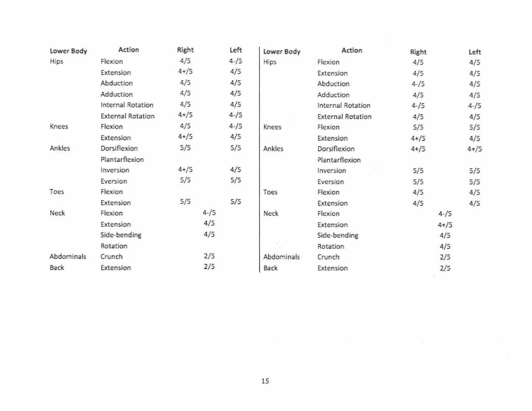
lower Body Action Right Left Lower Body Action Right left Hips Flex: on t./5 ~-/5 H"ps Flex. on 4/5 4/5
Extensior 4T/ 5 4/5 Extemion 4/5 4/5 Abtluction t./5 4/5 Abduction ~-/5 4/5
Add.Jction t./5 4/5 Adduction 4/5 4/5 Internal ~otation t./5 4/5 Internal ~otation t.-/5 4-/5
External Rot ation 4-+/5 ~-/5 ElCterna I Ro tation 4/5 4/5 Knees Fle.JUon t./5 t.-/5 Knees Flex.ton 5/5 5/5
Ex:temion 4T/5 4/5 Extension t.+/5 4/5 Ankle~ Dorsi flexion S/5 S/5 Ankles Dorsiflexion t.+/5 4+/ 5
Plan ta rfle.xion PI an ta r flexion
ln\'er~ion 4T/5 4/5 Inversion 5/5 5/S
Evers. on 5/5 S/5 Evers.on 5/5 S/5 Toe!:. Flex.on T()l!~ Flexion 4/5 4/5
Extension S/5 5/5 Extem.ion 4/5 4/ S Neck Fl(!~on 4-/5 Neck Flex.. on 4-/5
Exten~ion 4/5 Extension 4+/5 Side-hen ding 4/5 Sidf!-bendirg 4/ 5
Rotation Rotation 4/S Abdoninals Crunch 2/5 Abdoninals Crunch 2/5 Baclc. Extension 2/5 Ba~o( Extension 2/5
15

Grip dynamometry also revealed significant weakness compared to age norms.
Standardized test positioning was used with a Jamar® dynamomet er to assess grip
strength. According to the Rehabilitation Measures Database, normative values for women
aged 65-69 is 54.9 pounds of force with a 10.1 pound standard deviation. 1o The patient in
this case study generated 8 pounds of force with the right hand and 4 pounds with the left
hand; both grip strength values were significantly lower than the normative values.
Pinch strength was measured using a Sammons Preston Baseline® Pinch Gauge.
Normative data generated by faculty at the University of Wisconsin-Milwaukee for females
60-65 years old has been compiled. 11 Please, see Table 2 for these normative values. The
patient's initial tip pinch strength was 4 pounds with the right and 3 pounds with the left,
key pinch strength was 7 pounds on the right and 6 pounds on the left, and palmar grip
strength 4 pounds bilaterally. All of the dynamometry measurements were significantly
below normal for her age and gender.
16

Table 2. Pinch Dynamometry Normative Data for Females Age 60-65
Right Hand Right Hand Standard Left Hand Left Hand Standard (Pounds) Deviation (Pounds) (Pounds) Deviation (Pounds)
Tip Pinch 10.1 2.1 9.9 2
Key Pinch 15.5 2.7 14.1 2.5
Palmar 14.8 3.1 14.3 2.7
Pinch
Special tests were selected to assess the patient's balance, mobility, and fall risk. The
Berg Balance Scale (BBS) was administered to assess the patient's balance with various
tasks, allowing examiners to compute fall risk. The patient was instructed to perform both
static and dynamic balance activities of varying difficulty. A maximum score of 56 is ideal.
For patients with a history of falls and a score less than 51, or those with no history of falls
and a score less than 42, the BBS is predictive of falls with 91% sensitivity and 82%
specificity. A score of less than 40 on the BBS is associated with an almost 100% fall risk.
Normative scores for women ages 60-69 was 55 with a standard deviation of 2. The
patient in this case study scored 37, which means she had almost a 100% risk of falling at
the time of her initial evaluation.
17
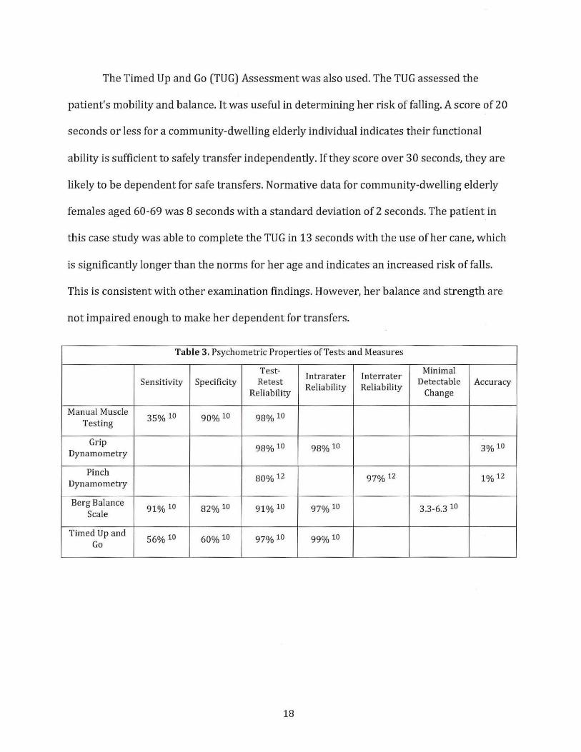
The Timed Up and Go (TUG) Assessment was also used. The TUG assessed the
patient's mobility and balance. It was useful in determining her risk of falling. A score of 20
seconds or less for a community-dwelling elderly individual indicates their functional
ability is sufficient to safely transfer independently. If they score over 30 seconds, they are
likely to be dependent for safe transfers. Normative data for community-dwelling elderly
females aged 60-69 was 8 seconds with a standard deviation of 2 seconds. The patient in
this case study was able to complete the TUG in 13 seconds with the use of her cane, which
is significantly longer than the norms for her age and indicates an increased risk of falls.
This is consistent with other examination findings. However, her balance and strength are
not impaired enough to make her dependent for transfers.
Table 3. Psychometric Properties of Tests and Measures
Test-Jntrarater Jnterrater
Minimal Sensitivity Specificity Retest
Reliability Reliability Detectable Accuracy
Reliability Change
Manual Muscle 35%10 90%10 98%10
Testing
Grip 98%10 98%10 3% 10 Dynamometry
Pinch 80% 12 97% 12 1 o/o 12 Dynamometry
Berg Balance 91 o/o 10 82% 10 91% 10 97% 10 3.3-6.3 10
Scale
Timed Up and 56%10 60% 10 97% 10 99% 10
Go
18

CHAPTER IV
DIAGNOSIS AND PROGNOSIS
The examination process confirmed initial clinical impressions and revealed
physical therapy diagnoses of bilateral upper and lower extremity weakness, decreased
muscle tone of her hands, bilateral grip myotonia, and impaired balance. These findings are
consistent with her medical diagnosis of MMD Type 2. It was determined that the patient
was in need of skilled physical therapy to improve her function. The plan of care included
balance and proprioceptive activities, a home exercise program, and manual therapy as
needed for treatment of pain. This plan of care was to be carried out twice weekly over the
duration of six weeks. This amount of time would allow her to learn her home exercise
program with instruction from a physical therapist to avoid compensation patterns,
develop her balance strategies and provide her with proprioceptive challenges under
clinical supervision to ensure safety, and explore pain management techniques to improve
her quality of life. Due to the progressive nature MMD Type 2, her prognosis and
rehabilitation potential are guarded. It was hypothesized that she would see some minor
improvements in her functional abilities and scores on tests and measures. Individuals
with muscular dystrophies are able to benefit from physical therapy services, and one goal
was to offer this patient a plan of care that would help her maintain or improve her current
level of function .
19

The patient's goals were to improve her ability to complete household activities,
increase her tolerance for walking, and to complete her self-cares with more ease. She also
wished to decrease her discomfort from muscular pain. The patient highly valued her
independence and interactions with her family, and wanted to maintain her function in
these areas. By improving her strength and balance, completing home activities, walking,
and self-care should become easier. The physical therapy goals and patient goals aligned.
Both were to maintain her function and to increase her strength and endurance to assist
with completion of her daily activities.
A plan for periodic reexamination was set in place. Continual reassessment of her
performance at each physical therapy session was used to track her progress. More formal
examinations were to be performed after three weeks and after six weeks of physical
therapy intervention. At these times, a TUG assessment and strength screening would be
performed to track her progress. A long term physical therapy program would include
tracking her progress in monthly intervals, offering her patient education, monitoring the
effectiveness of current treatment, and adjusting her plan of care as needed.
20

CHAPTER V
INTERVENTIONS
This patient was seen two days a week for one hour sessions over the course of
three weeks. Due to the patient's systemic involvement, exercise was maintained at
moderate intensity so that aerobic and strengthening effects would occur without
exacerbating the patient's fatigue or cardiovascular conditions. The patient was able to
continue a conversation during all of her exercises, which is consistent with moderate
intensity criteria. If the patient became breathless in conversation during activity,
expressed fatigue, or demonstrated integumentary color changes (e.g. cyanosis of the lips)
then exercise was paused and the patient would be instructed to rest for a few minutes
until the signs and symptoms dissipated. Our program followed the general guidelines
described by Pandya and Eichinger 6, as well as recommendations by the United States
Department of Health and Human Services7 and Ted Abresch8•
Week One
The first week's interventions included performing upper and lower extremity
strengthening exercises while teaching these to the patient for her to use as a home
exercise program. The patient demonstrated independence in these exercises after the first
week. With cuing and patient education, she was able to self-monitor and avoid
compensation patterns when completing the exercises. The goal of these exercises was to
21

increase her upper and lower extremity strength, resulting in increased functional
capabilities at home. The purpose of continuing the strengthening program was to
counteract the progressive nature of her disease by maintaining her functional strength for
as long as possible. The patient reported compliance with her home exercise program
during the course of her physical therapy treatment.
Upper Body Strengthening
The patient was instructed in bilateral upper extremity exercises against the
resistance of a Theraband®. The Theraband® selected was lime green in color and offered
medium resistance. The patient performed 12 repetitions of each exercise, 3 times per
week. The exercises included seated shoulder flexion, abduction, scaption, and bicep curls.
Due to her grip weakness, one end of the band was tied to the chair and the opposite end
was fashioned into a loop for her hand. Standing shoulder extension, internal rotation, and
external rotation were also performed. One end of the band was secured to a door and the
other was made into a loop for her hand.
Lower Body Strengthening
The bilateral lower extremity strengthening exercises were completed against
gravity with body weight resistance. The patient performed 15 repetitions of each exercise,
3 times per week. The exercises included seated hip flexion, seated long arc quads, and sit
to-stand transfers. While standing the patient performed heel and toe raises, marching,
mini squats to 45 degrees, hip abduction, hip extension, hamstring curls, and crossovers.
Crossovers were performed by bringing the leg into adduction and tapping the toe in front
of the contralateral foot, then repeating to the opposite side. The lower extremity exercises
22

were completed in parallel bars at the outpatient clinic, and with bilateral upper extremity
support on a countertop at home.
Weeks Two and Three
During the second and third weeks of therapy, more challenging strengthening and
balance exercises were performed under supervision. The goal of balance training was to
decrease the patient's likelihood of falling and to increase her mobility. The exercises
incorporated functional movements and elicited righting reactions. The purpose was to
train her body to respond to environments that challenged her balance by using
proprioceptive feedback and righting reactions to prevent falls. The patient was initially
apprehensive to perform these activities. The patient was educated on the benefits of
balance training and the safety measures used to protect her while she completed them,
which made her feel more comfortable and willing to perform the exercises. The patient
was compliant once she understood the purpose. The patient was adequately challenged, as
she was able to maintain balance for longer than 10 seconds but not over 30 seconds at a
time. The patient used the parallel bars often by tapping her hands on them to maintain
balance, however she did not have to grip them to maintain balance. She was unable to
multitask during the rocker board activities, and had to stop talking and focus specifically
on her balance. For the other exercises, she was able to multi task and maintain a
conversation while completing the task.
23
Balance Exercises
Her lower extremity home exercise program was completed with foam balance pads
under each foot. She also performed rocker board activities by shifting her weight in
anterior-posterior and side-to-side directions for 15 repetitions each. The patient also
performed 15 repetitions of weight shifting in diagonals with alternating lead leg. All of
these balance activities were performed in parallel bars with stand by assist. A gait belt was
donned for these activities.
Manual Therapy
Manual therapy techniques, including soft tissue massage and myofascial release,
were used throughout the 3-week duration of therapy to treat muscular pain as symptoms
arose. The patient did not tolerate deep pressure. Each time soft tissue massage was used,
light pressure was selected and as the patient accommodated pressure was gradually
increased to a moderate amount. The patient expressed relief following the use of manual
therapy and was happy with the results.
Aerobic Exercise
Aerobic exercise was incorporated into every physical therapy session. The pat ient
used the NuStep® and was progressed based on her presentation. At the initial evaluation,
the patient was able to complete 12.5 minutes on the NuStep® at level 2 resistance. By
discharge, the patient increased her duration to 40 minutes and resistance to level 5. The
length of time was increased first, then resistance was increased as the patient was able to
further progress. Conversation was maintained during the aerobic exercise to ensure
moderate intensity.
24

CHAPTER VI
OUTCOMES
The patient responded positively to treatment. She did not have symptom
exacerbation after her physical therapy sessions, and maintained compliance with her
home exercise program. The patient reported satisfaction with her therapy. She felt that it
was helping increase her functional abilities and understood the importance of continued
activity in relation to her diagnosis. She felt challenged by her exercises, but did not
experience set-backs in her condition after completing them.
At discharge, the patient demonstrated improvements in her strength, balance, and
aerobic capacity. The patient improved in her grip and pinch strength dynamometry
readings by 1-2 pounds of force bilaterally. Variable results were compiled from the
manual muscle testing strength assessment. The patient improved in both the TUG and
BBS. The patient completed the TUG in 12 seconds with her cane, and 11 seconds without
use of an assistive device. She scored 4 7/56 on the BBS. This 1 0-point improvement
exceeds the minimal detectable change criteria for the BBS (Table 3).10 This score indicates
that she is still at risk for falls, but she is no longer at a 100% risk. The patient
demonstrated increased aerobic capacity as well. She was able to increase her resistance
on the NuStep® from level 2 resistance to level 5, and increase the duration from 12.5
minutes to 40 minutes while maintaining a conversation.
25

The patient was discharged after three weeks for personal reasons. The student
physical therapist she had been working with was about to finish their clinical rotation, and
her care would have been transitioned over to one of the physical therapists at the facility.
Although she was assured the transition would be smooth and she would receive high
quality care, the patient decided she did not want to continue. The patient felt she was
ready to be discharged to complete her home exercise program on her own. She was
independent in her home exercise program and displayed the ability to self-manage her
care at this point. She would have benefitted from continued physical therapy intervention
for balance. To reduce her fall risk, it was recommended that the patient attend a balance
class that had just started in her community. The patient complied and signed up to start
this class immediately following her discharge from physical therapy. The patient is highly
motivated to prevent falls, and will likely comply with the plan that was developed.
The progressive nature of MMD Type 2 was discussed with the patient, and she was
aware of the implications of this. Since the patient has systemic involvement, she has a
multifaceted care team involved in her treatment. She maintains regular medical checkups
and had plans to continue with this after discharge.
26

CHAPTER VII
DISCUSSION
The patient made improvements in strength and balance as indicated by
dynamometry, TUG, and BBS measurements. Balance training and completion of a
strengthening home exercise program were integral to this success. Variable results were
compiled from the manual muscle testing strength assessment. The day before discharge,
the patient reported completing strenuous activity. This included spending time at a firing
range practicing with her pistol; during the discharge evaluation she reported lasting
fatigue from the day before.
Due to her daily changes in presentation, manual muscle testing results would be
more conclusive if completed at monthly intervals. Continued long-term assessment would
allow practitioners to track the progression of the disorder. Taking into account the
progressive nature of her disease, the patient was not expected to have large increases in
strength. The results obtained are promising, as she displayed positive changes in a short
3-week period of physical therapy intervention. This is likely due to greater efficacy of
muscle recruitment following regular exercise. If the patient continues her program, she
may continue to see small improvements. By maintaining her health through activity and
regular medical checkups, she may have a more positive future. Due to the progressive
nature of MMD Type 2, she is expected to experience decreases in her strength and
functional capabilities over time. Hopefully, the positive effects of exercise will help to slow
27

this process. The patient also displayed increases in her aerobic capacity, as shown by the
increased resistance and duration completed on the NuStep®. This will positively impact
her cardiovascular function and her ability to complete functional activities. This patient
responded similarly to patients described in literature.8
Future studies should focus on the short and long term impact of regular exercise on
patients with MMD Type 2. It would also be beneficial to study the effects of balance
training on fall risk for patients with MMD Type 2. The patient in this case study
experienced positive results from physical therapy intervention, which implicates the need
for further research. If positive results are consistently found for this patient population,
physical therapy intervention may become a more regular part of routine care for
individuals with MMD Type 2.
28
CHAPTER VIII
REFLECTION
In hindsight, this patient would have benefitted from a referral to a clinic with
specialists who have experience with MMD. Her care up to this point had been somewhat
disjointed. She saw a wide variety of specialists, but did not have a single provider with
experience treating MMD to tie the systemic manifestation together. When reviewing her
medical records, she had not received genetic testing or family counseling. The patient
would greatly benefit from this, as it would confirm her diagnosis and track the disease
through her family. If she was willing, she could also volunteer to be involved in research.
In future appointments, a cognitive assessment should be implemented to screen for
involvement. The Mini-Cog Test© would be an option to use for her. She did not present to
physical therapy with any signs or symptoms of cognitive delays, and she did not express
concern for this. However, changes in cognition commonly accompany the progression of
MMD. So, collecting baseline data and monitoring her cognition over time would be
beneficial.
The patient would have also benefitted from more formal patient education about
energy conservation and home safety. These were topics that were discussed in physical
therapy, however a home evaluation and ergonomic assessment were not performed.
29
Her family was supportive, and it would have been beneficial to include them in on
the patient care and education. Her husband did not attend any of the sessions, as he was at
work. Other family members had similar conflicts with work and school schedules. For
these reasons, family involvement in developing a plan of care was lacking and would be an
area of improvement for future appointments.
High quality evidence was applied to patient care through literature review and use
of well-known tests and measures. As stated in the decision making process for accepting
the patient for care, the interventions were indicated and helped the patient. Reflective
thinking and ongoing assessment led to small adaptations of her HEP, such as tying loops in
the Theraband® due to her grip weakness, so that her care was best suited to her individual
needs.
30

CHAPTER IV
CONCLUSION
The clinical implications of this case study support the completion of a routine
fitness program for individuals with MMD. Regular strengthening and aerobic exercises,
along with balance training, may benefit other individuals with MMD as well.
31

References
1. Myotonic dystrophy. U.S. National Library of Medicine.
https:/ jghr.nlm.nih.gov jconditionjmyotonic-dystrophy#genes. Accessed May 16,
2018.
2. Turner C, Hilton-Jones D. The myotonic dystrophies: diagnosis and
management. Journal of Neurology, Neurosurgery & Psychiatry 2010;81:358-
367. ht~L,Ljnnp.bmj.com/content/81/4/358.
3. Myotonic dystrophy. Genetic and Rare Diseases Information Center.
https:/ jrarediseases.info.nih.gov /diseases/10419 /myotonic-dystrophy. Accessed
May 16, 2018.
4. Hamman~n E, Lindberg C, Kjellby-Wendt G. Effects of a balance exercise programme
in myotonic dystrophy type 1: A pilot study. European journal of Physiotherapy.
2015;17(3):123-131. doi:10.3109/21679169.2015.1049204.
5. Dalton JC, Ranum LPW, Day JW. Myotonic Dystrophy Type 2. 2006 Sep 21 [Updated
2013 Jul 3]. In: Adam MP, Ardinger HH, Pagan RA, et al., editors. Gene Reviews®
[Internet]. Seattle (WA): University of Washington, Seattle; 1993-2018. Available
from: https: //www.ncbi.nlm.nih.gov /books /NBK1466{
6. Pandya S, Eichinger K. Role of Physical Therapy in the Assessment and Management
of Individuals with Myotonic Dystrophy. Myotonic Dystrophy Foundation.
32

7. Physical Activity Guidelines for Americans. U.S Department of Health and Human
Services. http: 1/health.gov /paguidelines.
8. Exercising with a Muscle Disease. Quest: MDA's Research and Health Magazine.
2009; 2: 1-14.
~/fwww.mda.org/sitesfdefault/filesjMDA Quest Excercise Package 2009.pdf.
9. Magee DJ. Orthopedic physical assessment. St. Louis, MO: Elsevier Saunders; 2014.
10. Rehabilitation Measures Database. Shirley Ryan Ability Lab.
https:j jwww.sralab.orgjrehabilitation-measures. Accessed November 4, 2017.
11. Mathiowetz V, Kashman N, Volland G, Weber K, Dowe M, Rogers S. Grip and Pinch
Strength: Normative Data for Adults.
https: //pdfs.semanticscholar.org.f70bb /13c89 24c91c61c4c0 141f4b45 308d4e0f4ca
.Illif.
12. Mathiowetz V, Weber K, Volland G, Kashman N. Reliability and validity of grip and
pinch strength evaluations. The journal of Hand Surgery. 1984;9(2):222-226.
doi: 10.1016/ sO 363-502 3(84 )80146-x.
33