outline coding resources
TRANSCRIPT

Coding in Ultrasound Imaging: Ensuring Compliance with Guidelines
and Optimizing Reimbursement
James M. Shwayder, M.D., J.D. Professor and Chair
Department of Obstetrics and Gynecology University of Mississippi
Jackson, Mississippi
Coding in Ultrasound Imaging: Ensuring Compliance with Guidelines
and Optimizing Reimbursement
James M. Shwayder, M.D., J.D.
Disclosures: None
Outline
• CPT coding • ICD-10 • Supervision requirements • Appropriate documentation and coding
• Obstetrical ultrasound • Gynecologic ultrasound • 3D/4D sonography
Coding Resources
• Procedures • Current Procedural Terminology • CPT® 2016
• Diagnosis • International Classification of
Diseases • ICD-10-CM
• Resources • ACOG, AMA, AIUM
Procedural Coding
• CPT book sets the rules • Descriptions are imperfect
ICD-10-CM Diagnosis Coding
• Diagnostic services during an encounter/visit • Sequence: diagnosis, condition, problem, or other
reason (symptom) for encounter/visit • Outpatient encounters for diagnostic tests and
procedures and the final report is available at the time of coding • Code any confirmed or definitive diagnosis
documented in the interpretation. • Do not code related signs and symptoms as additional
diagnosis
www.cdc.gov.nchs
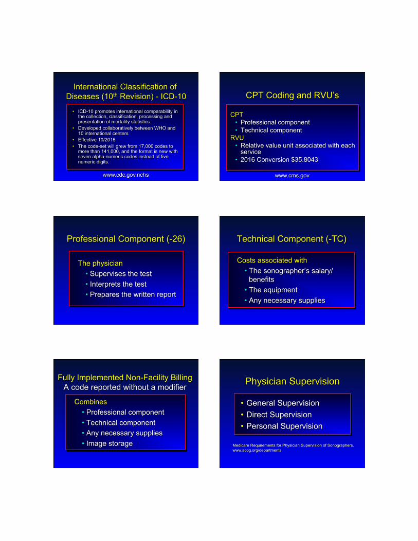
International Classification of Diseases (10th Revision) - ICD-10
• ICD-10 promotes international comparability in the collection, classification, processing and presentation of mortality statistics.
• Developed collaboratively between WHO and 10 international centers
• Effective 10/2015 • The code-set will grew from 17,000 codes to
more than 141,000, and the format is new with seven alpha-numeric codes instead of five numeric digits.
www.cdc.gov.nchs
CPT • Professional component • Technical component
RVU • Relative value unit associated with each
service • 2016 Conversion $35.8043
CPT Coding and RVU’s
www.cms.gov
Professional Component (-26)
The physician • Supervises the test • Interprets the test • Prepares the written report
Technical Component (-TC)
Costs associated with • The sonographer’s salary/
benefits • The equipment • Any necessary supplies
Fully Implemented Non-Facility Billing A code reported without a modifier
Combines • Professional component • Technical component • Any necessary supplies • Image storage
Physician Supervision
• General Supervision • Direct Supervision • Personal Supervision
Medicare Requirements for Physician Supervision of Sonographers. www.acog.org/departments

Physician Supervision General Supervision
• Procedure is furnished under the physician’s overall direction and control
• The physician’s presence is not required during the performance of the procedure.
• The training of the nonphysician personnel who perform the diagnostic procedure and equipment maintenance are the responsibility of the physician
Medicare Requirements for Physician Supervision of Sonographers. www.acog.org/departments
Physician Supervision Direct Supervision
• The physician must be present in the office suite and immediately available to furnish assistance and direction throughout the performance of the procedure.
• The physician’s in-room presence is not required during the performance of the procedure.
Medicare Requirements for Physician Supervision of Sonographers. www.acog.org/departments
Physician Supervision Personal Supervision
• Physician must be in attendance in the room during the performance of the procedure.
Medicare Requirements for Physician Supervision of Sonographers. www.acog.org/departments
Physician Supervision Personal Supervision of Gyn US
• Sonohysterography (ultrasound) • 76831 - TC
Medicare Requirements for Physician Supervision of Sonographers. www.acog.org/departments www.cms.gov
www.cms.gov
Medicare Fee Schedule Supervision Requirements
• 0 Procedure is not a diagnostic test or procedure is a diagnostic test that is not subject to the physician supervision policy.
• 1 Procedure must be performed under the general supervision of a physician.
• 2 Procedure must be performed under the direct supervision of a physician.
• 3 Procedure must be performed under the personal supervision of a physician.
• 9 Concept does not apply.

Coding – Ob Sonography 1st Trimester
• 76801 Ultrasound pregnant uterus, real time with image documentation, fetal and maternal evaluation, first trimester (< 14 weeks 0 days), transabdominal approach; single or first gestation
• 76802 ; each additional gestation. Add on code to 76801.
Coding – Ob Sonography Vaginal Sonography
• 76817 Ultrasound pregnant uterus, real time with image documentation, transvaginal
• No contingency for multiple gestations • If transvaginal examination is done in
addition to transabdominal obstetrical ultrasound exam, use 76817 in addition to the appropriate transabdominal code
Coding – Ob Sonography 2nd/3rd Trimester
• 76805 Ultrasound pregnant uterus, real time with image documentation, fetal and maternal evaluation, after first trimester (> 14 weeks 0 days), transabdominal approach; single or first gestation
• 76810 ; each additional gestation. • Add on code to 76805
Level 1 Scan
Survey • Viability (cardiac activity) • Fetal number • Fetal presentation • Amniotic fluid volume • Placental position Fetal biometry • BPD, HC, AC, FL, EFW
76805 Standard Content: Basic Scan
Survey • Viability (cardiac activity) • Fetal number • Fetal presentation • Amniotic fluid volume • Placental position Fetal biometry • BPD, HC, AC, FL, EFW Anatomic survey • Head, face and neck, chest, abdomen, spine,
extremities, gender Maternal anatomy • Cervix, adnexa, uterine anomalies
76805 Essential Elements of Anatomy
Head, face and neck • Cerebellum, choroid plexus, cisterna magna,
lateral ventricles, midline falx, lips Chest • 4-chamber cardiac view • Outflow tracts Abdomen • Stomach, kidney, bladder, cord insertion, cord
vessels (adrenal glands) Spine • Cervical, thoracic, lumbar, sacral Extremities • Legs and arms present or absent • (comment on inability to visualize all extremities)

Reddy et al. J Ultrasound Med 2014 May;33(5):745-57. Reddy et al. Am J Obstet Gynecol 2014 May;210(5):387-97. Reddy et al. Obstet Gynecol 2014 May;123(5):1070-82.
Inability to Visualize Anatomy
Obese women • Ultrasound at 20-22 weeks
• 2 weeks later than in the nonobese patient
Inability to Visualize Anatomy
• If fetal anatomy cannot be assessed completely
• Follow-up examination in 2-4 weeks • Comment on any limitation of the
exam • Follow-up • Only as clinically indicated
Coding – Ob Sonography 2nd/3rd Trimester
• 76811 Ultrasound pregnant uterus, real time with image documentation, maternal evaluation plus detailed fetal evaluation, transabdominal approach; single or first gestation
• 76812 ; each additional gestation. • Add on code to 76811
Detailed Anatomic Examination 76811
Performed when an anomaly is suspected on the basis of history, biochemical abnormalities, or the results of either the limited or standard [basic] scan
SMFM Statement on 76811
Because this code is assigned more RVUs than the basic obstetrical sonogram (76805), the SMFM believes the code describes an examination involving significantly more work, and requiring greater expertise than that required for 76805.

SMFM Statement on 76811
Additionally, sophisticated equipment, rather than typical office level ultrasound machines, will be required to obtain the necessary imaging detail.
SMFM Statement on 76811
The level of expertise required to perform this examination can generally only be obtained through the extended education beyond residency that is acquired in a fellowship in Maternal-Fetal Medicine or Radiology…Use of this code by general obstetricians should be the exception rather than the rule.
AIUM – 76811 Consensus Statement
• Previous fetus or child with a congenital, genetic, or chromosomal abnormality
• Known or suspected fetal anomaly or known growth disorder in current pregnancy
76811 Task Force. J Ultrasound Med 2014; 33:189-195.
AIUM – 76811 Consensus Statement
Fetal at increased risk for a congenital anomaly: • Maternal pregestational diabetes or gestational
diabetes before 24 weeks • High BMI (> 35 kg/m2) • Multiple gestation • Abnormal maternal serum analytes • Teratogen exposure • 1st trimester NT > 3.0 mm
76811 Task Force. J Ultrasound Med 2014; 33:189-195.
AIUM – 76811 Consensus Statement
Other conditions affecting the fetus: • Congenital infections • Maternal drug dependence • Isoimmunization • Oligohydramnios • Polyhydramnios
76811 Task Force. J Ultrasound Med 2014; 33:189-195.
76811
O35.8XX0 • Suspected fetal abnormality and
damage. O28.9 • Abnormal findings on antenatal
screening of mother E66.01 • Morbid obesity (BMI > 35)

Coding – Ob Sonography Limited study
• 76815 Ultrasound pregnant uterus, real time with image documentation, limited (e.g., fetal heart beat, placental location, fetal position and/or qualitative amniotic fluid volume), one or more fetuses
• Use 76815 only once per exam and not per element
Coding – Ob Sonography 76815 Limited Examination A limited examination is performed when a specific question requires investigation. For example, a limited examination could be performed to confirm fetal heart activity in a bleeding patient or to verify fetal presentation in a laboring patient. In most cases, limited sonographic examinations are appropriate only when a prior complete examination is on record.
Coding – Ob Sonography 2nd/3rd Trimester, Follow-up study
• 76816 Ultrasound pregnant uterus, real time with image documentation, follow-up (e.g., re-evaluation of fetal size by measuring standard growth parameters and amniotic fluid volume, re-evaluation of organ system(s) suspected or confirmed to be abnormal on a previous scan), transabdominal approach, per fetus
• Report 76816-59 for each additional fetus examined in a multiple pregnancy.
Coding – Ob Sonography 2nd/3rd Trimester
• What about the patient who presents for a repeat study later in the pregnancy?
• Code by status of indication • If new indication, use 76805 • If not new, use 76816
• Even if complete biometry and amniotic fluid assessment performed
Coding – Ob Sonography Biophysical Profile
• 76818 Fetal biophysical profile; with non-stress testing
• 76819 Fetal biophysical profile; without non-stress testing
Coding – Ob/Gyn Sonography Fetal Echocardiography
• 76825 Fetal initial (2D +/- m-mode) • 76826 F/U or repeat (2D +/- m-mode) • 76827 Doppler echo - initial • 76828 Doppler echo – F/U or repeat
• Add to 76825, 26826 • 93325 Color mapping
• Add to 76825, 76826, 76827, 76828

Coding – Ob/Gyn Sonography Fetal Evaluation
• 76820 Umbilical artery Doppler • 76821 Middle cerebral artery
Doppler
Coding – Ob/Gyn Sonography 3-D Rendering
• 76376 and 76377 3-D rendering with interpretation and reporting of computed tomography, magnetic resonance imaging, ultrasound, or other tomographic modality
• Add on codes to appropriate ultrasound code(s)
Coding – Ob/Gyn Sonography 3-D Rendering
• 76376 3-D rendering…not requiring image postprocessing on an independent workstation.
• 76377 3-D rendering…requiring image postprocessing on an independent workstation.
Coding in Ob-Gyn Sonography Modifiers
– 22 Unusual complexity – 26 Professional component – 52 Reduced services – 59 Distinct procedural service,
same day (e.g., referral for suspected fetal anomaly on the same day.
! Ob uses 76805 ! Consultant uses 76811-59
Coding – Ob Sonography Nuchal Translucency
• 76813 Ultrasound pregnant uterus, real time with image documentation, first trimester fetal nuchal translucency measurement, transabdominal or transvaginal; single or first gestation (List separately in addition to code for primary procedure)
Coding – Ob Sonography Nuchal Translucency
• 76814 Ultrasound pregnant uterus, real time with image documentation, first trimester fetal nuchal translucency measurement, transabdominal or transvaginal; each additional gestation (List separately in addition to code for primary procedure)

ICD-10 Codes
• Use all that apply • Prioritize • Note: Advanced maternal age may not be
accepted as an indication for ultrasound or amnio
Can use “suspected or known chromosomal abnormality” (O35.8XX0) • May use diagnosis as reflected on final
report
Coding - Gyn Ultrasound
• Vaginal sonography • Dimensions • Morphology • Dynamic studies • 3-D
• Abdominal sonography • Sonohysterography
76830 –Echography, transvaginal
• Complete evaluation of the female pelvic anatomy – vaginal study
• Elements • Description and measurements of uterus
and adnexal structures (cervix) • Measurement of the endometrium • Description of the cul-de-sac and fluid • Description of the bladder (if applicable) • Description of any pelvic pathology
Adnexa Ovaries
• Dimension • Length • Width • Depth
• Morphology • Motion • Doppler
• Fallopian Tubes • Usually not visualized
76856 – Gyn Abdominal (add to TVS)
• Complete evaluation of the female pelvic anatomy – abdominal study
• Elements • Description and measurements of
uterus and adnexal structures • Measurement of the endometrium • Measurement of the bladder
(when applicable) • Description of any pelvic pathology
76857 – Gyn Limited or follow-up • Ultrasound, pelvic (nonobstetric),
real-time with image documentation; limited or follow-up (e.g. for follicles)
• 76857 • Used if follow-up of urinary
bladder alone, i.e. post-void residual, imaged
• 51798 • Used for post-void residual non-
imaging: i.e. Bladder scan

CFD
CPA
93976 Coding Gyn Sonography Doppler Studies
• 93975 Duplex scan of A/V flow: Abdomen and pelvic – Complete
• 93976 Duplex scan of A/V flow: Abdomen and pelvic - Limited
Gyn ultrasound – 3D
76376
76942
• 76942 Ultrasonic guidance for needle placement imaging (supervision and interpretation)
76998 – Intraoperative Ultrasound
• Ultrasound guidance, intraoperative
• 76998 • Ultrasound guided follicular
aspiration • Ultrasound guided transfer • Ultrasound guided
insemination
76998 – Intraoperative Ultrasound
Ultrasound guidance, intraoperative
• Documentation may be incorporated into the operative report. A separate report is not required • Reimbursement for TC = 0.00
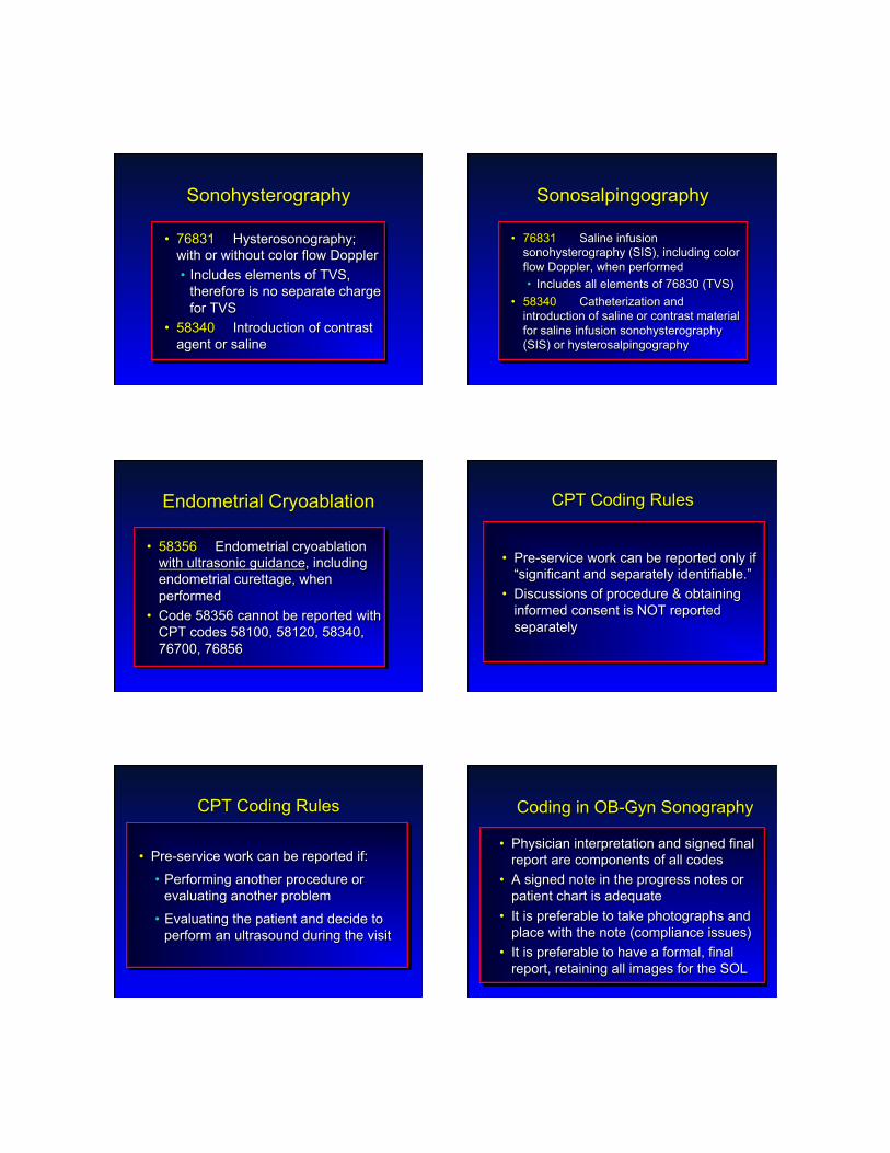
Sonohysterography
• 76831 Hysterosonography; with or without color flow Doppler • Includes elements of TVS,
therefore is no separate charge for TVS
• 58340 Introduction of contrast agent or saline
Sonosalpingography
• 76831 Saline infusion sonohysterography (SIS), including color flow Doppler, when performed • Includes all elements of 76830 (TVS)
• 58340 Catheterization and introduction of saline or contrast material for saline infusion sonohysterography (SIS) or hysterosalpingography
Endometrial Cryoablation
• 58356 Endometrial cryoablation with ultrasonic guidance, including endometrial curettage, when performed
• Code 58356 cannot be reported with CPT codes 58100, 58120, 58340, 76700, 76856
CPT Coding Rules
• Pre-service work can be reported only if “significant and separately identifiable.”
• Discussions of procedure & obtaining informed consent is NOT reported separately
CPT Coding Rules
• Pre-service work can be reported if:
• Performing another procedure or evaluating another problem
• Evaluating the patient and decide to perform an ultrasound during the visit
Coding in OB-Gyn Sonography
• Physician interpretation and signed final report are components of all codes
• A signed note in the progress notes or patient chart is adequate
• It is preferable to take photographs and place with the note (compliance issues)
• It is preferable to have a formal, final report, retaining all images for the SOL

CPT General Coding Rules
• The diagnosis code should demonstrate the medical necessity for the procedure
• Report only the procedures that were performed and documented
CPT Coding Rules
• Do not change the codes reported in order to obtain reimbursement for non-covered services.
• Report the highest valued procedure code first on the claim form.
James M. Shwayder, M.D., J.D. Professor and Chair Department of Obstetrics and Gynecology University of Mississippi Jackson, Mississippi
Thank You