ototoxicity, otosclerosis and otitis media in hearing … · ototoxicity, otosclerosis and otitis...
TRANSCRIPT

Ototoxicity, Otosclerosis and Otitis Media in Hearing aid Fitting
Kath Woolley M.Sc.North West School of Audiology

Definition of Ototoxicity
Damage to the ear- cochlea, auditory nerve or sometimes the vestibular system.

Drugs
Aminoglycosides e.g. Gentamycin ‘Mycin’
Loop Diuretics e.g. Furosemide
Chemotherapy Agents e.g. CisplatinPlatinum based

Symptoms

Discovery of Ototoxicity

Adverse Effect of Aminoglycosides
Damage to the vestibular system Headache Ear fullness Imbalance Inability to tolerate head movement Problems walking in dark
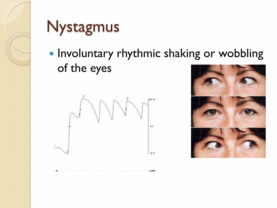
Nystagmus
Involuntary rhythmic shaking or wobbling of the eyes

Oscillopsia
Oscillo- to swing Opsis-vision Objects in sight appear to oscillate
Blur
Dizzy
Nausea

Acute Cochlear Damage
Tinnitus > 4000 Hz Low frequencies affected later Profound? Loss usually permanent

Ototoxicity Process Poorly understood
Kanamycin, Neomycin, Amikacin toxic to the cochlea
Free Radical

Other Ototoxicity
MercuryLead Styrene Manganese Xylene

The Risk of Noise and Chemicals Together
Noise alone- Risk factor 4.1
Solvent mixture alone- Risk factor 5.0
Noise and Toluene- Risk factor 10 to 27.5

Toluene
2.6 million tons produced annually

Variables in Ototoxicity
Bilateral, symmetrical, asymmetrical Time of onset Single dose. After completion of course. Monitor hearing for 6 months after. Benefits vs. risks Mouth vs. injection Susceptible Genetic link

Prevention of Ototoxicity Aminoglycoside, loop diuretics,
chemotherapy, aspirin, quinine – infusion rates, monitor drug levels, kidney history & hearing
Daily administration Lowest effective dose Other ototoxic agents High risk- alternatives Avoid noise for 6 months


General Monitoring High frequencies affected first- high
frequency audiometry No tinnitus monitoring Dizziness Handicap Inventory

Ototoxicity and Hearing Aid Fitting
Recruitment Tinnitus
Frequency selectivity reducedSpeech in noise unclearDead regions
Consider hearing aid output

What is Otosclerosis?
A disorder affecting collagen Cause? Remodelling faulty Oval window/round window Sensory, neural, mixed Gradual Progressive

Incidence Hereditary in 70% cases Dominant gene Virus? Unilateral 10-15% Caucasians Present in 10% of pop. Age of onset – approx. 30 years Females Worse in pregnancy

Symptoms & Diagnosis
Progressive conductive hearing loss
Carhart’s notch @ 2 kHz
Tinnitus in 4/5
Paracusis Willisi

Symptoms & DiagnosisSchwartze’s sign
Dizziness in 1/ 4 Weber lateralised to
affected ear Sometimes bluish cast
over eye whites Difficulty hearing when
chewing?

Risk Factors & Treatment
More common in White & Asian
Women Age-15 – 35 years +ve family history Drinking non-
fluoridated water-very controversial
Hearing aids BAHA Surgery Fenestration Stapedectomy Stapedotomy Fluoride therapy Oestrogen blockers Biphosphonates
Possible Complications of Surgery
Loss of hearing in 1 in 100
Dizziness
Taste disturbance
Reaction to ear dressings
Tinnitus

Considerations of Otosclerosis and Hearing Aid Fitting Type of hearing loss Conductive = more gain REM’s and conductive- careful
interpretation NAL- not for conductive losses BSA Guidance on REM’s (2007) Progressive- increase amplification

Otitis Media Eustachian tube
Otitis Media- inflammation of the middle ear
Eustachian tube dysfunctionair in middle ear absorbed-ve mepexudate fills up middle ear
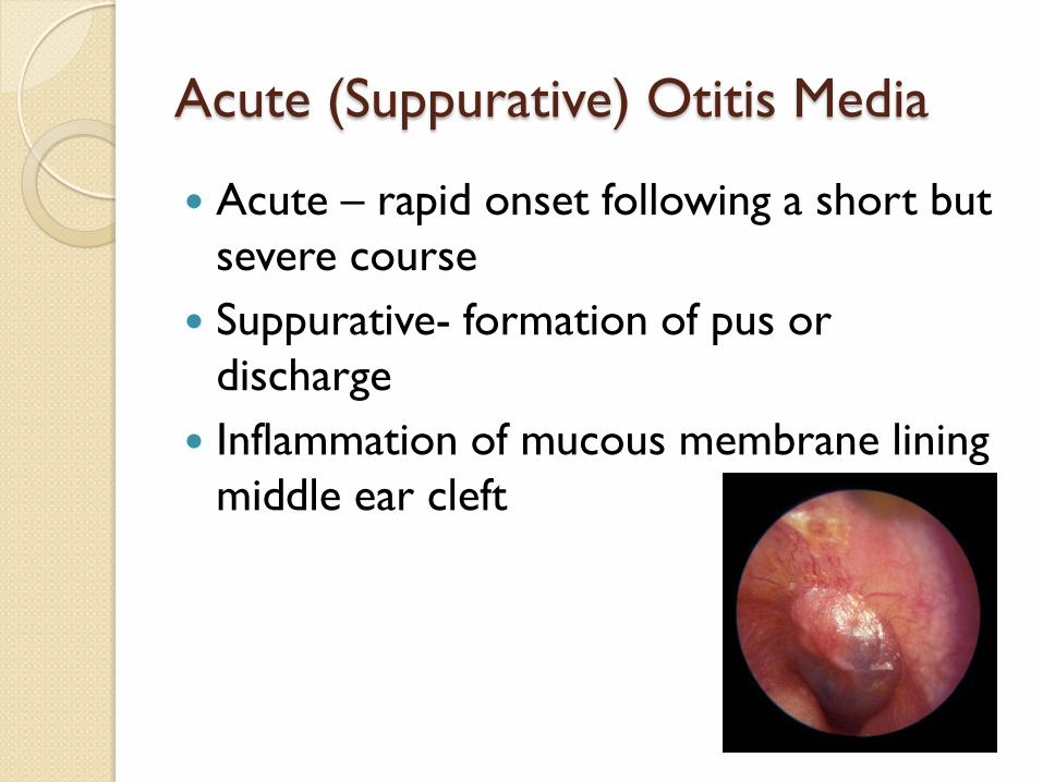
Acute (Suppurative) Otitis Media
Acute – rapid onset following a short but severe course
Suppurative- formation of pus or discharge
Inflammation of mucous membrane lining middle ear cleft

Bacteria Responsible
Adenovirus Rhinovirus Pneumococcus Haemophilus Influenzae Streptococcus Staphylococcus
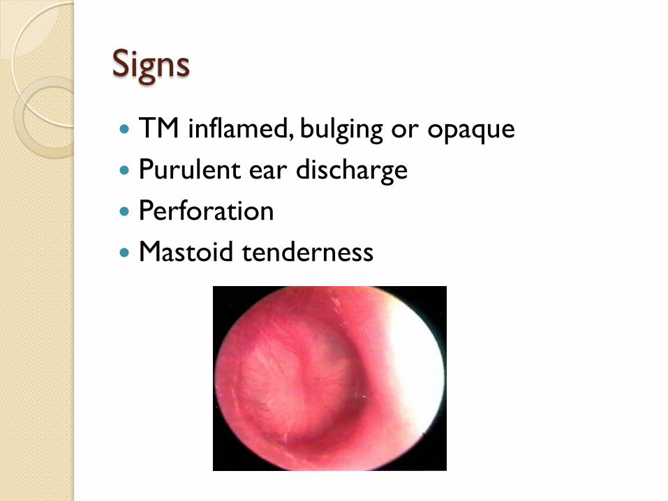
Signs
TM inflamed, bulging or opaque Purulent ear discharge Perforation Mastoid tenderness

Symptoms
Pain Temperature Earache Pressure build up Perforation Discharge containing pus escapes Antibiotics

Risk Factors Age Adenoids Frequent URTI Prematurity Craniofacial abnormalities Nurseries Poor socio-economic conditions Cold weather Pre-existing middle ear effusion

Chronic Suppurative Otitis Media
Chronic- long-standing Suppurative – formation of pus or
discharge Acute infection Irritation of lining of
middle earinfection
destroys bone Infectiongranulation

Bacteria responsible
Pseudomonas aeroginosa Staphylococcus aureaus Proteus species Klebsiella pneumonia Diptheroids
Symptoms: hearing loss; fever; vertigo; pain

‘Safe’Ear and ‘Unsafe Ear’
Perforation in pars tensa
No local destruction
Perforation in attic region (pars flaccida)
Infection of attic, antrum, mastoid
Possible cholesteatoma

Cholesteatoma Looks like an onion Produces osteolytic enzymes Complications include: Hearing loss Vertigo Headaches Facial nerve palsy Meningitis Epidural abscess
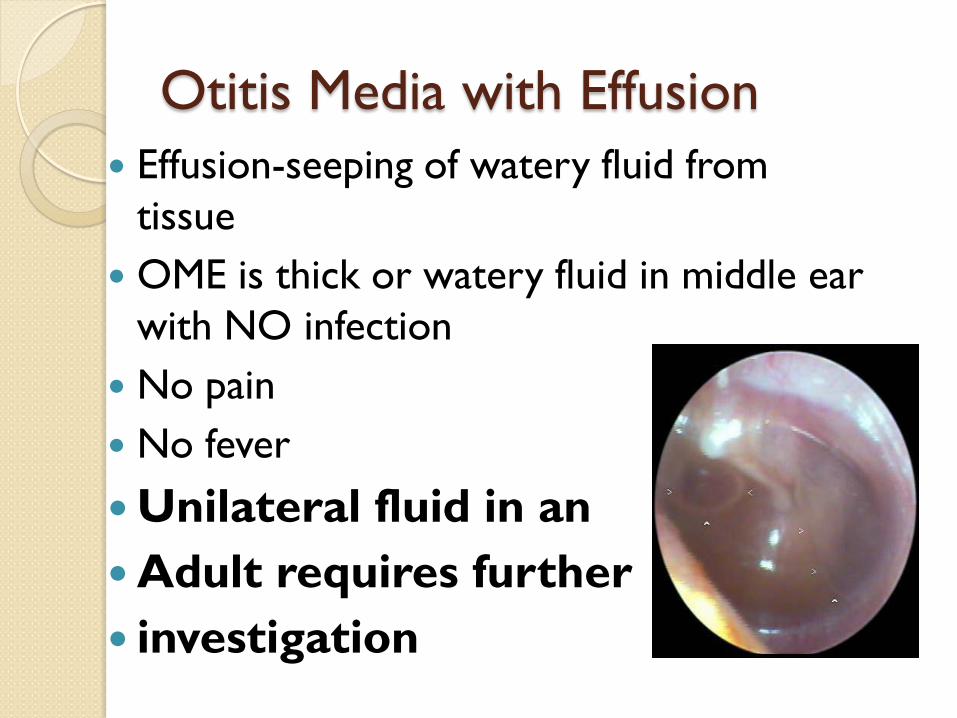
Otitis Media with Effusion Effusion-seeping of watery fluid from
tissue OME is thick or watery fluid in middle ear
with NO infection No pain No fever
Unilateral fluid in an Adult requires further investigation

Considerations of Otitis Media and Hearing Aid Fitting No active infection Conductive hearing loss and fluctuating Follow up fine tuning appointments Rigidity of TM and presence of fluid
increase the risk of feedback

Thank you for listening
Any questions?