otorhinolaryngologica italica 04-2013 interno.pdf · - references should be limited to the most...
TRANSCRIPT

Official Journal of the Italian Society of Otorhinolaryngology - Head and Neck SurgeryOrgano Ufficiale della Società Italiana di Otorinolaringologia e Chirurgia Cervico-Facciale
Otorhinolaryngologica Italica
Editorial BoardEditor-in-Chief: G. Paludetti President of S.I.O.: A. Serra Former Presidents of S.I.O. and Editors-in-Chief: I. De Vincentiis, D. Felisati, L. Coppo, G. Zaoli, P. Miani, G. Motta, L. Marcucci, A. Ottaviani, G. Perfumo, P. Puxeddu, M. Maurizi, G. Sperati, D. Passali, E. de Campora, A. Sartoris, P. Laudadio, E. Mora, M. De Benedetto, S. Conticello, D. Casolino, A. Rinaldi Ceroni, M. Piemonte, A. Staffieri, F. Chiesa, R. Fiorella, A. Camaioni
Editorial StaffEditor-in-Chief: G. Paludetti Deputy Editor: J. Galli Associate Editors: G. Almadori, F. OttavianiEditorial Coordinator: E. De CorsoEditorial Assistant: P. Moore Treasurer:L. de Campora
Italian Scientific BoardL. Bellussi, G. Danesi, C. Grandi, A. Martini, L. Pignataro, F. Raso, R. Speciale, I. Tasca
International Scientific BoardJ. Betka, P. Clement, M. Halmagyi, L.P. Kowalski, M. Pais Clemente, J. Shah, H. Stammberger, R. Laszig, G. O’Donoghue, R.J. Salvi, R. Leemans, M. Remacle, F. Marshal, H.P. Zenner
Editorial OfficeEditor-in-Chief: G. PaludettiDepartment of Head and Neck Surgery - OtorhinolaryngologyCatholic University of the Sacred Heart “A. Gemelli” Hospital L.go F. Vito, 1 - 00168 Rome, Italy Tel. +39 06 30154439 Fax + 39 06 3051194 [email protected]
Editorial Coordinator:E. De [email protected]
Argomenti di Acta Otorhinolaryngologica ItalicaEditor-in-Chief: G. Paludetti Editorial Coordinator: M.R. [email protected]
© Copyright 2013 bySocietà Italiana di Otorinolaringologia e Chirurgia Cervico-FaccialeVia Luigi Pigorini, 6/300162 Rome, Italy
PublisherPacini Editore SpAVia Gherardesca, 156121 Pisa, ItalyTel. +39 050 313011Fax +39 050 [email protected]
Acta Otorhinolaryngologica Italica is cited in Index Medicus, MEDLINE, PubMed Central, Science Citation Index Expanded, Scopus, DOAJ, Open-J Gate, Free Medical Journals, Index Copernicus, Socolar
Journal Citation Reports: Impact Factor 0.786
Acta Otorhinolaryngologica Italica is available on Google Scholar
Volume 33 – Number 4 – August 2013
Former Editors-in-Chief: C. Calearo†, E. de Campora, A. Staffieri, M. Piemonte, F. Chiesa
Information for authors including editorial standards for the preparation of manuscripts
Acta Otorhinolaryngologica Italica first appeared as “Annali di Laringologia Otologia e Faringologia” and was founded in 1901 by Giulio Masini. It is the official publication of the Italian Hospital Otology Association (A.O.O.I.) and, since 1976, also of the Società Italiana di Otorinolaringologia e Chirurgia Cervico-Facciale (S.I.O.Ch.C.-F.). The journal publishes original articles (clinical trials, cohort studies, case-control studies, cross-sectional surveys, and diagnostic test assessments) of interest in the field of otorhinolaryngology as well as case reports (unique, highly relevant and educationally valuable cases), case series, clinical techniques and technology (a short report of unique or original methods for surgical techniques, medical management or new devices or technology), editorials (including editorial guests – special contribution) and letters to the editors. Articles concerning scientific investigations and well prepared systematic reviews (including meta-analyses) on themes related to basic science, clinical otorhinolaryngology and head and neck surgery have high priority. The journal also publishes the official proceedings of the Italian Society, special columns and a calendar of events. Manuscripts must be prepared in accordance with the “Uniform Requirements for Manuscripts Submitted to Biomedical Journals” developed by the international committee of medical journal editors (http://www.icmje.org). Texts must be original and should not be presented simultaneously to more than one journal. All scientific papers are published in English (British spelling and grammar) with English and Italian titles, abstracts and key words. The text will be submitted to English language review by the publisher at no costs to the Authors. The Editor reserves the right to refuse any article not written in an appropriate English style. (The editorial staff will assist, if requested, all foreign Authors in the Italian translation of titles, key words and abstracts).Only papers strictly adhering to the editorial instructions outlined herein will be considered for publication. Acceptance is upon the critical assessment by experts in the field (Reviewers), the introduction of any changes requested and the final decision of the Editor-in-Chief.
Submission implies that publication has been approved by all co-authors, as well as, by the director of the Institute or Department where the work has been carried out. All manuscript and editorial communications should be sent online via the Editorial Manager. Please log on to the website http://actaitalica.it and follow the onscreen instructions for submission. First-time users are required to register themselves before making submissions. Paper copies of manuscripts will not be accepted. First submission should include (Checklist):- Cover letter (You should upload your cover letter at the “Comments
for the editors” section of the online submission process).- Permission form (Authorship responsibility - Financial disclosure-
Copyright Transfer Form). - Main Text: manuscript including title page, abstract, text (introduction,
methods, results, discussion, conclusion), acknowledgments, references, tables and figure legends).
- Figures. After submission, you will receive a confirmation of receipt of your manuscript via e-mail. You can also log into the website and check the status of your manuscript. The editorial staff will inform you via e-mail once a decision has been made. Permissions. Authorship responsibility - Financial disclosure- Copyright Transfer Form should be completed and signed by the Corresponding Author who accepts direct responsibility for the manuscript in agreement with all authors. It is not the role of editors to make authorship/contributorship decisions or to arbitrate conflicts related to authorship. The signed form can be electronically uploaded or sent by fax (0039 63051194) to the editorial office. Signatures can be submitted on multiple forms if Authors are not at the same institution. Accepted papers will not be sent for publication until this form has been completed and submitted. The journal holds literary rights to printed articles. The Authors are solely responsible for the contents of the article and for the statements made in their paper, and must specify that consent has been obtained from patients taking part in the investigations – or, in the case of paediatric patients, the guardian/s – and for the reproduction of any photographs. Written permission from the Authors to reproduce any material with copyright elsewhere must be obtained prior to submission. Authors must state that the material submitted has not been previously published, and is not under consideration (in whole or in part) elsewhere, and that it conforms with regulations currently in force regarding research ethics. For studies performed on laboratory animals, the Authors
must state that the relevant national laws or institutional guidelines have been adhered to. If an experiment on humans is described, a statement must be included that the work was performed in accordance with the principles of the 1983 Declaration of Helsinki. In this case, the research should have the approval of the relevant local ethical review body and such approval must be explicitly mentioned in the manuscript. Authors must state any information that may be perceived as potential conflict of interest. They should declare whether they have obtained other forms of personal or institutional financing – or if they are under contract – from Companies manufacturing products mentioned in the article. This declaration will be treated by the Editor as confidential while the paper is under review, and will not be made known to Reviewers. Accepted articles must include a declaration stating the source and nature of financing.
Article categories and manuscripts format.Acta Otorhinolaryngologica Italica publishes the types of articles defined below. When submitting your manuscript, please follow the instructions relevant to the applicable article category. Your manuscript will be returned to you if it does not meet these criteria. Original Articles. Original articles are full-length original report of clinical or basic science data, which has not yet been published, that cover topics in all areas of interest of otorhinolaryngology and that represent advanced information and a new contribution to biomedical literature. Article types include, but are not limited to: clinical trials, cohort studies, case-control studies, cross-sectional surveys, and diagnostic test assessments. An emphasis is given for higher levels of evidence. Both adult and pediatric problems are included.Text should be arranged as follow: title page, abstract, key words, main text (introduction, methods, results, discussion, conclusion), acknowledgments, references, tables (no more than 5), and figure legends. Text should not exceed 40,000 characters including space. Pages should be numbered, beginning with the title page without initiate every section on a separate page. All main text, with wide (2.5 cm) margins, must be double spaced throughout and typed in a 12 point font. Figures should be limited in number to 5. Please note:- The title page should contain: (1) A concise and informative
title (both English and Italian). (Subtitles should be avoided). (2) Running title not exceeding 100 characters including space. (3) The Authors’ names and surnames (the first name is indicated by an initial placed before the surname). The numbers of Authors should be commensurate with the complexity of the submitted material. (4) Name and address of the Institute or Institutes where the work was carried out; If the Authors are affiliated with different Institutes, the first Author and any others from the same Institute should be indicated with 1, the names of the Authors from another Institute with 2, and so on. (5) Name (written in full), surname and address of the corresponding Author, including telephone, fax numbers and e-mail address, to whom requests for reprints are to be addressed. (6) Key words (both Italian and English).
- The abstract (no longer than 300-400 words) in English and Italian should be clear and concise. The abstract should be unstructured (without headings). It should include an outline of the problem, method of study, results and significance of the research.
- Please use Microsoft Word™ preferably, saving files in .DOC or .RTF format. Any other programme can be used, including open source programmes: please always save files in .RTF format. Do not use, under any circumstances, graphical layout programmes such as Publisher™, Pagemaker™, Quark X-press™, Adobe Indesign™. Do not format the text in any way (avoid styles, borders, shading …); use only character styles such as italics, bold, underlined. Do not send the text in PDF. Notes to the text, indicated by asterisks or similar symbols, should appear at the bottom of the relevant page. Mathematical terms and formulae, abbreviations, and units of measure should conform to the standards set out in Science 1954;120:1078. Define acronyms at first mention in the text. Drugs should be referred to by their chemical name; the commercial name should be used only when absolutely unavoidable (capitalizing the first letter of the product name and giving the name of the pharmaceutical firm manufacturing the drug, town and country). In the text and legend of the tables, Authors must use, in the exact order, the following symbols: *, †, ‡, ¶, **, ††, ‡‡ …
- Acknowledgements and mention of any grants or other forms of financial support should appear at the end of the paper, before the list of references.

- References should be limited to the most essential and relevant references, identified in the text with numerals in brackets and listed at the end of the manuscript in the order in which they are cited. The format of the references listed should conform with the examples provided in N Engl J Med 1997;336:309-15. For papers with more than three authors only the first three Authors must be indicated, followed by et al. Abbreviate Journals names as in Index Medicus. Examples of the correct format for citation of references: Journal article (Chiesa A, Maroldi R, Perugini S et al. Il ruolo della tomografia assiale computerizzata nella patologia rinosinusale. Acta Otorhinolaryngol Ital 1981;1:173-94); Books (Smith DW. Recognizable patterns of human malformation. Third Edition. Philadelphia: WB Saunders Co.; 1982). Chapters from books or material from conference proceedings (Krmpotic-Nemanic J, Kostovis I, Rudan P. Aging changes of the form and infrastructure of the external nose and its importance in rhinoplasty. In: Conly J, Dickinson JT, editors. Plastic and reconstructive surgery of the face and neck. New York, NY: Grune and Stratton; 1972. p. 84.)
- Tables should be limited in number to 5. The same data should not be presented twice, both in the text and tables. Typewritten one to a page, and numbered consecutively with Roman numerals. Each table should have above a brief title and be self-explanatory. Table should be supplement the material in the text rather than duplicate. Insert any notes below. Explain all the abbreviation.
- Figure legends should allow the reader to understand the figures without reference to the text. All symbols used in figures should be explained. Do not use a separate sheet for each legend.
Systematic review (including meta-analysis): manuscripts should review topics of contemporary interest and importance. Review ideally should address controversial issues by expressing all different points of view. Critical assessments of literature and data sources on important clinical topics in otolaryngology-head and neck surgery or on basic research are required. The review should be comprehensive and authoritative as reflected by a contemporary bibliography. Reviews must not exceed a total of 50,000 characters including space, (including references, figure legends or table legends). The review should not exceed 100 references. Authors are also required to include no more than 10 images (figures and/or tables). The components of a review article are: (a) title page, including the manuscript title and all authors’ full names, academic degrees, institutional affiliations, and locations, designing the corresponding author. (b) an unstructured Abstract (without headings) no longer of 300 words. (b) Introduction that should outline explicitly the clinical problem and rationale for conducting the review. (c) Review articles do not require a Materials/Methods or Results section. Furthermore we recommend, particularly for meta-analyses, a Methods section that specifies the information sources and search strategy, inclusion and exclusion criteria, and potential biases in the review process, and a Results section that describes study selection and characteristics and statistical methods for summarizing data. (d) Discussion should interprets results in light of the total available evidence. (e) Conclusion to summarize key findings. Editorials: express opinions on current topics of interest. They are nearly always solicited by the Editor or by the members of editorial board, although unsolicited editorials may occasionally be considered. Editorial may provide commentary on an article in the issue of the Journal in which they appear or published elsewhere. Editorials are limited to 8000 characters including spaces with at least 10 references. They may include 1-2 figure or table and should have no more than 2 authors.Letters to the Editor: should be directed to the Editor regarding published material or information of timely interest and particularly if controversy exists. It should be brief not exceed 8000 characters including spaces. with no more of 10 references, and only 1 figure and/or table. If the letter is related to a previously published article on Acta Italica Otorhinolaryngologica, it must be submitted within 6 months of publication. Letters commenting on papers are sent to the authors of those papers for a response.Clinical Techniques and Technology: a short report on innovative solutions to clinical problems by unique methods: a) surgical techniques or medical management; b) new devices or technology. The manuscripts cannot be only theoretical but must include clinical data on 3 or more subjects. Submissions must have a title page, an unstructured abstract and key words. Manuscript length: no more than 15,000 characters including spaces, 5 references, and a total of 5 original and quality illustrations (figures and/or tables). Manuscripts should must conform to the following format: a) Introduction b) description of the clinical techniques or technology c) Discussion that should clearly indicate how the reported work fits with the current body of world literature.
Case Reports and Case series. Description of one or several patients with unique or unusual clinical situations. Please note that a “Low priority” will be assigned progressively to case reports and series. The key to an acceptable Case Report or series is the identification of a clinical wisdom that could benefit future patients. Manuscripts should be concise and focused on one topic of an educationally valuable case or series. Text should be conform to the following format: a) Title page b) Abstract and key words c) Introduction d) Description of the clinical case or cases c) Discussion that should clearly indicate how the reported work fits with the current body of world literature. Text length: no more than 10,000 characters including spaces with 10-15 references, and no more of 5 images (figures and/or tables). Should have no more than four authors.
Figures. All figures must be mentioned in the text and numbered consecutively with Arabic numerals. Upload figures in separate files from text and tables. Do not insert figures in the main document. Remove any information that can identify a patient. Software and format: preferably send images in .TIFF or .JPEG format, resolution at least 300 dpi (100 x 150 mm). Insert an extension that identifies the file format (example: .TIFF; .JPEG). If possible avoid .PPT (PowerPointTM files) and .DOC (images included in .DOC files). Black and white illustrations will be published without charge. Authors will be charged for color illustrations in print. The Publisher will provide, upon request, an estimate of the cost of color artwork.
Submitting revisions. If the Authors are invited to submit a revised paper, they should submit it online via the editorial manager following the instructions found there. The authors should submit a marked-up copy of the manuscript highlighting all the changes made by the authors after the original review (Authors can use the Track Changes function of Word or simply highlight where changes have been made). The authors must include a Letter to reviewers in the same file specifying point-by-point replies to queries at the beginning of the document. After acceptance. Galley proofs will be sent via mail to the corresponding author for final approval. The authors are required to carefully check the proofs and return them within 4 days of receipt. If the proofs are not received in time, the Authors will have to rely on the Editor’s corrections only. The Authors are responsible for mistakes that have been overlooked. Substantial changes in content, corrected values, title and authorship are not allowed without the approval of the editor. The date of receipt and the date of acceptance by the Scientific Committee will appear on each publication. The article may be published online first after receipt of the corrected proof. Papers are published free of charge, and a pdf of the final approved article will be sent without extra cost to the corresponding author. A pdf of all articles published are also available online at www.actaitalica.it/. Paper reprints will be provided only if requested by the authors (a reprint order form will be sent with proofs), and will be charged at extra cost. Cheques and money orders should be made out and sent to: Acta Otorhinolaryngologica Italica, Pacini Editore SpA, via Gherardesca 1 – 56121 Ospedaletto (Pisa), Italy.
Contact information.
Editorial Office: Acta Otorhinolaryngologica Italica, Università Cattolica del Sacro Cuore, Policlinico “A. Gemelli”, L.go F. Vito 1, 00168 Rome, Italy. Tel +39 06 30154439. Fax +39 06 3051194. E-mail [email protected]. For info: [email protected]
Subscriptions. Acta Otorhinolaryngologica Italica is published bimonthly and is sent, free of charge, to all members of the Society up to date with annual dues. The annual subscriptions for non members are: Italy: € 85; Elsewhere: € 95; Current issue: € 25. Back issues: € 35 (if available). Advertising and new subscriptions Acta Otorhinolaryngologica Italica Pacini Editore S.p.A. Via Gherardesca 1, 56121 Ospedaletto (Pisa), Italy. Tel. +39 050 313 011. Fax +39 050 313 0300. E-mail: [email protected]. Internet: www.pacinimedicina.it
For back issues contact. Società Italiana di Otorinolaringologia e di Chirurgia Cervico-Facciale, Via L. Pigorini 6, 00162 Rome, Italy. Tel. +39 06 44291164. Fax +39 06 44235157. E-mail: sioechcf@ sioechcf.it.
Journal printed with total chlorine free paper and water varnishing.
Printed by Pacini Editore S.p.A., Pisa (Italy) – July 2013
Subscribers’ data are treated in accordance with the provisions of the Legislative Decree, 30 June 2003, n. 196 – by means of computers operated by personnel, specifically responsible. These data are used by the Publisher to mail this publication. In accordance with Article 7 of the Legislative Decree no. 196/2003, subscribers can, at any time, view, change or delete their personal data or withdraw their use by writing to Pacini Editore SpA - Via A. Gherardesca 1, 56121 Ospedaletto (Pisa), Italy.
Review articleZenker’s diverticulum: exploring treatment options Il diverticolo di Zenker: un excursus sulle differenti opzioni terapeuticheA. Bizzotto, F. Iacopini, R. Landi, G. Costamagna . . . . . . . . . . . . . . . . . . . . . . . . . . . . . . . . . . . . . . . . . . . . . . . . . . . . pag. 219
Head and neckTransoral robotic surgery (TORS) for tongue base tumoursLa chirurgia robotica transorale (TORS) nel trattamento dei tumori della base linguaG. Mercante, P. Ruscito, R. Pellini, G. Cristalli, G. Spriano . . . . . . . . . . . . . . . . . . . . . . . . . . . . . . . . . . . . . . . . . . . . . » 230
The effect of substitution therapy on symptoms in patients with hypothyroidism following treatment for laryngeal and hypopharyngeal carcinomasL’effetto della terapia sostitutiva sulla sintomatologia dei pazienti affetti da ipotiroidismo in corso di trattamento per carcinoma della laringe o dell’ipofaringeA.M. Lo Galbo, I.M. Verdonck-De Leeuw, P. Lips, D.J Kuik, C.R. Leemans, R. De Bree . . . . . . . . . . . . . . . . . . . . . . » 236
AudiologyEffect of vitamin B12 deficiency on otoacoustic emissions Effetti del deficit della vitamina B12 sulle otoemissioni acusticheR. Karli, A. Gül, B. U gur . . . . . . . . . . . . . . . . . . . . . . . . . . . . . . . . . . . . . . . . . . . . . . . . . . . . . . . . . . . . . . . . . . . . . . . » 243
RhinologyEvaluation of total oxidative stress parameters in patients with nasal polypsValutazione degli indici di stress ossidativo cellulare totale in pazienti affetti da poliposi nasaleF. Bozkus, I. San, T. Ulas, I. Iynen, Y. Yesilova, Y. Guler, N. Aksoy . . . . . . . . . . . . . . . . . . . . . . . . . . . . . . . . . . . . . . . » 248
VestibologyDirection-fixed paroxysmal nystagmus lateral canal benign paroxysmal positioning vertigo (BPPV): another form of lateral canalolithiasisLa vertigine parossistica posizionale benigna (VPPB) con nistagmo parossistico a direzione fissa: un’altra forma di canalolitiasi lateraleL. Califano, A. Vassallo, M.G. Melillo, S. Mazzone, F. Salafia . . . . . . . . . . . . . . . . . . . . . . . . . . . . . . . . . . . . . . . . . . . » 254
Sleep disordersIdentification of obstructive sites and patterns in obstructive sleep apnoea syndrome by sleep endoscopy in 614 patientsIdentificazione dei siti di ostruzione e dei pattern di chiusura mediante “Sleep endoscopy” in 614 pazienti affetti da sindrome delle apnee ostruttive durante il sonnoF. Salamanca, F. Costantini, A. Bianchi, T. Amaina, E. Colombo, F. Zibordi . . . . . . . . . . . . . . . . . . . . . . . . . . . . . . . . » 261
Paediatric otorhinolaryngologyManagement of otolaryngological manifestations in mucopolysaccharidoses: our experienceTrattamento delle manifestazioni otorinolaringoiatriche nella mucopolisaccaridosi: nostra esperienzaM. Mesolella, M. Cimmino, E. Cantone, A. Marino, M. Cozzolino, R. Della Casa, G. Parenti, M. Iengo . . . . . . . . . . » 267
Clinical techniques and technologyAssessment of skills using a virtual reality temporal bone surgery simulatorValutazione delle competenze nella chirurgia dell’osso temporale con un simulatore della realtà virtualeR. Linke, A. Leichtle, F. Sheikh, C. Schmidt, H. Frenzel, H. Graefe, B. Wollenberg, J.E. Meyer . . . . . . . . . . . . . . . . . » 273
Case series and reportsExtended-pedicle peroneal artery perforator flap in intraoral reconstructionLembo perforante peroniero con estensione del peduncolo vascolare nelle ricostruzioni endoraliA. Baj, G.A. Beltramini, M. Demarchi, A.B. Giannì . . . . . . . . . . . . . . . . . . . . . . . . . . . . . . . . . . . . . . . . . . . . . . . . . . . » 282
Mucoepidermoid carcinoma of the tonsil: a very rare presentationCarcinoma mucoepidermoide della tonsilla: una presentazione molto raraS.J. Jarvis, V. Giangrande, P.A. Brennan . . . . . . . . . . . . . . . . . . . . . . . . . . . . . . . . . . . . . . . . . . . . . . . . . . . . . . . . . . . . » 286
Notiziario SIO . . . . . . . . . . . . . . . . . . . . . . . . . . . . . . . . . . . . . . . . . . . . . . . . . . . . . . . . . . . . . . . . . . . . . . . . . . . . . . . . » 289
Calendar of events - Italian and International Congresses and Courses . . . . . . . . . . . . . . . . . . . . . . . . . . . . . . . . . » 292
219
ACTA oTorhinolAryngologiCA iTAliCA 2013;33:219-229
Review article
Zenker’s diverticulum: exploring treatment options Il diverticolo di Zenker: un excursus sulle differenti opzioni terapeutiche
A. BIZZOTTO1, F. IACOPINI2, R. LANDI1, G. COSTAMAGNA1
1 Digestive Endoscopy Unit, Catholic University, Rome, Italy; 2 Gastroenterology and Digestive Endoscopy Unit, S. Giuseppe Hospital, Albano Laziale, Rome, Italy
SUMMAry
Zenker’s diverticulum is an acquired sac-like outpouching of the mucosa and submucosa layers located dorsally at the pharyngoesophageal junction through Killian’s dehiscence. it is the most common type of oesophageal diverticula with a reported prevalence ranging between 0.01 to 0.11% and typically occurs in middle-aged and elderly patients. Predominant symptoms are dysphagia and regurgitation. Treatment is recommended for symptomatic patients and considering the aetiopathogenesis of the disease demands myotomy of the cricopharyngeal muscle. Myotomy may be pursued through either open surgical or endoscopic techniques. Management of Zenker’s diverticulum has dramatically progressed during past decades. open surgery with cricopharyngeal myotomy has long been the conventional treatment with satisfactory results, but is associated with high complication rates. Since Zenker’s diverticulum mainly affects frail elderly patients, less invasive treatments are indicated. in recent years, endoscopic repair of Zenker’s diverticulum has been found to be a viable safe and ef-fective alternative to surgery and gained widespread acceptance. Endoscopic stapled diverticulotomy is generally the preferred approach, but flexible endoscopy is a valuable option, particularly for high-risk patients. The literature is mainly based on retrospective case series or comparative case series, and the optimal treatment modality has not yet been established. The choice between the different approaches depends on local expertise and preferences. Based on retrospective literature results, appropriate technique selection dictated by the size of the diverticulum and the patient’s conditions is however desirable.
KEy WorDS: Zenker’s diverticulum • Cricopharyngeal muscle • Myotomy • Diverticulectomy • Endoscopic stapling diverticulotomy • Flexible endoscopy
riASSUnTo
Il diverticolo di Zenker è una estroflessione sacciforme della mucosa e sottomucosa che si sviluppa a livello della parete posteriore della giunzione faringoesofagea attraverso il triangolo di Killian. Il diverticolo di Zenker è il più frequente tra i diverticoli del tratto gastrointe-stinale superiore con prevalenza compresa tra 0,1 e 0,11%. Colpisce prevalentemente pazienti di età medio-avanzata. Sintomi prevalenti di presentazione sono la disfagia ed il rigurgito. Il trattamento è indicato per i pazienti sintomatici e, considerando le recenti acquisizioni sulla eziopatogenesi, sottende la miotomia chirurgica o endoscopica del muscolo cricofaringeo. Nel corso delle ultime decadi la gestione del diverticolo di Zenker ha subito una notevole evoluzione. Accanto alla tradizionale exeresi chirurgica, efficace ma gravata da alto tasso di complicanze, si sono affermate altre forme di trattamento meno invasive e maggiormente indicate in pazienti compromessi per età o comorbidità. La sezione del setto sotto guida endoscopica (diverticolotomia) si è dimostrata una sicura ed efficace opzione terapeutica. La diverticolotomia endoscopica con suturatrice meccanica (endostapler) è attualmente la tecnica che prevale, ma una valida alternativa è rappresentata dalla endoscopia flessibile in particolare nei pazienti ad alto rischio. Resta ancora da definire tuttavia quale sia il trattamen-to ottimale per il diverticolo di Zenker ed attualmente la scelta tra l’una o l’altra tecnica dipende di fatto dalle preferenze e abilità locali. Alla luce dei dati presenti in letteratura, basati esclusivamente su studi retrospettivi, la dimensione del diverticolo e le condizioni cliniche del paziente dovrebbero guidare nella scelta della procedura terapeutica più appropriata.
PArolE ChiAVE: Diverticolo di Zenker • Muscolo cricofaringeo • Miotomia • Diverticolectomia • Diverticolotomia endoscopica con suturatrice meccanica • Endoscopia flessibile
Acta Otorhinolaryngol Ital 2013;33:219-229
Introduction Zenker’s diverticulum (ZD), also known as hypopharyn-geal diverticulum, is an acquired sac-like outpouching of the mucosa and submucosa layers originating from the pharyngoesophageal junction. it consists in a typical pul-sion diverticulum (false diverticulum) occurring dorsally at the pharyngoesophageal wall through a locus minoris resistentiae (the Killian’s dehiscence) bounded by the
propulsive oblique inferior pharyngeal constrictor muscle and the transversal fibres of the cricopharyngeal muscle (contributing to the upper oesophageal sphincter) 1. The first description of Zenker’s diverticulum dates back to 1769 by ludlow 2. A century after that report, a german pathologist, Friedrich Albert von Zenker, recognized and further characterized the physiopathology of this peculiar entity, since then deserving the eponym 3.

A. Bizzotto et al.
220
Although a complete understanding of the pathogen-esis of ZD has not yet been reached, it is generally ac-cepted that ZD is the landing place of a disorder of the upper oesophageal sphincter opening. ZD occurs due to increased intraluminal pressure in the oropharynx dur-ing swallowing, against an inadequate relaxation of the cricopharyngeal muscle, and subsequent incomplete opening of the UES, causing the protrusion of the mucosa through an area of relative weakness at the dorsal pharyn-goesophageal wall 4.Treatment options encompass open surgery or transoral rigid or flexible endoscopy and are aimed at eliminating functional outflow obstruction and restore continuity at the pharyngoesophageal junction through myotomy with or without resection of the diverticulum (diverticulecto-my) or diverticulopexy 5. Changes in treatment modalities during the last decades reflect better understanding of the underlying pathophysiologic mechanism over the years 6. The present paper provides a review of the management of ZD. Mostly based on retrospective series, the current literature shows heterogeneous results. in clinical prac-tice, the management and therapeutic approach to ZD is far from being standardized and the optimal treatment option remains unsettled. none of the available studies demonstrates substantial superiority of one technique over another, and the choice between different approach-es is made according to local expertise 7. Though less-invasive procedures may sometimes be the sole option, for instance in older multi-morbid patients unfit for sur-gery, the best procedure should be defined according to precise factors 7 other than local practice, and a tailored approach based on the size of the diverticulum, patient conditions and ability to withstand surgical complica-tions is advisable 7-9.
Epidemiology, clinical presentation and pathophysiology Zenker’s diverticula typically present in middle-aged adults and elderly individuals, especially during the seventh and eighth decades of life, with a 1.5-fold male predominance. There is a geographical variation in its occurrence, and ZD is more frequent in northern Europe 10. The estimated an-nual incidence is 2 per 100,000 with prevalence between 0.01 and 0.11% 1 11. however, although Zenker’s diverticula are the most common type that cause symptoms 4, its in-cidence and prevalence may be underestimated as many diverticula may remain clinically silent, and many elderly patients with small pouches and minimal symptoms may not seek medical advice 1. As ZD is directly related to ag-ing, the prevalence of ZD is expected to increase due to the increased aging population. Classical symptoms of Zenker’s diverticulum are pro-gressive oropharyngeal dysphagia (usually to solids and liquids), regurgitation (often hours after ingestion) of
undigested food debris due to food entrapment in the di-verticulum, pharyngeal stasis of secretion, chronic cough, chronic aspiration, halitosis, sensation of a lump in the throat, hoarseness, whistling and cervical borborygmi 1.The patient may note food on the pillow upon awaken-ing in the morning. Although small diverticula may not cause symptoms, larger diverticula usually are sympto-matic. Both the inability of the sphincter to fully open and the extrinsic compression from the pouch itself are likely to explain the dysphagia experienced by patients 4. With very large diverticula, a gurgling swelling in the neck can occasionally be detected on palpation. Secondary conse-quences and potential complications of ZD include ab ingestis pneumonia secondary to aspiration, medication ineffectiveness, malnourishment and unintentional weight loss. other reported complications of untreated ZD are diverticulitis, peptic ulceration, bleeding, iatrogenic per-forations during passage of endoscopes or nasogastric tubes, fistulas and vocal cord paralysis 1 11. Cancer, probably a result of chronic irritation and in-flammation due to food and liquid stasis, has rarely been reported in association with Zenker diverticula, with an incidence of 0.5% 12. Malignancy should be suspected if there is a sudden change in the severity of symptoms, such as severely worsening dysphagia or aphagia or develop-ment of alarm symptoms (haemoptysis, haematemesis or local pain) 1 13.A barium swallow study is the mainstay in diagnosis of Zenker’s diverticulum, which allows determination of its size and location, but careful endoscopic evaluation is mandatory to rule out malignancy 6 12.Though it is widely accepted that the primary cause of a Zenker’s diverticulum appears to be impaired relaxa-tion of the upper oesophageal sphincter, generating an abnormally increased pharyngeal intrabolus pressure, as corroborated by manometric investigations 14, ZD is like-ly to be a multifactorial disorder. The noncompliant cri-copharyngeal muscle shows structural changes in terms of histological reduction in muscle component combined with qualitative fibre alterations, increase in fibrotic tis-sue and significant increase of the collagen to elastin ra-tio 14 15. The aging process might play a role because of the loss of tissue elasticity and the decrease in muscle tone. Some authors postulate an anatomical predisposition 12. This belief is reinforced by the evidence of rare famil-ial cases in addition to geographical and racial differenc-es 11 12, and further supported by the results of morpho-metric and anthropometric studies of the Killian’s triangle showing that the dimension of the triangle correlates with anthropometric features 16. This might account for the ge-ographical variations in incidence of ZD and for its male predominance. Because gastroesophageal reflux contrib-utes to cricopharyngeal dysfunction, a relation between gastro-oesophageal reflux disease and ZD has finally been assumed 11, but never been consistently investigated.

Zenker’s diverticulum: exploring treatment options
221
What is the aim of treatment? The primary therapeutic aim is to create a communicating door between the diverticulum and the oesophageal lu-men by transecting the septum to eliminate the diverticu-lum reservoir, restore outflow continuity at the pharyn-goesophageal segment allowing clearance of ingested bolus and subsequently relief symptoms and prevent re-currence 5. Treatment should be reserved for symptomatic patients with or without associated complications 11 17, while small asymptomatic diverticula do not need treat-ment as the risk of severe adverse complications, cancer and aspiration is low 6. According to the current focus on the contribution of cricopharyngeal muscle in the genesis of ZD, treatment imposes mytomy of the cricopharyngeal muscle indepen-dently of the additional procedure (creation of a plain oe-sophagodiverticulostomy, diverticulectomy or suspension diverticulopexy) 6. Division of cricopharyngeal muscle fibres (even without diverticulectomy) reduces the UES resting pressure and normalizes both UES opening (relax-ation) and intrabolus pressure as demonstrated by pharyn-goesophageal manometry 4 8 9 15. Since both the cricopharyngeal muscle and the upper mus-cular cuff of the oesophagus appear to be involved in the pathogenesis of ZD, some authors advocate the extension of the myotomy for 2-3 cm into the muscularis propria of the oesophagus below the cricopharyngeal muscle 15. in their opinion, extended myotomy to the oesophageal muscle potentially reduces the risk of recurrence. This raises however doubt as to whether it is associated with an increased risk of mediastinum exposure and perforation or vascular injury, especially in case of huge floating or plunging diverticula.
Treatment optionsin the general trend versus less invasive approaches, new techniques and new devices have been implemented, and transoral endoscopic treatment 18 and flexible endosco-py 19 20 have gained in popularity over open surgery with a concurrent decrease in mortality and morbidity. Treat-ment procedures for ZD encompass open cricopharyngeal myotomy with diverticulectomy or diverticulopexy or diverticular inversion, myotomy alone 21, endoscopic sta-ple-assisted oesophagodiverticulostomy 18 22, endoscopic Co2-laser myotomy 23, endoscopic harmonic scalpel di-verticulotomy 24 and flexible endoscopic diverticuloto-my 25. As already mentioned, the evolution in surgical and endoscopic treatment reflects the better understanding of underlying mechanisms, and it is a widespread belief that myotomy should always be part of treatment 6. Diverti-culectomy, diverticulopexy or inversion alone without myotomy are no longer acceptable given the high rate of long-term recurrence in the absence of cricopharyngeal myotomy 26.
Surgical techniquesThe management of patients with pharyngeal pouch may be either conservative (for smaller than 1 cm, asympto-matic diverticula) or surgical through an incision in the neck (open) or mouth (endoscopic). Surgery – either open or minimally invasive – is the main therapeutic approach. A) Open surgery: Surgical repair of ZD, based on a tran-scervical access, consists in stapled or hand-sewn diverti-culectomy or diverticulopexy or inversion with concur-rent crycopharyngeal myotomy or even myotomy alone for small diverticula. The operation is usually performed under general anaesthesia, but can also be performed un-der local anaesthesia or C5-C6 superselective spinal an-aesthesia 27. The patient is positioned in a supine position with a small pillow under his shoulders and the head hy-perextended and slightly turned to the right side. The left later neck incision is made ventrally to the sternocleido-mastoid muscle. Following division of the subcutaneous tissue and platysma, the pharynx and cervical oesophagus are exposed by retracting the sternocleidomastoid and carotid sheath laterally and the larynx and thyroid gland medially. once the pouch is identified and completely dis-sected from the surrounding loose connective tissue and the neck of the pouch displayed, transection (myotomy) of the cricopharyngeal muscle and proximal fibres of the oesophageal muscle is performed for a length of about 5 cm on the cervical oesophagus 27 28. Following myot-omy the ZD is: 1) surgically excised (diverticulectomy) or 2) uplifted and retracted as far as possible towards the prevertebral fascia and suspended as high as possible by suture to the prevertebral fascia or posterior pharyn-geal wall (diverticulopexy) with the collar of the sac in a non-dependent position or finally, 3) inverted into the oesopageal lumen and oversewn (diverticulum inversion or invagination) 27-29. in case of minute diverticula, once the myotomy is performed, the marsupialized diverticu-lum disappears becoming a part of the freed mucosa 28. During the surgical procedure, care must be taken to not injure the following anatomical structures: the recurrent laryngeal nerve running in the tracheoesophageal groove, the external laryngeal nerve that runs deep to the superior thyroid artery, the descending hypoglossal nerve and the cervical cutaneous nerve 28. A drain is placed, the subcu-taneous space and platysma borders are sutured and the skin incision is closed. The drain is removed after 24 to 48 hours 28. intravenous broad-spectrum antibiotics are usually administered perioperatively and continued for 1 week after surgery 29.All studies of the different open surgical approaches are retrospective, and few are comparative where selection criteria for the choice of treatment are either not stated or unclear. The following surgical algorithm may how-ever be drawn from the available literature: small (1 cm) symptomatic pouches are very likely well suited to myo-

A. Bizzotto et al.
222
tomy alone, moderate-sized diverticula (1 to 4 cm) are best treated by myotomy with suspension or inversion, and larger pouches probably warrant diverticulectomy with myotomy 28 30.B) Rigid endoscopy: Though ZD can affect young adults as well, it is primarily a disease of the elderly, often affected by significant comorbidities and a minimally-invasive en-doscopic approach avoiding the need for a neck incision, thus offering potential advantages. The rationale is that a septum containing the cricopharyngeal muscle divides the diverticulum sac from the oesophagus. By endoscopi-cally dividing this party wall, the cricopharyngeal muscle is divided, and the diverticulum is marsupialized and be-comes a unique cavity with the oesophagus, eliminating food entrapment and relieving the outflow obstruction. A number of endoscopic options to section the septum using operating laryngoscopes and laparoscopic instruments are available that are characterised by shorter operative time, reduced hospital stay, quicker resumption of oral intake and lower complication rates; moreover, they are as effec-tive as open surgery. B1) Endoscopic stapling diverticulotomy: in 1993, Col-lard 18 in Belgium, and simultaneously Martin hirsch 31 in England, proposed a transoral single-stage cut and suture technique using a laparoscopic stapler introduced through a rigid endoscope, namely the bivalved Karl Storz Weerda diverticuloscope. The patient is positioned supine with the neck fully extended. The procedure requires general anaesthesia with orotracheal intubation. The bivalved la-ryngoscope in the closed position is carefully introduced into the oesophageal inlet under direct vision or better under video endoscopic monitoring. The diverticuloscope is then slowly withdrawn and with the opened self-re-tracting valves accommodated to expose the party wall between the diverticulum and the oesophageal lumen so that the anterior blade of the diverticuloscope is placed inside the oesophagus while the posterior blade intubates the diverticulum. The diverticuloscope is advanced until the bottom of the diverticulum is exposed. The common wall and the cricopharyngeus are set between the two lips of the diverticuloscope. An endoscopic linear stapler is introduced through the diverticuloscope down to the sep-tum so that the cartridge blade is in the oesophagus and the anvil blade in the diverticulum. The diverticulostomy is created by simultaneously cutting and sealing togeth-er the anterior wall of the ZD and the posterior wall of the oesophagus with a double (or triple) row of staples along the cutting edges with minor leakage, perforation, mediastinitis or bleeding rates 32 33. Care must be taken to avoid diverticular perforation while placing the stapler. Attention must be paid in proper selection of patients to avoid leaving a significant residual septum in smaller di-verticula (which may lead to persistent symptoms) given the non-functional protruding end of the stapler 32. The fact that the stapler anvil extends beyond the end of the
staples and the staples extend beyond the razor cut entails that the stapler leaves some residual pouch, usually about 1.5 cm 33. The technique is consequently not indicated for diverticula smaller than 3 cm. however, the end of the stapling device can be trimmed to reduce the length of its non-functional distal tip and to subsequently allow advancement of the blade to the bottom of the diverticu-lum 34. The use of retraction sutures (with an Endostitch suturing device) through the lateral edges of the common wall to provide proximal tension on the cricopharyngeal bar and easier delivery of the septum fully into the jaws of the endoscopic stapling device has been successfully described 35-37. Endoscopic staple-assisted oesophagodi-verticulostomy has gained widespread acceptance and is often considered the first-line choice for treatment of ZD. The technique has become the most frequent surgi-cal intervention for pharyngeal pouch performed in EnT practice in UK 38. B2) Endoscopic carbon dioxide laser diverticulostomy: En-doscopic Co2 laser-assisted diverticulostomy, first intro-duced in 1981 by van overbeek 12, is a sutureless technique where the septum is divided by Co2
laser. The principle of
the laser endoscopic technique is to perform a full-length mucosal incision and complete myotomy of the common wall that separates the diverticulum from the oesophagus. The procedure is performed under general anaesthesia with endotracheal intubation. once the diverticuloscope is ac-commodated and the tissue bridge is properly exposed, an operating microscope with a 400 mm lens and attached Co2 laser micromanipulator is focused on the common wall visualized through the diverticuloscope. Using the laser at 5 to 10 Watts in continuous mode, the spur is tran-sected at the midline down to the bottom of the diverticu-lum, with care taken not to leave residual common wall. The cricopharyngeal muscle fibres appear as they retract laterally during division 39 40. Visualization of targeted tis-sue through the microscope and the precise laser beam control enabled by the micromanipulator device allow ex-cellent exposure and the precision required to section the common wall down to the bottom of the diverticulum sac without the view being impaired by instruments 39 41. Car-bon dioxide laser endoscopic diverticulotomy can also be achieved with thinner diverticuloscopes than those required for the stapler-assisted technique, keeping a good view of the diverticular threshold 40. Microendoscopic laser tech-niques seem suitable to treat small-moderate sized diver-ticula or as a complementary technique in addition to endo-scopic stapling when the pouch is considered too small to be (further) cut by the stapler 17 39 42. With regard to concerns over less secure mucosal closure achieved with this suture-less technique, the Co2 laser has a high-energy, high-focus beam providing high cutting power while minimizing later-al thermal tissue damage, arguably ensuring rapid healing and mucosal coverage of cut surfaces 39 41 42. Peretti et al. 43 have interestingly reported on endoscopic Co2 laser cri-

Zenker’s diverticulum: exploring treatment options
223
copharyngeal myectomy for medium-sized ZD. The partial myectomy of the posterior part of the cricopharyngeal mus-cle is achieved by entirely sectioning the posterior part of the muscle itself, following two vertical paramedian lines, and then removing the in-between portion of the muscle fibres up to the external fascial layer. B3) Harmonic scalpel: More recently, using the Weerda diverticuloscope with the patient under general endotra-cheal anaesthesia, the section of the party wall between the diverticulum and the oesophagus has been achieved using a harmonic scalpel (harmonic Ace). The harmonic scalpel, or Ultracision (Ethicon Endo-Surgery, Cincinnati, ohio), is used in laparoscopic surgery to simultaneously cut and coagulate tissues with minimal thermal spread to adjacent tissues. The harmonic scalpel blade operates ul-trasonically, causing protein denaturation such that vessels are sealed and tamponaded while providing adequate and effective timely haemostasis. This sutureless technique has been shifted to ZD repair as an additional tool for perform-ing a cricopharyngeal myotomy with success and minimal complications. in particular, diverticulostomy with the ultrasonic scalpel has proved effective for small ZD (≤ 2 cm). The smaller diameter of the harmonic scalpel allows it to be manoeuvred and positioned within small divertic-ula. in addition, the harmonic scalpel’s cutting surface ex-tends to its distal tip, allowing it to perform endoscopic oe-sophagodiverticulostomy in patients with shallow pouches that could not be adequately treated with the stapling de-vice 24 44-46. The use of the harmonic scalpel technique with a soft diverticuloscope has recently been described 46.
Freehand, cap-assisted or diverticuloscope-assisted flexible endoscopyin addition to surgical techniques, evolution in flexible endoscopy paved the way for its use in the treatment of ZD. in 1995, two landmark papers 19 20 indicated that flex-ible endoscopy was a possible option for ZD. Flexible en-doscopy shares the same principles as rigid endoscopy: the septum between the diverticulum and the oesophagus contains the cricopharyngeal muscle, while by dividing the septum and creating a common cavity a myotomy is automatically added 6. high-risk elderly patients are ex-pected to benefit the most from flexible endoscopic diver-ticulotomy 11. The procedure can be safely perfomed in the endoscopy suite, in the inpatient or outpatient setting, does not require general anaesthesia and is rapid and ef-fective 25 47. Some centres offer this option to all ZD pa-tients 47, although most authors recommend reserving it for a subset of selected patients, especially highly morbid patients and older individuals who are poor surgical can-didates with head and neck anatomy that make rigid en-doscopic access difficult 11 17. The technique can be either “freehand” or combined with a variety of different accesso-ries (hood, cap, overtube) to obtain a better exposure of the
septum, stabilize the position and protect the oesophageal and diverticular wall against thermal injury 48-57. Patients are placed in a left lateral decubitus position, either in con-scious sedation or under general anaesthesia with propofol or endotracheal intubation according to local practice 47 53. Antibiotic prophylaxis is not routinely administered. The procedure is usually done with a standard flexible endo-scope and starts with initial endoscopic examination with suction of possible retained material from the diverticu-lum. A standard large bore (16-18 Fr) nasogastric tube is generally inserted (over a guidewire) in the oesopha-gus for the aforementioned purpose. Transparent caps or oblique-end hoods attached to the tip of the flexible endoscope can further stabilize the position 54-56. A novel device for exposing, stretching and fixing the septum, and optimizing the operative field is the soft diverticuloscope (ZD overtube; Cook Endoscopy, Winston−Salem, north Carolina, USA) 52. This double duck-billed transparent soft-rubber overtube has two distal flaps of 40 mm and 30 mm that respectively protect the anterior oesophageal and posterior diverticular wall. The overtube is advanced over the endoscope up to a black marker indicating the aver-age distance (16 cm) between the septum and teeth line. Under endoscopic vision the septum is displayed and the position of the overtube can be further adjusted 53. once the septum is properly exposed, different cutting methods can be applied. incision can be done using needle-knives, monopolar forceps, argon plasma coagulation or a hook-knife 55-58. With the needle-knife, the predominant cutting technique, the septum is divided through blended current or pure coagulation current. The incision is caudally di-rected by moving the tip of the endoscope, hence the tip of the needle, from the middle at the top of the septum towards the basis of the ZD recess, indifferently from the inside of the diverticulum towards the posterior oesopha-geal wall or in the opposite direction 50-54 56. The wound edges of the ZD spur separate immediately after incision. The incision has to be cautiously balanced to prevent me-diastinal perforations due to excessive incision (beyond the inferior border of the diverticulum) and to be complete (not too short) 11. An incomplete cricopharyngeal myoto-my may account for the higher recurrence rates associated with flexible endoscopy. ideally, ZD should be reduced to < 1 cm left 48. Bleeding at the site of incision can be lo-cally controlled. Some endoscopists routinely place one or more metal endoclips at the incision basis to secure the oesophageal and diverticula margins, thereby preventing microperforations 25 53. Concerns over perforation risks as-sociated with a sutureless section have led some authors to adopt a clip-assisted (clip and cut) technique where, prior to dissection with a needle-knife in the middle, two endoclips are placed on either side of the ZD bridge 59. Several authors describe limited incisions in a single ses-sion in short-term repeat procedures, and reserving one-session diverticulotomy for small diverticula 11.

A. Bizzotto et al.
224
Technical and clinical success of treatment optionsTreatment of ZD has dramatically evolved over the past years. An external surgical approach has for long been the conventional treatment modality with satisfactory clini-cal success rates ranging between 80-100% 17. The Mayo Clinic reported excellent or good outcome in 93% of 888 patients treated with open surgery 21, but complication and mortality rates are not negligible and have been reported to be as high as 30% and 3%, respectively 60 61. Major complications (requiring intensive medical treatment, blood transfusion, surgery or intensive care unit admis-sion) include pharyngocutaneous fistulas, parapharyngeal abscess, mediastinitis, perforation, pneumomediastinum, oesophageal stricture, wound infection, significant bleed-ing requiring operative revision, vocal cord paralysis, aspiration pneumonia, and death. Minor complications consist of transient recurrent laryngeal nerve paralysis, postoperative fever and temporary subcutaneous emphy-sema suggestive of microperforation. in a literature re-view by Zbaren et al. 62, mediastinitis and stenosis were reported in up to 9.5% and 7.1%, respectively, of external approach cases. Cutaneous fistulas and recurrent laryn-geal nerve paralysis were described in 19% and 12.9%, respectively. Among the available transcervical modali-ties, only diverticulectomy removes the pouch allowing histopathological examination of the diverticulum sac 1. however, this technique is associated with a higher risk of pharyngocutaneous fistula (up to 30%), transient or permanent recurrent nerve paralysis, and oesophageal strictures. Some authors suggest therefore diverticulum inversion as an effective, less traumatic and less compli-cated surgical treatment modality 29. however, after either inversion or suspension of the sac, no further inspection of the diverticulum mucosa is possible for early detection of malignancy, and this has to be kept in mind in case of larger long-standing diverticula in which the risk of malignant degeneration is reported to be higher 62 63. As already mentioned about the aetiology of the disease, my-otomy is a crucial part of the ZD treatment whatever the attitude towards the pouch is. Although very effective at mid-term, ZD resection without myotomy predisposes to the development of postoperative salivary fistula and to long-term recurrence of the pouch, probably due to persistence of high intrapharyngeal pressure against the posterior pharyngeal wall 26. Data reported in the relevant literature indicate recurrence in 3-19% of diverticular re-sections, 6-15% of cases with diverticulum inversion, and up to 7% for diverticular suspension 29. According to the available literature, lacking in high quality comparative studies, the choice between transcervical surgical options may be best dictated by the size of the ZD in the context of the patient’s conditions. Diverticulectomy is advisable for ZD larger than 5-6 cm and in younger patients given
the risk, though low, of malignant degeneration, while di-verticulum inversion or suspension are suitable to small-moderate sized (up to 4 cm) diverticula, and patients with small, but symptomatic, pouches can be adequately man-aged with myotomy alone 17 28-30 65.As already pointed out, since ZD affects frail elderly pa-tients, who are more often than not poor surgical candi-dates, less invasive treatments are desirable. The first at-tempt in 1917 to introduce an endoscopic approach was promptly abandoned due to high complications and mor-tality rates. An endoscopic approach for the treatment of ZD was again attempted in 1960 with satisfactory results, but due to concerns over possible leak with mediastinitis surgeons were reluctant and the endoscopic technique did not gain acceptance 66. it was not until 1993 that a rigid endoscopic approach with endostapler was definitively introduced and became increasingly popular 34 35. Endo-scopic stapling of pharyngeal pouch is less invasive, very safe and effective, and has become, as supported by the abundant literature, the first-line surgical treatment with clinical success rates that favourably compare with open surgery 17. large studies demonstrated endostapling to be effective in 90-100% of cases 5, with acceptable persis-tent symptomatic relief during long-term follow-up 27. Myotomy, the crucial aspect of ZD treatment, is unavoid-ably a part of the procedure. Endoscopic stapler-assisted diverticuloesophagostomy has a lower rate of major com-plications (fistula, iatrogenic perforation and mediastini-tis, persistent recurrent laryngeal nerve injury) up to 4 % on average, with < 1% mortality. Minor adverse events include sore throat, gingival or mucosal tear, dental inju-ry, transient vocal cord palsy, subcutaneous emphysema and foreign body sensation or stenosis due to staples 67. Antibiotics are not routinely given nor is a ngT routinely inserted. The distinct advantages of endostapling over standard open-neck technique encompass, as reported in several series 27 60 68 69 and in a recent meta-analysis involving 585 patients 70, the absence of skin incision, shorter operative time, minimal or absent post-operative pain, quicker resumption of oral intake (within 24 hours), reduced hospital stay calculated from the day of opera-tion until discharge (24-48 hours), resulting in lower total hospital charges, as well as a lower rate of overall com-plications. An additional advantage lies in case of repeat procedures, for persistent or recurrent symptoms, that can successfully be carried out through a transoral ap-proach (rigid or flexible), while an open approach may pose a major technical challenge 27. review of the litera-ture highlights mean recurrence rates of about 6% (range 0-22%) consistent with the mean recurrence rate of 5% reported for external approaches 68. The above-mentioned meta-analysis 70 reports a clinical success rate in terms of resolved or significantly improved symptoms of 91% with a recurrence rate as high as 12.8% and a technical success rate in 92% of cases. This relatively high level

Zenker’s diverticulum: exploring treatment options
225
of recurrence may reflect incomplete sectioning of the fi-bres of the septum by the stapler. Determining the point at which stapler division of the septum should end is a critical issue, as a division that is too shallow will lead to persisting symptoms, while a division that is too deep increases the risks of perforation with mediastinitis. The same circumstance accounts for the high long-term recur-rence rates recorded by Bonavina et al. 27 in the subgroup of patients treated for small (< 3 cm) diverticula, when recurrence rates were stratified according to ZD size. in small diverticula, a portion of the septum may remain un-divided. Diverticula smaller than 3 cm represent a formal contraindication to an endosurgical approach because too shallow to properly accommodate the anvil of the stapler and allow complete transection of the septum; recurrence may occur in > 35% of patients. Endoscopic stapling di-verticulotomy is better suited to medium-sized divertic-ula (3-5 cm) 9 in accordance with the existing literature. on the other hand, diverticula longer than 6 cm represent a relative contraindication to endoscopic treatment as the residual pouch may be too large to allow easy clearance of the common cavity upon swallowing 71. Moreover open surgical diverticulectomy with myotomy provides radicality, eliminating any theoretical risk of carcinoma, and this has to be borne in mind when considering the potential of malignant evolution in residual pouches af-ter endoscopic treatment 72. Despite early reports over higher complication rates with endoscopic Co2 laser 73
and ultrasonic cutting 74 in the management of ZD, some authors suggest a possible complementary role of these techniques for dividing the residual septum when en-dostapling fails to be complete or when introduction of a stapling gun results in poor access or a poor surgical view 44 70 75. After rigid endoscopic or surgical treatment, a persistent septum or a residual pouch can still be evident on barium swallow examination. Persistent symptoms may be due to other underlying swallowing abnormali-ties and/or inadequate myotomy without any correlation between the size of the residual pouch and symptomatic recurrence. There is general agreement in the literature that the assessment of treatment outcome and the need for further treatment has to be clinically prompted by patient’s symptoms 1 17. rigid endoscopy is not always technically feasible and may require conversion to open surgery in about 5% of cases. A recent review of the lit-erature reports technical success rates ranging from 70% to 100% 17. The main reasons for technical failure are im-possible or inadequate exposure of the diverticulum due to the patient’s anatomy such as retrognathia, teeth pro-trusion, rigid cervical kyphosis, insufficient neck motil-ity, inability to hyperextend the neck or to open the mouth wide. Moreover, rigid endoscopy requires general anaes-thesia with endotracheal intubation, and not all patients are fit for surgery or able to withstand general anaesthe-sia. last but not least, in addition to anatomical or clini-
cal considerations, smaller or very deep diverticula are not amenable to rigid endoscopy since those conditions impair accommodation of the rigid diverticuloscope and stapler in the pouch. in case of small diverticula, the anvil of the stapler cannot be properly placed and cricopharyn-geal muscle cannot adequately be resected. other factors predictive of success or failure are short necks, decreased hyomental distance, large osteophytes, obesity, redundant mucosa 6 11 17 76 77 and the radiological characteristics of the diverticulum 78. in the general trend towards a minimalist approach, flex-ible endoscopy is an attractive alternative and may over-come some of the technical limitations of open surgery and rigid endoscopy, as well as some of the constraints related to ZD size and the patient’s conditions. Flexible endoscopy is usually performed in the endoscopic unit, under conscious sedation with midazolam and/or opiates, and is optimal for frail elderly patients, unfit for surgery, who are most likely to benefit from a brief procedure with-out the need for general anaesthesia and for neck hyper-extension 47. As for rigid endoscopy, flexible endoscopic treatment focuses on releasing the cricopharyngeal spasm by performing a cricopharyngeal myotomy and restoring the outflow continuity. Assuming no complications, it al-lows a rapid (normally after 24 hr) oral diet resumption and fast hospital discharge (usually 12-48 hr in an inpa-tient setting and 6 hours in the ouptatient setting) with success and complication rates similar to endostapling. in case of symptom persistence or recurrence the procedure is safely and easily repeatable. in a recent retrospective paper 79 assessing flexible endoscopy versus endostapler the authors reported similar outcomes in terms of hospital stay, dysphagia symptom score improvement and com-plication rates, but a significantly longer procedure time for endostapling versus flexible endoscopy. Several case series published since 1995 demonstrate the efficacy and safety of flexible endoscopy with clinical success rates ranging from 56% to 100% 19 47-59. The lower clinical suc-cess rate of 56% reported in one series 53 is very likely due to the fact that clinical remission was assessed according to the presence or absence of a pool of symptoms and not only dysphagia. When considering the sole series in which success is defined according to dysphagia, clinical success rises to 84-100% 19 48 49 51 52 54-57. Moreover in some cases outcome was assessed after one treatment session 52 53 57, while in other series it was determined after multiple treat-ment sessions 19 48-50 55 56 58. Unlike rigid endoscopy, and in particular endostapling, the technique of flexible ZD sep-totomy is neither univocal nor standardized. As already mentioned, different cutting techniques exist that can be variably combined with different accessories. The optimal cutting technique is unknown as prospective randomized trials are lacking and the choice is mainly based on the endoscopist’s personal experience and preferences. The needle-knife, even if more difficult to master without ad-

A. Bizzotto et al.
226
ditional devices 47, is the most frequently used cutting de-vice often in combination with a cap 53 56, hood 54 55 or soft diverticuloscope 52 53 to achieve a more stabilized position and optimal view of the operative field. There are no sig-nificant differences in clinical outcomes with the use of one or other accessory. Flexible endoscopy is associated in the available literature with a clinical recurrence rate of 20% 11. The incision must not extend beyond the inferior border of the diverticulum as this may cause mediastinal perforation, but a transection that is too short may lead to incomplete cricopharyngeal myotomy and subsequently account for the higher clinical recurrence rates reported for flexible endoscopy. The depth of septotomy is a major technical issue. Unfortunately, when the incision is made from up to down the inferior border may be difficult to en-doscopically define, and there are no objective parameters or reliable anatomical landmarks (except for muscular fi-bres) to guide the endoscopist. This prompted repici et al. 57 to expand the use of the hook-knife, from endoscopic mucosal dissection, to ZD septal dissection and assess its safety and efficacy. The direction of the incision is in-verted, from the bottom to the top. The cut appears to be more controlled and precise, reducing the risk of uninten-tional blunted dissection and perforations. The authors reported 1 perforation out of 35 treatments (2.8%) and a very good clinical remission rate (up to 93%). While the technical advantage should correspond to a reduced risk of perforation, the small sample size and short follow-up time did not allow for definitive conclusions. With regard to complication rates, perforations ranging from mediastinitis or cervical abscesses to microperforation (presenting as self-limited subcutaneous cervical emphy-sema) and bleeding have been reported in 0-27% (me-dian 4%) and 0-10% of cases, respectively 17. other pos-sible complications of flexible endoscopic treatment of ZD are transient fever with leukocytosis, throat pain, and sedation-related adverse events. Although routinely per-formed by some authors, post-procedural water-soluble contrast studies have limited sensitivity for detection of small perforations and do not correlate with symptomatic response to endoscopic therapy or with recurrences 11.
Flexible endoscopy is an appealing safe and effective minimally-invasive treatment option for ZD, with good clinical outcome, acceptable recurrence, and complica-tion rates. The most suitable candidates are older and high-risk patients who are unfit for surgery, even though some authors extend the indication to all symptomatic patients referred for treatment.When comparing the technical and clinical results from the available literature on ZD treatment, it should be criti-cally pointed out that a direct comparison between studies and results may be inappropriate because data are neither
homogenous nor standardized with regard to multiple variables. of note in this regard: symptoms collection (dysphagia, dysphagia plus regurgitation, a pool of symp-toms), symptom assessment (objective dysphagia scores, subjective grading of symptoms relief and satisfaction), choice of one or other technique (sequential, diverticulum size, clinical conditions, local policy), definition of clini-cal success (total disappearance of symptoms, disappear-ance plus improvement), success and recurrence meas-urements (after one session or multiple sessions), and the variable length of follow-up (the shortest follow-up schedules being reported for treatment via flexible endos-copy in contrast with the more recent series of transorally treated patients and with a historical cohort of surgically treated patients).none of the available studies, based on retrospective case series, unequivocally demonstrates the substantial supe-riority of one treatment modality over another, and al-though rigid endoscopy is the preferred and most frequent approach, the choice between different options depends on local expertise and preferences.
ConclusionsThe present paper provides an overview of the literature for the main surgical and endoscopic treatment modali-ties for ZD. Many treatment alternatives exist, reflecting the fact that none has gained proven superiority. All ap-proaches have been shown to work in the hands of expe-rienced surgeons, otolaryngologists, and gastrointestinal endoscopists, and the optimal treatment policy remains highly debated.There are no randomized studies comparing the different surgical and endoscopic approaches, few are compara-tive, and selection criteria for the choice of treatment are either not stated or unclear. The large number of retro-spective case series does not allow the possibility to draw firm conclusions, although some general indications have emerged. in particular, small-medium (up to 5 cm) sized diverticula are best treated endoscopically, with ZD up to 3 cm best amenable by flexible endoscopy, while very large diverticula may still benefit from open surgical ex-cision, especially in younger, good surgical candidates. repeat procedures, in case of treatment failure or sympto-matic recurrence, can easily and successfully be achieved through flexible or rigid endoscopy.randomized comparative trials of the general approaches to treatment are long overdue, but are hardly feasible. The low prevalence of the disease, the minimum number need-ed to treat and the highly concentrated local expertise, would make it difficult to enrol and randomize candidates between the various treatment options.

Zenker’s diverticulum: exploring treatment options
227
References1 Siddiq MA, Sood S, Strachan D. Pharyngeal pouch (Zenk-
er’s diverticulum). Postgrad Med J 2001;77:506-11. 2 ludlow A. A case of obstructed deglutition from a preter-
natural dilatation of, and bag formed in, the pharynx. Med observ Enq 1769;3:85-101.
3 Zenker FA, von Ziemssen h. Krankheiten des Oesophagus. in: Von Ziemssen h, editor. Handbuch der speciale Pa-thologie und Therapie. leipzig, germany: FC Vogel; 1877. p. 1-87.
4 Cook iJ, gabb M, Panagopoulos V, et al. Pharyngeal (Zenk-er’s) diverticulum is a disorder of upper esophageal sphinc-ter opening. gastroenterology 1992;103:1229-35.
5 Costantini M, Zaninotto g, rizzetto C, et al. Oesoph-ageal diverticula. Best Pract res Clin gastroenterol 2004;18:3-17.
6 herbella FA, Patti Mg. Modern pathophysiology and treat-ment of esophageal diverticula. langenbecks Arch Surg 2012;397:29-35.
7 Sen P, lowe DA, Farnan T. Surgical interventions for phar-yngeal pouch. Cochrane Database Syst rev 2005; (3): CD004459.
8 Zaninotto g, narne S, Costantini M, et al. Tailored approach to Zenker’s diverticula. Surg Endosc 2003;17:129-33.
9 rizzetto C, Zaninotto g, Costantini M, et al. Zenker’s diver-ticula: feasibility of a tailored approach based on diverticu-lum size. J gastrointest Surg 2008;12:2057-65.
10 Klockars T, Sihvo E, Mäkitie A. Familial Zenker’s diverticu-lum. Acta otolaryngol 2008;128:1034-6.
11 Ferreira lE, Simmons DT, Baron Th. Zenker’s diverticula: pathophysiology, clinical presentation, and flexible endo-scopic management. Dis Esophagus 2008;21:1-8.
12 van overbeek JJ. Pathogenesis and methods of treat-ment of Zenker’s diverticulum. Ann otol rhinol laryngol 2003;112:583-93.
13 Bradley PJ, Kochaar A, Quraishi MS. Pharyngeal pouch car-cinoma: real or imaginary risks? Ann otol rhinol laryngol 1999;108:1027-32.
14 Zaninotto g, Costantini M, Boccù C, et al. Functional and morphological study of the cricopharyngeal mus-cle in patients with Zenker’s diverticulum. Br J Surg 1996;83:1263-7.
15 Venturi M, Bonavina l, Colombo l, et al. Biochemi-cal markers of upper esophageal sphincter compli-ance in patients with Zenker’s diverticulum. J Surg res 1997;70:46-8.
16 Anagiotos A, Preuss SF, Koebke J. Morphometric and an-thropometric analysis of Killian’s triangle. laryngoscope 2010;120:1082-8.
17 Dzeletovic i, Ekbom DC, Baron Th. Flexible endoscopic and surgical management of Zenker’s diverticulum. Expert rev gastroenterol hepatol 2012;6:449-65; quiz 466.
18 Collard JM, otte JB, Kestens PJ. Endoscopic stapling tech-nique of esophagodiverticulostomy for Zenker’s diverticu-lum. Ann Thorac Surg 1993;56:573-6.
19 ishioka S, Sakai P, Maluf Filho F, et al. Endoscopic incision of Zenker’s diverticula. Endoscopy 1995;27:433-7.
20 Mulder CJ, den hartog g, robijn rJ, et al. Flexible endo-scopic treatment of Zenker’s diverticulum: a new approach. Endoscopy 1995;27:438-42.
21 Payne WS. The treatment of pharyngoesophageal diver-ticulum: the simple and complex. hepatogastroenterology 1992;39:109-14.
22 Peracchia A, Bonavina l, narne S, et al. Minimally invasive surgery for Zenker diverticulum: analysis of results in 95 consecutive patients. Arch Surg 1998;133:695-700.
23 Krespi y, Kacker A, remacle M. Endoscopic treatment of Zenker’s diverticulum using CO2 laser. otolaryngol head neck Surg 2002;127:309-14.
24 Fama AF, Moore EJ, Kasperbauer Jl. Harmonic scalpel in the treatment of Zenker’s diverticulum. laryngoscope 2009;119:1265-9.
25 Case DJ, Baron Th. Flexible endoscopic management of Ze-nker diverticulum: the Mayo Clinic experience. Mayo Clin Proc 2010;85:719-22.
26 gutschow CA, hamoir M, rombaux P, et al. Management of pharyngoesophageal (Zenker’s) diverticulum: which tech-nique? Ann Thorac Surg 2002;74:1677-83.
27 Bonavina l, Bona D, Abraham M, et al. Long-term results of endosurgical and open surgical approach for Zenker diver-ticulum. World J gastroenterol 2007;13:2586-9.
28 Simić A, radovanović n, Stojakov D, et al. Surgical experi-ence of the national institution in the treatment of Zenker’s diverticula. Acta Chir iugosl 2009;56:25-33.
29 Mantsopoulos K, Psychogios g, Künzel J, et al. Eval-uation of the different transcervical approaches for Zenker diverticulum. otolaryngol head neck Surg 2012;146:725-9.
30 Aly A, Devitt Pg, Jamieson gg. Evolution of surgical treat-ment for pharyngeal pouch. Br J Surg 2004;91:657-64.
31 Martin-hirsch DP, newbegin CJ. Autosuture GIA gun: a new application in the treatment of hypopharyngeal diverticula. J laryngol otol 1993;107:723-5.
32 luna rA, Collard JM. Transoral stapled diverticulotomy. rev Col Bras Cir 2009;36:268-70.
33 richtsmeier WJ. Endoscopic management of Zenker di-verticulum: the staple-assisted approach. Am J Med 2003;115(Suppl. 3A):175S-178S.
34 Philippsen lP, Weisberger EC, Whiteman TS, et al. Endo-scopic stapled diverticulotomy: treatment of choice for Ze-nker’s diverticulum. laryngoscope 2000;110:1283-6.
35 Cook rD, huang PC, richstmeier WJ, et al. Endoscopic staple-assisted esophagodiverticulostomy: an excellent treatment of choice for Zenker’s diverticulum. laryngoscope 2000;110:2020-5.
36 Bonavina l, rottoli M, Bona D, et al. Transoral stapling for Zenker diverticulum: effect of the traction suture-assisted technique on long-term outcomes. Surg Endosc 2012;26:2856-61.
37 nicholas BD, Devitt S, rosen D, et al. Endostitch-assisted endoscopic Zenker’s diverticulostomy: a tried approach for difficult cases. Dis Esophagus 2010;23:296-9.
38 Siddiq MA, Sood S. Current management in pharyngeal pouch surgery by UK otorhinolaryngologists. Ann r Coll Surg Engl 2004;86:247-52.

A. Bizzotto et al.
228
39 Chang CW, Burkey BB, netterville Jl, et al. Carbon di-oxide laser endoscopic diverticulotomy versus open di-verticulectomy for Zenker’s diverticulum. laryngoscope 2004;114:519-27.
40 helmstaedter V, Engel A, huttenbrink KB, et al. Carbon di-oxide laser endoscopic diverticulotomy for Zenker’s divertic-ulum: results and complications in a consecutive series of 40 patients. orl J otorhinolaryngol relat Spec 2009;71:40-4.
41 Kos MP, David EF, Mahieu hF. Endoscopic carbon dioxide laser Zenker’s diverticulotomy revisited. Ann otol rhinol laryngol 2009;118:512-8.
42 Verhaegen VJ, Feuth T, van den hoogen FJ, et al. Endoscop-ic carbon dioxide laser diverticulostomy versus endoscopic staple-assisted diverticulostomy to treat Zenker’s diverticu-lum. head neck 2011;33:154-9.
43 Peretti g, Piazza C, Del Bon F, et al. Endoscopic treatment of Zenker’s diverticulum by carbon dioxide laser. Acta otorhi-nolaryngol ital 2010;30:1-4.
44 May JT iV, Padhya TA, McCaffrey TV. Endoscopic repair of Zenker’s diverticulum by harmonic scalpel. Am J otolaryn-gol 2011;32:553-6.
45 Sharp DB, newman Jr, Magnuson JS. Endoscopic manage-ment of Zenker’s diverticulum: stapler assisted versus Har-monic Ace. laryngoscope 2009;119:1906-12.
46 hondo Fy, Maluf-Filho F, giordano-nappi Jh, et al. Endo-scopic treatment of Zenker’s diverticulum by harmonic scal-pel. gastrointest Endosc 2011;74:666-71.
47 Mulder CJ, Costamagna g, Sakai P. Zenker’s diverticu-lum: treatment using a flexible endoscope. Endoscopy 2001;33:991-7.
48 Mulder CJ. Zapping Zenker’s diverticulum: gastroscopic treatment. Can J gastroenterol 1999;13:405-7.
49 Mulder CJ. Zenker’s diverticulum: treatment with a flexible endoscope. gastrointest Endosc 1999;50:596-7.
50 hashiba K, de Paula Al, da Silva Jg, et al. Endoscopic treatment of Zenker’s diverticulum. gastrointest Endosc 1999;49:93-7.
51 Al-Kadi AS, Maghrabi AA, Thomson D, et al. Endoscopic treatment of Zenker diverticulum: results of a 7-year experi-ence. J Am Coll Surg 2010;211:239-43.
52 Evrard S, le Moine o, hassid S, et al. Zenker’s divertic-ulum: a new endoscopic treatment with a soft diverticulo-scope. gastrointest Endosc 2003;58:116-20.
53 Costamagna g, iacopini F, Tringali A, et al. Flexible en-doscopic Zenker’s diverticulotomy: cap-assisted tech-nique vs. diverticuloscope-assisted technique. Endoscopy 2007;39:146-52.
54 Sakai P, ishioka S, Maluf-Filho F, et al. Endoscopic treatment of Zenker’s diverticulum with an oblique-end hood attached to the endoscope. gastrointest Endosc 2001;54:760-3.
55 Christiaens P, De roock W, Van olmen A, et al. Treatment of Zenker’s diverticulum through a flexible endoscope with a transparent oblique-end hood attached to the tip and a mo-nopolar forceps. Endoscopy 2007;39:137-40.
56 Vogelsang A, Preiss C, neuhaus h, et al. Endotherapy of Ze-nker’s diverticulum using the needle-knife technique: long-term follow-up. Endoscopy 2007;39:131-6.
57 repici A, Pagano n, romeo F, et al. Endoscopic flexible treatment of Zenker’s diverticulum: a modification of the needle-knife technique. Endoscopy 2010;42:532-5.
58 rabenstein T, May A, Michel J, et al. Argon plasma coagula-tion for flexible endoscopic Zenker’s diverticulotomy. Endos-copy 2007;39:141-5.
59 Tang SJ, Jazrawi SF, Chen E, et al. Flexible endoscopic clip-assisted Zenker’s diverticulotomy: the first case series (with videos). laryngoscope 2008;118:1199-205.
60 Chang Cy, Payyapilli rJ, Scher rl. Endoscopic staple di-verticulostomy for Zenker’s diverticulum: review of litera-ture and experience in 159 consecutive cases. laryngoscope 2003;113:957-65.
61 Bonafede JP, lavertuP, Wood Bg, et al. Surgical outcome in 87 patients with Zenker’s diverticulum. laryngoscope 1997;107:720-5.
62 Zbären P, Schär P, Tschopp l, et al. Surgical treatment of Ze-nker’s diverticulum: transcutaneous diverticulectomy versus microendoscopic myotomy of the cricopharyngeal muscle with CO2 laser. otolaryngol head neck Surg 1999;121:482-7.
63 Bowdler DA, Stell PM. Carcinoma arising in posterior phar-yngeal pulsion diverticulum (Zenker’s diverticulum). Br J Surg 1987;74:561-3.
64 yekebas E, Eisenberger CF, Jaekel KT, et al. [Squamous epithelial carcinoma in a Zenker diverticulum]. Chirurg 2000;71:1513-6. german.
65 leporrier J, Salamé E, gignoux M, et al. [Zenker’s diver-ticulum: diverticulopexy versus diverticulectomy]. Ann Chir 2001;126:42-5. French.
66 hillel AT, Flint PW. Evolution of endoscopic surgical thera-py for Zenker’s diverticulum. laryngoscope 2009;119:39-44
67 Sen P, Bhattacharyya AK. Endoscopic stapling of pharyn-geal pouch. J laryngol otol 2004;118:601-6.
68 Wasserzug o, Zikk D, raziel A, et al. Endoscopically stapled diverticulostomy for Zenker’s diverticulum: re-sults of a multidisciplinary team approach. Surg Endosc 2010;24:637-41.
69 Smith Sr, genden EM, Urken Ml. Endoscopic sta-pling technique for the treatment of Zenker diverticulum vs standard open-neck technique: a direct comparison and charge analysis. Arch otolaryngol head neck Surg 2002;128:141-4.
70 leong SC, Wilkie MD, Webb CJ. Endoscopic stapling of Zenker’s diverticulum: establishing national baselines for auditing clinical outcomes in the United Kingdom. Eur Arch otorhinolaryngol 2012;269:1877-84.
71 narne S, Cutrone C, Bonavina l, et al. Endoscopic diverticu-lotomy for the treatment of Zenker’s diverticulum: results in 102 patients with staple-assisted endoscopy. Ann otol rhi-nol laryngol 1999;108:810-5.
72 Acharya A, Jennings S, Douglas S, et al. Carcinoma arising in a pharyngeal pouch previously treated by endoscopic sta-pling. laryngoscope 2006;116:1043-5.
73 Miller Fr, Bartley J, otto rA. The endoscopic management of Zenker diverticulum: CO2 laser versus endoscopic sta-pling. laryngoscope 2006;116:1608-11.
74 Whited C, lee WT, Scher r. Evaluation of endoscopic har-monic diverticulostomy. laryngoscope 2012;122:1297-300.
Zenker’s diverticulum: exploring treatment options
229
75 Stoeckli SJ, Schmid S. Endoscopic stapler-assisted diver-ticuloesophagostomy for Zenker’s diverticulum: patient satisfaction and subjective relief of symptoms. Surgery 2002;131:158-62.
76 Bloom JD, Bleier BS, Mirza n, et al. Factors predicting en-doscopic exposure of Zenker’s diverticulum. Ann otol rhi-nol laryngol 2010;119:736-41.
77 Visosky AM, Parke rB, Donovan DT. Endoscopic management of Zenker’s diverticulum: factors predic-
tive of success or failure. Ann otol rhinol laryngol 2008;117:531-7.
78 Tsikoudas A, Eason D, Kara n, et al. Correlation of radio-logic findings and clinical outcome in pharyngeal pouch sta-pling. Ann otol rhinol laryngol 2006;115:721-6.
79 repici A, Pagano n, Fumagalli U, et al. Transoral treatment of Zenker diverticulum: flexible endoscopy versus endoscop-ic stapling. A retrospective comparison of outcomes. Dis Es-ophagus 2011;24:235-9.
Address for correspondence: Alessandra Bizzotto, Endoscopia Dige-stiva Chirurgica, Policlinico Universitario “A. gemelli”, l.go A. ge-melli 8, 00168 rome, italy. E-mail: [email protected]
received: December 1, 2012 - Accepted: February 15, 2013

230
ACTA oTorhinolAryngologiCA iTAliCA 2013;33:230-235
Head and neck
Transoral robotic surgery (TORS) for tongue base tumoursLa chirurgia robotica transorale (TORS) nel trattamento dei tumori della base lingua
G. Mercante, P. ruscito, r. Pellini, G. cristalli, G. sPrianoDepartment of otolaryngology Head and neck surgery, regina elena national cancer institute, rome, italy
SummAry
in recent years, transoral robotic surgery (TorS) has been used for the removal of pharyngeal and laryngeal cancers with the objective to improve functional and aesthetic outcomes without worsening the survival. This prospective single-centre cohort study described TorS in selected tumours of the tongue base in order to assess safety, efficacy and functional outcome of the procedure. From october 2010 to February 2012, TorS was performed in 13 consecutive patients affected by T1-T2 tumours of the base of the tongue. This procedure was applicable in all cases. The clinical stage demonstrated 8 T1 tumours and 5 T2 tumours. neck node metastases were clinically evident in 6 cases (7 n0, 1 n1, 4 n2b and 1 n2c). The final pathology report confirmed malignancy in all cases (11 squamous cell carcinoma and 2 mucoepidermoid carcinoma). negative-margin resections were obtained in all cases but one with close margins. Synchronous lymph node neck dissections were performed in 7 cases (6 monolateral, 1 bilateral). Patients underwent temporary tracheostomies for a mean time of 6 days. A naso-gastric feeding tube was positioned in 10/13 (76.9%) patients for a mean time of 7.5 days. The average time to carry out the TorS procedure was 95 min (set-up time 25 min; TorS 70 min). no deaths occurred. Surgical complications were observed in 4 cases (postoperative bleedings in 3 cases and intraoperative anaphylactic shock in 1 case). median hospital stay was 9 days. All patients had good functional outcomes. Adjuvant treatment was indicated in 5/13 cases (35.4%). TorS represents a good tool for staging and treating neoplasm of the base of the tongue. The transoral removal is safe and can radically remove limited oropharyngeal tumours of the tongue base with good functional outcomes. The operating costs can be relatively high but they are related to the number of procedures per year, although the advantages to patients seem to justify the procedure. TorS can represent the definitive treatment in selected T1-T2 cases of base of the tongue tumours without adverse features and allow the possibility for the deintensification of adjuvant treatments.
Key wordS: Robotic surgery • Minimal Invasive • Transoral • Oropharyngeal cancer
riASSunTo
Negli ultimi anni, la chirurgia robotica transorale (TORS) è stata utilizzata per l’asportazione di neoplasie di faringe e laringe con l’obiet-tivo di migliorare i risultati funzionali ed estetici senza peggiorare la sopravvivenza. Abbiamo eseguito uno studio prospettico di coorte in soggetti affetti da tumore orofaringeo localizzato a livello della base lingua con l’intento di verificare la sicurezza, l’efficacia e i gli esiti funzionali della procedura. Dall’ottobre 2010 a febbraio 2012, 13 pazienti consecutivi, affetti da tumore T1-T2 della base lingua, sono stati sottoposti a TORS. La procedura è stata sempre tecnicamente eseguibile. Lo stadio clinico era: T1 in 8 casi e T2 in 5 casi. Linfonodi metastatici latero-cervicali erano evidenti in 6 casi (7 N0, 1 N1, 4 N2b and 1 N2c). L’esame istologico definitivo ha confermato la diagnosi di neoplasia maligna in tutti i casi (11 carcinomi squamosi e 2 carcinomi muco epidermoidi). I margini di resezioni sono sempre stati negativi ad eccezione di un caso con margini “close”. Uno svuotamento latero-cervicale sincrono è stato eseguito in 7 casi (6 monolate-rali, 1 bilaterale). I pazienti sono stati sottoposti a tracheotomia temporanea per un tempo medio di 6 giorni. Si sono alimentati tramite sondino naso-gastrico 10 su 13 (76,9%) pazienti per una media di 7,5 giorni. La procedura è durata mediamente 95 minuti (25 minuti per la preparazione e 70 minuti per la chirurgia TORS intesa dall’incisione al termine del tempo chirurgico). Non si sono verificati decessi. Abbiamo osservato complicanze in 4 casi: sanguinamento nel postoperatorio in 3 casi e shock anafilattico in 1 caso. Il ricovero è durato mediamente 9 giorni. Tutti hanno riportato buoni risultati funzionali. Un trattamento adiuvante nel postoperatorio era indicato in 5 su 13 (35,4%). Dallo studio si può concludere che la TORS rappresenta un valido strumento al fine di stadiare e trattare le neoplasie della base lingua. L’asportazione trans orale è sicura e consente di asportare in maniera radicale i tumori di ridotte dimensioni localizzati nella base lingua ottenendo buoni risultati funzionali. I costi possono essere elevati, tuttavia, essi sono legati al numero di procedure annue e i vantaggi per i pazienti sembrano giustificare tale procedura. La TORS può rappresentare il trattamento definitivo in selezionati tumori T1-T2 (≤ 3cm) della base lingua ed in assenza di fattori prognostici sfavorevoli. Questa procedura sembra poter consentire la riduzione dei trattamenti adiuvanti.
PArole ChiAve: Chirurgia robotica • Mini-invasiva • Transorale • Cancro orofaringeo
Acta Otorhinolaryngol Ital 2013;33:230-235

TORS for tongue base tumours
231
IntroductionOpen surgical approaches to the oropharynx can be as-sociated with morbidities such as cosmetic deformity, malocclusion and dysphagia. Therefore, a trend toward using radiotherapy and concurrent chemotherapy as a pri-mary modality in case of oropharyngeal cancer has been observed in the last few decades 1 2. However, evidence of a clear advantage of concurrent chemoradiotherapy over using combined treatment (primary surgery followed by radiotherapy or chemoradiotherapy) 3 is still lacking, while toxicity of intensive chemoradiotherapy causing se-vere dysphagia with dependence on a gastrostomy tube has been well documented 1 4. In recent years, transoral robotic surgery (TORS) has been used for the removal of pharyngeal and laryngeal cancers with the objective to improve functional and aesthetic out-comes without worsening survival 5-7.Based on reports in transoral laser surgery (TOLS), the benefits of the transoral approach to the pharyngo-laryn-geal lumen are well known 8 9. TORS allows a clearer and wider view of the surgical field and better 3D visualiza-tion of structures than TOLS, enabling access to the tu-mour via a smaller approach than the external one. An-other advantage of TORS is the use of miniaturized tools. This allows mimicking standard surgical instruments and arm movements, with tremor filtration. In addition, it permits a frontal view and reaches “blind corners” of the pharyngo-laryngeal complex, not-perpendicularly po-sitioned to the visual line due to the possibility to use a 30° telescope 10. One of the objectives of this study is to evaluate whether acceptable overall functional outcomes in case of tongue base tumours are obtained using TORS. Most reports describe the use of TORS in radical tonsil-lectomy 5 11 and partial laryngectomy 12. Few authors have focused on tongue base neoplasms, most likely due to the difficulty to recruit eligible cases 6 13-18. The aim of this study was to demonstrate the feasibility, efficacy, functional outcomes and costs in a consecutive series of T1-T2 (≤ 3 cm) patients with tumours of the base of the tongue treated with TORS.
Materials and methods
Patient characteristicsData were collected from a group of consecutive patients who underwent TORS for tumours of the base of the tongue at the Department of Otolaryngology, Head Neck Surgery of the Regina Elena National Cancer Institute in Rome, Italy. The study was a prospective, single-centre cohort trial. The local ethics committee approval was ob-tained to perform a clinical trial using the da Vinci Robot (Intuitive Surgical Inc., Sunnyvale, CA) for the resection of head and neck tumours.
Inclusion criteria was the presence of a T1-T2 (≤ 3 cm) oropharyngeal tumour of the base of the tongue, histo-logically-proven, that was amenable to transoral radical “en bloc” resection. Decision making to treat or not these patients by TORS was made in a tumour board counseling upon clinical evaluation and magnetic resonance imaging (MRI) or computed tomography scan, in case of impos-sibility to obtain MRI, that was used for detecting tumour extension. Patients with a tumour of the tongue base with superficial extension or infiltration into intrinsic muscles ≤ 3 cm were included in the study, while infiltration of the extrinsic muscle by the tumour (cT4a) represented a contraindication to this surgery. Patients with a mouth opening < 2.5 cm and/or distant metastasis were excluded from the study. Presence of nodal metastasis or previous treatment for head neck malignancy did not influence the procedure on the primary tumour. Neck dissection (ND) was always indicated for patients with squamous cell car-cinoma staged as T1 and T2, while ND was not indicated for patients with mucoepidermoid carcinoma (low grade), in accordance with NCCN guidelines 19. Informed con-sent form was obtained by all patients after attending a counselling session on the alternatives to surgery. Patients were followed up every 2 months for the first year, every 3 months for the second and third year and every 6 months thereafter. At each visit, history and clinical examination were performed, including flexible endoscopy. PET-CT scan and MRI of the tongue base were performed every 6 months for the first two years and every year thereafter.
Tumour managementSurgery started by positioning a temporary tracheostomy and NGFT. A tracheostomy tube was placed at the begin-ning of surgery before the TORS time for preventing dif-ficulty during intubation and allowing better tumour ex-posure. When indicated, elective or therapeutic ND was performed during the same procedure, prior to TORS pharyngectomy. The TORS technique applied for tumours of the base of the tongue was the same as described by Moore et al. 20.Exposure to the base of tongue was achieved by using a Feyh-Kastenbauer retractor. Radical surgery was assessed by the presence of intraoperative negative margins at the frozen section exam.Postoperative (p.o.) radiation or concurrent radiochemo-therapy were recommended if adverse features were pre-sent, including: positive or close (< 0.5 cm) surgical mar-gins, 2 or more metastatic lymph nodes and whether there was extracapasular spread in the cervical lymph nodes.
Outcome measuresDemographic, clinicopathological, and follow-up data were collected. Global time of TORS procedure, TORS setup time, TORS operative time were recorded. Hospital stay was also registered. Intraoperative and p.o. compli-

G. Mercante et al.
232
cations were reported. The recovery to normal breathing (removal of tracheostomy tube) and swallowing (removal of NGFT) were reported. The long-term results were as-sessed by interviewing each patient about their breathing, speech and swallowing preoperatively at 1 month post-operatively and 3 months after radiotherapy or chemora-diotherapy, when performed. Functional assessment of breathing was done after evaluating a breathing score (BS) (CS 0 = normal breathing; 1 = minor dyspnoea, 2 = gross dyspnoea). Functional assessment of speech was based on a communication score (CS) (CS 0 = normal speech; 1 = minor dysphonia, 2 = gross dysphonia). Functional assessment of swallowing was measured recording a dys-phagia score (DS) (DS 0 = normal swallowing; 1 = minor dysphagia; 2 = gross dysphagia). The functional impair-ment was considered “minor” when it was felt by the pa-tient as abnormality without affecting the daily routine, while as “major” when he/she had to change habits. The role of TORS in reducing or avoiding p.o. adjuvant treat-ment was also considered. The cost analysis was carried out taking into consideration “Da Vinci” surgical robot-related direct costs.
ResultsFrom October 2010 to February 2012, TORS was per-formed in 13 consecutive patients affected by T1-T2 (≤3 cm) tumours of the base of the tongue at the Department of Otolaryngology Head Neck Surgery of the Regina Ele-na National Cancer Institute in Rome, Italy. Mean age of patients was 60.8 years (range 43-76; SD 9.7), 9 patients (69.2%) were male and 4 (30.8%) female. Table I shows the characteristics of patients. TORS rep-resented the primary treatment for the oropharyngeal tumour in all cases, except in one already treated with chemoradiotherapy for squamous cell carcinoma of the base of the tongue. One patient was previously irradiated for an oral cavity cancer and another patient had a previ-ous total laryngectomy for laryngeal cancer.Preoperative biopsies tested positive for malignancies in all cases evidencing carcinoma. The final pathology re-port confirmed malignancy in 13 cases with: 11 squamous cell carcinomas and 2 mucoepidermoid carcinomas. Clear resection margins were obtained in all cases except in one with close margins (< 0.5 cm). The average intraoperative blood loss was 105 mL (range 15-420 mL). Blood loss was measured by evaluating the mL of blood present in the aspiration system.ND was indicated in the 11 patients with squamous cell carcinomas, while it was not performed in case of mu-coepidermoid carcinoma as suggested by NCCN guide-lines 19. ND was performed only in 7 cases (6 monolater-ally and 1 bilaterally) synchronously to the treatment of the primary tumour except in 4 cases (all cN0) for the fol-lowing reasons: 1 case already dissected, 1 case delayed
for intraoperative arrhythmia, 1 case for intraoperative anaphylactic shock and 1 case for previous irradiation of the neck.Table II shows clinical staging by TNM classification. Pathological findings were as follows: 7 cases of stage I (7 pT1 cN0), 1 case of stage II (1 pT2 pN0), 1 case of stage III (1 pT2 pN1), 4 cases of stage IV (2 pT1 pN2b and 2 pT2 pN2b).All patients underwent temporary tracheostomy, except the one with previous total laryngectomy. A NGFT was positioned in 10 (76.9%) patients; it was not used in the patient with previous total laryngectomy and in 2 cases who refused it.Global time of the TORS procedure averaged 95 min (set-up time 25 ± 7 min; TORS time 70 ± 18 min).One gram (g) of intravenous paracetamol 2 or 3 times daily was delivered for the first p.o. 48 hours. Antibiotic therapy with amoxicillin and clavulanic acid 2.2 g intra-venously, twice a day, was maintained for 1 week after surgery. No deaths occurred. Surgical complications were observed in 4 cases (3 cases of p.o. bleeding on p.o. day 4, 6 and 14 and 1 case of intraoperative anaphylactic shock). The patient with p.o. bleeding on day 6 required surgical revision with transoral cauterization of small vessels of the tongue base for a 250 mL blood loss, while the other 2 patients with p.o. bleeding did not require further surgery because of a 50 and 60 mL blood loss that spontaneously
Table I. Clinical characteristics of patients.
Characteristic Patients, N (%)SexFemale 4 (30.8)Male 9 (69.2)
HistologySquamous cell carcinoma 11 (84.6)Mucoepidermoid carcinoma (low grade) 2 (15.4)
Neck dissectionYes 7 (53.8)No 6 (46.2)
Adjuvant treatmentNo 8 (61.5)Radiotherapy 1 (7.7)Radiochemotherapy 4 (30.8)
Table II. Clinical staging by TNM classification.
Clinical classification cT1 cT2 TotalcN0 6 1 7cN1 0 1 1cN2a 0 0 0cN2b 1 3 4cN2c 1 0 1cN3 0 0 0Total 8 5 13
TORS for tongue base tumours
233
stopped in 30 min. No cases of blood transfusion were registered. Median hospital stay was 9 days (range 3-30 days).The tracheostomy cuff placed at the time of surgery was deflated on the first postoperative day and, then, substi-tuted with an uncuffed tube the second postoperative day. The patient was decannulated when he was able to tolerate the cannula plugged for 24 hours consecutively. The aver-age time of tracheostomy dependence was 6 days (range: 2-14 days). All patients were discharged from the hospital without tracheostomy tube. Breathing was considered as normal (BS = 0) by all patients before and after surgery. Speech evaluation did not show any difference before (CS = 0 in 12 cases and CS = 2 in the case with total laryngectomy) and after treatments (CS = 0 in 12 cases and CS = 2 in the laryngectomized case). The 76.9% of patients had a NGFT for enteral feeding and avoidance of aspiration or bleeding. All patients started nutrition with liquids 48 hours after surgery. The NGFT was removed when patient could tolerate both liquid and soft diet with-out aspiration. NGFT was placed for a mean time of 7.5 days (range: 3-18 days). Before surgery, DS was 0 in 6 cases, 1 in 6 cases and 2 in 1 case. After treatments (sur-gery alone or surgery followed by adjuvant treatment), DS was 0 in 9 cases and 1 in 2 cases.Return to oral function and normal diet was achieved in all patients and none of the patients complained of voice or breathing problems after surgery. Adjuvant treatment was indicated in 5 of 13 cases (38.4%) with malignancy: 4 cases had concomitant postoperative chemoradiotherapy (all stage IVa), 1 cases had postoperative radiotherapy alone (stage III). One pa-tient with clinical stage IVa did not receive any adjuvant treatment after he was re-staged as stage II according to the pTNM.The cost for a single TORS procedure at our Institution was about €2500 per procedure during the period of study, as calculated by the administration of the Institute.The average follow-up time was 16 months (range, 8-27 months). This period is short for comparison with other treatments, however, all patients are being followed pro-spectively and oncologic data will be reported in the fu-ture as follow-up time increases. At last follow-up, all patients were alive without evidence of loco-regional dis-ease or distant metastasis, except for one who died for causes unrelated to the neoplasia.
DiscussionThe use of TORS started in animal models in 2003. It was first applied in humans in 2005 for vallecula cyst 21. In 2006, O’Malley and colleagues published the first three cases of tumour of the base of the tongue excised by TORS 22. They demonstrated that the Da Vinci Robot provided excellent visualization and enabled transoral re-moval of the tumour while preserving key structures and
nerves. In addition, they showed that it further allowed a complete resection with negative surgical margins and without complications. Recently, many authors published reports using TORS for head and neck cancer. Weinstein and colleagues described the use of TORS in supraglottic laryngectomy 23 and radical tonsillectomy 11 in patients with squamous cell carcinoma. Desai et al. reported re-sults in 7 cases with oropaharyngeal and laryngeal tu-mours using the robotic system combined with flexible carbon dioxide laser 24. We analyzed the use of the Da Vinci system for radical treatment of oropharyngeal tumours, localized in the base of the tongue in order to gain an homogeneous group of patients with similar sites and sizes of tumours, whose excision would have previously required trans-mandibu-lar or trans-pharyngeal approach. All the cases consisted of T1 and T2 smaller than 3 cm because we aimed to: (1) verify the feasibility of TORS as primary treatment in case of malignancy, (2) allow “en bloc” resection with free margins, (3) avoid reconstruction and (4) increase the learning curve before approaching more challenging cases. A longer follow-up time is required to confirm the data about the local control published by Weinstein et al. 16. The routine use of tracheostomy and NGFT was the surgeon’s first choice before opting for this new surgical approach. It was decided to begin the procedure in the saf-est way guaranteeing respiration and nutritional status in order to avoid complications such as extubation, haemor-rhage and weight loss. Temporary tracheostomy was used in all cases. However, it was not required in patients who already had a total laryngectomy. In the Weinstein study on radical tonsillectomy, 20/27 patients were extubated at the end of the TORS 11, Genden did not perform any tra-cheostomies in 20 cases treated with TORS 5. The use of tracheostomy in the literature is less frequent than that ob-served in our study, even though the published papers fo-cused on tonsil tumours and the authors preferred to extu-bate patients 24-48 hours after surgery. A great advantage in performing a temporary tracheostomy before TORS consisted in obtaining better exposure of the oropharynx. This may not justify routine use of the tracheostomy, but it can be taken into consideration for candidates opting to undergo TORS without good oropharyngeal exposure. We maintained the NGFT in the 77% of cases for a mean time of 7.5 days to avoid that the swallowing movement could facilitate bleeding. All patients started swallowing saliva on the first postoperative (p.o.) day and water on the second p.o. day. No patient complained about swallowing and all were discharged without NGFT. Other authors de-scribed discharging patients earlier from hospital and the use of percutaneous gastrostomy for a longer period than the NGFT reported in our study 15 20 25 26.Direct comparisons across these first reported functional outcomes is not straightforward. Performing tracheosto-mies avoided complications at the moment of extubation

G. Mercante et al.
234
and was useful in 2 of 3 cases of p.o. bleedings (patients bleeding on p.o. day 4 and 6). Conversely, NGFT did not prevent haemorrhage, as expected, but could guarantee enteral feeding as a proper way of nutrition. Complica-tions related to TORS in the present series are not negli-gible. To date, 3 cases of haemorrhage represented 23% of the risk of complication related to TORS, even if only one case required surgical revision for haemostasis; the case of anaphylactic shock can hardly be related to TORS procedure, since it has never been described before and occurred following intra-operative drug administration.We registered a global time of TORS of 95 min for the resection of tongue base lesions. A comparison with other statistics is difficult because other authors consider all the oropharyngeal subsites. The mean hospital stay was 9 days ranging from 3 days to 30 days in a patient submitted to excision of the tongue base extended to the supraglottis.The day before endoscopic surgery, external access to the base of the tongue has always required a transmandibular and/or transcervical approach with consequent high mor-bidity and poor cosmetic and functional outcomes 2 28.TORS for tongue base neoplasms contributed to improv-ing all these aspects offering a substantial advantage for the patient. Future studies should validate the procedure in terms of oncologic outcomes.One of the main criticisms against using robotic surgery is related to the cost of the procedure. At our hospital the cost per procedure is about €2500, considering that 400 “Da Vinci” procedures a year are carried out in 4 departments (urologic, gynaecologic, abdominal and otolaryngology). When considering this cost alone versus traditional surgi-cal techniques (endoscopic or open), the amount seems excessive, but the true comparison should be done be-tween using a surgical transcervical or transmandibular approach to using non-surgical options like radiation or chemoradiation. Saving hospitalization time itself could be enough to balance the costs of using the robotic sys-tem, but the advantages related to the less invasive pro-cedure, the best recovery and the functional outcomes justify the expense. The advantages of TOLS compared to the external approach are already well known. Future studies should confirm whether robotic surgery represents the main field for tongue base cancers because the alter-natives (external approach or non-surgical options) seem to be related to a high incidence of complications and se-quelae 2 27-29.TORS, as surgical treatment, allowed to assess the patho-logical staging of the primary lesion in all patients. Neck dissections only added further information on the patho-logical nodal stage in 7 of 13 cases. TORS and neck dis-sections permitted the fine staging based on the patho-logical examination of the specimen. TORS represented the definitive treatment in 8 cases with oropharyngeal tu-mours of the base of the tongue without adverse events. TORS and neck dissection selected patients for postop-
erative adjuvant treatment sparing radiochemotherapy in one patient with clinical stage IVa who did not receive any adjuvant treatment after he was re-staged as stage II ac-cording to the pTNM. A great advantage of TORS is rep-resented by the chance to give another option to patients with T1 and T2 (≤ 3 cm) who would have required more aggressive surgery or chemoradiotherapy as non-surgical treatment.
ConclusionsTORS represents a good tool for staging and treating neo-plasms of the base of the tongue. The procedure is safe and can radically remove limited oropharyngeal tumours of the tongue base with good functional outcomes. Costs may be high but are related to the number of procedures carried out per year, although the advantages for patients seem to justify performing the procedure. TORS can rep-resent the definitive treatment in selected T1-T2 cases of oropharyngeal tumours of the base of the tongue without adverse features and allow the possibility to deintensify adjuvant treatments.
AcknowledgmentsThe authors acknowledge Ms. Tania Merlino for her or-thographic corrections and grammatical suggestions.
References1 Forastiere AA, Trotti A. Radiotherapy and concurrent
chemotherapy: a strategy that improves locoregional con-trol and survival in oropharyngeal cancer. J Natl Cancer Inst 1999;91:2065-6.
2 Parsons JT, Mendenhall WM, Stringer SP, et al. Squamous cell carcinoma of he oropharynx: surgery, radiation therapy, or both. Cancer 2002;94:2967-80.
3 Koch WM. Head and neck surgery in the era of organ pres-ervation therapy. Semin Oncol 2000;27(4 Suppl 8):5-12.
4 Caudell JJ, Schaner PE, Meredith RF, et al. Factors associ-ated with long-term dysphagia after definitive radiotherapy for locally advanced head-and-neck cancer. Int J Radiat On-col Biol Phys 2009;73:410-5.
5 Genden EM, Shaun Desai BA, et al. Transoral robotic surery for the management of head and neck cancer: preliminary experience. Head Neck 2009;31:283-9.
6 Weinstein GS, O’Malley BW, Cohen MA, et al. Transoral robotic surgery for advanced oropharyngeal carcinoma. Arch Otolaryngol Head Neck Surg 2010;136:1079-85.
7 Dowthwaite SA, Franklin JH, Palma DA, et al. The role of transoral robotic surgery in themanagement of oro-pharyngeal cancer: a review of the literature. ISRN Oncol 2012;2012:945162.
8 Peretti G, Piazza C, Cocco D. Transoral CO2 laser treatment for Tis-T3 glottic cancer: the university of Brescia experi-ence of 595 patients. Head Neck 2010;32:977-83.
9 Hinni ML, Salassa JR, Grant DG, et al. Transoral laser mi-

TORS for tongue base tumours
235
crosurgery for advanced laryngeal cancer. Arch Otolaryngol Head Neck Surg 2007;133:1198-204.
10 Weinstein GS, O’Malley BV, Hockstein NG. Transoral ro-botic surgery: supraglottic laryngectomy in canine model. Laryngoscope 2005;115:1315-9.
11 Weinstein GS, O’Malley BV, Sneyder W, et al. Transoral robotic surgery. Arch Otolaryngol Head Neck Surg 2007;133:1220-6.
12 Park YM, Lee WJ, Lee JG, et al. Transoral robotic surgey (TORS) in laryngeal and hypopharyngeal cancer. J Lapar-oendosc Adv Surg Tech 2009;19:361-7.
13 Cohen MA, Weinstein GS, O’Malley BW, et al. Transoral robotic surgery and human papillomavirusstatus: oncolo on-cologic results. Head Neck 2011;33:573-80.
14 Genden EM, Kotz T, Tong CCL, et al. Transoral robotic re-section and reconstruction for head and neck cancer. Laryn-goscope 2011;121:1668-74.
15 White HN, Moore EJ, Rosenthal EL, et al. Transoral robot-ic-assisted surgery for head and neck squamous cell carci-noma: one- and 2-year survival analysis. Arch Otolaryngol Head Neck Surg 2010;136:1248-52.
16 Weinstein GS, Quon H, Newman HJ, et al. Transoral ro-botic surgery alone for oropharyngeal cancer: an analy-sis of local control. Arch Otolaryngol Head Neck Surg 2012;138:628-34.
17 Cognetti DM, Luginbuhl AJ, Nguyen AL, et al. Early adop-tion of transoral robotic surgical program: preliminary out-comes. Otolaryngology - Head and Neck Surgery, published online 10 April 2012.
18 Van Abel KM, Moore EJ, Carlson ML, et al. Transoral robot-ic surgery using the thulium:yag laser: a prospective study. Arch Otolaryngol Head Neck Surg 2012;138:158-66.
19 NCCN Guidelines: clinical practice guidelines in oncology. Head and Neck Cancer. Version I.2012.
20 Moore EJ, Olsen KD, Kasperbauer JL. Transoral robotic surgery for oropharyngeal squamous cell carcinoma: a pro-spective study of feasibility and functional outcomes. Laryn-goscope 2009;119:2156-64.
21 McLeod IK, Melder PC. DaVinci Robot assisted exci-sion of a vallecular cyst: a case report. Ear Nose Throat J 2005;84:170-2.
22 O’Malley BV, Weinstein GS, Sneyder W, et al. Tronsoral ro-botic surgery (TORS) for base of the tongue neoplasm. La-ryngoscope 2006;115:1465-72.
23 Weinstein GS, O’Malley BV, Sneyder W, et al. Transoral ro-botic surgery supraglottic laryngectomy. Ann Otol Rhynol Laryngol 2007;116:19-23.
24 Desai SC, Sung CK, Joung DW, et al. Traoral robotic sur-gery using a carbon dioxide flexible laser for tumor of the upper aerodigestive tract. Laryngoscope 2008;118:2187-9.
25 Iseli TA, Kulbersh BD, Iseli CE, et al. Functional outcomes after transoral robotic surgery for head and neck cancer. Otolaryngol Head Neck Surg 2009;141:166-71.
26 Weinstein GS, O’Malley BWJr, Desai SC, et al. Transoral robotic surgery: does the ends justify the means? Curr Opin Otolaryngol Head Neck Surg 2009;17:126-31.
27 Harrison LB, Zelefsky MJ, Armstrong JG, et al. Perfor-mance status after treatment for squamous cell cancer of the base of tongue a comparison of primary radiation ther-apy versus primary surgery. Int J Radiat Oncol Biol Phys 1994;30,4:953-7.
28 Fein DA, Lee WR, Amos WR, et al. Oropharyngeal carci-noma treated with radiotherapy: a 30-year experience. Int J Radiat Oncol Biol Phys 1994;34,2:289-96.
29 Dallan I, Castelnuovo P, Vicini C, et al. The natural evolu-tion of endoscopic approaches in skull base surgery: robotic-assisted surgery? Acta Otorhinolaryngol Ital 2011;31:390-4.
Address for correspondence: Giuseppe Mercante, Department of Otolaryngology, Head and Neck Surgery, Regina Elena National Cancer Institute, via Elio Chianesi 53, 00144 Rome, Italy. Tel. +39 06 52666770. Fax +39 06 52662015. E-mail: [email protected]
Received: August 16, 2012 - Accepted: March 6, 2013
236
ACTA oTorhinolAryngologiCA iTAliCA 2013;33:236-242
Head and neck
The effect of substitution therapy on symptoms in patients with hypothyroidism following treatment for laryngeal and hypopharyngeal carcinomasL’effetto della terapia sostitutiva sulla sintomatologia dei pazienti affetti da ipotiroidismo in corso di trattamento per carcinoma della laringe o dell’ipofaringe
A.M. Lo GALbo1, I.M. Verdonck-de Leeuw1, P. LIPs2, d.J kuIk3, c.r. LeeMAns1, r. de bree1
departments of 1 otolaryngology / Head and neck surgery, 2 Internal Medicine, endocrine section and 3 clinical epidemiology and biostatistics, Vu university Medical center, Amsterdam, The netherlands
SummAry
hypothyroidism is a well-known complication following treatment of laryngeal or hypopharyngeal carcinomas, and may cause various psychological and physical problems that negatively affect quality of life. The aim of this study was to evaluate the effect of substitution therapy on symptoms in patients with hypothyroidism. A study-specific questionnaire on physical and psychological problems (before and after substitution therapy) was sent to 70 patients who had been treated between 1977 and 2008 with clinical or subclinical hypothyroid-ism. ninety-four percent returned the questionnaire. Symptoms on energy levels were reported most often (67% always tired and 70% lack of energy). moodiness and emotional and physical symptoms were reported more often in substituted (sub)clinical hypothyroidism. Substitution therapy resulted in an improvement of energy (P = 0.013), sense of general interest and enjoyment (P = 0.022) and a reduction of puffy face (P = 0.041). most symptoms in patients with thyroid dysfunction do not improve after substitution therapy. nevertheless, due to its impact on health-related quality of life and the low burden of substitution therapy, screening for hypothyroidism and subsequent substitution therapy remains important.
Key wordS: Health-related quality of life • Hypothyroidism • Substitution therapy • Laryngeal neoplasms • Hypopharyngeal neoplasms • head and neck cancer
riASSunTo
L’ipotiroidismo è una delle possibili complicanze a seguito del trattamento di un carcinoma della laringe o dell’ ipofaringe e può essere alla base di disturbi fisici e psicologici con conseguente impatto negativo sulla qualità di vita del paziente. Lo scopo del nostro studio è stato quello di valutare l’effetto della terapia sostitutiva nei pazienti con ipotiroidismo. Abbiamo sottoposto un questionario studio-specifico a 70 pazienti trattati nel peiodo compreso fra il 1977 e il 2008 e affetti da ipotiroidismo clinico o subclinico, con l’intento di evidenziare le relative problematiche fisiche e psicologiche (prima e dopo il trattamento). Il 94% dei pazienti ha risposto al questionario. La sintomatologia maggiormente riferita è stata quella relativa allo stato di spossatezza del paziente (67% sempre stanco e 70% scarse energie). Disordini dell’umore, sintomi psicologici e sintomi fisici sono stati riportati più spesso in casi di ipotiroidismo (sub)clinico in terapia sostitutiva. La terapia sostitutiva migliorava esclusivamente il senso di spossatezza (P = 0,013), il senso di generico interesse attivo e apprezzamento per le attività comuni (P = 0,022), e la riduzione del gonfiore del viso (P = 0,041). In conclusione la terapia sostitutiva risulta inefficace nel modificare l’andamento della maggior parte dei sintomi dei pazienti affetti da una disfunzione tiroidea. Tuttavia, in considerazione dell’impatto della terapia sulla qualità di vita del paziente e tenendo presente la scarsa invasività della terapia stessa, lo screening per l’ipotiroidismo e la relativa terapia sostitutiva restano indicati.
PArole ChiAve: Qualità della vita • Ipotiroidismo • Terapia sostitutiva • Neoplasie laringee • Neoplasie ipofaringee • Tumori del distretto testa-collo
Acta Otorhinolaryngol Ital 2013;33:236-242
IntroductionHypothyroidism is a well-known complication following treatment of laryngeal and hypopharyngeal carcinomas due to the close anatomic proximity of the thyroid gland to the larynx, hypopharynx and cervical lymph nodes. Depending on the extent of treatment, the reported inci-
dence of hypothyroidism varies between 27-78%, with the highest incidence found after combination therapy of surgery and radiotherapy 1 2. Surgery is believed to cause damage to the vasculature of the thyroid gland, while radiation can cause damage by inducing fibrosis within the capsule of the thyroid gland 3 4. Thyroid dys-

Substitution therapy in patients with hypothyroidism following treatment for laryngeal and hypopharyngeal carcinomas
237
function may cause various psychological and physi-cal symptoms that negatively affect the quality of life. The main psychological symptoms include anxiety, dysphoria, emotional instability, depression, insomnia and cognitive dysfunction 5. Physical symptoms consist of weight gain, cold intolerance, dry skin, constipa-tion, lack of energy and fatigue 6 7. Several studies have shown that subclinical hypothyroidism is a risk factor for coronary disease and atherosclerosis 8-10. In patients treated for laryngeal or hypopharyngeal cancer, weight gain and cold intolerance were significantly increased and associated with hypothyroidism 11. In patients with clinical hypothyroidism substitution, therapy with thy-roxin is considered to be safe and inexpensive. Treat-ment of subclinical hypothyroidism is controversial. Gulseren et al. found significant improvements of anxi-ety, depression and fatigue both in clinical and subclini-cal hypothyroidism 5. However, Jaeschke et al. found a lack of symptom response in subclinical hypothyroid-ism and suggested that symptoms were nonspecific and might not be caused by (subclinical) hypothyroidism 6 7. Walsch et al. discovered that small changes in thyroxin dosage, with the aim of achieving serum TSH concen-trations in the lower reference range, did not change well-being or quality of life 10. Razvi et al. stated that substitution of subclinical hypothyroidism leads to a significant improvement of cardiovascular risk factors and fatigue 8. A recent Cochrane review did not find statistically significant improvement of health-related quality of life and symptoms when comparing treated and untreated patients diagnosed with subclinical hy-pothyroidism 12. Because substitution therapy requires lifelong monitoring with subsequent problems of labe-ling, multi-drug therapy and thyroid hormone-induced osteoporosis, Jaeschke et al. suggest watchful waiting in case of subclinical hypothyroidism 6 7. In the past, several questionnaires have been used scor-ing symptoms associated with hypothyroidism. McMil-lan et al. suggest using a questionnaire focusing on the impact of hypothyroidism on personally applicable life domains of the patient, by asking questions regarding work, spare time, family life and social life 13. Others suggest using a disease-specific questionnaire geared towards symptoms including physical complaints, emo-tional problems, decreased energy and/or well-being, and cognitive decline 6 7 14. In the present study, we use a study-specific questionnaire based on the disease-spe-cific questionnaires. The number of questions regarding physical symptoms, energy levels, moodiness and emo-tional symptoms has been narrowed down to make the questionnaire easier for patients.The aim of this study is to evaluate the importance of various aspects of hypothyroidism and the effect of sub-stitution therapy on the quality of life in patients who
have been treated for laryngeal or hypopharyngeal carci-nomas, and who have subsequently developed hypothy-roidism.
Materials and methodsThis study was conducted by systematically question-ing a group of patients previously treated for laryngeal or hypopharyngeal cancer by surgery, with or without radiotherapy or (chemo)radiotherapy, and subsequently developed clinical or subclinical hypothyroidism. The databases from a cross-sectional study and an ongoing prospective study (started in 2004) were used 11. Patients with psychiatric disorders or total thyroidectomy were excluded. All patients who had developed overt hypo-thyroidism or subclinical hypothyroidism before March 2008 were included. A total of 37 patients with clinical hypothyroidism and 60 patients with subclinical hypo-thyroidism were identified. Thirty-eight patients were from our previous reported cross-sectional study; at that time no routine monitoring of thyroid function was per-formed and thus the chronology of development of the hypothyroidism could not be evaluated. The remaining 32 patients were from a prospective study in which thy-roid function was performed every 6 months after cancer treatment. The median time to development of hypothy-roidism in the latter group was 10 months.Hypothyroidism was defined as overt (clinical) if basal thyroid-stimulating hormone (TSH) was increased and free thyroxin (FT4) was decreased. Hypothyroidism was defined as subclinical if the basal TSH was increased and free FT4 was normal. Reference values in this study were TSH 0.3-4.5 mU/l and FT4 11.0-24.0 pmol/l. Pa-tients with overt hypothyroidism and those with sub-clinical hypothyroidism characterized by TSH >10.0 mU/l were referred to an endocrinologist for substitution therapy. Patients with subclinical hypothyroidism and TSH < 10.0 mU/l were not treated unless clear symptoms of hypothyroidism existed. Twenty-seven patients had died before questionnaires were sent. The study cohort thus consisted of 70 patients. Of the 42 patients with sub-clinical hypothyroidism, 7 patients were staged with T2, 4 patients with T3, 8 patients with T4 and 9 patients were treated for recurrence. Of these patients, 30 were treat-ed by radiation only and 12 by total laryngectomy with hemithyroidectomy and postoperative radiotherapy. Of the 28 patients with clinical hypothyroidism, 7 patients were staged with T1, 15 patients with T2, 5 patients with T3, 6 patients with T4 and 9 patients were treated for recurrence after (chemo)radiotherapy. Of these patients, 16 received radiotherapy only and 12 patients total lar-yngectomy, hemithyroidectomy and postoperative radio-therapy. As only routine clinical data and questionnaires were used, approval by the medical ethics committee was considered not to be necessary.

A.M. Lo Galbo et al.
238
A study-specific questionnaire was used. Patients were given a list of questions that queried physical symptoms, energy levels, moodiness and emotional symptoms before and after substitution therapy (Paragraph 1). Patients with subclinical hypothyroidism, who were not treated with substitution therapy, filled out the first part of the ques-tionnaire only. Patients filled in the questionnaire and rat-ed how they felt before and after substitution therapy on a scale of 1 (no symptoms) to 5 (extremely large number of symptoms), retrospectively. There was adequate space available for comments. Patients were asked to return the questionnaire anonymously. The questionnaire was first sent out in March 2008. In May 2008, the questionnaire was sent out once more to all patients except those who had returned the questionnaire including their name.
ResultsPrior to sending the questionnaire out, the patients were divided in two groups; overt hypothyroidism (n = 28) and subclinical hypothyroidism (n = 42). Because of the small number of patients, scores 2 and 3 as well as 4 and 5 were grouped for statistical analysis. A score change, the dif-ference in symptoms-score before and after substitution,
was noted. Wilcoxon signed ranks test and McNemar test were used to compare the two groups. A chi-square test was used to test for differences in improvement after sub-stitution therapy.Sixty-six patients returned the questionnaire (response rate of 94%). Thirty-four patients received substitution therapy for clinical or subclinical hypothyroidism and 32 patients were not treated for subclinical hypothyroidism. Symptoms on energy levels and moodiness and emotional symptoms were reported most often before substitution therapy was started. A lack of energy was the most fre-quently reported complaint (70%) following treatment of a laryngeal or hypopharyngeal carcinoma. Many pa-tients mentioned symptoms of always being tired (67%) and an increased need for sleep (64%). A substantial number of patients mentioned being slower physically (58%), slower movements (58%), difficulty concentrat-ing (56%) and cold intolerance (53%) (Table I). Besides their responses to the items included in the questionnaire, 9 patients reported other symptoms after oncological treatment such as headache, heavy breasts, numbness of fingers and hands, swollen feet, pale hands or feet, itchy hands and irritability. Five patients specifically reported significant improvement after substitution therapy. Two
Table I. Symptoms after treatment for laryngeal or hypopharyngeal cancer in patients who developed hypothyroidism.
Symptoms NO YES
(n) (%) (n) (%)Physical symptoms
Weight gain 41 62.1 25 37.9
Puffy face 48 72.7 18 27.3
Cold intolerance 31 47.0 35 53.0
Dry skin 33 50.0 33 50.0
Dry hair 39 59.1 27 40.9
Swollen hands 56 82.8 10 17.2
Muscle weakness 37 56.1 29 43.9
Energy levels
Always tired 22 33.3 44 66.7
Increased need of sleep 24 36.4 42 63.6
Lack of energy 20 30.3 46 69.7
Physically slower 28 42.4 39 57.6
Mentally slower 37 56.1 29 43.9
General exhaustion 33 50.0 33 50.0
Less interest in going out 37 56.1 29 43.9
Slower movements 28 42.4 39 57.6
Moodiness and emotional symptoms
Feeling frustrated 35 53.0 31 47.0
Feeling depressed 38 57.6 29 42.4
Difficulty concentrating 29 43.9 37 56.1
Decreased sense of general interest and enjoyment 32 48.5 34 51.5
NO = score 1 = no symptomsYES = score 2+3+4+5 = few symptoms+average number of symptoms+large number of symptoms+extremely large number of symptoms

Substitution therapy in patients with hypothyroidism following treatment for laryngeal and hypopharyngeal carcinomas
239
patients reported an inability to compare symptoms be-fore and after substitution therapy, as their hypothyroid-ism was directly diagnosed and treated, postoperatively. In comparison to the group of patients that did not receive treatment, the patients that were treated with substitution therapy had, prior to treatment, more often complained of muscle weakness and a decreased sense of general interest and enjoyment (p = 0.012 and p = 0.011). Dif-ferences were also seen in symptoms such as difficulty concentrating (p = 0.030), slower movements (p = 0.039) and cold intolerance (p = 0.046) (Table II). For 60-94% of patients in the group that received substitution thera-py, symptoms did not improve following substitution. Six patients (18%) specifically mentioned no improve-ment in symptoms after substitution therapy was started. Reported improvement in symptoms after substitution therapy included lack of energy (7 patients improved one score-change; 2 patients improved two score-changes); decreased sense of general interest and enjoyment (5 pa-tients improved one score-change; 4 patients improved two score-changes); puffy face (2 patients improved one score-change; 2 patients improved two score-changes and 1 patient improved three score-changes) and being physi-cally slower (8 patients improved one score-change; 1
patient improved two score-changes). A non-significant trend towards improvement of symptoms was found in favour of the treated group considering dry skin and gen-eral exhaustion (7 patients improved one score-change on both symptoms) and slowing of movements (6 patients improved one score-change) (Table III).
Discussion
The present study shows the prevalence of a wide variety of symptoms in patients diagnosed with hypothyroidism after treatment of laryngeal or hypopharyngeal carcinoma with laryngectomy and radiotherapy. A lack of energy, al-ways being tired, an increased need of sleep and being physically slower are reported most often. These symp-toms however, are often mentioned after cancer treatment and are therefore not specific to hypothyroidism. Watt et al. stressed that symptoms related to general well-being are more relevant, whereas clinicians tend to focus on physical symptoms characteristic for hypothyroidism 14.
It is arguable whether thyroxin substitution should be ad-ministered in case of subclinical hypothyroidism. A recent Cochrane review did not show improvement in symptoms after substitution therapy in patients with subclinical hy-
Table II. Symptoms in patients with substituted hypothyroidism (HT) (overt and subclinical) compared with non-substituted subclinical HT after treatment for laryngeal or hypopharyngeal cancer.
Symptoms Substituted HT (n = 34)
Non-substituted HT (n = 32)
Score (1-5) 1 2+3 4+5 1 2+3 4+5 PPhysical symptoms
Weight gain 21 61.7% 12 35.3% 1 2.9% 20 62.5% 9 28.1% 3 9.4% 0.533
Puffy face 24 70.6% 4 11.8% 6 17.6% 24 75.0% 8 25.0% 0 0 0.072
Cold intolerance 14 41.2% 11 32.4% 9 26.5% 17 53.0% 13 40.6% 2 6.3% 0.046Dry skin 16 47.0% 10 34.0% 8 23.5% 17 53.0% 10 31.3% 5 15.6% 0.435
Dry hair 20 58.8% 9 26.5% 5 14.7% 19 59.3% 13 40.6% 0 0 0.149
Swollen hands 28 82.4% 5 14.7% 1 2.9% 28 87.5% 4 12.5% 0 0 0.364
Muscle weakness 16 47.0% 9 26.5% 9 26.5% 21 65.6% 10 31.3% 1 3.1% 0.012Energy levels
Always tired 11 32.4% 11 32.4% 12 35.3% 11 34.4% 13 40.6% 8 25.0% 0.453
Increased need of sleep 11 32.4% 13 38.2% 10 34.0% 13 40.6% 11 34.4% 8 25.0% 0.563
Lack of energy 10 34.0% 13 38.2% 11 32.4% 10 31.3% 15 46.9% 7 21.9% 0.427
Physically slower 14 41.2% 10 34.0% 10 34.0% 14 43.8% 15 46.9% 3 9.4% 0.118
Mentally slower 18 52.9% 9 26.5% 7 20.6% 19 59.3% 12 37.5% 1 3.1% 0.086
General exhaustion 16 47.0% 10 34.0% 8 23.5% 17 53.0% 12 37.5% 3 9.4% 0.194
Less interested in going out 17 50.0% 9 26.5% 8 23.5% 20 62.5% 10 31.3% 2 6.3% 0.071
Slower movements 13 38.2% 11 32.4% 10 34.0% 15 46.9% 15 46.9% 2 6.3% 0.039Moodiness and emotional symptomsFeeling frustrated 16 47.0% 10 34.0% 8 23.5% 19 59.3% 8 25.0% 5 15.6% 0.320
Feeling depressed 18 52.9% 8 23.5% 8 23.5% 20 62.5% 8 25.0% 4 12.5% 0.256
Difficulty concentrating 11 32.4% 14 41.2% 9 26.5% 18 56.3% 11 34.4% 3 9.4% 0.030Decreased sense of general interest and enjoyment 12 35.3% 11 32.4% 11 32.4% 20 62.5% 9 28.1% 3 9.4% 0.011
Score1: no symptoms; score 2: few symptoms; score 3: average number of symptoms; score 4: large number of symptoms; score 5: extremely large number of symptoms P difference: the difference in symptoms between substituted HT and non-substituted HT

A.M. Lo Galbo et al.
240
pothyroidism 12. However, over the long term, it is com-monly assumed that subclinical hypothyroidism evolves into clinical hypothyroidism, and it is reported that high serum levels of TSH are a risk factor for thyroid carcino-ma 15. Moreover, subclinical hypothyroidism may have a deleterious effect on wound healing and flap survival, and is associated with elevated cholesterol levels and conges-tive heart failure 16. It is therefore suggested that thyroid hormone substitution be administered in case of subclini-cal hypothyroidism, or at least when TSH >10.0 mU/l.On the other hand, in head and neck cancer patients an association between development of hypothyroidism and improved survival has been recently reported by Nelson et al. 16. In a retrospective study, they found that patients who develop secondary hypothyroidism after treatment have increased overall survival and recurrence-free survival compared to patients who did not become hypothyroid.16 In view of the growing body of evidence supporting a per-missive role for thyroid hormone in the growth of certain solid tumours, this may be especially relevant for indi-viduals with a prior or current cancer diagnosis 17.
Only very limited data are available on quality of life in patients with hypothyroidism after treatment of laryngeal or hypopharyngeal carcinoma. Because head and neck cancer patients may already have a diminished quality of life, data on quality of life in other patients with hypothy-roidism may not be applicable to patients with hypothy-roidism due to treatment of laryngeal or hypopharyngeal carcinoma. Our results can improve patient counselling and also provide guidance in observing symptoms attrib-uted to hypothyroidism after oncological treatment and in evaluating subsequent response to substitution therapy.One limitation of our study is the lack of a control group of patients treated for a laryngeal or hypopharyngeal car-cinoma without hypothyroidism. The second limitation is that we do not know whether other comorbidities, other than psychiatric disorder and thyroid disorder, were pre-sent which may cause symptoms similar to those found in hypothyroidism. Moreover, due to the anonymous char-acter of the survey no real distinction could be made be-tween clinical and subclinical hypothyroidism treated by substitution therapy for the entire group. Because most
Table III. Improvement in symptoms after substitution therapy.
Symptoms
Score change (n = 34)
Meanscore change P
-3 -2 -1 0 1 2 3
Improvement
Physical symptoms
Weight gain 1 2 26 4 1 0.029 0.660
Puffy face 29 2 2 1 0.265 0.041
Cold intolerance 4 23 3 4 0.206 0.118
Dry skin 1 1 4 20 7 1 0.059 0.816
Dry hair 2 2 28 2 0.118 0.234
Swollen hands 1 32 1 0.029 0.655
Muscle weakness 1 1 28 4 0.029 0.738
Energy levels
Always tired 3 24 7 0.118 0.206
Increased need of sleep 1 1 24 5 3 0.235 0.103
Lack of energy 1 24 7 2 0.294 0.013
Physically slower 1 24 8 1 0.235 0.051
Mentally slower 1 27 5 1 0.147 0.187
General exhaustion 1 2 26 5 0.029 0.763
Less interested in going out 1 1 28 3 1 0.059 0.588
Slower movements 3 24 6 1 0.147 0.166
Moodiness and emotional symptoms
Feeling frustrated 5 23 3 3 0.118 0.329
Feeling depressed 3 26 3 2 0.118 0.271
Difficulty concentrating 1 1 24 5 3 0.206 0.143
Decreased sense of general interest and enjoyment 2 23 5 4 0.324 0.022Score change: the difference between symptom score (score 1: no symptoms; score 2: few symptoms; score 3: average number of symptoms;4: large number of symptoms; score 5: extremely large number of symptoms) before and after substitution therapy. Positive number means improvement and negative number means detoriation of symptoms after substituation therapy.
Substitution therapy in patients with hypothyroidism following treatment for laryngeal and hypopharyngeal carcinomas
241
patients with subclinical hypothyroidism were not treated, we might expect the vast majority of the treated group of patients to consist of those with clinical hypothyroid-ism. The third limitation is that our survey was a retro-spective study in which patients were asked to score their symptoms before and after substitution therapy. It may have been difficult for patients to score their symptoms due to the time interval, and a longitudinal, prospective study would reveal more reliable data. We included pa-tients with clinical or subclinical hypothyroidism, with and without substitution therapy. Patients with subclini-cal hypothyroidism probably have fewer symptoms and therefore may not benefit from thyroxin substitution as stated by the recent Cochrane review 12. Therefore, the ef-fect of substitution therapy in patients with clinical hy-pothyroidism maybe greater than the effect found in our entire group, consisting of both clinical and subclinical hypothyroidism patients who have received substitution therapy.We found at diagnosis that most symptoms were very similar in patients with treated or non-treated hypothy-roidism. Jaeschke et al. found a higher frequency of symptoms such as dry skin, weight gain, feeling physi-cally slower, a lack of energy, an increased need of sleep and feeling tired in overt hypothyroidism, by comparison to subclinical hypothyroidism 6 7. The present study found that patients more often reported cold intolerance, mus-cle weakness, slower movements, difficulty concentrating and a decreased sense of general interest and enjoyment. Despite thyroxin substitution treatment, most patients still reported several symptoms attributable to hypothy-roidism, as also reported by Romijn et al. and the recent Cochrane review on subclinical hypothyroidism 5 8 12 18. Because data on thyroxin dosage and thyroid function after substitution were not available for all patients, it is possible that some patients received suboptimal substitu-tion of hypothyroidism 19. However, these symptoms may also be caused by other comorbidities or might be due to another sequela of cancer and its treatment. Moreover, the symptoms that are erroneously considered to be classical hypothyroid symptoms may be difficult to treat 14.Screening for hypothyroidism and substitution therapy following treatment of a laryngeal or hypopharyngeal car-cinoma remains of the utmost importance. Fairly simple screening procedures, such as thyroid function testing and patient questionnaires, may improve at least some aspects of the quality of life for patients.15
ConclusionsIn patients treated for laryngeal or hypopharyngeal can-cer several symptoms can be observed that may be caused by hypothyroidism. Substitution therapy only improves a few symptoms such as a puffy face, a lack of energy, a decreased sense of general interest and enjoyment and be-
ing physically slower. Although improvement of quality of life may be limited, substitution therapy of clinical and subclinical (if TSH > 10.0 mU/l) hypothyroidism is ad-vised because of the easy and inexpensive treatment and potential prevention of wound healing and cardiovascular problems. Prospective randomized studies should include both the symptoms and response to substitution therapy of larger groups of patients diagnosed with either clinical or sub-clinical hypothyroidism.
References1 Léon X, Gras JR, Pérez A, et al. Hypothyroidism in patients
treated with total laryngectomy. A multivariate study. Eur Arch Otorhinolaryngol 2002;259:193-6.
2 Mercado G, Adelstein DJ, Saxton JP, et al. Hypothyroidism: a frequent event after radiotherapy and after radiotherapy with chemotherapy for patients with head and neck carci-noma. Cancer 2001;92:2892-7.
3 Ozawa H, Saitou H, Mizutari K, et al. Hypothyroidism after radiotherapy for patients with head and neck cancer. Am J Otolaryngol 2007;28:46-9.
4 Tell R, Sjödin H, Lundell G, et al. Hypothyroidism after ex-ternal radiotherapy for head and neck cancer. Int J Radiat Oncol Biol Phys 1997;39:303-8.
5 Gulseren S, Gulseren L, Hekimsoy Z, et al. Depression, anxi-ety, health-related quality of life, and disability in patients with overt and subclinical thyroid dysfunction. Arch Med Research 2006;37:133-9.
6 Jaeschke R, Guyatt G, Gerstein H, et al. Does treatment with L-thyroxine influence health status in middle-aged and older adults with subclinical hypothyroidism? J Gen Intern Med 1996;11:744-9.
7 Jaeschke R, Guyatt G, Cook D, et al. Spectrum of quality of life impairment in hypothyroidism. Qual Life 1994;3:323-7.
8 Razvi S, Ingoe L, Keeka G, et al. The beneficial effects of L-thyroxine on cardiovascular risk factors, endothe-lial function, and quality of life in subclinical hypothyroid-ism: randomized, crossover trial. J Clin Endocrinol Metab 2007;92:1715-23.
9 Rodondi N, Newman AB, Vittinghoff E, et al. Subclinical hy-pothyroidism and the risk of heart failure, other cardiovascu-lar events, and death. Arch Intern Med 2005;165:2460-6.
10 Walsh JP, Ward LC, Burke V, et al. Small changes in thy-roxine dosage do not produce measurable changes in hypo-thyroid symptoms, well-being, or quality of life: results of a double-blind, randomized clinical trial. J Clin Endocrinol Metab 2006;91:2624-30.
11 Lo Galbo AM, de Bree R, Kuik DJ, et al. The prevalence of hypothyroidism after treatment for laryngeal and hy-popharyngeal carcinomas: are autoantibodies of influence? Acta Otolaryngol 2007;127:312-7.
12 Villar HCCE, Saconato H, Valente O, et al. Thyroid hormone replacement for subclinical hypothyroidism (Review). The Cochrane Library 2009;4:1-56.
13 McMillan CV, Bradley C, Razvi S, et al. Evaluation of new measures of the impact of hypothyroidism on quality of life

A.M. Lo Galbo et al.
242
Address for correspondence: Remco de Bree, Department of Otola-ryngology / Head and Neck Surgery, VU University Medical Cen-ter, De Boelelaan 1117, 1081 HV, Amsterdam, The Netherlands. Tel. +31 20 4443689. Fax +31 20 4443688. E-mail: [email protected]
Received: August 25, 2012 - Accepted: January 10, 2013
and symptoms: the ThyDQoL and ThySRQ. Value Health 2008;11:285-94.
14 Watt T, Groenvold M, Rasmussen AK, et al. Quality of life in patients with benign thyroid disorders. A review. Eur J Endo-crinol 2006;154:501-10.
15 de Bree R, Lips P, Leemans R. The need for patients’ en-docrine function vigilance following treatment of head and neck cancer. Curr Opin Otolaryngol Head Neck Surg 2008;16:154-7.
16 Nelson M, Hercbergs A, Rybicki L, et al. Association be-tween development of hypothyroidism and improved survival
in patients with head and neck cancer. Arch Otolaryngol Head Neck Surg 2006;132:1041-6.
17 Hercbergs A, Davis P. Hypothyroidism in the patient with cancer: how much thyroid supplementation is “safe”? Arch Otolaryngol Head Neck Surg 2006;133:625-6.
18 Romijn JA, Smit JW, Lamberts SW. Intrinsic imperfec-tions of endocrine replacement therapy. Eur J Endocrinol 2003;149:91-7.
19 Bunevicius R, Kazanavucius G, Zalinkevicius R, et al. Effects of thyroxine as compared with thyroxine plus triiodothyronine in patients with hypothyroidism. New Eng J Med 1999;340:424-9.
243
ACTA oTorhinolAryngologiCA iTAliCA 2013;33:243-247
Audiology
Effect of vitamin B12 deficiency on otoacoustic emissions Effetti del deficit della vitamina B12 sulle otoemissioni acustiche
R. KARLI1, A. GÜL2, B. UGUR3 1 Department of Otorhinolaryngology, School of Medicine, Ondokuz Mayıs University Samsun, Turkey; 2 Training and Research Hospital Department of Otorhinolaryngology, Diyarbakir Turkey; 3 Department of Otorhinolaryngology, School of Medicine, Karaelmas University, Zonguldak, Turkey
SUMMAry
The aim of this study was to investigate the possible association between otoacoustic emission (oAE) values and cochlear function in pa-tients with vitamin B12 deficiency and no evidence of symptomatic hearing loss. Two groups were studied: group 1: patients with vitamin B12 deficiency; group 2: a matched control group of patients with normal vitamin B12 levels. There was no evidence of symptomatic hearing loss in either group. Transiently evoked oAEs (TEoAEs) and spontaneous oAEs (SoAEs) were recorded. A comparative analysis of the studied parameters revealed that results at TEoAE 1000, SoAEs 1500 and SoAEs 4000 hz were somewhat lower in the vitamin B12 deficient group compared with the control group. According to our findings, there was a significant association between vitamin B12 deficiency and cochlear dysfunction. We recommend that routine vitamin B12 serum levels be determined when evaluating patients for symptomatic hearing loss.
KEy WorDS: Vitamin B12 deficiency • Hearing loss • Otoacoustic emission
riASSUnTo
L’obiettivo del nostro lavoro è stato lo studio della funzione cocleare tramite le otoemissioni acustiche(OAE) in pazienti con deficit della vitamina B12 senza evidenza di ipoacusia sintomatica. Abbiamo diviso i pazienti in due gruppi; Gruppo 1: pazienti con deficit di vitamina B12; gruppo 2: pazienti con livelli normali di vitamina B12 (controllo). I pazienti di entrambi i gruppi non riferivano ipoacusia soggetti-va. Sono state registrate le otoemissioni acustiche transienti (TEOAEs) e le otoemissioni spontanee (SOAEs). Un’analisi comparativa dei parametri studiati ha rivelato che le TEOAEs a 1000, le SOAEs a 1500 e 4000 Hz erano relativamente più basse nel gruppo con deficit di vitamina B12 che nel gruppo di controllo. Sulla base dei nostri dati è possibile stabilire un’associazione significativa fra il deficit di vitamina B12 e la disfunzione cocleare. Per tale motivo riteniamo sia indicato di routine il dosaggio della vitamina B12 sierica nei pazienti affetti da ipoacusia di tipo cocleare.
PArolE ChiAVE: Deficit della vitamina B 12 • Ipoacusia • Otoemissioni acustiche
Acta Otorhinolaryngol Ital 2013;33:243-247
Introduction
in humans, vitamin B12 deficiency may affect haematologi-cal, gastrointestinal and neurological systems. neurological damage is the result of pathological changes, which may eventually lead to demyelination, axonal degeneration and eventual neuronal death 1 2. Electrophysiological studies in patients with vitamin B12 deficiency have revealed the pres-ence of sensory motor axonopathy as a result of neurologi-cal deficits such as myelopathy, myeloneuropathy, peripheral neuropathy, axonal demyelinating and optic neuropathy 3. hearing loss is a major health problem, which results in a decrease in income, workforce and quality of life. Some studies have reported that hearing loss, tinnitus and noise-induced hearing loss (nihl) are associated with vitamin
B12 deficiency, with a possible relationship between vitamin B12 deficiency and dysfunction of the auditory pathway 4 9 10. however, other investigations have failed to find any strong evidence to support such an association 5 23 24. herein, we aimed to determine the relationship between vitamin B12 deficiency and cochlear function, in particu-lar at the level of the outer inner hair cells.
Materials and methods Fifty-three patients were included in the study. The vita-min B12 deficiency group (group 1) comprised 33 pa-tients between 18 and 69 years with a mean age of 44.6 [females, 19 (57.6%); males, 14 (42.4%)]. The remaining 20 patients in the control group (group 2) were between

R. Karli et al.
244
20 and 57 years with a mean age of 42.4 [females, 18 (54.5%); male, 15 (45.5%)]. This prospective study in-cluded 33 patients (66 ears) with vitamin B12 deficiency and normal hearing. All subjects (patients and controls) gave informed consent prior to inclusion in the study. The control group included 20 healthy subjects (40 ears) with normal hearing and normal vitamin B12 levels. Pure tone audiometric threshold averages in the (0.5, 1, 2, 4, 6 khz) lower than 30 dB hl were accepted in the study.To ensure reliability of results, all patients underwent an otoscopic examination before performing the test. Patients with a clean external ear channel and normal eardrum were included. Pure tone audiogram (PTA), TEoAE and SoAEs measurements were performed bilaterally in both groups. Subsequently, the average of the oAE values obtained for each single patient was calculated. Patients in whom emis-sions could not be obtained or with remarkable medical histories (i.e. family history of hearing impairment or his-tory of noise exposure, prescription of ototoxic drugs) were excluded. During the study, background noise was lower than 50 dB. oAEs were measured with the DPE choport il-o292 (otodynamics ltd.) through the use of an adult probe. TEoAEs were measured at 1000, 1500, 2000, 3000 and 4000 hz using standard techniques. PTAs were meas-ured at 250, 500, 1000, 2000, 4000 and 6000 hz. The same device was used in all cases for measurement of TEoAEs. A nonlinear mode of click stimulation of intensity (75+/-85) dB SPl was produced. The presence of TEoAE measure-ment method is the normal case of standard; reconstructed form reproducibility (correlation) value was accepted only when better than 70% 15. in analysis of TEoAE measures, parameters of reproduci-bility percentage, response (emission strength) value (dB) and S/n rate (signal to noise ratio) were evaluated. The averages and standard deviations (SD) of the oAE results were calculated. The differences between the oAE results of the B12 deficiency group and the control group were compared statistically using the Mann Whitney U test.
SOAE otoacoustic emissions (oAEs) are evident as sound ema-nating from the healthy cochlea, which may be record-ed using a sensitive microphone placed in the ear canal. Without an external stimulus individuals emit sounds that can be recorded by the external ear, but which not are be-lieved to originate from outer hair cells of the cochlea. These spontaneously-emitted sounds are called spontane-ous otoacoustic emission (SoAE) 16. This type of emis-sion is narrow-band low intensity acoustic signals. Their incidence is around 38% in a population with normal hearing 17.
TEOAE A commonly-studied oAE is elicited in response to a brief transient sound, such as a click or a tone burst. This
is referred to as transient-evoked otoacoustic emission (TEoAE), and can be measured as electromotive activity emitted in outer hair cells of cochlea after a specific latent period in response to an acoustic stimulus from external ear. This type of emission is widely used in clinical prac-tice. TEoAE is present in all ears with normal hearing and almost normal cochlear function 19.
Statistical analysisAll analyses were conducted using SPSS 15.0 (SPSS® for Windows 15.0, Chicago, USA). The data were distributed non-parametrically according to the one-sample Kolmog-orov-Smirnov test. Comparisons of all parameters were done using the Mann Whitney U test. Variables were ex-pressed as mean ± SD. A two tailed p < 0.05 was consid-ered statistically significant.
Results The results for the two groups and comparison with the Mann Whitney U test are shown in Tables i-iV. in the vi-tamin B12 (group 1), the following values were obtained: min: 121 pg/ml, max: 157 pg/ml (mean 140.52 pg/ml). in group 2: min: 210 pg/ml, max: 435 pg/ml (mean 262.5 pg/ml). There were no significant differences in terms of gender or age between the two groups. The PTA thresholds in decibels are presented in Table i. There was no statistically significant difference between the groups in terms of audiological results (P > 0.05). A statistically significant decrease in the emission val-ues was observed at 1 khz in TEoAEs (P < 0.05) (Ta-bles ii, iii) and 1500-4000 hz in SoAEs (P < 0.05) (Ta-ble iV) between the vitamin B12 deficiency and control groups.
Discussion oAEs are sounds recorded in the external ear, generated by the outer hair cells 7. The measurement of oAEs pro-vides an indicator of the functioning of the peripheral auditory organ. Both the cochlea components of hear-ing loss can be monitored, and a subclinical dysfunction of hearing presenting no audiological evidence can be detected by oAE tests, which are objective and non-invasive 8. According to the findings of our study, the results of TE-oAE 1000 and SoAEs 1500, 4000 hz were lower in the vitamin B12 deficiency group. This finding suggests that vitamin B12 deficiency has a negative effect on hearing at the cochlear level. The significant results at different frequencies in our TEoAE and SoAEs analysis depends on the characteristics of these types of emission. in pre-vious studies, the incidence of SoAES was around 30-40% 18 17 and with a clinical value that was more limited than TEoAE 20 21.
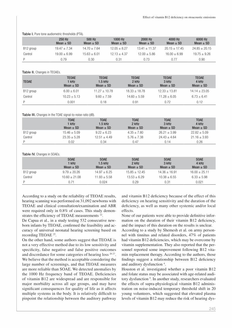
Effect of vitamin B12 deficiency on otoacoustic emissions
245
According to a study on the reliability of TEoAE results, hearing scanning was performed on 31,092 newborns with TEoAE and clinical consultation/examination and ABr were required only in 0.8% of cases. This study demon-strates the efficiency of TEoAE measurements 22. De Capua et al., in a study testing 532 consecutive new-born infants by TEoAE, confirmed the feasibility and ac-curacy of universal neonatal hearing screening based on recording TEoAE 25.on the other hand, some authors suggest that TEoAE is not a very effective method due to its low sensitivity and specificity, false negative and false positive outcomes and discordance for some categories of hearing loss 26 27.We believe that the method is acceptable considering the large number of screenings, and that TEoAE measures are more reliable than SoAE. We detected anomalies by the 1000 hz frequency band of TEoAE. Deficiencies of vitamin B12 are widespread and are responsible for major morbidity across all age groups, and may have significant consequences for quality of life as it affects multiple systems in the body. it is relatively difficult to pinpoint the relationship between the auditory pathway
and vitamin B12 deficiency because of the effect of this deficiency on hearing sensitivity and the duration of the deficiency, as well as many other systemic and/or local effects. none of our patients were able to provide definitive infor-mation on the duration of their vitamin B12 deficiency, and the impact of this duration on the results is unclear. According to a study by Shemesh et al. on army person-nel with tinnitus and related disorders, 47% of patients had vitamin B12 deficiencies, which may be overcome by vitamin supplementation. They also reported that the per-sonnel reported some improvement following B12 vita-min replacement therapy. According to the authors, those findings suggest a relationship between B12 deficiency and auditory dysfunction 4. houston et al. investigated whether a poor vitamin B12 and folate status may be associated with age-related audi-tory dysfunction 9. in another study, researchers evaluated the effects of supra-physiological vitamin B12 adminis-tration on noise-induced temporary threshold shift in 20 young volunteers, which suggested that elevated plasma levels of vitamin B12 may reduce the risk of hearing dys-
Table I. Pure tone audiometric thresholds (PTA).
250 HzMean ± SD
500 HzMean ± SD
1000 HzMean ± SD
2000 HzMean ± SD
4000 HzMean ± SD
6000 HzMean ± SD
B12 group 19.47 ± 7.34 14.70 ± 7.64 12.05 ± 8.27 13.41 ± 11.37 20.15 ± 17.45 24.85 ± 20.15
Control 19.00 ± 6.99 15.63 ± 6.01 12.13 ± 4.37 12.00 ± 5.86 16.00 ± 6.99 19.75 ± 9.26
P 0.79 0.30 0.31 0.73 0.77 0.90
Table II. Changes in TEOAEs.
TEOAETEOAE 1 kHz
Mean ± SD
TEOAE 1.5 kHz
Mean ± SD
TEOAE 2 kHz
Mean ± SD
TEOAE 3 kHz
Mean ± SD
TEOAE 4 kHz
Mean ± SD
B12 group 6.00 ± 6.01 11.27 ± 10.78 18.33 ± 18.78 12.33 ± 13.81 14.14 ± 23.05
Control 10.23 ± 5.13 9.60 ± 7.59 14.60 ± 5.59 11.28 ± 6.05 6.73 ± 6.41
P 0.001 0.18 0.91 0.72 0.12
Table III. Changes in the TOAE signal-to-noise ratio (dB).
TOAE 1 kHz
Mean ± SD
TOAE 1.5 kHz
Mean ± SD
TOAE 2 kHz
Mean ± SD
TOAE 3 kHz
Mean ± SD
TOAE 4 kHz
Mean ± SDB12 group 15.46 ± 5.09 9.22 ± 6.23 4.35 ± 7.80 26.21 ± 3.99 22.82 ± 5.09Control 23.35 ± 5.28 12.51 ± 4.49 5.76 ± 7.38 24.43 ± 4.64 21.16 ± 3.93P 0.02 0.34 0.47 0.14 0.26
Table IV. Changes in SOAEs.
SOAE 1 kHz
Mean ± SD
SOAE 1.5 kHz
Mean ± SD
SOAE 2 kHz
Mean ± SD
SOAE3 kHz
Mean ± SD
SOAE 4 kHz
Mean ± SDB12 group 9.79 ± 20.26 14.97 ± 6.25 15.85 ± 12.45 14.36 ± 16.91 16.00 ± 25.11
Control 10.60 ± 21.08 11.93 ± 5.58 13.53 ± 6.29 10.38 ± 6.55 6.33 ± 5.98
P 0.71 0.024 0.29 0.31 0.021

R. Karli et al.
246
function resulting from noise exposure in healthy young subjects 10. Another study investigated the effect of vita-mins A, E, B12 and folic acid on nihl and found that vitamin A, E and folic acid levels were normal while the levels of vitamin B12 were low in individuals with hear-ing loss due to noise 11. As a result, the authors recom-mend routine measurement of vitamin B12 in control of people working in noisy environment 11. Although a number of studies, as noted above, support a negative effect of vitamin B12 deficiency on hearing function, others contend no such relationship 1 5. in a study in in-dividuals over the age of 60, folate and vitamin B12 values in those with normal hearing at both speaking frequency and high frequency were compared to those with hearing loss. A significant decrease in folate lev-el in those with age-related hearing loss (Arhl) was found, but a significant relationship with vitamin B12 levels was not found. As a result, it was emphasized that folate deficiency might be significant in age-related hearing loss 23. in another study in 2956 patients with presbycusis over the age of 50 years, serum folate, vi-tamin B12 and homocysteine levels were not found to play a role in hearing loss 24. As mentioned above, despite many studies, the effects of this widespread nutritional deficiency on the auditory pathway remain unknown 6. Vitamin B12 is integral to the myelination process, and its lack or deficiency results in abnormal myelin formation or even complete demyeli-nation 12. Furthermore, animal experiments of B12 defi-ciency have revealed neuropathological effects 13. Medial efferent fibres of the medial olivocochlear bundle origi-nate in the medial nuclei of the superior olivary complex. The large myelinated fibres of the bundle project mainly contralaterally to the synapses at the base of the outer hair cells 14.Uğur and colleagues reported a contralateral suppres-sion effect in children with type 1 diabetes mellitus who presented with no evidence of symptomatic neuropathy. The findings point to the presence of a dysfunction in the medial olivocochlear efferent system in diabetic children. This may be regarded as an early, central manifestation of diabetic neuropathy 7. Vitamin B12 deficiency is known to have negative effects on hearing by affecting myelinization at any level of the retrocochlear area of the auditory system. More detailed studies are needed to determine the exact nature of these effects.
Conclusions There appears to be a correlation between vitamin B12 deficiency and cochlear function. The level of vitamin B12 in the blood should not be ignored in assessment of auditory function.
References 1 Babior BM, Bunn hF. Megaloblastic anemias. in: Braun-
wald E, Fauci AS, Kasper Dl, et al, eds. Harrison’s princi-ples of internal medicine. 15th ed. new-york: Mcgraw-hill; 2001. p. 674-80.
2 Molloy A, Cawley n, Ali E, et al. A pernicious leucoen-cephalopathy. ir Med J 2009;102:292-94.
3 Puri V, Chaudhry n, goel S, et al. Vitamin B12 deficiency: a clinical and electrophysiological profile. Electromyogr Clin neurophysiol 2005;45:273-84.
4 Shemesh Z, Attias J, ornan M, et al. Vitamin B12 deficiency in patients with chronic- tinnitus and noise-induced hearing loss. Am J otolaryngol 1993;14: 94-9.
5 Berner B, odum l, Parving A. Age-related hearing impair-ment and B vitamin status. Acta otolaryngol 2000;120:633-7.
6 green r. Indicators for assessing folate and vitamin B12 sta-tus and for monitoring the efficacy of intervention strategies. Am J Clin nutr 2011;94:666-72.
7 Ugur AK, Kemaloglu y K, Ugur MB, et al. Otoacoustic emissions and effects of contralateral white noise stimula-tion on transient evoked otoacoustic emissions in diabetic children. int J Pediatr otorhinolaryngol 2009;73:555-9.
8 Plinkert PK, Bootz F, Vossieck T. Influence of static mid-dle ear pressure on transiently evoked otoacoustics emis-sions and distortion products. Eur Arch otorhinolaryngol 1994;251:95-9.
9 houston DK, Johnson MA, nozza rJ, et al. Age-related hearing loss, vitamin B-12, and folate in elderly women. Am J Clin nutr 1999;69:564-71.
10 Quaranta A, Scaringi A, Bartoli r, et al. The effects of ‘su-pra-physiological’ vitamin B12 administration on temporary threshold shift. int J Audiol 2004;43:162-5.
11 gök U, halifeoğlu i, yildiz M. The levels of vitamins A, E, B12 and folic acid in noise-induced hearing loss. Kulak Bu-run Bogaz ihtis Derg 2004;12:60-4.
12 Vasconcelos oM, Poehm Eh, McCarter rJ, et al. Potential outcome factors in subacute combined degeneration: review of observational studies. J gen intern Med 2006;21:1063-8.
13 Agamanolis DP, Chester EM, Victor M, et al. Neuropathol-ogy of experimental vitamin B12 deficiency in monkeys. neu-rology 1976;26:905-14.
14 robertson D. Brainstem location of efferent neurons project-ing to the guinea pig cochlea. hear res 1985;20:79-84.
15 Kemp DT, ryan S, Bray P. A guide to the effective use of otoacoustic emissions. Ear hear 1990;11:93-105.
16 Kaga K, Akamatsu y, nakamura M, et al. Spontaneous otoacoustic emissions in two infants. oto-laryngologica 2009;129:399-404.
17 Kuroda T. Clinical investigation on spontaneous otoa-coustic emission (SOAE) in 447 ears. Auris nasus larynx 2007;34:29-38.
18 Bilger rC, Matthies Ml, hamel Dr, et al. Genetic implica-tions of gender differences in the prevalence of spontaneous otoacoustic emissions. J Speech hear res 1990;33:418-32.
19 nakamura M, yamasoba T, Kaga K. Changes in otoacoustic emissions in patients with idiopathic sudden deafness. Audi-ology 1997;36:121-35.

Effect of vitamin B12 deficiency on otoacoustic emissions
247
20 Kemp DT. Otoacoustic emissions, their origin in cochlear function, and use. Br Med Bull 2002;63:223-41.
21 Probst r, lonsbury-Martin Bl, Martin gK. A review of otoacoustic emissions. J Acoust Soc Am 1991;89:2027-67.
22 Berninger E, Westling B. Outcome of a universal newborn hearing-screening programme based on multiple transient-evoked otoacoustic emissions and clinical brainstem re-sponse audiometry. Acta otolaryngol 2011;131:728-39.
23 lasisi Ao, Fehintola FA, yusuf oB. Vitamin B12, and folate in the elderly. otolaryngol head neck Surg 2010;143:826-30.
24 gopinath B, Flood VM, rochtchina E, et al. Serum homo-
cysteine and folate concentrations are associated with preva-lent age-related hearing loss. J nutr 2010;140:1469-74.
25 De Capua B, De Felice C, Costantini D, et al. Newborn hearing screening by transient evoked otoacoustic emis-sions: analysis of response as a function of risk factors. Acta otorhinolaryngol ital 2003;23:16-20.
26 olusanya Bo, Bamigboye BA. Is discordance in TEOAE and AABR outcomes predictable in newborns? int J Pediatr otorhinolaryngol 2010;74:1303-9.
27 Pedersen l, Møller Tr, Wetke r, et al. Neonatal hearing screening. A comparison of automatic auditory brainstem audiometry and otoacoustic emissions. Ugeskr laeger 2008;170:642-6.
received: February 10, 2012 - Accepted: May 29, 2012
Address for correspondence: rifat Karli, ondokuz Mayıs Universi-tesi Tıp Fakültesi , KBB kliniği, Samsun / Turkey. Tel. +90 530 327 73 67 22. E-mail: [email protected]

248
ACTA oTorhinolAryngologiCA iTAliCA 2013;33:248-253
Rhinology
Evaluation of total oxidative stress parameters in patients with nasal polypsValutazione degli indici di stress ossidativo cellulare totale in pazienti affetti da poliposi nasale
F. Bozkus1, I. san1, T. ulas2, I. Iynen1, y. yesIlova3, y. Guler1, n. aksoy4
1 Department of otolaryngology Head and neck surgery, 2 Department of Internal Medicine, 3 Department of Dermatology, 4 Department of Biochemistry, Harran university, Faculty of Medicine, sanliurfa, Turkey
SummAry
oxidative stress, an imbalance between reactive oxygen species production and antioxidative defense activity, is believed to have a role in the development and pathogenesis of nasal polyps (nPs). Based on this assumption, several known oxidants and antioxidants have been studied in patients with nPs. The purpose of this study was to evaluate the association between oxidative stress parameters with a more valid and reliable method in patients with nPs. Seventy-three patients with nPs, septal deviations and middle concha hypertrophies were recruited. Patients were divided into two groups; group 1 (n = 38) consisted of patients with nPs, and group 2 (n = 35) included patients with septal deviations and middle concha hypertrophies. Polyp specimens were taken from all patients who underwent endoscopic surgery for nPs. Control specimens were obtained from patients who underwent an operation for septoplasty or middle concha hypertrophy. Blood and tissue samples were obtained to assess total oxidant status (ToS), total antioxidant status (TAS) and oxidative stress index (oSi). Compared to group 2, group 1 had significantly higher ToS and oSi and lower TAS levels both in serum and tissue samples (p < 0.001 for all). in group 1, tissue ToS and oSi levels were significantly higher, and TAS levels were significantly lower than in serum (p < 0.001 for all), whereas no significant difference was found in ToS, oSi and TAS levels either in serum or tissue samples in group 2 (p = 1.0; p = 1; p = 0.208, respectively). in group 1, serum oSi levels were significantly correlated with age (r = 0.442, p = 0.005). our study demonstrated that oxidative stress, both in serum and tissues in patients with nPs, was higher than in patients without nPs. our study differs from previ-ous studies in that we used a more reliable method that measures both ToS and TAS.
Key wordS: Nasal polyp • Reactive oxygen species • Oxidative stress • Total antioxidant status • Total oxidant status • Aging • Pathogenesis
riASSunTo
Lo stress ossidativo cellulare, risultato dell’equilibrio tra produzione ed eliminazione di specie reattive dell’ossigeno, è considerato un agente patogenetico di rilievo coinvolto nello sviluppo della poliposi nasale (NPs). Per tale ragione diversi fattori ossidanti e i relativi antiossidanti sono stati oggetti di studio nei pazienti affetti da poliposi naso-sinusale. Scopo del nostro studio è stato quello di esaminare l’associazione tra gli indici di stress ossidativo cellulare in pazienti affetti da poliposi nasale attraverso metodiche affidabili e standardiz-zate. Sono stati arruolati complessivamente 73 pazienti affetti da poliposi nasosinusale, deviazione del setto nasale e ipertrofia dei turbinati medi con concha bullosa. Tali pazienti sono stati divisi in due gruppi: nel gruppo 1 (n = 38) sono stati inseriti pazienti affetti da poliposi nasale, nel gruppo 2 (n = 35) sono stati inclusi, invece, i pazienti con deviazione del setto e ipertrofia dei turbinati medi con concha bul-losa. I campioni di polipi nasali sono stati raccolti e analizzati dai pazienti sottoposti a chirurgia endoscopica funzionale nasale durante l’intervento chirurgico. I campioni di controllo, invece, sono stati raccolti ed analizzati nei pazienti sottoposti ad intervento chirurgico di settoplastica e riduzione di concha bullosa. I prelievi ematici e istopatologici sono stati utilizzati per la valutazione di Status Ossidativo Totale (TOS), Status Antiossidativo Totale (TAS) e Indice di Stress Ossidativo (OSI). Nel gruppo 1 rispetto al gruppo 2 sono stati misurati, sia a livello sierico che tissutale, più alti livelli di TOS e OSI e più bassi livelli di TAS con una differenza in tutti casi statisticamente si-gnificativa (p < 0,001). Inoltre nel gruppo 1 i livelli tissutali di TOS e OSI erano significativamente più alti rispetto a quelli sierici mentre i livelli di TAS erano significativamente più bassi (p < 0.0001) mentre nessuna differenza significativa è stata osservata fra i livelli di TOS, OSI e TAS tissutale e sierici nel gruppo 2. Nel gruppo 1, i livelli serici di OSI risultavano correlati in maniera significativa con l’età (r = 0,442, p = 0,005). In conclusione il nostro studio dimostra che lo stress ossidativo cellulare è più elevato in pazienti affetti da poliposi nasale rispetto ai controlli, sia a livello sistemico che localmente a livello tissutale. A differenza di altri studi precedenti è stata utilizzata una metodica di determinazione di TOS e TAS più affidabile e standardizzata.
PArole ChiAve: Poliposi nasale • Specie reattive dell’ossigeno • Stress ossidativo • Status antiossidativo totale • Status ossidativo totale • Invecchiamento • Patogenesi
Acta Otorhinolaryngol Ital 2013;33:248-253

Oxidative stress and nasal polyps
249
Introduction Nasal polyp (NP) is a chronic inflammatory disease of the upper respiratory tract; however, its pathophysiology remains poorly understood. There are no aetiological fac-tors that explain the pathogenesis of NPs, though inflam-mation continues to be a major factor 1 2. Many studies have noted that histopathological abnormities of NP are closely related to the infiltration of inflammatory cells. As a result of inflammation, neutrophils activate and mi-grate to the inflammatory area and exert their bactericidal effects by producing reactive oxygen species (ROS) 3-7. Once the balance between ROS production and antioxi-dative defense activity is disrupted, oxidative stress can occur that may result in cell injury or death, subsequent tissue damage and, finally, chronic disease 8-10.
Studies investigating the role of ROS and antioxidants in nasal polyposis have found strong evidence for the in-volvement of oxidative stress in the pathogenesis of the condition 7 11-23. However, several of the oxidants and anti-oxidant enzymes that were evaluated in these studies may not be sufficient to accurately quantify oxidative stress. To clarify the results of these studies, we examined patients who underwent endoscopic surgery for NPs and assessed oxidative stress using a more valid and reliable method that measures total oxidant status (TOS) and total antioxi-dant status (TAS) both in serum and tissue.
Materials and methodsStudy design and patients This cross-sectional study was conducted at the Otolar-yngology Head and Neck Surgery Department of Harran University School of Medicine, Sanliurfa, Turkey. Prior to subject recruitment, the study protocol was reviewed and approved by the University’s ethics committee, in ac-cordance with the ethical principles for human investiga-tions, as outlined by the Second Declaration of Helsinki. Written informed consent was obtained from all patients. From May 2010 to March 2011, 73 consecutive patients with NPs, septal deviations and middle concha hypertro-phies were recruited for the study. Patients were divided in two groups; group 1 (n = 38) consisted of patients with NPs, and group 2 (n = 35) was composed of patients with septal deviations and middle concha hypertrophies with computed tomography (CT) scores comparable to those of the study population. The diagnosis was based on anterior rhinoscopy, endoscopic examination and paranasal sinus CT. Findings on preop-erative CT were graded according to the Lund–Mackay system. The mucosal abnormalities were graded as 0 (no abnormality), 1 (partial opacification), or 2 (total opacifi-cation) for each sinus group. The ostiomeatal complexes were scored bilaterally as 0 (not occluded) or 2 (occlud-ed). The maximal CT grading score was 24 24. Polyp spec-
imens were taken from all patients who underwent endo-scopic surgery for NPs, and control specimens (mucosal punctates from lateral lamella of the middle turbinate) were acquired from patients who underwent an operation for septoplasty or middle concha hypertrophy. The exclusion criteria included the following: recent acute infectious illness; any evidence of liver, kidney, or respiratory disease; diabetes mellitus; malignancy; any inflammatory or infiltrative disorder; recent use (within 2 weeks) of any systemic or local drug with antioxidant properties; preoperative drug use such as parenteral or oral corticosteroids and antibiotics for at least 4 weeks prior to sampling; and regular alcohol use or alcohol use within the previous 48 hours. No patient had a history of nasal allergy, asthma, or acetylsalicylic acid sensitivity. All showed negative results to a skin prick test with com-mon airborne allergens, which was evaluated by the same experienced dermatologist to prevent interobserver vari-ability. During interventions, tissue samples from polyps and turbinates were collected, fixed in formalin and fro-zen (-80o C) until the date of analysis.
Biochemical analysisAll blood samples were drawn after a 12-hour overnight fasting from a large antecubital vein without interrup-tion of venous flow using a 19-gauge butterfly needle connected to a plastic syringe. Twenty ml of blood was drawn, with the first few ml being discarded. Ten ml were used for baseline routine laboratory tests. The residual content of the syringe was transferred imme-diately to polypropylene tubes, which were then centri-fuged at 3,000 rpm for 10 min at 10 to 18°C. Supernatant plasma samples were stored in plastic tubes at –80°C until assayed. As serum markers of oxidant stress, TOS was measured and the oxidative stress index (OSI) was calculated. TAS was measured as an indicator of anti-oxidant status. These parameters were also studied and calculated in tissues. Tissues were homogenized in sa-line solution using a homogenizer. After centrifugation at 10,000 g for approximately 60 min, the clear super-natant was taken. TAS and TOS were measured in this fraction and OSI was calculated.
Measurement of total oxidant status Serum TOS was measured using a novel automated meth-od developed by Erel 25. Oxidants present in the sample oxidize the ferrous ion-o-dianisidine complex to fer-ric ion. The oxidation reaction is enhanced by glycerol molecules, which are abundantly present in the reaction medium. The ferric ion generates a colored complex with xylenol orange in an acidic medium. The colour intensity, which can be measured spectrophotometrically (V-530; Jasco®, Tokyo, Japan), is related to the quantity of oxidant molecules present in the sample. The assay is calibrated with hydrogen peroxide, and the results are expressed in

F. Bozkus et al.
250
terms of micromolar hydrogen peroxide equivalents per liter (μmol H
2O
2 equiv/l).
Measurement of total antioxidant status Serum TAS was measured using a novel automated meth-od developed by Erel 26. In this technique, hydroxyl radi-cal, the most potent biological radical, is produced. In the assay, the ferrous ion solution in reagent 1 is mixed with the hydrogen peroxide present in reagent 2. Sequentially-produced radicals, such as the hydroxyl radical-produced brown-colored dianisidinyl radical cation, are also potent radicals. This method allows the measurement of the sam-ple’s antioxidative effect against potent free radical reac-tions that are initiated by the hydroxyl radical. The assay has excellent precision values of more than 97%. The re-sults are expressed as mmol Trolox equiv/l.
Oxidative stress indexThe OSI was defined as the ratio of TOS to TAS level. A standardized value does not exist for OSI levels, which were used only for comparisons. For the calculation, TAS units were changed to mmol/l, and the OSI value was calculated according to the following formula: OSI (arbi-
trary unit) = TOS (μmol H2O
2 equiv./l)/TAS (mmol Trolox
equiv/l) 25 26.
Other variablesSerum urea, creatinine, fasting blood glucose, aspartate aminotransferase, alanine aminotransferase, triglycerides, total cholesterol, and high-density and low-density lipo-protein cholesterol levels were determined using commer-cially available assay kits (Abbott®, Abbott Park, North Chicago, Illinois, USA) with an auto-analyzer (Abbott®, Abbott Park, North Chicago, Illinois, USA).
Statistical analysis All statistical analyses were performed using SPSS for Windows version 17.0 (SPSS, Chicago, IL, USA). The Kolmogorov-Smirnov test was used to test the normality of data distribution. The data were expressed as arithmetic means and standard deviations. The chi-square test was used to compare categorical variables between groups. The independent sample T-test and Mann Whitney-U tests were used to compare continuous variables between the two groups. One-way ANOVA with a post-hoc Bonfer-
roni test was used in normally dis-tributed continuous data between all groups. Pearson’s and Spear-man’s rank correlation analyses were used to examine the asso-ciation of demographic and bio-chemical variables with oxidative stress parameters in group 1. A two-sided p value < 0.05 was con-sidered statistically significant.
ResultsThe biochemical and demograph-ic characteristics of all patients are presented in Table I. There were no significant differences in gender, age or biochemical values in the two groups (p > 0.05 for all). Renal and the liver function tests, cholesterol, triglyceride lev-els, and the CT scores were simi-lar between the two groups.
Comparison of total antioxidant status levelsCompared to group 2, group 1 had significantly lower TAS levels both in serum and tissues (both p < 0.001). In group 1, tis-sue TAS levels were significantly lower than in serum (p < 0.001). In group 2, there was no signifi-
Table I. Comparison of demographic and biochemical characteristics.
Group 1(n = 38)
Group 2(n = 35) p
Gender, male/female 17/21 16/19 NSa
Age, years 38.68 ± 8.44 36.26 ± 8.48 NSb
Glucose, mg/dL 82.20 ± 10.20 79.00 ± 9.76 NSb
CT scores 13.37 ± 2.85 12.29 ± 2.55 NSb
Urea, mg/dL 25.24 ± 6.18 23.49 ± 4.46 NSb
Creatinine, mg/dL 0.68 ± 0.25 0.71 ± 0.22 NSb
ALT, U/mL 24.03 ± 4.54 25.17 ± 3.75 NSb
AST, U/mL 26.15 ± 5.48 25.43 ± 5.88 NSb
Total cholesterol, mg/dL 202.13 ± 24.54 194 ± 36.22 NSc
LDL cholesterol, mg/dL 131.70 ± 36.93 141.70 ± 32.82 NSc
Triglyceride level, mg/dL 205.16 ± 55.46 197.32 ± 61.37 NSc
All measurable values are given as the mean ± standard deviation. NS: non-significant, CT: computed tomography, ALT: alanine aminotransferase, AST: aspartate aminotransferase, LDL: low-density lipoproteinChi-squarea, independent sample T testb and Mann Whitney-Uc tests were used
Table II. Comparison of oxidative stress parameters in all patients.
Group 1(n = 38)
Group 2(n = 35) p
TOS, μmol H2O
2 equiv/l Serum
Tissuep
21.34 ± 5.9828.59 ± 3.98
< 0.001
17.38 ± 4.1416.72 ± 2.49
> 0.05
< 0.001< 0.001
TAS, mmol Trolox equiv/l SerumTissue
p
0.94 ± 0.140.13 ± 0.03
< 0.001
1.10 ± 0.141.16 ± 0.12
> 0.05
< 0.001< 0.001
OSI, arbitrary unitSerumTissue
p
2.33 ± 0.653.12 ± 0.43
< 0.001
1.89 ± 0.451.76 ± 0.29
> 0.05
< 0.001< 0.001
All measurable values are given as the mean ± standard deviation. TOS: total oxidant status, TAS: total antioxidant status, OSI: oxidative stress indexOne-way ANOVA with a post-hoc Bonferroni* test was used

Oxidative stress and nasal polyps
251
cant difference in TAS levels either in serum or tissues (p = 0.208).
Comparison of total oxidant status and oxidative stress index levelsCompared to group 2, group 1 had significantly higher TOS and OSI levels both in serum and tissues (both p < 0.001). In group 1, the TOS and OSI levels were sig-nificantly higher in tissue than in serum (p < 0.001 for all). In group 2, there was no significant difference in the TOS or OSI levels either in serum or tissues (both p = 1.0). All OSI levels in serum and tissue are presented in Figure 1. In bivariate analysis, the serum OSI levels were signifi-cantly correlated with age in group 1 (r = 0.442, p = 0.005) (Fig. 2). However, no significant correlations were ob-served between oxidative stress parameters and gender, serum fasting glucose, lipid parameters, urea, creatinine, AST or ALT levels (p > 0.05 for all).
DiscussionThe main findings of this study are summarized as fol-lows: (i) oxidative stress both in serum and tissues of pa-tients with NPs was higher than in patients without NPs, (ii) serum OSI levels were significantly correlated with age and (iii) our study was able to provide important new insights and data because it assessed oxidative stress pa-rameters using a more reliable method that measures both TOS and TAS. Measuring different oxidant and antioxidant molecules is impractical, and oxidant and antioxidant effects are additive. Because there are numerous oxidants and anti-oxidants in the body, measuring total oxidant-antioxidant status is more valid and reliable. When only a few pa-rameters are measured, levels may remain unchanged or decrease, even though the actual oxidant status increased or vice versa 25-27. In the light of this information, we used TOS and TAS levels in our study. ROS are suspected to have a major function in the pathogenesis of NPs 7 11-23. Studies have reported similar results, showing an increase in oxidative stress mark-ers and deterioration in antioxidant scavenging systems. These studies provide strong evidence that oxidative stress plays a role in the pathogenesis of NPs 12. Dogru et al. and Uneri et al. investigated the role of ROS in the development of NPs and reported that ROS may play a significant role in the pathogenesis of NPs 7 13. Dagli et al. compared 31 patients and 19 control subjects to investigate the role of ROS and antioxidants in NPs, and showed that the increase in MDA was higher in NP tissue and the blood of patients with NPs than in a con-cha bullosa group 18. According to their results, in polyp tissue, the levels of an oxidant (malondialdehyde) were increased, and the levels of antioxidants (glutathione and α-tocopherol) were decreased compared with con-
trol tissues from inferior turbinates. In another study performed by Okur et al., the investigators demon-strated the production of ROS in the neutrophils of NPs and determined a very high ratio of ROS production in NPs 19. Uneri at al. also reported high ROS levels in NP samples and showed a strong relationship between tis-sue damage and NP. Moreover, Cheng et al. studied the expression of superoxide dismutase 1 and 3 and found higher levels in polyp tissues. Such investigations indi-cate that ROS may be important in the pathogenesis of NP 13 14.
Fig. 2. Relationship between serum oxidative stress index and age (r = 0.442, p = 0.005).
Fig. 1. Oxidative stress index levels in serum and tissue.

F. Bozkus et al.
252
Recent studies have also confirmed the role of oxidative stress in the development of NPs. Park et al. suggested the possibility that [6]-gingerol may play an important role in inhibiting the production of the extracellular matrix in the development of NPs through an antioxidant effect 28. The same authors performed another study and showed that nicotinamide adenine dinucleotide phosphate oxi-dase and ROS have a role in myofibroblast differentia-tion and collagen production of transforming growth factor-β1-induced nasal polyp-derived fibroblasts, and that these processes are inhibited by the elimination of ROS 29. Moon et al. have also demonstrated the ROS de-rived from NADPH oxidases, and transforming growth factor-β-1 have been implicated in the pathogenesis of hypoxia-induced NPs 30. As confirmation, Jeanson et al. have shown the existence of an unfolded protein re-sponse in nasal epithelial cells that is linked to oxidative stress leading to interleukin-8 and leukotriene-B4 secre-tions. They concluded that these mechanisms may par-ticipate in chronic inflammation in NPs 31. In our study, oxidative stress both in serum and tissue in patients with NPs was higher than in patients without NPs. Our study results on tissue and serum samples support the previous studies as it provides a more reliable quantification of oxidative stress parameters.The frequency of NPs increases with age and seems to occur more often in men. The prevalence of this condi-tion increases with age in both sexes to reach a peak in those aged 50 years or older. In the aging process, ROS production increases, while degradation is impaired, and thus oxidative stress accumulates 32-34. In our study, serum OSI levels were significantly correlated with age but not correlated with gender in patients with NPs. Although not definitive, our study suggests that the increase in oxida-tive stress inherent in the aging process may contribute to the formation of NPs.
ConclusionsThe findings of the present study demonstrate that oxida-tive stress is increased in patients with NPs. The important therapeutic potential to repair the harmful effects of oxida-tive stress on nasal tissues with antioxidant agents will only emerge with an improved understanding of the role of oxi-dative stress in NPs. The sample size was relatively small, and the design of the study was cross-sectional which are limitations of the present study. Therefore, large, prospec-tive cohort studies are needed to address this issue.
AcknowledgementsThis study was conducted at the Otolaryngology Head and Neck Surgery, Internal Medicine, Dermatology and Biochemistry Departments of Harran University School of Medicine, Sanliurfa. This study was approved by the Institutional Review Board.
References 1 Watkins DN, Lewis RH, Basclain KA, et al. Expression and
localization of the inducible isoform of nitric oxide synthase in nasal polyp epithelium. Clin Exp Allergy 1998;28:211-9.
2 Sarisoy BA, Eken M, Oktay AZ, et al. Myeloperoxydase ex-pression in the pathogenesis of nasal polyps. Indian J Otolar-yngol Head Neck Surg 2011;63:260-3.
3 Bachert C, Gevaert P, Holtappels G, et al. Nasal polyposis: from cytokines to growth. Am J Rhinol 2000;14:279-90.
4 Norlander T, Brönnegård M, Stierna P. The relationship of nasal polyps, infection, and inflammation. Am J Rhinol 1999;13:349-55.
5 Nelson HS. Advances in upper airway diseases and allergen immunotherapy. J Allergy Clin Immunol 2004;113:635-42.
6 Shukla GK, Mahajan A, Pandey S, et al. A study of free radi-cals and scavenging enzyme in tonsillitis. Boll Chim Farm 1996;135:653-5.
7 Doğru H, Delibaş N, Döner F, et al. Free radical damage in nasal polyp tissue. Otolaryngol Head Neck Surg 2001;124: 570-2.
8 Döner F, Delibaş N, Doğru H, et al. Malondialdehyde levels and superoxide dismutase activity in experimental maxillary sinusitis. Auris Nasus Larynx 1999;26:287-91.
9 Cross CE, Halliwell B, Borish ET, et al. Oxygen radicals and human disease. Ann Intern Med 1987;107:526-45.
10 Halliwell B, Gutteridge JM, Cross CE. Free radicals, anti-oxidants, and human disease: where are we now? J Lab Clin Med 1992;119:598-620.
11 Taysi S, Uslu C, Yilmaz A, et al. Lipid peroxidation and some antioxidant enzymes in nasal polyp tissue. Cell Biochem Funct 2006;24:461-5.
12 Sagit M, Erdamar H, Saka C, et al. Effect of antioxidants on the clinical outcome of patients with nasal polyposis. J Lar-yngol Otol 2011;125:811-5.
13 Uneri C, Oztürk O, Polat S, et al. Determination of reactive oxygen species in nasal polyps. Rhinology 2005;43:185-9.
14 Cheng YK, Hwang GY, Lin CD, et al. Altered expression profile of superoxide dismutase isoforms in nasal polyps from nonallergic patients. Laryngoscope 2006;116:417-22.
15 Cekin E, Ipcioglu OM, Erkul BE, et al. The association of oxidative stress and nasal polyposis. J Int Med Res 2009;37:325-30.
16 Cheng YK, Tsai MH, Lin CD, et al. Oxidative stress in non-allergic nasal polyps associated with bronchial hyperrespon-siveness. Allergy 2006;61:1290-8.
17 Karlidağ T, Ilhan N, Kaygusuz I, et al. Roles of free radicals, nitric oxide, and scavenging enzymes in nasal polyp develop-ment. Ann Otol Rhinol Laryngol 2005;114:122-6.
18 Dagli M, Eryilmaz A, Besler T, et al. Role of free radi-cals and antioxidants in nasal polyps. Laryngoscope 2004;114:1200-3.
19 Okur E, Inanc F, Yildirim I, et al. Malondialdehyde level and adenosine deaminase activity in nasal polyps. Otolaryngol Head Neck Surg 2006;134:37-40.
20 Giannessi F, Ursino F, Fattori B, et al. Expression of 3-nitro-tyrosine, a marker for peroxynitrite, in nasal polyps of non-atopic patients. Med Sci Monit 2010;16:172-9.

Oxidative stress and nasal polyps
253
21 Woo HJ, Bae CH, Song SY, et al. Expression of glutaredox-in-1 in nasal polyps and airway epithelial cells. Am J Rhinol Allergy 2009;23:288-93.
22 Bugdayci G, Kaymakci M. Nitrite/nitrate and malondialde-hyde levels in nasal polyp. Cell Mol Biol (Noisy-le-grand). 2008;54(Suppl):1043-5.
23 Kang BH, Huang NC, Wang HW. Possible involvement of ni-tric oxide and peroxynitrite in nasal polyposis. Am J Rhinol 2004;18:191-6.
24 Lund VJ, Kennedy DW. Staging for rhinosinusitis. Otolaryn-gol Head Neck Surg 1997;117:35-40.
25 Erel O. A new automated colorimetric method for measuring total oxidant status. Clin Biochem 2005;38:1103-11.
26 Erel O. A novel automated method to measure total antioxi-dant response against potent free radical reactions. Clin Bio-chem 2004;37:112-9.
27 Ulas T, Buyukhatipoglu H, Kirhan I, et al. The effect of day and night shifts on oxidative stress and anxiety symptoms of the nurses. Eur Rev Med Pharmacol Sci 2012;16:594-99.
28 Park SA, Park IH, Cho JS, et al. Effect of [6]-gingerol on myofibroblast differentiation in transforming growth factor
beta 1-induced nasal polyp-derived fibroblasts. Am J Rhinol Allergy 2012;26:97-103.
29 Park IH, Park SJ, Cho JS, et al. Role of reactive oxygen species in transforming growth factor beta1-induced alpha smooth-muscle actin and collagen production in nasal pol-yp-derived fibroblasts. Int Arch Allergy Immunol 2012;159: 278-86.
30 Moon YM, Kang HJ, Cho JS, et al. Nox4 mediates hypoxia-stimulated myofibroblast differentiation in nasal polyp-de-rived fibroblasts. Int Arch Allergy Immunol 2012;159:399-409.
31 Jeanson L, Kelly M, Coste A, et al. Oxidative stress induces unfolding protein response and inflammation in nasal poly-posis. Allergy 2012;67:403-12.
32 Shimosawa T. [Increasing oxidative stress in aging]. Nihon Rinsho 2005;63:994-9.
33 Settipane GA, Chafee FH. Nasal polyps in asthma and rhi-nitis. A review of 6,037 patients. J Allergy Clin Immunol 1977;59:17-21.
34 Lund VJ. Diagnosis and treatment of nasal polyps. BMJ 1995;311:1411-4.
Address for correspondence: Ferhat Bozkus, Harran University School of Medicine, Department of Otolaryngology Head and Neck Surgery, Yenisehir Campus, 63000, Sanliurfa, Turkey. Tel. (0414) 3183027. Fax (0414) 3183192. E-mail: [email protected]
Received: October 1, 2012 - Accepted: December 15, 2012

254
ACTA oTorhinolAryngologiCA iTAliCA 2013;33:254-260
Vestibology
Direction-fixed paroxysmal nystagmus lateral canal benign paroxysmal positioning vertigo (BPPV): another form of lateral canalolithiasisLa vertigine parossistica posizionale benigna (VPPB) con nistagmo parossistico a direzione fissa: un’altra forma di canalolitiasi laterale
L. CaLifano, a. VassaLLo, M.G. MeLiLLo, s. Mazzone, f. saLafia Departmental Unit of audiology and Phoniatrics, surgical sciences Department, “G. Rummo” Hospital, Benevento, italy
SummAry
Benign paroxysmal positioning vertigo (BPPV) is the most frequent vertiginous syndrome. it is caused either by free-floating otoliths in the semicircular canals (canalolithiasis) or by otoconial debris adhering to a canal cupula (cupulolithiasis). The posterior canal is the most frequently involved (80%), while the lateral canal is involved less frequently (15%), and the rarest conditions are anterior canalolithiasis and apogeotropic posterior canalolithiasis (5%). The main diagnostic sign of lateral canal BPPV is paroxysmal horizontal bidirectional positioning nystagmus evoked through Pagnini-mcClure’s test (head roll in the yaw plane in supine position). in the geotropic variant, which is more frequent, the fast phase of the nystagmus is directed towards the lowermost ear, when the patient lies on the affected side or on the healthy side; in the apogeotropic variant, which is less frequent, the fast phase is directed always toward the uppermost ear, regard-less of which side the patient lies on. Paroxysmal nystagmus is more intense on the affected side in the geotropic form, and more intense on the healthy side in the apogeotropic form. The authors describe five cases of another primitive and rare form of lateral BPPV, defined as “direction-fixed paroxysmal nystagmus lateral canal BPPV”, which has previously been described by other authors as a transitory step observed during the transformation from an apogeotropic into a geotropic form. it is characterized by typical BPPV symptoms and diag-nosed by the presence of a paroxysmal horizontal unidirectional positioning nystagmus, evoked through Pagnini-mcClure’s test, which is apogeotropic on the affected side and geotropic on the healthy side. in the reported cases, direction-fixed horizontal paroxysmal nystagmus was always transformed into a typical geotropic form. The clinical features and pathophysiology of direction-fixed nystagmus lateral canal BPPV are discussed.
Key wordS: BPPV • Lateral canalolithiasis • Horizontal canalolithiasis • Direction-fixed nystagmus • Lateral geotropic canalolithiasis • lateral Apogeotropic canalolithiasis
riASSunTo
La vertigine parossistica posizionale benigna (VPPB) è la sindrome vertiginosa di più frequente osservazione. Essa è determinata dalla presenza di otoliti liberi nei canali semicircolari (canalolitiasi) o da detriti otolitici adesi alla cupula canalare (cupulolitiasi). Il canale se-micircolare posteriore è quello più frequentemente interessato (80%), seguito da quello laterale (15%), mentre molto più rare sono le forma da canalolitiasi anteriore e da canalolitiasi posteriore apogeotropa (5%). Il segno diagnostico primario della VPPB laterale è il nistagmo parossistico orizzontale bidirezionale da posizionamento evocato dalla manovra diagnostica di Pagnini-McClure (rotazione rapida della testa in posizione supina sul piano frontale): nella più frequente forma geotropa, il nistagmo batte verso il lato posto in basso, sia quando il paziente decombe sul lato malato, ove esso è più intenso, sia quando decombe sul lato sano; nella forma apogeotropa, il nistagmo, inve-ce, batte verso il lato posto in alto, sia quando il paziente decombe sul lato malato, ove esso è meno intenso, sia quando decombe sul lato sano. Gli Autori descrivono una nuova forma primitiva di canalolitiasi laterale, definita “canalolitiasi laterale con nistagmo parossistico a direzione fissa”, precedentemente descritta da altri Autori come una forma transitoria osservabile durante la trasformazione di una forma apogeotropa in una forma geotropa. Essa è caratterizzata dalla presenza di un nistagmo parossistico orizzontale unidirezionale da posizionamento, evocato dalla manovra diagnostica di Pagnini-McClure, apogeotropo quando il paziente decombe sul lato malato, geo-tropo quando decombe sul lato sano. Nei casi descritti, la canalolitiasi laterale con nistagmo parossistico a direzione fissa è stata sempre convertita in una forma geotropa. Gli aspetti clinici e fisiopatologici di tale forma sono discussi.
PArole ChiAVe: BPPV • Canalolitiasi laterale • Canalolitiasi orizzontale • Canalolitiasi laterale con nistagmo parossistico a direzione fissa • Canalolitiasi laterale geotropa • Canalolitiasi laterale apogeotropa
Acta Otorhinolaryngol Ital 2013;33:254-260
Direction-fixed paroxysmal nystagmus
255
IntroductionBenign paroxysmal positional vertigo (BPPV) is the most common vertiginous syndrome, characterized by recur-rent short-lasting episodes of vertigo when the patient changes his/her position. It is usually caused by free-floating otoconia dislodged from the utricle in the semi-circular canals (canalolithiasis). Less frequently, otoliths adhere to a semicircular canal cupula, rendering it heavier than the surrounding endolymph (cupulolithiasis). Both conditions are able to change the firing rate of the vestibu-lar nerve 1 2.There are several known variants of BPPV 3: posterior canalolithiasis and cupulolithiasis, including their rare variants, namely short arm BPPV 4 or posterior apogeo-tropic canalolithiasis 5; anterior canalolithiasis; geotropic lateral canalolithiasis, caused by the presence of otoliths in the non-ampullary arm; apogeotropic lateral canalolith-iasis, caused by either otoliths in the ampullary arm or by lateral cupololithiasis.The most frequent form is posterior canal BPPV (80% of observed cases), followed by both geotropic and apo-geotropic lateral canal BPPV (15%); anterior canal BPPV and apogeotropic posterior canal BPPV are less frequent (5%). The main diagnostic sign of lateral canal BPPV is paroxysmal horizontal bidirectional positioning nystag-mus, evoked through Pagnini-McClure test (head roll in the yaw plane in supine position) 6-8; in the geotropic form the fast phase of the nystagmus always beats towards the lowermost ear and the affected side is where nystagmus is the strongest; instead, in apogeotropic lateral canal BPPV paroxysmal nystagmus is directed always toward the up-permost ear and the affected side is where the nystagmus is the weakest.This study reports five cases of lateral canal BPPV which we define as “direction-fixed paroxysmal nystagmus lat-eral canal BPPV” because horizontal paroxysmal posi-tioning nystagmus, evoked through Pagnini-Mc Clure’s test, is always directed towards the healthy side, when the patient is positioned either on the affected side (apogeo-tropic nystagmus) or the healthy one (geotropic nystag-mus): thus, nystagmus is unidirectional. A similar form was first reported by Vannucchi 9 who observed it as a transitory step of the transformation from an apogeotropic lateral canal BPPV to a geotropic lateral canal BPPV after a certain number of head rotations from side to side in supine position.The present research for the first time describes a form of “direction-fixed paroxysmal nystagmus lateral canal BPPV” since its diagnosis.
Materials and methodsFrom September 2010 to June 2012, we observed 1234 BPPV, 925 from the posterior canal (75%), 37 from the
anterior canal (3%), 272 from the latera1 canal (22%) of which l89 were geotropic lateral canal BPPV (69.5%), 78 apogeotropic lateral canal BPPV (28.7%) and 5 “di-rection-fixed paroxysmal nystagmus lateral canal BPPV” (1.8%). Herein, we report clinical data for only these five patients,: three males and two females, with an mean age of 52.8 years; before our observation they had suffered from positional vertigo for a period of 1 to 10 days and none had used vestibular suppressant medications for at least 24 hours before the visit or had undergone physical therapy for BPPV in another centre.Patients underwent pure tone audiometry and a bed-side vestibular examina-tion in sitting and supine positions to evaluate spontaneous and evoked nystagmus: gaze-evoked nystagmus – head shaking induced nystagmus (HSIN) – vibration induced nystagmus - paroxysmal positioning nystagmus through Dix-Hallpike test and Pagnini-McClure test. Paroxysmal horizontal positioning nystagmus as evoked by Pagnini-Mc Clure test was considered the primary di-agnostic sign of lateral canal BPPV.In addition, the following secondary signs of lateraliza-tion were considered: • in the sitting position: pseudo-spontaneous nystag-
mus 10 - Leaning and Bowing nystagmus 11 - HSIN;• inthesupineposition:thesittingtosupineevokedpo-
sitional horizontal nystagmus 12.Transformation from an apogeotropic to a geotropic form (geotropization) was attempted through head pitching manoeuvre (HPM) in the sitting position 13-15 and, when HPM failed, through side to side rotations in the supine position 16.Caloric stimulation was performed according to Fitzger-ald-Hallpike’s technique (FH) 17. It was performed during the first observation and before that patients underwent therapeutic maneuvers. Canal paresis was evaluated through Jongkees’s formu-la 18, assuming as significant a labyrinthine prevalence > 25%.Patients underwent Gufoni’s liberatory manoeuvre 19 af-ter geotropization occurred; it was performed five times towards the healthy side. “Forced prolonged position” on the healthy side 20 was then prescribed for the next 8-10 hours. A successful outcome was defined as the absence of paroxysmal horizontal positioning nystagmus evoked through Pagnini-Mc Clure test during the 24 hour-follow-up examination. Nystagmus was observed in darkness, through infrared videonystagmoscopy.Due to a relapse of the disease six months after the first observation, Patient 1 underwent high resolution com-puted tomography (CT) and magnetic resonance imaging (MRI) of the inner ear (1.5 Tesla). MRI generated three-dimensional models of the inner ear. Because of the be-nign course of their disease, the other four patients did not undergo neuroimaging.
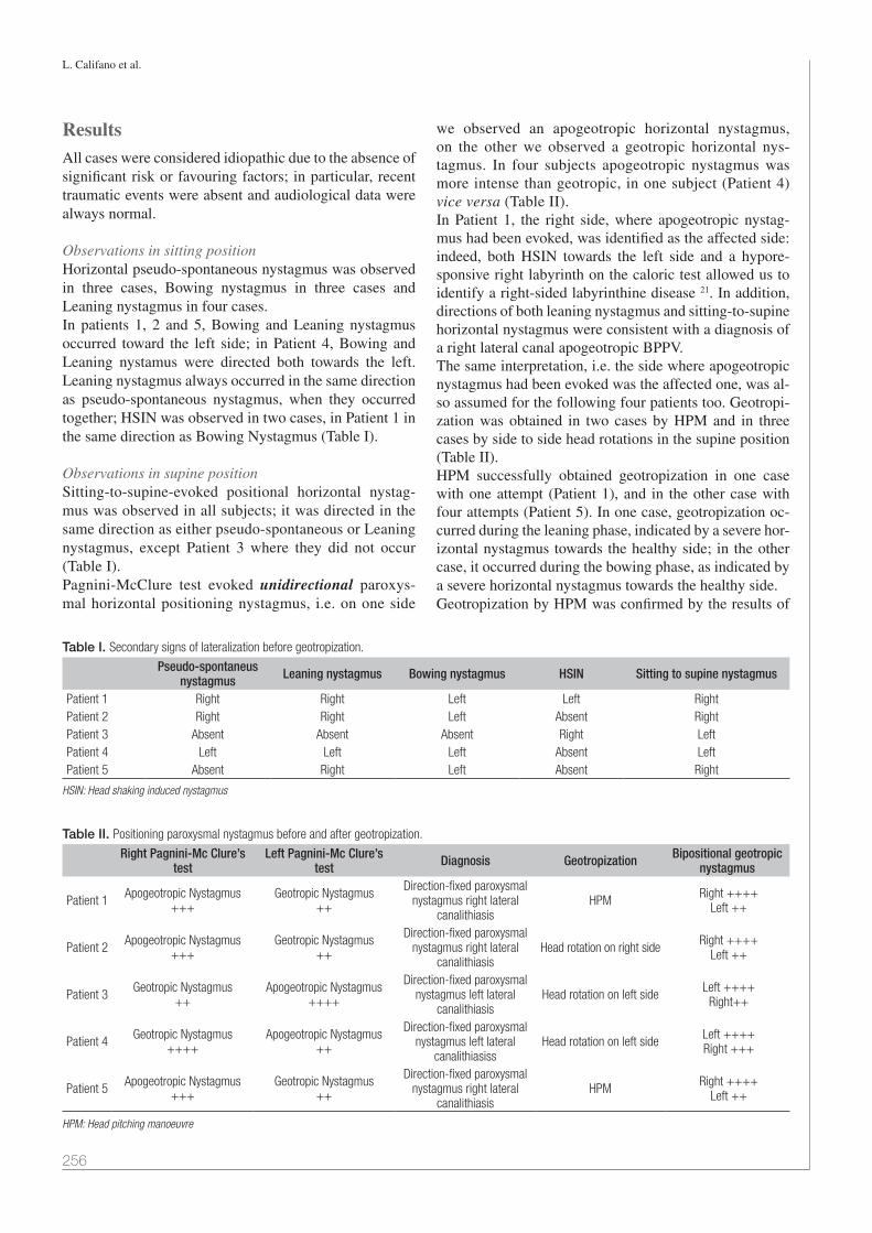
L. Califano et al.
256
ResultsAll cases were considered idiopathic due to the absence of significant risk or favouring factors; in particular, recent traumatic events were absent and audiological data were always normal.
Observations in sitting position Horizontal pseudo-spontaneous nystagmus was observed in three cases, Bowing nystagmus in three cases and Leaning nystagmus in four cases.In patients 1, 2 and 5, Bowing and Leaning nystagmus occurred toward the left side; in Patient 4, Bowing and Leaning nystamus were directed both towards the left. Leaning nystagmus always occurred in the same direction as pseudo-spontaneous nystagmus, when they occurred together; HSIN was observed in two cases, in Patient 1 in the same direction as Bowing Nystagmus (Table I).
Observations in supine positionSitting-to-supine-evoked positional horizontal nystag-mus was observed in all subjects; it was directed in the same direction as either pseudo-spontaneous or Leaning nystagmus, except Patient 3 where they did not occur (Table I).Pagnini-McClure test evoked unidirectional paroxys-mal horizontal positioning nystagmus, i.e. on one side
we observed an apogeotropic horizontal nystagmus, on the other we observed a geotropic horizontal nys-tagmus. In four subjects apogeotropic nystagmus was more intense than geotropic, in one subject (Patient 4) vice versa (Table II).In Patient 1, the right side, where apogeotropic nystag-mus had been evoked, was identified as the affected side: indeed, both HSIN towards the left side and a hypore-sponsive right labyrinth on the caloric test allowed us to identify a right-sided labyrinthine disease 21. In addition, directions of both leaning nystagmus and sitting-to-supine horizontal nystagmus were consistent with a diagnosis of a right lateral canal apogeotropic BPPV.The same interpretation, i.e. the side where apogeotropic nystagmus had been evoked was the affected one, was al-so assumed for the following four patients too. Geotropi-zation was obtained in two cases by HPM and in three cases by side to side head rotations in the supine position (Table II).HPM successfully obtained geotropization in one case with one attempt (Patient 1), and in the other case with four attempts (Patient 5). In one case, geotropization oc-curred during the leaning phase, indicated by a severe hor-izontal nystagmus towards the healthy side; in the other case, it occurred during the bowing phase, as indicated by a severe horizontal nystagmus towards the healthy side.Geotropization by HPM was confirmed by the results of
Table I. Secondary signs of lateralization before geotropization.
Pseudo-spontaneus nystagmus Leaning nystagmus Bowing nystagmus HSIN Sitting to supine nystagmus
Patient 1 Right Right Left Left RightPatient 2 Right Right Left Absent RightPatient 3 Absent Absent Absent Right LeftPatient 4 Left Left Left Absent LeftPatient 5 Absent Right Left Absent Right
HSIN: Head shaking induced nystagmus
Table II. Positioning paroxysmal nystagmus before and after geotropization.
Right Pagnini-Mc Clure’s test
Left Pagnini-Mc Clure’s test Diagnosis Geotropization Bipositional geotropic
nystagmus
Patient 1 Apogeotropic Nystagmus+++
Geotropic Nystagmus++
Direction-fixed paroxysmal nystagmus right lateral
canalithiasis HPM Right ++++
Left ++
Patient 2 Apogeotropic Nystagmus+++
Geotropic Nystagmus++
Direction-fixed paroxysmal nystagmus right lateral
canalithiasisHead rotation on right side Right ++++
Left ++
Patient 3 Geotropic Nystagmus++
Apogeotropic Nystagmus++++
Direction-fixed paroxysmal nystagmus left lateral
canalithiasisHead rotation on left side Left ++++
Right++
Patient 4 Geotropic Nystagmus++++
Apogeotropic Nystagmus++
Direction-fixed paroxysmal nystagmus left lateral
canalithiasissHead rotation on left side Left ++++
Right +++
Patient 5 Apogeotropic Nystagmus+++
Geotropic Nystagmus++
Direction-fixed paroxysmal nystagmus right lateral
canalithiasisHPM Right ++++
Left ++
HPM: Head pitching manoeuvre

Direction-fixed paroxysmal nystagmus
257
Pagnini-McClure’s test, which showed the typical par-oxysmal horizontal positioning geotropic nystagmus on both the affected and healthy sides.In three cases (Patients 2, 3 and 4), geotropization was obtained by side to side head rotations in the supine po-sition; on average, four attempts were necessary, with a progressive reduction in intensity of apogeotropic nystag-mus; geotropization caused intense geotropic nystagmus 1-3 seconds after the acquisition of the lateral position on the affected side, where apogeotropic nystagmus had been previously evoked.Post-geotropization observation confirmed the initial
diagnosis of the affected side: in Patients 1, 2 and 5 the right semicircular canal was involved; in Patients 3 and 4 the left one was involved, as correctly assumed at ini-tial examination (Table II). After geotropization occurred, secondary signs of lateralization changed their direction, except HSIN (Table III). A significant labyrinthine preva-lence on the caloric test (left labyrinth prevalence > 25%) was present before treatment in only Patient 1. Gufoni’s manoeuver and the “Forced prolonged position” on the healthy side were able to successfully treat all patients who presented a negative Pagnini-McClure test at the control.
Patient 1 relapsed six months later, presenting the same clinical picture of the first observation; both CT scans and MRI did not show significant alterations of the morphology of his semicircular canals (Fig. 1). All other subjects are still disease-free.
DiscussionThe geotropic form is the most frequent form of lateral canal BP-PV. It is caused by free-floating otoconia in the non-ampullary arm of the lateral semicircular ca-nal (Fig. 2). When the patient is lying down on the affected side, the position of the affected canal causes an excitatory ampullopetal endolymphatic flow which pro-vokes a horizontal paroxysmal geotropic nystagmus; when the patient is lying down instead on the healthy side, the position of the affected canal causes an inhibitory ampullofugal endolymphatic flow, which pro-vokes a nystagmus in the opposite direction of the previous one, and therefore again geotropic.The apogeotropic form is rarer; it is usually caused by either free-
Table III. Secondary signs of lateralization after geotropization.
Pseudo-spontaneous nystagmus Leaning nystagmus Bowing nystagmus HSIN Sitting to supine
nystagmusPatient 1 Left Left Right Left LeftPatient 2 Left Left Absent Absent LeftPatient 3 Right Right Absent Right RightPatient 4 Right Right Left Absent RightPatient 5 Absent Left Right Absent Left
HSIN: Head shaking induced nystagmus
Fig. 2. Geotropic left lateral canalolithiasis (left view): (a) on the affected side otoliths in the non-ampullary arm cause ampullopetal flow; (b) on the healthy side otoliths in the non-ampullary arm cause ampullofugal flow (inner arrows).
Fig. 1. MRI 3D reconstrution of patient’s 1 right inner ear. No significant morphological alterations are pre-sent (a) Front view. (b.) Right view. A: Anterior semicircular canal; L: Lateral semicircular canal; P: Posterior semicircular canal.
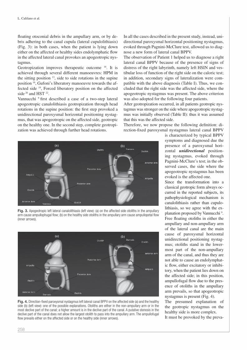
L. Califano et al.
258
floating otoconial debris in the ampullary arm, or by de-bris adhering to the canal cupula (lateral cupulolithiasis) (Fig. 3): in both cases, when the patient is lying down either on the affected or healthy sides endolymphatic flow in the affected lateral canal provokes an apogeotropic nys-tagmus. Geotropization improves therapeutic outcome 16. It is achieved through several different manoeuvres: HPM in the sitting position 13, side to side rotations in the supine position 16, Gufoni’s liberatory manoeuvre towards the af-fected side 19, Forced liberatory position on the affected side 20 and HST 22.Vannucchi 9 first described a case of a two-step lateral apogeotropic canalolithiasis geotropization through head rotations in the supine position: the first step provoked a unidirectional paroxysmal horizontal positioning nystag-mus, that was apogeotropic on the affected side, geotropic on the healthy one. In the second step, complete geotropi-zation was achieved through further head rotations.
In all the cases described in the present study, instead, uni-directional paroxysmal horizontal positioning nystagmus, evoked through Pagnini-McClure test, allowed us to diag-nose a new form of lateral canal BPPV.The observation of Patient 1 helped us to diagnose a right lateral canal BPPV because of the presence of signs of distress of the right labyrinth, namely left HSIN and ves-tibular loss of function of the right side on the caloric test; in addition, secondary signs of lateralization were com-patible with the above diagnosis (Table I). Thus, we con-cluded that the right side was the affected side, where the apogeotropic nystagmus was present. The above criterion was also adopted for the following four patients. After geotropization occurred, in all patients geotropic nys-tagmus was stronger on the side where apogeotropic nystag-mus was initially observed (Table II): thus it was assumed that this was the affected side.Therefore, we now propose the following definition: di-rection-fixed paroxysmal nystagmus lateral canal BPPV
is characterized by typical BPPV symptoms and diagnosed due the presence of a paroxysmal hori-zontal unidirectional position-ing nystagmus, evoked through Pagnini-McClure’s test; in the ob-served cases, the side where the apogeotropic nystagmus has been evoked is the affected one.Since the transformation into a classical geotropic form always oc-curred in the reported subjects, its pathophysiological mechanism is canalolithiasis rather than cupulo-lithiasis, so we agree with the ex-planation proposed by Vannucchi 9.Free floating otoliths in either the ampullary and non-ampullary arm of the lateral canal are the main cause of paroxysmal horizontal unidirectional positioning nystag-mus; otoliths stand in the lower-most part of the non-ampullary arm of the canal, and thus they are not able to cause an endolymphat-ic flow, either excitatory or inhibi-tory, when the patient lies down on the affected side; in this position, ampullofugal flow due to the pres-ence of otoliths in the ampullary arm prevails, so that apogeotropic nystagmus is present (Fig. 4). The presumed explanation of the geotropic nystagmus on the healthy side is more complex.It must be provoked by the preva-
Fig. 3. Apogeotropic left lateral canalolithiasis (left view): (a) on the affected side otoliths in the ampullary arm cause ampullophugal flow; (b) on the healthy side otoliths in the ampullary arm cause ampullopetal flow (inner arrows).
Fig. 4. Direction-fixed paroxysmal nystagmus left lateral canal BPPV on the affected side (a) and the healthy side (b) (left view): one of the possible explanations. Otoliths are either in the non-ampullary arm or in the most declive part of the canal; a higher amount is in the declive part of the canal. A putative stenosis in the declive part of the canal does not allow the largest otolith to pass into the ampullary arm. The ampullofugal flow prevails either on the affected side or on the healthy side (inner arrows).

Direction-fixed paroxysmal nystagmus
259
lence of an ampullofugal flow in the affected lateral canal, according to Ewald’s second law. The endolymphatic flow, caused by the head rotation, could push otoliths of both the ampullary and non-ampul-lary arms toward the utricle, but ampullofugal flow could also result from the algebraic sum of an ampullopetal flow, caused by otoliths in the ampullary arm, and preva-lent ampullofugal flow, due to a higher amount of otoliths in the declive part of the canal that are pushed toward the utricle (Fig. 5).Side to side rotations progressively move otoliths from the anterior to the posterior aspect of the lateral canal and geotropization is achieved when all otoliths shift from the ampullary to the non-ampullary arm.This event is always characterized by the occurrence of a sudden paroxysmal geotropic nystagmus on the affected side, which begins after a few seconds of latency from the acquisition of the lateral position. Latency is related to the time that otoliths take to fall down due to the force of gravity.Geotropization through HPM is characterized by the same dynamics (Fig. 6): complete geotropization is achieved when all the debris moves from the anterior arm to the
posterior arm of the lateral canal by head-pitching move-ments; in this case, however, geotropization must be con-firmed through Pagnini-McClure test.Why does a direction-fixed paroxysmal nystagmus lateral canal BPPV happen? According to Vannucchi’s multi-step interpretative model, it is possible that the patient has in-stinctively self-undergone a “forced prolonged position”, which partially moved otoliths from the ampullary arm to the lowermost part of the non-ampullary arm. Here, we draw attention to Patient 1’s intriguing history: he relapsed six months after our first observation, the ocu-lomotor pattern was the same of the first observation and we again diagnosed a direction-fixed paroxysmal nystag-mus lateral canal BPPV. The repetition of the event in the same terms as the first time let us think that putative alterations of lateral ca-nal morphology, stenoses, obturation, or anatomic vari-ations 23 could be possible reasons for the entrapment of otoliths in both the anterior and posterior aspects of the lateral canal, causing endolymphatic currents respon-sible of the oculomotor pattern of the direction-fixed paroxysmal nystagmus lateral canal BPPV. However, neither CT scans or MRI showed significant alterations of the morphology of the semicircular canals (Fig. 1), but this could be related to the fact that current imaging techniques offer insufficient sensitivity in detecting sub-tle canal alterations.What is the importance of “direction-fixed paroxysmal nystagmus lateral canal BPPV”?First of all, a new form of lateral canal BPPV has been identified; the transformation into a geotropic form, al-ways occurring in the patients described in the present study, sustains, in our opinion, more effectively the hy-pothesis of “canalolithiasis” rather than “cupulolithiasis” as the pathophysiological mechanism of most lateral ca-nal BPPV.The second consideration has practical relevance: an er-roneous diagnosis of acute unilateral vestibular loss of function could be made if only directional characteristics of positional nystagmus were evaluated. Patients usually arrive in the emergency room still in the supine position, with severe neurovegetative symptoms and, sometimes, a complete vestibular examination cannot be performed; furthermore, pseudo-spontaneous nystagmus and/or sit-ting to supine positioning nystagmus, with direction oppo-site to the nystagmus evoked through Pagnini-McClure’s test, could be interpreted as a variable direction nystag-mus, sign of a possible central vestibular disease, which requires complex and expensive diagnostic procedures, which are unnecessary if a correct diagnosis is made.
ConclusionsBPPV was previously considered as a simplex phenom-enon, caused by otoconial material adhering to the cupula
Fig. 6. Geotropization through the head pitching manoeuvre in sitting position.
Fig. 5. Geotropization of a direction-fixed paroxysmal nystagmus left lateral canal BPPV by side-to-side head rotations in supine position (left view). (a) decubitus on the affected side; (b) intermediate position during rotation; (c) decubitus on the healthy side; (d) Complete geotropization is achieved when all otoliths pass in the non-ampullary arm; the endolymphatic flow on the af-fected side becomes ampullopetal (inner arrows).

L. Califano et al.
260
of the posterior canal, making it heavier. However, it is a more complex condition, since otolith deposition and drift can involve all the tracts of the semicircular canals and occur simultaneously in different parts of a single canal or in multiple canals, causing, depending on the location and size of otoconial debris, several and sometimes atypical oculomotor patterns which do not always need to be con-sidered as indicators of central nervous system diseases.“Direction-fixed paroxysmal nystagmus lateral canal BP-PV” is the rarest form of lateral canal BPPV; its pathogen-esis is still unclear and the observed cases are still too few to allow definitive conclusions. Finally, both the knowledge of all possible BPPV sub-types as well as careful and complete vestibular exami-nation, in both sitting and supine positions, can prevent specialists from making erroneous diagnoses.
References1 Raiguru SM, Rabbitt RD. Afferent responses during experi-
mentally induced semicircular canalithiasis. J Neurophysiol 2007;97:2355-63.
2 Valli P, Botta L, Zucca G, et al. Simulation of cupulolithi-asis and canalolithiasis by an animal model. J Vestib Res 2008;18:89-96.
3 Bhattacharyya N, Baugh RF, Orvidas L, et al. Clinical prac-tice guideline: Benign paroxismal positional vertigo Otolar-yngology–Head and Neck Surgery 2008;139:47-81.
4 Cherian N, Oas J. Short-Arm BPPV. Does It Really Exist? http://www.aro.org/archives/2006/2006_1255.html 30th Mid-winter Meeting, Feb 2006; Abstract 1255.
5 Vannucchi P, Pecci R, Giannoni B. Posterior semicircu-lar canal benign paroxysmal positional vertigo presenting with torsional downbeating nystagmus: an apogeotropic variant. Int J Otolaryngol 2012; article ID 413603, 9 pages, doi:10.1155/2012/413603
6 Cipparrone L, Corridi G, Pagnini P. Cupulolitiasi. In: Boots-Formenti Editors. V Giornata Italiana di Nistagmografia Cli-nica, Milano;1985. p. 36-53.
7 McClure A. Lateral canal BPPV. J Otolaryngol 1985;14:30-5.8 Pagnini P, Nuti D, Vannucchi P. Benign paroxysmal vertigo
of the horizontal canal. ORL J Otorhinolaryngol Relat Spec 1989;51:161-70.
9 Vannucchi P, Pecci R. About nystagmus transformation in a case of apogeotropic lateral semicircular canal be-nign paroxysmal positional vertigo. Int J Otolaryngol 2011;2011:687921.
10 Asprella-Libonati G. Pseudo-spontaneous nystagmus: a new sign to diagnose the affected side in lateral semicircular canal benign paroxysmal positional vertigo. Acta Otorhi-nolaryngol Ital 2008;28:73-8.
11 Lee S-H, Choi K-D, Jeong S-H et al. Nystagmus during neck flexion in the pitch plane in benign paroxysmal posi-tional vertigo involving the horizontal canal. J Neurol Sci 2007;256:75-80.
12 Nuti D, Vannucchi P, Pagnini P. Benign paroxysmal position-al vertigo of the horizontal canal: a form of canalolithiasis with variable clinical features. J Vest Res 1996;6:173-84.
13 Califano L, Melillo MG, Mazzone S, et al. Converting apoge-otropic into geotropic lateral canalolithiasis by head-pitch-ing manoeuvre in the sitting position Acta Otorhinolaryngol Ital 2008;28:287-91.
14 Asprella-Libonati G. Lateral semicircular canal benign paroxysmal positional vertigo diagnostic signs Acta Otorhi-nolaryngol Ital 2010;30:222.
15 Califano L. Converting apogeotropic into geotropic lateral canalolithiasis by head-pitching manoeuvre in the sitting po-sition Acta Otorhinolaryngol Ital 2010;30:224.
16 Nuti D, Vannucchi P, Pagnini P. Lateral canal BPPV: which is the affected side? Audiological Medicine 2005;3:16-20.
17 Fitzgerald G, Hallpike CS. Studies on human vestibular function: I. Observations on the directional preponderance of caloric nystagmus resulting from cerebral lesions. Brain 1942;65:115-37.
18 Jongkees L. The value of the caloric test of the labyrinth Arch Otorhinolaryngol 1948;48:402-17.
19 Gufoni M, Mastrosimone L. Trattamento con manovra di ri-posizionamento per la canalolitiasi orizzontale Acta Otorhi-nolaryngol Ital 1998;18:363-7.
20 Vannucchi P, Giannoni B, Giuffreda P, et al. The therapy of benign paroxysmal positional vertigo of the lateral semicir-cular canal In: Versino M, Zambardieri D. Editors. Interna-tional Workshop on Eye Movements. Pavia: 1994; p. 321-4.
21 Strupp M, Brandt T, Steddin S. Horizontal canal benign paroxysmal positioning vertigo: reversible ipsilateral ca-loric hypoexcitability caused by canalolithiasis? Neurology 1995;45:2072-6.
22 Oh SY, Kim JS, Jeong SH, et al. Treatment of apogeotrop-ic benign positional vertigo: comparison of therapeutic Head-shaking and modified Semont Maneuver. J Neurol 2009;256:1330-6.
23 Horii A, Kitahara T, Osaki Y, et al. Intractable benign par-oxysmal positioning vertigo: long-term follow-up and inner ear abnormality detected by three-dimensional magnetic resonance imaging. Otol Neurotol 2010;31:250-5.
Address for correspondence: Luigi Califano, via A. Lepore a4 bis, 82100 Benevento, Italy. Tel. +39 0824 311467. E-mail: [email protected], [email protected]
Received: November 27, 2012 - Accepted: February 6, 2013

261
ACTA oTorhinolAryngologiCA iTAliCA 2013;33:261-266
Sleep disorders
Identification of obstructive sites and patterns in obstructive sleep apnoea syndrome by sleep endoscopy in 614 patientsIdentificazione dei siti di ostruzione e dei pattern di chiusura mediante “Sleep endoscopy” in 614 pazienti affetti da sindrome delle apnee ostruttive durante il sonno
F. Salamanca, F. coStantini, a. Bianchi, t. amaina, E. colomBo, F. ZiBordidepartment of otolaryngology, S. Pio X hospital, milan, italy
SummAry
The aim of this study was to analyze and report sites and patterns of obstruction observed during sleep endoscopy in a large group of pa-tients and suggest consequent therapeutic prescriptions. 614 consecutive patients who approached the Centre for Diagnosis and Treatment of respiratory Sleep Disorders underwent sleep endoscopy. We used propofol to induce sleep, monitoring the value of bispectral index to evaluate the depth of sedation. For each patient, we recorded obstruction sites,obstruction patterns and the effects of the mandibular pull-up manoeuvre on both obstruction and snoring. We ascertained that, in almost all patients, the noise of snoring was generated at the oropharyn-geal level. The obstruction at the oropharyngeal level, either in isolation or in combination with other structures, is far more common. The mandibular pull-up manoeuvre was effective in reducing or resolving the obstruction in a large number of patients, even though their Ahi values were high. For those patients having an Ahi over 15, we point out the various therapeutic indications gained from the sleep endos-copy examinations. Drug-induced (propofol) sleep endoscopy can be considered be a safe procedure, easily practicable, valid and reliable; we therefore consider it a fundamental clinical investigation that can be essential when choosing treatment.
Key WorDS: Sleep endoscopy • Obstructive sleep apnoea syndrome • Mandibular pull-up manoeuvre • Oral device
riASSunTo
Abbiamo sottoposto a Sleep Endoscopy 614 pazienti consecutivi che si sono rivolti al nostro Centro per la Diagnosi e Cura dei Disturbi Repiratori del Sonno, allo scopo di rilevare e riportare i siti ed i pattern di ostruzione osservati nel corso di questo esame in un ampio gruppo di pazienti e di riferire le relative prescrizioni terapeutiche. È stato utilizzato il propofol per indurre il sonno, controllando il valore di BIS (Bispectral Index) per valutare la profondità della sedazione; per ogni paziente sono stati riportati i siti ed i pattern di ostruzione, oltre ai risultati della manovra di avanzamento mandibolare, sia sull’ostruzione che sul russamento. Abbiamo rilevato che in quasi tutti i pazienti il rumore del russamento era generato a livello orofaringeo. Anche l’ostruzione a livello orofaringeo, sia isolata che associata ad altre strutture è risultata di gran lunga la più comune. La manovra di avanzamento mandibolare è risultata efficace e quindi in grado di ridurre o risolvere l’ostruzione in un gran numero di pazienti, anche tra quelli con valori di AHI elevati. Per i soggetti con indice di AHI superiore a 15 vengono anche riportate le varie indicazioni terapeutiche ricavate dall’esame di Sleep Endoscopy. La Sleep Endoscopy eseguita con propofol si è dimostrata una procedura sicura, di facile realizzazione, in grado di fornire risultati validi ed affidabili; nella nostra opinione al momento si tratta di un elemento essenziale nella scelta del trattamento.
PArole ChiAve: Sleep endoscopy • Sindrome delle apnee ostruttive del sonno • Manovra di avanzamento mandibolare • Oral device
Acta Otorhinolaryngol Ital 2013;33:261-266
Introduction
At present, diagnosis of obstructive sleep apnoea syn-drome (OSAS) is codified and fundamentally based on the results of polysomnography in conjunction with a set of other data: anamnesis, objective examination of upper airways and utilization of Epworth’s sleep scale to evalu-ate daytime sleepiness. The breathing obstruction that oc-
curs while sleeping is the consequence of dynamic phe-nomena inside the upper airways, which are presumable but not foreseeable on the basis of the above-mentioned data. Muller’s manoeuvre, which was introduced by Sher et al. 1 in 1985, as an attempt to simulate the obstruction events during wakefulness, has been for a long time the sole resource for the specialists who wanted to challenge the surgical approach to this pathology.

F. Salamanca et al.
262
When dealing with a patient subjected to nasal continu-ous positive airway pressure (nCPAP), it is not important to precisely distinguish the level and the characteristic of the obstruction, since the prosthesis, if used correctly, will be effective. Knowledge of site and pattern of the obstruc-tion (i.e. level and dynamic of obstruction) can be deci-sive when choosing treatments: surgical, orthodontic or a combination of the two. As far as the site of origin of snoring is concerned, it is now almost certain that the noise is generated at the level of the palate and contiguous oropharyngeal anatomical structures; however, even in the case of simple snorers, the observation of snoring can contribute to planning treatment.In 1991, Croft and Pringle 2 introduced sleep endoscopy in their practice, an endoscopic examination performed during a short term hypnotic (midazolam) induced sleep; they also proposed a grading system based on the results of the test 3, which is still in use. Later, Guerin et al. 4 reported on sleep endoscopy cases carried out by employ-ing intravenous anaesthetic (propofol), which proved to be particularly suitable due to its pharmacokinetic char-acteristics. Notwithstanding a few initial doubts, mainly due to the fact that the pharmacologically-induced sleep is different from natural sleep, the validity of drug-induced sleep endoscopy (DISE) is commonly agreed upon. The main discrepancy between the two conditions consists in a different degree of oropharynx and tongue muscle re-laxation and in a possible central action of the drug.Berry et al. 5 demonstrated the validity of this method by comparing the effects of propofol on several differ-ent types of subjects: normal, simple and apnoeic snor-ers, finding that none of the normal individuals became symptomatic after the injection of propofol. Furthermore, Rabelo et al. 6 compared the polysomnography results of natural sleep to those induced by propofol in the same 15 patients. They concluded that the drug does not signifi-cantly interfere with sleep breathing aspects, although the neurological structure of sleep is partially altered (i.e. the REM phase is not reached).We performed this examination on a large number of pa-tients suffering from respiratory sleep troubles to deter-mine the utility of sleep endoscopy in identifying the sites and patterns of obstruction in view of appropriate therapy.
Materials and methods614 consecutive patients suffering from sleep breathing disorders were subjected to sleep endoscopy from 2005 to July 2011; the cohort was composed of 497 males (80.9%) and 117 females (19%) with an average age of 50.7 years (range 18 to 80) and an average body mass index (BMI) of 27.3 (range 17.8 to 47)Before the examination, each patient was subjected to complete anaesthesiological evaluation.
During the examination, oxygen saturation, heart rate and blood pressure were monitored. We utilized a Pentax flexible video endoscopy system with a CMOS distal sensor and a one-directional micro-phone positioned close to the mouth of the patient to re-cord snoring.All video and audio data were recorded and stored on a HHD video recorder as AVI files.Pharmacological sleep was obtained through propofol. The choice of the drug was determined on the basis of its pharmacokinetic characteristics: short half-life, reduced accumulation in adipose tissue, absence of respiratory de-pression and negligible effects on muscular tone.Our process in executing DISE consists in a first phase of induction using an infusion pump at incremental speed, and in a second phase of maintenance with possible dos-age adjustments.The process demanded continuous neurophysiological monitoring achieved through the adoption of the bispec-tral index (BIS), a non-invasive method which indicates sleep depth. BIS utilises sensors applied on the forehead of the patient recording an EEG tracing. The tracing is processed to give a direct measurement of the degree of sedation expressed by a whole number between 0 and 100. We noticed that BIS optimal value, when executing DISE, was between 45 and 60. As soon as an adequate de-gree of sedation was reached, the respiratory phenomena was observed .During the examination, (usually in its initial phase), probable central apnoeas might be noticed; they were characterized by the absence of respiratory chest-abdomi-nal movements that could be revealed by laying a hand on the patient’s abdomen.When snoring or obstructive apnoeas appeared, the ob-servation could usefully be started. In most cases, at the end of each apnoeic episode, an arousal phase occurred; this is a rapid solution of the obstruction associated with the restart of snoring and the much slower increment of oxygen values. During sleep endoscopy it is important to be able to achieve the minimum oxygen saturation values reported in polysomnography in order to confirm the va-lidity of the observation so as to reflect the spontaneous sleep condition.During the examination, while the respiratory space was at its minimum, we performed the mandibular pull-up (MPU) manoeuvre, which is capable of predicting the effectiveness of a jaw propulsion oral device (OD). The manoeuvre consists of moving the jaw forward by 6-8 mm, pulling-up the lower dental arch, but avoiding to reach the articular unblock. By doing so, it was possible to evaluate the increase in the air space of the areas under examination while breathing improved or snoring disap-peared. Through a continuous EEG recording in the 10 first patients under examination, we noticed that such a manoeuvre did not cause changes of the graph indicating
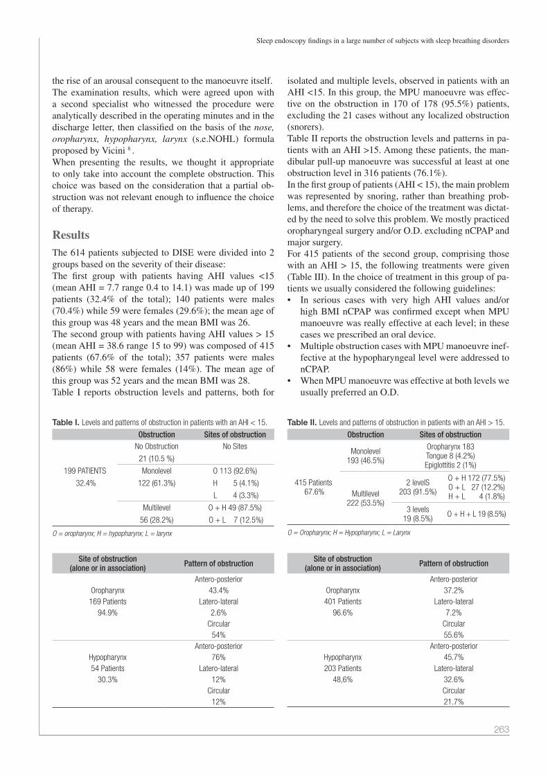
Sleep endoscopy findings in a large number of subjects with sleep breathing disorders
263
the rise of an arousal consequent to the manoeuvre itself.The examination results, which were agreed upon with a second specialist who witnessed the procedure were analytically described in the operating minutes and in the discharge letter, then classified on the basis of the nose, oropharynx, hypopharynx, larynx (s.e.NOHL) formula proposed by Vicini 8 .When presenting the results, we thought it appropriate to only take into account the complete obstruction. This choice was based on the consideration that a partial ob-struction was not relevant enough to influence the choice of therapy.
ResultsThe 614 patients subjected to DISE were divided into 2 groups based on the severity of their disease:The first group with patients having AHI values <15 (mean AHI = 7.7 range 0.4 to 14.1) was made up of 199 patients (32.4% of the total); 140 patients were males (70.4%) while 59 were females (29.6%); the mean age of this group was 48 years and the mean BMI was 26.The second group with patients having AHI values > 15 (mean AHI = 38.6 range 15 to 99) was composed of 415 patients (67.6% of the total); 357 patients were males (86%) while 58 were females (14%). The mean age of this group was 52 years and the mean BMI was 28.Table I reports obstruction levels and patterns, both for
isolated and multiple levels, observed in patients with an AHI <15. In this group, the MPU manoeuvre was effec-tive on the obstruction in 170 of 178 (95.5%) patients, excluding the 21 cases without any localized obstruction (snorers). Table II reports the obstruction levels and patterns in pa-tients with an AHI >15. Among these patients, the man-dibular pull-up manoeuvre was successful at least at one obstruction level in 316 patients (76.1%).In the first group of patients (AHI < 15), the main problem was represented by snoring, rather than breathing prob-lems, and therefore the choice of the treatment was dictat-ed by the need to solve this problem. We mostly practiced oropharyngeal surgery and/or O.D. excluding nCPAP and major surgery.For 415 patients of the second group, comprising those with an AHI > 15, the following treatments were given (Table III). In the choice of treatment in this group of pa-tients we usually considered the following guidelines:• In serious cases with very highAHI values and/or
high BMI nCPAP was confirmed except when MPU manoeuvre was really effective at each level; in these cases we prescribed an oral device.
• MultipleobstructioncaseswithMPUmanoeuvreinef-fective at the hypopharyngeal level were addressed to nCPAP.
• WhenMPUmanoeuvrewaseffectiveatbothlevelsweusually preferred an O.D.
Table I. Levels and patterns of obstruction in patients with an AHI < 15.
Obstruction Sites of obstructionNo Obstruction No Sites
21 (10.5 %)
199 PATIENTS Monolevel O 113 (92.6%)
32.4% 122 (61.3%) H 5 (4.1%)
L 4 (3.3%)
Multilevel O + H 49 (87.5%)
56 (28.2%) O + L 7 (12.5%)
O = oropharynx; H = hypopharynx; L = larynx
Site of obstruction(alone or in association) Pattern of obstruction
Antero-posteriorOropharynx 43.4%169 Patients Latero-lateral
94.9% 2.6%Circular
54%Antero-posterior
Hypopharynx 76%54 Patients Latero-lateral
30.3% 12%Circular
12%
Table II. Levels and patterns of obstruction in patients with an AHI > 15.
Obstruction Sites of obstruction
Monolevel193 (46.5%)
Oropharynx 183 Tongue 8 (4.2%)Epiglottitis 2 (1%)
415 Patients 67.6% Multilevel
222 (53.5%)
2 levelS203 (91.5%)
O + H 172 (77.5%)O + L 27 (12.2%)H + L 4 (1.8%)
3 levels19 (8.5%) O + H + L 19 (8.5%)
O = Oropharynx; H = Hypopharynx; L = Larynx
Site of obstruction(alone or in association) Pattern of obstruction
Antero-posteriorOropharynx 37.2%401 Patients Latero-lateral
96.6% 7.2%Circular55.6%
Antero-posteriorHypopharynx 45.7%203 Patients Latero-lateral
48,6% 32.6%Circular21.7%

F. Salamanca et al.
264
• The combination of oropharyngeal surgery (with orwithout tonsillectomy) and O.D. was the choice when the MPU manoeuvre was effective only at the hy-popharyngeal level
• In young patientswith a large tonsillarmass (grade3-4), we performed only tonsillectomy
• Whenreductionofsnoringnoisewasfundamentalforthe patient, we preferred oropharyngeal surgery even when the MPU manoeuvre was effective on obstruc-tion
• In the presence of oropharyngeal and laryngeal ob-struction, the treatment was surgery for both levels when nCPAP was refused.
In the choice of treatment, we had to consider also that in Italy, an oral device is not refused by National Health Care Service, while nCPAP and surgery are.
DiscussionOn the basis of our experience, a close look at BIS data and clinical observation of the sedation progress permits achieving a stable sleep state, to best obtain reliable data, without necessarily applying target controlled infusion. (TCI) 7. We believe that the s.e.NOHL formula as modi-fied by Vicini 8 can be considered a valid solution for the synthetic survey of the results, inasmuch as it contains all fundamental data. In our practice, we add information about the effectiveness of the MPU manoeuvre.We accepted, for further simplification, the hypothesis suggested by some authors to consider the hypopharyn-geal level as the lower (retro-lingual) segment of the oro-pharynx. Particular attention was given to the distinction
between primary obstruction due to epiglottis collapse, from laryngeal obstruction consequent to the fall of the tongue base, in consideration of several therapeutic im-plications.The most significant finding our research led to is that oropharyngeal obstruction widely prevails on the other ar-eas; in fact, it is present in 96% of all cases, alone (50% of cases) or in association with other obstruction areas when facing with multiple obstructions. Isolated hypopharyn-geal obstruction seems to be rare (2.2%), while specific single obstruction due to the epiglottis is exceptional. These data are only partially in accordance with those published by other authors (Table IV); the disagreements are mainly due to the fact that the cases considered in the different studies were not homogeneous, especially con-sidering severity of disease. The obstruction pattern is an important component of each single level and provides fundamental information when choosing treatment, especially when surgical. DISE has earned substantial importance since this is the only tech-nique that is able to characterize the obstruction dynam-ics; Muller’s manoeuvre has not proven to be as reliable.Campanini et al. 13, in their 2010 publication, ascertained a disagreement in 76% of cases between the Muller’s ma-noeuvre and sleep endoscopy data as far as the obstruc-tion is concerned, especially at the hypopharyngeal level; another 49% disagreement of the kind of the obstruction, and a tendency to overestimate the antero-posterior pat-tern were noticed. Moreover, this manoeuvre does not per-mit to observe possible obstruction at the laryngeal level, which occurred in 7.4% of our cases. At the oropharyngeal level, the circular pattern was present in more than half of patients (54% of subjects with mild pathology and in 55.6 % of patients with moderate to serious pathology). We must remember that, at this level, the obstruction pattern is notably influenced by the presence, especially when vo-luminous, of the palatine tonsils. In fact, a latero-lateral pattern can be observed almost exclusively in patients af-fected by grade 2-3 tonsillar hypertrophy. In addition, at both levels, but mostly at the hypopharyngeal level, when the intensity grew worse, the antero-posterior pattern fre-quency was reduced in favour of the circular pattern.At the hypopharyngeal level, if the indication was OD, the pattern was irrelevant since it was verified that MPU was
Table III. Therapies administered in patients with an AHI >15.
147 nCPAP (35.5%)109 Oral Device (O.D.) (26.4%)104 Oropharyngeal surgery (with or without tonsillectomy)+ O.D. (25%) 30 Oropharyngeal surgery (with or without tonsillectomy) (7%) 3 Tonsillectomy (0.7%) 1 Epiglottoplasty(0.35%) 4 Epiglottoplasty + O.D. (1%) 4 Epiglottoplasty + UP3 (0.35%) 12 Weight loss (2.7%) 1 TORS (0.35%)
Table IV. Comparison of sites of obstruction in literature.
No obstruction Monolevel obstruction Multilevel obstructionPalate Tongue/hypopharyngeal Total
Present study (n = 614) 23 (3.7%) 296 (48.2%) 15 (2.9%) 311 (51.1%) 278 (45.2%)Hamans study 9 (n = 70) 4 (5.5%) 23 (31.9%) 20 (27,8%) 43 (59.7%) 23 (31.9%)Hessel study 10 (n = 340) 74 (21.7%) 8 (2.4%) 82 (24.1%) 205 (60.3%)Quinn study 11 (n = 50) 35 (70%) 4 (8%) 369 (78%) 11 (22%)Pringle study 3 (n = 70) 33 (47.1%) 9 (13%) 42 (60%) 28 (40%)Carrasco study 11 (n = 51) 33.20% 7.80% 41% 59%
Sleep endoscopy findings in a large number of subjects with sleep breathing disorders
265
effective in all types of obstruction. Moreover, the MPU manoeuvre was particularly effective to solve obstruction in patients with an AHI index < 15 (95.5%).As already described by Battagel et al. 14, the MPU ma-noeuvre was often effective even in those obstructions at the oropharyngeal level, not only in those at the tongue base. This was probably due to an indirect traction mecha-nism on front pillars, in the absence of a true anatomical obstruction such as that represented by a large tonsillar mass. Even in those cases in which the manoeuvre was effective on “simple” snorers (28.5%), the mechanism probably was the same.In addition, we observed that in the second group of seriously-affected patients the effectiveness of the MPU manoeuvre decreased, although in many cases it could be considered successful, at least at the hypopharyngeal level (76%). This result is predictable if we take into considera-tion that the mandibular pull-up manoeuvre is effective in enlarging the antero-posterior diameter of the upper air-ways. It should be kept in mind that, even if rarely, MPU can worsen breathing because of the appearance of an ob-struction at the epiglottal level.
ConclusionsWe confirm that DISE is a safe procedure, easily prac-ticable, and is valid and reliable, as already reported in other publications. Although we are aware that the condi-tions of the examination do not fully correspond to those that occur during physiological sleep, we believe that it is presently the most rational method to observe the phe-nomenon of snoring and the obstruction in its dynamism while sleeping. Though this procedure is well accepted by patients who easily understand its usefulness, we feel that this examination, when dealing with simple snoring, can be avoided, at least initially. In fact, we ascertained that in all patients the noise of snoring as such, in that it disturbs his/her partner, is generated at oropharyngeal level (soft palate, tonsils, lateral pharyngeal walls). Other kinds of noises, possibly originating by other structures like hypopharyngeal walls or epiglottis, when present, only achieve modest levels of sound poignancy and do not seem strong enough to disturb his/her partner.Certainly there is a clear indication for DISE when ex-pecting significant surgical intervention and in the analy-ses of therapeutic failures. The information that can be obtained during sleep endoscopy from the MPU manoeu-vre is invaluable for its ability to predict the efficacy of treatment, and only through this manoeuvre is it possible to identify those individuals for whom the application of an OD might be ineffective or worsen the condition.One limit of the method is represented by the difficulty to gain a valid systematization of the parameters observed. To overcome the inevitable subjectivity of the examiner, it would be desirable to command either numeric values
or graphics to better specify the sites of obstruction and especially its pattern.The identification of such elements would undoubtedly be useful in understanding this diagnostic technique and, fur-thermore, would produce a better standardization of the data, which is fundamental when evaluating therapeutic outcomes.The results of our study confirm that DISE is a basic clini-cal element and can be an important test for the choice of the treatment; it is likely to become increasingly relevant over time as OSAS surgery continues to evolve. Our in-tention at present is to verify the appropriateness of our therapeutic indication on the basis of the results of the treatment.
References1 Sher AE, Thorpy MJ, Shprintzen RJ, et al. Predictive val-
ue of Muller maneuver in selection of patients for uvulo-palatopharyngoplaty. Laryngoscope 1985;95:1483-7.
2 Croft CB, Pringle M. Sleep nasendoscopy: a technique of assessment in snoring and obstructive sleep apnea. Clin Oto-laryngol Allied Sci 1991;16:504-9.
3 Pringle MB, Croft CB. A grading system for patients with obstructive sleep apnea: based on sleep nasendoscopy. Clin Otolaryngol Allied Sci 1993;18:480-4.
4 Guerin P, Lasenecal L. Pharyngeal and bronchial endoscop-ic study in the diagnosis and treatment of sleep apnea syn-drome. Presse Med 1992;11:249-52.
5 Berry S, Roblin G, Williams A, et al. Validity of sleep nasen-doscopy in the investigation of sleep related breathing disor-ders. Laryngoscope 2005;115:538-40.
6 Rabelo FAW, Braga A, Küpper DS, et al. Propofol-induced sleep: polysomnographic evaluation of patients with ob-structive sleep apnea and controls. Otolaryngol Head Neck Surg 2010;142:218-24.
7 Roblin G, Williams AR, Whittet H. Target-controlled infu-sion in sleep endoscopy. Laryngoscope 2001;111:175-6.
8 Vicini C, Editor. Textbook of Snoring and Sleep Apnea Sur-gery. Lucca: Eureka 2007.
9 Hamans E, Meeus O, Boudewyns A, et al. Outcome of sleep endoscopy in obstructive sleep apnea: the Antwerp experi-ence. B-ENT 2010;6:97-103.
10 Hessel NS, de Vries N. Role of uvulopalatopharyngoplasty after diagnostic workup with polysomnography and sleep en-doscopy: a report of 136 snoring patients. Eur Arch Otoorhi-nolaryngol 2003;260:91-5.
11 Quinn SJ, Daly N, Ellis PDM. Observation of the mecha-nism of snoring using sleep nasendoscopy. Clin Otolaryngol 1995;20:360-4.
12 Carrasco llatas M, Dalmau Galofre J, Lopez Martinez R, et al. Our findings in the sleep endoscopy exams. Acta Otorino-laringol Esp 2005;56:17-21.
13 Campanini A, Canzi P, De Vito A, et al. Awake versus sleep endoscopy: personal experience in 250 OSAHS patients. Acta Otorhinolaryngol Ital 2010;30:73-7.
14 Battagel JM, Johal A, Kotecha BT. Sleep nasendoscopy as a
F. Salamanca et al.
266
predictor of treatment success in snorers using mandibular advancement splints. Journ Laryngol Otol 2005;119:106-12.
15 Passali D, Caruso G, Arigliano LC, et al. Database appli-cation for patients with obstructive sleep apnoea syndrome. Acta Otorhinolaryngol Ital 2012;32:252-5.
16 Piumetto E, Sammartano AM, Meinardi G, et al. Diagnostic and therapeutic iter in paediatric OSAS: personal experi-ence. Acta Otorhinolaryngol Ital 2011;31:149-53.
17 Mantovani M, Minetti A, Torretta S, et al. The velo-uvulo-pharyngeal lift or “roman blinds” technique for treatment of snoring: a preliminary report. Acta Otorhinolaryngol Ital 2012;32:48-53.
18 Passali FM, Bellussi L, Mazzone S, et al. Predictive role of nasal functionality tests in the evaluation of patients before nocturnal polysomnographic recording. Acta Otorhinolaryn-gol Ital 2011;31:103-8.
Address for correspondence: Fabrizio Costantini, Casa di Cura S. Pio X, via F. Nava 31, 20159 Milan, Italy. Tel. +39 02 69516442. E-mail: [email protected]
Received: December 3, 2012 - Accepted: March 6, 2013
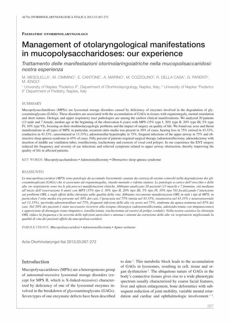
267
ACTA oTorhinolAryngologiCA iTAliCA 2013;33:267-272
Paediatric otorhinolaryngology
Management of otolaryngological manifestations in mucopolysaccharidoses: our experienceTrattamento delle manifestazioni otorinolaringoiatriche nella mucopolisaccaridosi: nostra esperienza
M. Mesolella1, M. CiMMino1, e. Cantone1, a. Marino1, M. Cozzolino2, r. Della Casa1, G. Parenti1, M. ienGo1
1 University of naples “Federico ii”, Department of otorhinolayngology, naples, italy; 2 University of naples “Federico ii” Department of Pediatry, naples, italy
SummAry
mucopolysaccharidoses (mPSs) are lysosomal storage disorders caused by deficiency of enzymes involved in the degradation of gly-cosaminoglycans (gAgs). These disorders are associated with the accumulation of gAgs in tissues with organomegaly, mental retardation and short stature. otologic and upper respiratory tract pathologies are among the earliest clinical manifestations. We analyzed 20 patients (13 male and 7 female, median age at the beginning of the observation 6 years) with mPS (35% type i, 30% type ii, 20% type iii, 5% type iV, 10% type Vi), focusing on their otorhinolaryngologic problems and the impact of surgery on quality of life. We found ear, nose and throat manifestations in all types of mPS; in particular, recurrent otitis media was present in 30% of cases, hearing loss in 75% (mixed in 43.33%, conductive in 43.33%, sensorineural in 13.33%), adenotonsillar hypertrophy in 75%, frequent infections of the upper airway in 75% and ob-structive sleep apnoea syndrome in 45% of cases. Fifty percent of patients required surgical therapy (adenotonsillectomy, adenoidectomy with insertion of middle ear ventilation tubes, tonsillectomy, tracheotomy and exeresis of vocal cord polyps). in our experience the EnT surgery reduced the frequency and severity of ear infections and relieved symptoms related to upper airway obstruction, thereby improving the quality of life in affected patients.
KEy WordS: Mucopolysaccharidoses • Adenotonsillectomy • Obstructive sleep apnoea syndrome
riASSunTo
Le mucopolisaccaridosi (MPS) sono patologie da accumulo lisosomiale causate da carenza di enzimi coinvolti nella degradazione dei gli-cosaminoglicani (GAGs) che si associano ad organomegalia, ritardo mentale e ridotta statura. Le patologie a carico dell’orecchio e delle alte vie respiratorie sono tra le più precoci manifestazioni cliniche. Abbiamo analizzato 20 pazienti (13 maschi e 7 femmine, età mediana all’inizio dell’osservazione 6 anni) con MPS (35% tipo I, 30% tipo II, 20% tipo III, 5% tipo IV, 10% tipo VI) focalizzando l’attenzione sui problemi ORL e sugli effetti della chirurgia sulla qualità della vita. Abbiamo riscontrato manifestazioni ORL in tutti i tipi di MPS; in particolare l’otite media era presente nel 30% dei casi, l’ipoacusia nel 75% (mista nel 43.33%, trasmissiva nel 43.33% e neurosensoriale nel 13.33%), ipertrofia adenotonsillare nel 75%, frequenti infezioni delle alte vie aeree nel 75%, sindrome da apnea notturna nel 45% dei casi. Nel 50% dei pazienti è stato necessario ricorrere alla terapia chirurgica (adenotonsillectomia, adenoidectomia con timpanocentesi e apposizione di drenaggio trans-timpanico, tonsillectomia, tracheotomia ed exeresi di polipo cordale). Nella nostra casistica la chirurgia ORL riduce la frequenza e la severità delle infezioni auricolari e attenua i sintomi da ostruzione delle alte vie respiratorie migliorando la qualità di vita dei pazienti affetti da mucopolisaccaridosi.
PArolE ChiAVE: Mucopolisaccaridosi • Adenotonsillectomia • Apnee notturne
Acta Otorhinolaryngol Ital 2013;33:267-272
Introduction
mucopolysaccaridoses (mPSs) are a heterogeneous group of autosomal-recessive lysosomal storage disorders (ex-cept for mPS ii, which is X-linked-recessive) character-ized by deficiency of one of the lysosomal enzymes in-volved in the breakdown of glycosaminoglycans (gAgs). Seven types of one enzymatic defects have been described
to date 1. This metabolic block leads to the accumulation of gAgs in lysosomes, resulting in cell, tissue and or-gan dysfunction 2. The ubiquitous nature of gAgs in the body’s connective tissues gives rise to a wide phenotypic spectrum usually characterized by coarse facial features, liver and spleen enlargement, bone deformities with sub-sequent reduction of joint mobility, variable mental retar-dation and cardiac and ophthalmologic involvement 1 3.

M. Mesolella et al.
268
Ear, nose and throat (EnT) disorders are extremely fre-quent, mostly in mPS i, ii and Vi, and are often the ear-liest clinical manifestations of these diseases 4. indeed, mPS patients display an increased risk of otitis media with effusion (omE) due to the pathologic deposition of gAgs in the post-nasal space, eustachian tubes and middle ear 5. Sensorineural hearing loss, whose aetiology remains unclear, is believed to result from infiltration of the cochlear duct, stria vascularis and cochlear nerve af-ferents 4. nevertheless, in most mPS Vi patients deficits are conductive in nature 6. other common EnT disor-ders are: adenotonsillar hypertrophy, almost universal in mPS 5, chronic recurrent rhinitis and persistent copious nasal discharge 7. These conditions, in addition to nasal dysmorphism, mandibular abnormalities, tracheomalacia, thickened vocal cords, macroglossia and redundant tissue in the upper airway (uA) can contribute to uA complica-tions and to obstructive sleep apnoea (oSA) 6-8.Although patients with mPS may improve airway ob-struction with more conservative treatment approaches in-cluding positive airway pressure devices (CPAP/BiPAP), management often requires early adenotonsillectomy and in extreme cases tracheostomy to ensure a patent airway in the short- or long-term 5. Before the advent of haemat-opoietic stem cell transplantation (hSCT) and especially enzyme replacement therapy (ErT), the main focus of treatment of mPS i, ii, and Vi was prevention and man-agement of complications. recently, much progress has been achieved in the treatment of mPS, and hSCT has been used in patients with mPS to correct the enzyme deficiency. Although many studies reveal that hSCT can change the natural course of the disease, increasing life expectancy and improving many systemic abnormali-ties 9 10, it is a high-risk procedure with high morbidity/mortality. moreover, its indication depends on the type of mPS, patient’s clinical picture, age and presence of neurological impairment 11-13. ErT, currently considered an efficient therapeutic method, is based on the periodic replacement of the defective enzyme, leading to higher gAg degradation in tissues and organs and promoting significant improvement in some clinical features. how-ever, the influence of ErT on pathological manifestations is still not well understood, and long-term data on its ef-ficacy is not yet available 14 15.We designed an observational study in order to define the incidence of EnT problems in patients with mPSs and to establish the impact of surgical treatment. We also ob-served the role of surgery in improving the clinical pheno-type and the quality of life of patients affected by mPSs.
Materials and methodsThe present study includes 20 patients with mPS (7 [35%] mPS i, 6 [30%] mPS ii, 4 [20%] mPS iii, 1 [5%] mPS iV and 2 [10%] mPS Vi), 13 male and 7 female (median age
at the beginning of the observation 6 years, ranging from 1 to 16), observed at the department of Paediatric medi-cine of Federico ii university in naples from June 1999 to June 2009. The median age at diagnosis of mPS was 3 years and the median age of presentation to an otolaryn-gologist was 12 months. The average length of follow-up was 8.4 years.We performed flexible fibre otoscopy, rhinopharyngola-ryngoscopy, tympamograms, audiograms in patients able to cooperate (n = 15) in addition to polysomnography.The following was considered:1 The number of OME and upper respiratory tract infec-
tions (URTIs) episodes per year.2 Adenoid hypertrophy (grade 1: adenoid tissue occupies
only the upper segment in the rhinopharyngeal cavity; grade 2: adenoid tissue is confined to the upper half of the rhinopharyngeal cavity; grade 3: adenoid tissue is extended over the rhinopharynx with obstruction of choanal openings and partial closure of tube ostium; grade 4: tube ostium and lower choanal border could not be observed) 16.
3 Tonsillar hypertrophy (0: tonsils entirely within the tonsillar fossa; 1: tonsils occupying less than 25% of the lateral dimension of the oropharynx as measured between the anterior tonsillar pillars; 2: tonsils occu-pying less than 50% of the lateral dimension of the oropharynx; 3: tonsils occupying less than 75% of the lateral dimension of the oropharynx; 4: tonsils occupy-ing 75% or more of the lateral dimension of the oro-pharynx).
4. Audiometric test.5. Type of tympanograms (Type A: static compliance
[SA] ≥ 0.2 cm3, tympanic peak pressure [TPP] -200 → +100 mm h
2o, type B: SA < 0.2 cm3, type C: SA ≥ 0.2
cm3, TPP ≤ 200 mm h2o).
6. OSAS (number of obstructive apnoea and hypopnoea events per hour of sleep: apnoea-hypopnoea index Ahi and oxygen saturation: Sat % o
2).
Eight (40%) of our patients (3 mPS i, 4 mPS ii and 1 mPS Vi) were treated with ErT; of these, 4 patients (1 mPS i, 2 mPS ii and 1 mPS Vi) underwent surgery, in 2 cases before starting enzyme therapy and in the remaining 2 cases one year after initiating therapy.in 10 patients (50%) surgical therapy was required (Ta-ble i): adenotonsillectomy in 5 patients (25%), adenoid-ectomy in 3 patients (15%), tonsillectomy in 2 patients (10%), insertion of middle ear ventilation tubes in 3 pa-tients (15%), tracheotomy in 1 patient (5%) and exeresis of vocal cord polyps in 1 patient (5%). Flexible fibre-optic laryngoscopy was performed before surgery to familiar-ize the anaesthesiologist with the anatomy that would be encountered.We finally considered the role of EnT surgery on improve-ment in quality of life, estimated through parental opinion and clinical manifestations. in particular, the severity of

Otolaryngological manifestations in mucopolysaccharidoses
269
respiratory tract and/or otological infections was evalu-ated according to an infection score system considering the type of infection, presence of systemic symptoms, im-pairment of daily activities, improvement of instrumental date, need for therapy and/or hospital admission and dura-tion of disease (Table ii). A visual analogue scale (VAS) is a psychometric response scale used for subjective characteristics or attitudes that cannot be directly measured. A VAS is usually a horizon-tal line, 10 cm in length, anchored by picture descriptors. Patients indicate the line point that they feel best repre-sents perception of their current state (pain, ability to do daily activity) before and after surgery.
Results18 of 20 patients (90%) showed at least one EnT mani-festations (Table iii). in particular, 6 patients (30%) had a history of chronic and recurrent omE ≥ 5 episodes in a year. hearing loss was demonstrated by audiogram and ABr in 15/20 (75%) patients: in 6 (43.33%) patients mixed hypoacusia was present, conductive hypoacusia in 6 (43.33%) and sensorineural hypoacusia in 3 (13.33%) cases. We found type A tympanograms in 6 patients, type B in 10 and type C in 4 patients. Adenoid hypertrophy
Table I. Surgical procedures performed.
Patients 1 5 6 8 11 12 14 16 19 20
Adenoidectomy X X X
Tonsillectomy X X
Adenotonsillectomy X X X X X
Insertion of middle ear ventilation tubes X X X
Tracheotomy X
Exeresis of polyps of the vocal cords X
Table II. Total score ≤ 5: mild respiratory tract and/or otological infection; score 6-11: moderate respiratory tract and/or otological infection; score ≥ 12: severe respiratory tract and/or otological infection.
Score
Type of infectionRhinitis 0
Rhinitis + otitis and/or tonsillitis 1
Pneumonia 2
Systemic symptoms
Absent 0
Slight fever and/or some aches 1
Definite elevation of temperature 2
Daily activity
Not limited 0
Some limitation 1
Severely incapacitated 2
Therapy
Local 0
Systemic (oral administration) 1
Systemic (intravenous administration) 2
Hospitalization
No 0
Single entry followed by home therapy 1
Admission 2
Resolution
< 7 days 0
7-10 days 1
> 10 days 2
Fig. 1. Comparison between otorhinolaryngological manifestations before and after surgery.

M. Mesolella et al.
270
(degree ≥ 2) was present in 15 of 20 (75%) cases; ton-sillar hypertrophy (degree ≥ 1) was present in 17 of the 20 patients; uA obstruction and oSA was present in 8 cases. 15 patients (75%) had a history of urTis, such as rhinosinusitis, pharyngotonsillitis and laryngitis. in 9 pa-tients (45%), oSA was documented by positive results of polysomnnography that demonstrated apnoea-hypopnea indexes (Ahi) ranging from 18 to 31, and an oxygen satu-ration ranging from 74% to 85%.Ten patients underwent surgical treatment, and none re-quired prolonged intubation. Figure 1 shows the com-parison between EnT manifestations before and after surgery.At least 30% (3 patients) of the patients obtained an improvement in quality of life. indeed, VAS scores af-ter surgery compared to VAS score before surgery sig-nificantly improved (median value 7.0 ± 1.5 vs. 3.6 ± 1.6). The severity of respiratory and EnT infections, measured with the infection score system also showed improvement (median infection score before surgery 13.7 ± 4.6 vs. 3.9 ± 2.4 after surgery). one patient (10%) still presented oSA after adenotonsillectomy because of macroglossia and dysmorphism. All patients are still alive at the time of writing.
DiscussionmPSs are lysosomal storage disorders with deficiency of enzymes involved in the degradation of gAgs. The structures of the head and neck are nearly always in-volved in mPSs, often at an early age, and as a result, otolaryngologists are commonly the first clinicians to whom these individuals present. our study confirms the importance of EnT management in the multidisciplinary approach to these patients. Because of the high incidence of ear disorders, and their effects on language develop-ment and quality of life, routine otologic and audiologic evaluation must be obtained 17. hearing loss affects al-most all mPS patients, which is characterized by both conductive and sensorineural involvement. in fact, in the present study, hearing loss was present in 75% of cases, in agreement with literature data, in which the percent-age varies from 59.7% to 89% 18. While it is reported that conductive hearing loss can be improved after adenoid-ectomy and insertion of middle ear ventilation tubes 19, amplification is typically required to overcome what is generally permanent sensorineural loss. As mentioned above, our data confirm these observations as about 60% of cases experienced improvement in hearing after surgi-cal treatment.While otologic manifestations have an effect on quality of life, upper-airway obstruction contributes more to mor-bidity and mortality. The accumulation of mucopolysac-charides can cause alterations of normal airway function,
so prompt diagnosis is crucial. Changes in soft tissues in-cluding tonsils, adenoids, tongue and lingual tonsils are responsible for most respiratory problems. As the disease progresses, pharyngomalacia and tracheomalacia may develop and become severe, leading to significant airway obstruction 20.upper airway obstruction may range from varying degrees of oSA to life-threatening airway emergencies. however, airway evaluation is very difficult, typically non-uniform among different providers and varies from case to case 21. in the present study, we found upper airway obstruction in 75% of cases while literature data describes percentages varying from 38% 22 to 48% 18 to 92% 23. Currently, treat-ment of airway obstruction is controversial, but the accu-mulation of gAgs in the adenoids and tonsils, with result-ing hypertrophy, makes these structures frequent targets of surgical intervention. Therefore, in more severe forms, such as purulent, recurrent and chronic cases, adenoidec-tomy should be performed without delay 16. oSA initially can be helped by tonsillectomy and adenoidectomy, but many patients require nocturnal oxygen treatment later 24 or, in more severe cases, tracheotomy. in the light of the elevated anaesthetic risk of this population due to copi-ous secretions, temporomandibular joint arthritis, difficult or failed intubations, macroglossia, abnormal laryngeal anatomy and subglottic narrowing 15, in our centre, it is common practice to perform and record a bronchoscopy examination with a flexible fibre-optic bronchoscope be-fore surgery 5 6 to evaluate the extent and severity of air-way infiltration.Among 20 mPS patients, 10 (50%) underwent surgery (Table i): adenotonsillectomy (5 patients, 25%), ade-noidectomy (3 patients, 15%), tonsillectomy (2 patients, 10%), insertion of middle ear ventilation tubes (3 patients, 15%), tracheotomy (1 patient, 5%) and exeresis of vocal cord polyps (1 patient, 5%).oSA evaluation should begin with history and physical examination, but the degree of pre and post-operative ob-struction should be studied with polysomnography and rhino-oro-laryngoscopy 23 25. For these reasons, we per-formed a pre- and post-operative sleep study that dem-onstrated improvement of Ahi ranging from 7 to 15 after surgery, with oxygen saturation ranging from 81% to 87% compared to preoperative results (Ahi ranging from 18 to 31, and oxygen saturation ranging from 74% to 85%). one patient (10%), in contrast, still presented oSA after adenotonsillectomy because of his macroglossia and dys-morphism.The study was also designed to examine the quality of life of patients affected by mPSs using a VAS score, measured before and after surgery, which demonstrated a significant improvement after treatment from score 5 (60%) and score 4 (40%) to score 3 (75%) and score 2 (25%).

Otolaryngological manifestations in mucopolysaccharidoses
271
Conclusionsin conclusion, because of the high incidence of EnT man-ifestations in mPS, otolaryngologists have a crucial role in the multidisciplinary approach to diagnosis and man-agement of many subjects with this disorder. moreover, despite the fact that ErT has made an important contribu-tion to improving the quality of life of these patients, EnT surgery remains a fundamental therapeutic procedure for reducing the frequency and severity of ear infections and for relieving the symptoms of upper airway obstruction, even if these interventions are not definitive.
References1 neufeld EF, muenzer J. The mucopolysaccharidoses. in:
Scriver Cr, Beaudet Al, Sly WS, et al., editors. The meta-bolic and molecular bases of inherited disease. 8th ed. new york: mcgraw-hill; 2001. pp. 3421-52.
2 muenzer J. The mucopolysaccharidoses: a heterogeneous group of disorders with variable pediatric presentations. J Pediatr 2004;144(Suppl 5):27-34.
3 Wenger dA. Insights into the diagnosis and treatment of lysosomal storage disorders. Arch neurol 2003;60:322-8.
4 ruckenstein mJ, macdonald rE, Clarke JTr, et al. The management of otolaryngological problems in the muco-polysaccharidoses: a retrospective review. J otolaryngol 1991;20:177-83.
5 Simmons mA, Bruce iA, Penney S, et al. Otorhinolaryngo-logical manifestations of the mucopolysaccharidoses. int J Pediatr otorhinolaryngol 2005;69:589-95.
6 giugliani r, harmatz P, Wraith JE. Management guidelines for mucopolysaccharidosis VI. Pediatrics 2007;120:405-18.
7 Wraith JE, Scarpa m, Beck m, et al. Mucopolysaccharido-sis type II (Hunter syndrome): a clinical review and recom-mendations for treatment in the era of enzyme replacement therapy. Eur J Pediatr 2008;167:267-77.
8 nashed A, Al-Saleh S, gibbons J, et al. Sleep-related breath-ing in children with mucopolysaccharidosis. J inherit metab dis 2009;32:544-50.
9 Vellodi A, young EP, Cooper A, et al. Bone marrow trans-plantation for mucopolysaccharidosis type I: experience of two British centres. Arch dis Child 1997;76:92-9.
10 Wraith JE, Scarpa m, Beck m, et al. Mucopolysaccharido-sis type II (Hunter syndrome): a clinical review and recom-mendations for treatment in the era of enzyme replacement therapy. Eur J Pediatr 2008;167:267-77.
11 mckinnis EJ, Sulzbacher S, rutledge JC, et al. Bone marrow transplantation in Hunter syndrome. J Pediatr 1996;129:145-8.
12 Aldenhoven m, Boelens JJ, de Konong TJ. The clinical out-come of Hurler syndrome after stem cell transplantation. Biol Blood marrow Transplant 2008;14:485-98.
13 muenzer J, Beck m, Eng Cm, et al. Multidisciplinary man-agement of Hunter syndrome. Pediatrics 2009;124:1228-39.
14 Viana gm, de lima no, Cavaleiro r, et al. Mucopolysac-charidoses in northern Brazil: Targeted mutation screen-ing and urinary glycosaminoglycan excretion in patients undergoing enzyme replacement therapy. genet mol Biol 2011;34:410-5.
15 Wold Sm, derkay CS, darrow dh, et al. Role of the pedi-atric otolaryngologist in diagnosis and management of chil-
Table III. Otorhinolaryngological manifestations.
Pt MPS Otitis mediaNo. of episodes
per year
Hypoacusia Tympanograms Degree adenoid hypertrophy
Degree tonsillar hypertrophy
(URTIs)No. of episodes
per year
OSASAHI
1 I 7 Conductive B 4 2 6 312 I 2 Mixed C 2 1 53 I 2 Mixed B 2 1 54 I 2 Sensorineural A 2 1 55 I H/S 6 Conductive B 3 2 6 226 I S 3 Mixed B 3 2 67 I 0 Sensorineural A 2 0 28 II 3 Mixed C 1 3 5 259 II 1 - A 2 0 310 II 2 Sensorineural A 2 2 511 II 4 Mixed B 3 3 6 2812 II 6 Conductive B 4 4 7 2913 II 2 - C 1 1 514 III B 7 Conductive B 3 3 6 1815 III B 0 - A 1 2 2 1916 III B 7 Conductive B 4 3 7 2617 III B 1 - A 1 0 218 IV A 1 - C 2 1 319 VI 3 Mixed B 1 3 620 VI 8 Conductive B 3 3 7 21

M. Mesolella et al.
272
dren with mucopolysaccharidoses. int J Pediatr otorhinola-ryngol 2010;74:27-31.
16 Cassano P, gelardi m, Cassano m, et al. Adeniod tissue rhi-nopharyngeal obstruction grading based on fiberendoscopic findings: a novel approach to therapeutic management. int J Pediatr otorhinolaryngol 2003;67:1303-9.
17 motamed m, Thorne S, narula A. Treatment of otitis media with effusion in children with mucopolysaccharidoses. int J Pediatr otorhinolaryngol 2000;53:121-4.
18 Schwartz iVd, ribeiro md, mota Jg, et al. A clinical study of 77 patients with mucopolisaccharidosis type II. Acta Pae-diatrica 2007;96:63-70.
19 Peck JE. Hearing loss in Hunter’s syndrome - mucopolysac-charidosis II. Ear and hearing 1984;5:243-6.
20 Pelley CJ, Kwo J, hess dr. Tracheomalacia in an adult with respiratory failure and Morquio syndrome. respir Care 2007;52:278-82.
21 yeung Ah, Cowan mJ, horn B, et al. Airway management in children with mucopolysaccharidoses. Arch otolaryngol head neck Surg 2009;135:73-9.
22 Bredenkamp JK, Smith mE, dudley JP, et al. Otolaryngo-logic manifestations of the mucopolysaccharidoses. Ann otol rhinol laryngol 1992;101:472-8.
23 leighton SE, Papsin B, Vellodi A, et al. Disordered breath-ing during sleep in patients with mucopolysaccharidoses. int J Pediatr otorhinolaryngol 2001;58:127-38.
24 Wraith JE. The mucopolysaccharidoses: a clinical review and guide to management. Arch dis Child 1995;72:263-7.
25 Santamaria F, Andreucci mV, Parenti g, et al. Upper airway obstructive disease in mucopolysaccharidoses: polysomnog-raphy, computed tomography and nasal endoscopy findings. J inherit metab dis 2007;30:743-9.
received: July 25, 2011 - Accepted: march 10, 2012
Address for correspondence: massimo mesolella, via g. Filangieri 72, 80121 naples, italy. Tel. +39 081 404545; +39 081 7463582. Fax +39 081 7463582. E-mail: [email protected]

273
ACTA oTorhinolAryngologiCA iTAliCA 2013;33:273-281
Clinical techniques and technology
Assessment of skills using a virtual reality temporal bone surgery simulatorValutazione delle competenze nella chirurgia dell’osso temporale con un simulatore della realtà virtuale
R. Linke1, A. LeichtLe2, F. Sheikh3, c. Schmidt4, h. FRenzeL1, h. GRAeFe5, B. WoLLenBeRG1, J.e. meyeR5
1 University of Luebeck, department of oto-Rhino-Laryngology and Facial Plastic Surgery, Luebeck, Germany; 2 clinics of the city of Bielefeld, department of oto-Rhino-Laryngology, Bielefeld, Germany; 3 University of california-San diego, department of medicine (cardiology division), School of medicine, BSB 5042, La Jolla, california, USA; 4 clinics of the city of koeln, Germany; 5 clinics St. Georg hamburg, department of oto-Rhino-Laryngology and Facial Plastic Surgery, hamburg, Germany
SummAry
Surgery on the temporal bone is technically challenging due to its complex anatomy. Precise anatomical dissection of the human temporal bone is essential and is fundamental for middle ear surgery. We assessed the possible application of a virtual reality temporal bone surgery simulator to the education of ear surgeons. Seventeen EnT physicians with different levels of surgical training and 20 medical students performed an antrotomy with a computer-based virtual temporal bone surgery simulator. The ease, accuracy and timing of the simulated temporal bone surgery were assessed using the automatic assessment software provided by the simulator device and additionally with a modified Final Product Analysis Scale. Trained EnT surgeons, physicians without temporal bone surgical training and medical students were all able to perform the antrotomy. however, the highly trained EnT surgeons were able to complete the surgery in approximately half the time, with better handling and accuracy as assessed by the significant reduction in injury to important middle ear structures. Trained EnT surgeons achieved significantly higher scores using both dissection analysis methods. Surprisingly, there were no significant differ-ences in the results between medical students and physicians without experience in ear surgery. The virtual temporal bone training system can stratify users of known levels of experience. This system can be used not only to improve the surgical skills of trained EnT surgeons for more successful and injury-free surgeries, but also to train inexperienced physicians/medical students in developing their surgical skills for the ear.
KEy WordS: Virtual reality operation simulator • Temporal bone laboratory • Ear surgery • Skills lab
riASSunTo
La chirurgia dell’osso temporale è difficile dal punto di vista tecnico a causa della sua complessa anatomia. La precisa dissezione ana-tomica è però essenziale e fondamentale per la chirurgia dell’orecchio medio. Nel nostro studio abbiamo valutato il possibile utilizzo di un simulatore virtuale nella chirurgia dell’osso temporale per la formazione di chirurghi in questo campo. Diciassette medici ORL con diversi livelli di formazione chirurgica e venti studenti di medicina hanno eseguito una antrotomia con un simulatore virtuale di chirurgia temporale ossea collegato ad un PC. La leggerezza, l’accuratezza e il tempo impiegato per l’intervento simulato all’osso temporale sono stati valutati utilizzando sia un software automatico di valutazione collegato al dispositivo simulatore sia una scala modificata per l’analisi finale del prodotto. I chirurghi ORL esperti, i medici senza esperienza chirurgica dell’osso temporale ma anche gli studenti di medicina sono stati tutti in grado di eseguire l’antrotomia. Tuttavia i chirurghi ORL altamente qualificati erano in grado di completare l’intervento in circa la metà del tempo, con una migliore maneggevolezza del trapano e una maggiore precisione, come dimostrato dalla significativa riduzione di lesioni a importanti strutture dell’orecchio medio. I chirurghi ORL con maggiore esperienza hanno ottenuto punteggi signifi-cativamente più alti con entrambi i metodi di analisi. Sorprendentemente non sono state trovate differenze significative nei risultati ottenuti dagli studenti di medicina e dai medici senza esperienza nella chirurgia dell’orecchio. L’utilizzo del sistema virtuale per interventi all’osso temporale è in grado di stratificare gli utenti in base ai livelli noti di esperienza. Questo sistema può essere utilizzato non solo per miglio-rare le competenze chirurgiche di operatori esperti per interventi di maggior successo e senza lesioni, ma anche per aiutare i medici senza esperienza e gli studenti di medicina a sviluppare le loro competenze chirurgiche all’orecchio.
PArolE ChiAvE: Simulatore operativo di realtà virtuale • Laboratorio dell’osso temporale • Chirurgia dell’orecchio • Competenze di laboratorio
Acta Otorhinolaryngol Ital 2013;33:273-281
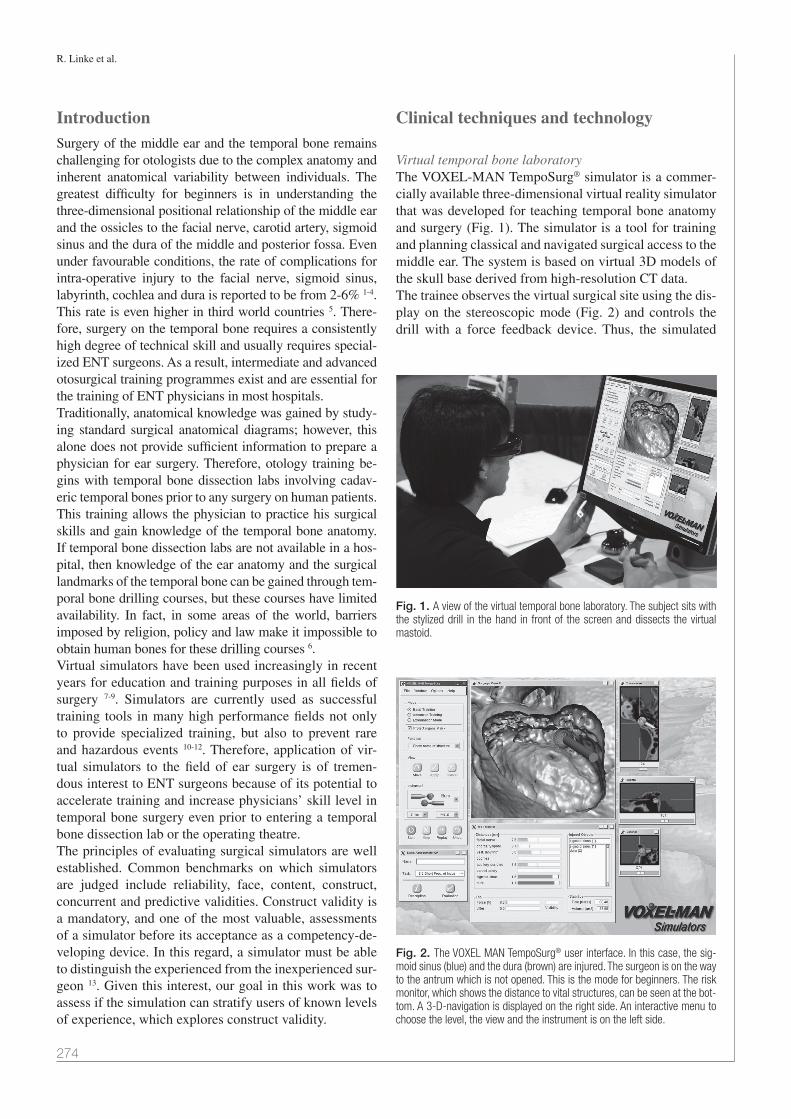
R. Linke et al.
274
IntroductionSurgery of the middle ear and the temporal bone remains challenging for otologists due to the complex anatomy and inherent anatomical variability between individuals. The greatest difficulty for beginners is in understanding the three-dimensional positional relationship of the middle ear and the ossicles to the facial nerve, carotid artery, sigmoid sinus and the dura of the middle and posterior fossa. Even under favourable conditions, the rate of complications for intra-operative injury to the facial nerve, sigmoid sinus, labyrinth, cochlea and dura is reported to be from 2-6% 1-4. This rate is even higher in third world countries 5. There-fore, surgery on the temporal bone requires a consistently high degree of technical skill and usually requires special-ized ENT surgeons. As a result, intermediate and advanced otosurgical training programmes exist and are essential for the training of ENT physicians in most hospitals. Traditionally, anatomical knowledge was gained by study-ing standard surgical anatomical diagrams; however, this alone does not provide sufficient information to prepare a physician for ear surgery. Therefore, otology training be-gins with temporal bone dissection labs involving cadav-eric temporal bones prior to any surgery on human patients. This training allows the physician to practice his surgical skills and gain knowledge of the temporal bone anatomy. If temporal bone dissection labs are not available in a hos-pital, then knowledge of the ear anatomy and the surgical landmarks of the temporal bone can be gained through tem-poral bone drilling courses, but these courses have limited availability. In fact, in some areas of the world, barriers imposed by religion, policy and law make it impossible to obtain human bones for these drilling courses 6.Virtual simulators have been used increasingly in recent years for education and training purposes in all fields of surgery 7-9. Simulators are currently used as successful training tools in many high performance fields not only to provide specialized training, but also to prevent rare and hazardous events 10-12. Therefore, application of vir-tual simulators to the field of ear surgery is of tremen-dous interest to ENT surgeons because of its potential to accelerate training and increase physicians’ skill level in temporal bone surgery even prior to entering a temporal bone dissection lab or the operating theatre. The principles of evaluating surgical simulators are well established. Common benchmarks on which simulators are judged include reliability, face, content, construct, concurrent and predictive validities. Construct validity is a mandatory, and one of the most valuable, assessments of a simulator before its acceptance as a competency-de-veloping device. In this regard, a simulator must be able to distinguish the experienced from the inexperienced sur-geon 13. Given this interest, our goal in this work was to assess if the simulation can stratify users of known levels of experience, which explores construct validity.
Clinical techniques and technology
Virtual temporal bone laboratoryThe VOXEL-MAN TempoSurg® simulator is a commer-cially available three-dimensional virtual reality simulator that was developed for teaching temporal bone anatomy and surgery (Fig. 1). The simulator is a tool for training and planning classical and navigated surgical access to the middle ear. The system is based on virtual 3D models of the skull base derived from high-resolution CT data.The trainee observes the virtual surgical site using the dis-play on the stereoscopic mode (Fig. 2) and controls the drill with a force feedback device. Thus, the simulated
Fig. 1. A view of the virtual temporal bone laboratory. The subject sits with the stylized drill in the hand in front of the screen and dissects the virtual mastoid.
Fig. 2. The VOXEL MAN TempoSurg® user interface. In this case, the sig-moid sinus (blue) and the dura (brown) are injured. The surgeon is on the way to the antrum which is not opened. This is the mode for beginners. The risk monitor, which shows the distance to vital structures, can be seen at the bot-tom. A 3-D-navigation is displayed on the right side. An interactive menu to choose the level, the view and the instrument is on the left side.

Virtual reality temporal bone surgery simulator
275
procedure closely mimics the real procedure with respect to the patient’s orientation and the surgeon’s view, work-ing direction and haptic feedback. The trainee can choose the view angle and magnification as well as the type, size and rotation speed of the drill. In addition, the user can navigate with three orthogonal cross-sectional views. A set of models derived from individualized patient cas-es with labelled organs at risk are provided for surgical training. During the early training phase, the organs at risk are coloured both in the 3D surgical site and the cross-sectional views, and alarms can be set to indicate when organs at risk are injured. These functions enable different levels of training support. It is also possible to record the dissection procedure, and the unit can create an error log and measure various parameters.
Study designWe performed a validation study using the computer-based virtual ear surgery simulator in an academic train-ing programme. Seventeen physicians from the ENT de-partment and 20 medical students consented to perform an antrotomy using the simulator. Of the physicians, 10 were residents without any experience in ear surgery, while seven (six consultants and one resident) had vary-ing levels of experience and had previously performed mastoidectomies and tympanoplasties. Residents with-out experience in ear (temporal bone) surgery did have at least two years of prior experience in other forms of ENT surgery (e.g. tonsillectomy, surgery of the nasal septum or the paranasal sinuses, surgery of tumours of the neck, insertion of grommets).The task was first explained to all subjects in the same manner. Subjects were told that they could try the sim-ulator programme in a task referred to as the first step. Subjects were asked to write their names with metal and diamond burrs of two different sizes on an animation of a plane face. The pathogenesis of otitis media and mastoiditis was ex-plained to the students. All subjects without experience in ear surgery were shown a model of the middle ear and the temporal bone. The antrotomy and mastoidectomy procedures were explained. The landmarks, delicate ear structures and surgical procedure were illustrated by a three dimensional model. All subjects without experience in ear surgery were shown two images. The first image illustrated the human temporal bone, which was dissected without corticalis and gave the view of partially opened mastoid cells. The second image illustrated the aim of the procedure by showing a completed antrotomy with a wide open antrum and a view of the short process of the incus, dura, sinus with a thinned out bony hull, labyrinth and angle of Citelli. The principles of the techniques of ear surgery were ex-plained to all subjects without experience in temporal bone surgery. Special instructions were given including
a discussion of the differences between diamond and metal burrs, and advice was provided to always choose the largest possible burr, to avoid drilling without seeing the top of the instrument and to always use the maximum burr rotation speed. All subjects without experience in ear surgery were instructed in the anatomy of the area again immediately prior to drilling, including the linea tempora-lis, Henle’s spine, tympanic membrane, dorsal wall of the outer ear canal, position of the approximated antrum and sigmoid sinus as well as the dura. All subjects were also instructed that credit would be giv-en for smooth and steady drill action, avoidance of dam-age to important ear structures and progress made in the task. A film of the drilling procedure was recorded on the computer as well as an electronic record of the prepara-tion.
Assessment of the preparationThe performance of subjects was measured using two dif-ferent methods; a newly developed custom rating scale for the special situation of a virtual dissection that is based on the Final Product Analysis Scale as well as the automat-ic assessment software provided by the computer-based simulator.
Modified Final Product Analysis ScaleThe easiest way to assess surgical skills is, in addition to the surgical time, to assess the surgical outcome. This is, ultimately, essential for the patient. The Final Product Analysis Scale is a method to assess the outcome of an operation or a dissection, which was first described for a GI procedure 14. We modified this scale for the conditions set by an ear operation and especially for the virtual real-ity operation simulator. The score was designed to rate the subject’s achievement of the basic objective of the opera-tion rather than perfect completion.We awarded points for the presentation of important struc-tures and restricted the operation time to 20 min (Table I). Points were deducted when subjects injured important structures. Distinctions were made as to whether the violation took place during the dissection when using a metal or dia-mond burr. This distinction was made because there is a big difference if an injury takes place with the metal or the diamond burr. If someone touches the ossicles, for example, with the diamond burr, the patient gets a senso-rineural hearing loss of about 40 dB. Touching the chain with the cutting burr leads to deafness as a rule. In the case of injury of the dura, the tympanic membrane or the sigmoid sinus, the damage is much smaller if the injury is performed with the diamond. Opening of the cochlea is a major error; if the facial nerve is cut, the patient has a facial palsy for life. It makes no difference if this hap-pens by dissection with a diamond or a metal cutting burr. Therefore, no distinction was made for these items.
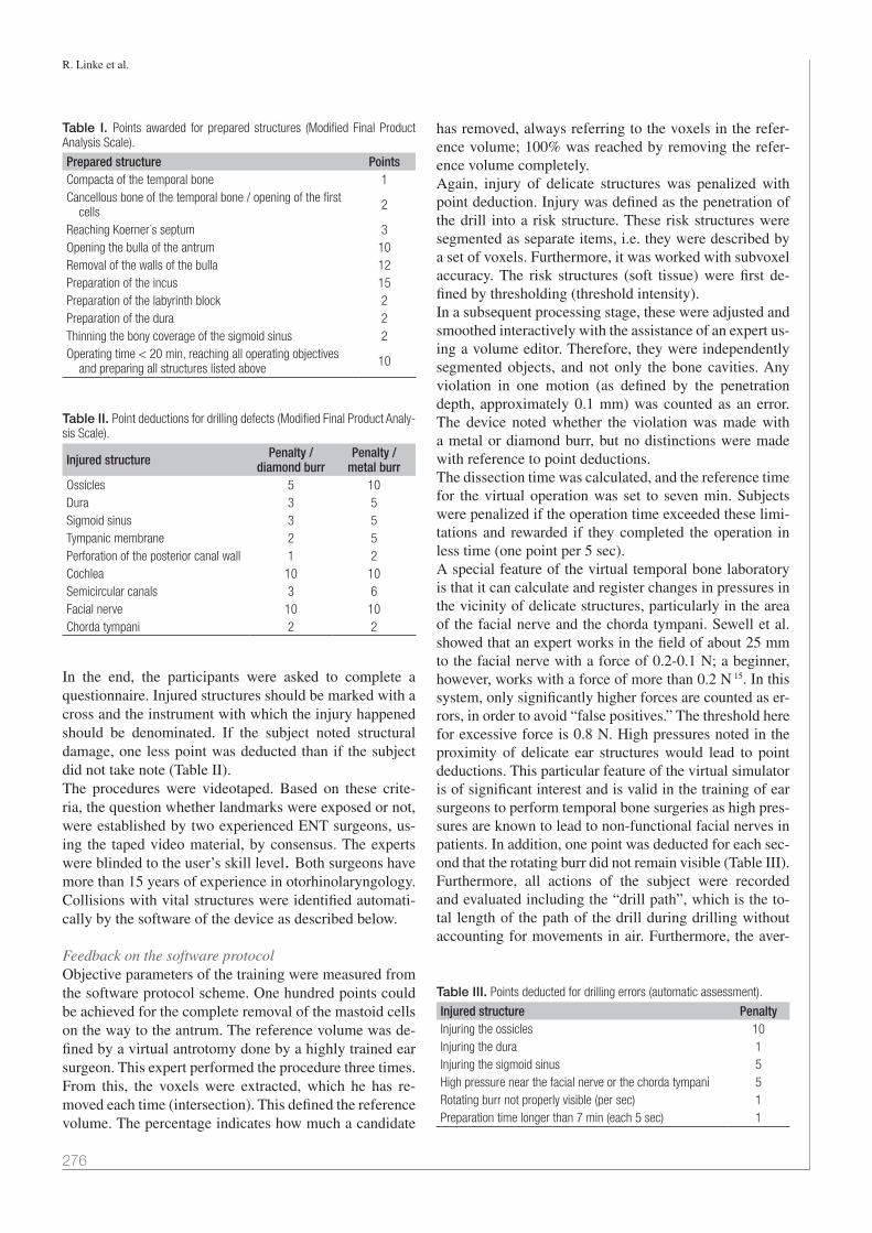
R. Linke et al.
276
In the end, the participants were asked to complete a questionnaire. Injured structures should be marked with a cross and the instrument with which the injury happened should be denominated. If the subject noted structural damage, one less point was deducted than if the subject did not take note (Table II). The procedures were videotaped. Based on these crite-ria, the question whether landmarks were exposed or not, were established by two experienced ENT surgeons, us-ing the taped video material, by consensus. The experts were blinded to the user’s skill level. Both surgeons have more than 15 years of experience in otorhinolaryngology. Collisions with vital structures were identified automati-cally by the software of the device as described below.
Feedback on the software protocolObjective parameters of the training were measured from the software protocol scheme. One hundred points could be achieved for the complete removal of the mastoid cells on the way to the antrum. The reference volume was de-fined by a virtual antrotomy done by a highly trained ear surgeon. This expert performed the procedure three times. From this, the voxels were extracted, which he has re-moved each time (intersection). This defined the reference volume. The percentage indicates how much a candidate
has removed, always referring to the voxels in the refer-ence volume; 100% was reached by removing the refer-ence volume completely.Again, injury of delicate structures was penalized with point deduction. Injury was defined as the penetration of the drill into a risk structure. These risk structures were segmented as separate items, i.e. they were described by a set of voxels. Furthermore, it was worked with subvoxel accuracy. The risk structures (soft tissue) were first de-fined by thresholding (threshold intensity). In a subsequent processing stage, these were adjusted and smoothed interactively with the assistance of an expert us-ing a volume editor. Therefore, they were independently segmented objects, and not only the bone cavities. Any violation in one motion (as defined by the penetration depth, approximately 0.1 mm) was counted as an error. The device noted whether the violation was made with a metal or diamond burr, but no distinctions were made with reference to point deductions. The dissection time was calculated, and the reference time for the virtual operation was set to seven min. Subjects were penalized if the operation time exceeded these limi-tations and rewarded if they completed the operation in less time (one point per 5 sec).A special feature of the virtual temporal bone laboratory is that it can calculate and register changes in pressures in the vicinity of delicate structures, particularly in the area of the facial nerve and the chorda tympani. Sewell et al. showed that an expert works in the field of about 25 mm to the facial nerve with a force of 0.2-0.1 N; a beginner, however, works with a force of more than 0.2 N 15. In this system, only significantly higher forces are counted as er-rors, in order to avoid “false positives.” The threshold here for excessive force is 0.8 N. High pressures noted in the proximity of delicate ear structures would lead to point deductions. This particular feature of the virtual simulator is of significant interest and is valid in the training of ear surgeons to perform temporal bone surgeries as high pres-sures are known to lead to non-functional facial nerves in patients. In addition, one point was deducted for each sec-ond that the rotating burr did not remain visible (Table III). Furthermore, all actions of the subject were recorded and evaluated including the “drill path”, which is the to-tal length of the path of the drill during drilling without accounting for movements in air. Furthermore, the aver-
Table I. Points awarded for prepared structures (Modified Final Product Analysis Scale).
Prepared structure PointsCompacta of the temporal bone 1Cancellous bone of the temporal bone / opening of the first
cells 2
Reaching Koerner´s septum 3Opening the bulla of the antrum 10Removal of the walls of the bulla 12Preparation of the incus 15Preparation of the labyrinth block 2Preparation of the dura 2Thinning the bony coverage of the sigmoid sinus 2Operating time < 20 min, reaching all operating objectives
and preparing all structures listed above 10
Table II. Point deductions for drilling defects (Modified Final Product Analy-sis Scale).
Injured structure Penalty / diamond burr
Penalty / metal burr
Ossicles 5 10Dura 3 5Sigmoid sinus 3 5Tympanic membrane 2 5Perforation of the posterior canal wall 1 2Cochlea 10 10Semicircular canals 3 6Facial nerve 10 10Chorda tympani 2 2
Table III. Points deducted for drilling errors (automatic assessment).
Injured structure Penalty Injuring the ossicles 10Injuring the dura 1Injuring the sigmoid sinus 5High pressure near the facial nerve or the chorda tympani 5Rotating burr not properly visible (per sec) 1 Preparation time longer than 7 min (each 5 sec) 1

Virtual reality temporal bone surgery simulator
277
age volume removed in an internal time step of 20 msec was also measured. The device also recorded the angle of drilling. These metrics have not been validated previously and, therefore, are not included in the evaluation scheme, except for output as a measured value. It was assumed that they could be meaningful, as (i) experienced surgeons will be more targeted and operate with less movement in the air, (ii) they are goal orientated, (iii) and they bit more with the equator than grow with the tip as inexperienced colleagues.
Our experience The study was approved by the human ethics committee of the University of Luebeck (AZ-12-021). There are no objections to the participation of medical students and qualified doctors in a virtual temporal bone lab. Data were analyzed with a one- and two-way ANOVA followed by Fisher’s Exact Test using Stat View 5.0 software. Differ-ences between groups were considered significant when p < 0.05.The results show that all experienced ENT surgeons were able to complete the aim of the operation and open the antrum and to intraoperatively dissect both the dura and the sigmoid sinus. Nine of ten residents succeeded in entering the antrum. Eight residents (80%) explored the sigmoid sinus, but only three (30%) explored the du-ra. Additionally, 18 (90%) students explored the sigmoid sinus, but only 11 (55%) explored the dura. It should be noted that none of the subjects injured the cochlea, semi-circular canals, facial nerve or chorda tympani. In addi-tion, no injury was observed to the ossicles when the sur-gical procedure was performed by the experienced ENT surgeons. However, the rate of injury to the ossicles was 50% in residents and 60% in students. This suggests that the experienced ENT surgeons performed significantly better than the residents (p < 0.05) and students (p < 0.01). In contrast to the students, a majority of the resi-dents immediately before entering the antrum changed to the diamond burr to avoid injuring the ossicles. While
only one experienced ENT surgeon injured the sigmoid sinus, 50% of residents and 45% of students damaged the sigmoid sinus. Interestingly, there was a high rate of injury to the dura, with 70% of the experienced ENT surgeons inducing injury compared with 20% and 45% by residents and students, respectively. In addition, no experienced ENT surgeon perforated the posterior wall of the outer ear canal. However, two residents (20%) and nine students (45%, Table IV) did perforate the posterior wall of the outer ear canal. Excessive force near deli-cate ear structures was observed in 60% of residents and 55% of students, but in none of the experienced ENT surgeons. This parameter further illustrated that the experienced ENT surgeons were significantly better at handling the drill compared to residents (p < 0.05) and students (p < 0.05).Our results further show that dissection times signifi-cantly differed between subject groups. Experienced ENT surgeons required half the time for the surgical procedure when compared to residents without ear surgery experi-ence and students. Differences in procedure times be-tween the experienced ENT surgeons and both students and residents were highly significant (p < 0.001), while no significant variability was detected between residents and students. A significant difference was observed between groups in the automatic score assigned by the software provided with the virtual device. The difference between the expe-rienced ENT surgeons and both the students and residents was again highly significant (p < 0.001), but no signifi-cant difference was noted between residents and students. Scores measured by the Modified Final Product Analysis Scale further validated that the more experienced ENT surgeons scored significantly higher than residents and students (p < 0.05). Again, there were no differences in scores between residents and students. We also show that there was no difference in the removal of temporal bone volume (p > 0.15), length of the drill path (p > 0.45) or drill angle (p > 0.5) between subject
Table IV. Preparation of landmarks and damage to the posterior wall of the outer ear canal.
Experienced ear surgeons (n = 7)
Residents without experience in ear surgery
(n = 10)
Students (n = 20)
Preparation of the dura* 7 (100%) 3 (30%) 11 (55%)Thinning of the bony hull of the sigmoid sinus* 7(100%) 8 (80%) 18 (90%)Entry to the antrum* 7(100%) 9 (90%) 20 (100%)Damage to the posterior wall of the outer ear canal* 0 (0%) 2 (20%) 9 (45%)Injury of the auditory ossicles* 0 (0%) 5 (50%) 12 (60%)Injury of the auditory ossicles with metal burr* 0 (0%) 1 (10%) 9 (45%)Injury of the sigmoid sinus* 1 (10%) 5 (50%) 9 (45%)Injury of the dura* 5 (70%) 2 (20%) 9 (45%)Luebeck-Score** 51.0 (± 2.9) 41.5 (± 8.7) 43.7 (± 7.8)
* number of incidences (percentage); ** mean (standard deviation)

R. Linke et al.
278
groups. However, the volume per strike was significantly higher in experienced ENT surgeons when compared to residents and students (p < 0.001). In addition, it should be noted that the rotating burr was not properly visible for a significantly longer period of time when used by residents compared to experienced ENT surgeons and students (p < 0.01). Otherwise, no significant differences were found between ENT specialists and students (Ta-ble V). Figures 3 and 4 show representative examples of the electronic protocol of the completed antrotomy performed by an experienced ENT surgeon and by an advanced resi-dent without experience in ear surgery, respectively. Subjects were also asked about their im-pressions of the virtual device for train-ing. Students expressed appreciation for the possibility of self-assessment and thought that the system was suitable for use in surgical specialization and was valuable as an initial training tool to prepare for surgical skills. Experi-enced ENT surgeons found the model to be similar to human temporal bone and suitable for training purposes.
DiscussionTemporal bone surgery is complex and requires extensive surgical skills. The first step in acquiring the credentials for ear surgery is the dissection of cadaver temporal bones in a temporal bone lab. Human temporal bone specimens natu-rally provide an excellent resemblance to the anatomic details of the human tem-
poral bone in living people. In fact, post-mortem defects in these bone specimens are almost negligible and can be advantageous since they also provide a breadth of ana-tomical variability between individual specimens, reflect-ing the inherent variability between individuals. On the other hand, disadvantages of using these cadaver prepara-tions include the inability to study pathological changes to the ear, the lack of anatomical reproducibility in the specimens, and the lack of specimen availability.
Table V. Parameters recorded by the device.
Experienced ear surgeons
(n = 7)
Residents without experience in ear
surgery (n = 10)
Students (n = 20)
Preparation time (min:sec)** 06:36 (± 0:38) 14:55 (± 8:47) 15:05 (± 6:27)
Automatic score** 68.7 (± 30.5) -80.4 (± 82.0) -52.7 (± 61.8)
Injury of the auditory ossicles* 0 (0%) 5 (50%) 12 (60%)
Injury of the auditory ossicles with metal burr* 0 (0%) 1 (10%) 9 (45%)
Injury of the sigmoid sinus* 1 (10%) 5 (50%) 9 (45%)
Injury of the dura 5 (70%) 2 (20%) 9 (45%)
Removed volume (ml)** 1.35 (± 0.30) 1.11 (± 0.22) 1.38 (± 0.24)
Excessive force near delicate structures* 0 (0%) 6 (60%) 11 (55%)
Rotating burr not properly visible (sec)** 0.4 (± 0.8) 4.1 (± 5.4) 1.4 (± 2.8)
Length of the drill path (mm)** 2542 (± 1270) 2439 (± 1682) 3170 (± 1944)
Drill angle (°)** 46.0 (± 5.5) 47.7 (± 6.9) 46.3 (± 6.6)
Volume per strike (mm3)** 0.77 (± 0.19) 0.39 (± 0.26) 0.43 (± 0.20)
* number (percentage); ** mean (standard deviation)
Fig. 3. Example of an electronic protocol of a completed antrotomy performed by an experienced ear surgeon. The major part (95%) of the reference volume is removed. The sigmoid sinus and the dura are explored. The antrum is wide open with a free view of the incus (brown). The virtual opera-tion was finished within the time limit. The verdict on the procedure in general is “passed”.

Virtual reality temporal bone surgery simulator
279
Artificial temporal bone models have been developed over the last two decades in Europe, especially in Germany 16-19. A number of virtual temporal bone models have been devel-oped over the last several years 15 20-24. The VOXEL MAN Tempo Surg® simulator is a commercially available system for training on bone drilling interventions in middle ear sur-gery and offers the possibility to learn about temporal bone anatomy as well as to perform procedures with relatively re-alistic visual and haptic feedback. The system is based on a volumetric, high-resolution model of the temporal bone derived from a CT. Interactive volume-cutting methods are performed using a multivolume scheme cut where regions are modelled independently. Data volumes can also be meas-ured using voxelization techniques. The so-generated tempo-ral bone model shows all anatomical landmarks and organs of risk such as the sigmoid sinus, dura, posterior wall of the outer ear canal, labyrinth block, facial nerve and incus 20.Compared to classical methods and artificial temporal bones, training using a virtual temporal bone lab provides
a number of major advantages. It allows physicians to train with temporal bones, which exhibit normal and path-ological conditions, and the system can adapt to the user’s existing skill level. Another advantage of the electronic simulation is the ability to infinitely repeat the procedure. It also allows the user to correct for mistakes by undoing a step and starting over again from a point before the er-ror was committed. The system can also be used by many trainees and provides the possibility of self-assessment. Because users can record procedures, it allows for cor-rection or evaluation at a later time. This virtual simulator can also decrease the required investment in bone drill-ing labs and the consumption of cadaveric material can be substantially reduced or even omitted. Results achieved from temporal bone dissection labs can be difficult to recapitulate in human specimens 18 25 26. We developed a “Modified Final Product Analysis Scale” to assess performance. The software included with the
VOXEL MAN Tempo Surg® simulator can provide automatic assessment of the user’s surgical skills. A limitation of the automatic evaluation is that it only deter-mines the ratio of the volume removed from the mastoid to the reference vol-ume. The device does not note whether the antrum is in fact open at the end of the procedure. In this study, the experi-enced ENT surgeons never injured the chain of ossicles, injured the sigmoid sinus less frequently than other subjects and needed only half the operation time compared with other subjects to perform the temporal surgery. This is also reflect-ed in a significantly better score calculat-ed using both the self-developed Modi-fied Final Product Analysis Scale as well as the automatic analysis software pro-vided by the device. These results further validate that the performance in the vir-tual surgery closely resembles the level of skill and performance observed in the operating room.Interestingly, the more experienced ENT surgeons injured the dura more often. This could be due to the lack of an in-crease in the intensity of blood flow in this region in the simulator, which is characteristic for the bone in the vicin-ity of the dura. More importantly, the ex-perienced ear surgeon is looking for the dura as a landmark. In reality, the dura does not disrupt immediately if it comes in contact with the drill. The drill pushes the dura in front of it; the dura is not li-able to be fixed to the bone. Thus, these
Fig. 4. Example of an electronic protocol of a completed antrotomy performed by a resident. Two-thirds (74%) of the reference volume is removed. Points were deducted because excessive force was applied in the area of the facial nerve and of the chorda tympani. The antrum is wide open, but the auditory ossicles were injured twice and the sigmoid sinus once. The rotating burr was not properly visible for 17 sec. The virtual operation was not finished within the time limit. The verdict on the procedure in general is “failed”.
R. Linke et al.
280
models should be further developed to account for these observed differences between the simulator and real op-erations. Experienced ENT surgeons did not apply a high pressure with the drill near delicate ear structures, unlike half of the residents and students. These results suggest that the virtual device is very sensitive to major technical errors in drilling, which is also a good indicator of the level of surgical skill of the prospective ENT surgeon.Operating room training often consists of guidance and supervision of a theoretically highly educated but more or less inexperienced surgeon (trainee) by a more experi-enced colleague. Even when a supervisor is present, there is always a high risk when a surgical trainee operates on a patient requiring complex surgery. Operation time is becoming increasingly important as medicine becomes commercialized. The atmosphere in the operating room is accompanied by a time limitation and a high level of expense. It has been calculated that surgical residents cost the operating room $1,000 per training hour in addition to the regular cost of the operation 27. In addition, individual surgical steps cannot be practiced repeatedly prior to sur-gery. Thus, the virtual surgery simulator offers the possi-bility of partial replacement of expensive operation room time and supervision by an expensive academic teacher with electronic supervision. One option for the future is preoperative simulation of the operation of selected and particularly difficult patient cases on a personal com-puter 28. In this way, even experienced ear surgeons could simulate complex procedures before the real intervention, saving time and avoiding complications. In line with these thoughts, a training model for paranasal sinus surgery is currently in development 29.Surprisingly, no significant difference was noted between the residents without experience in middle ear surgery and the medical students. In Germany, a classical education in ENT surgery begins with adenotomy and tonsillectomy. Middle ear surgeries are the pinnacle of ENT surgeries and are performed by especially committed medical specialists during the culmination and height of their ENT training. It is obvious that ear surgery occupies a special position in ENT surgery and we show that surgical education in other sub-specializations has no influence on the physician’s per-formance at the beginning of ear surgery training. In our study we observed a dichotomic distribution sepa-rating skilled and non-skilled surgeons. This first evalua-tion of the virtual temporal bone laboratory by means of a standard operation revealed its construct validity and its suitability as a training model for temporal bone surgery in principle. Surgery training is a continuous process where different intermediate skill levels are observed. Therefore further studies on construct validity are required. These should include surgeons of different levels, e.g. complete-ly inexperienced surgeons, inexperienced physicians who just had a temporal bone dissection course, surgeons with
intermediate experience (< 200 operations), experienced surgeons (> 200 and < 1000 operations) and experts (> 1000 operations).The next step is to assess the efficacy of the system as a learning tool. Future work should investigate the learn-ing curves on the simulator with single-subject repeated measure design. One possibility is to study whether there is a significant difference between the first and subsequent sessions of the inexperienced subjects in terms of oper-ating time and surgical outcome. Furthermore, every at-tempt of the inexperienced surgeons could be compared with the data from the group of experienced surgeons to determine the point at which there is no significant differ-ence between the two groups. Another experiment might assess the performance of trainees over time to see if the simulator performance improves with increasing clinical experience, as confirmed by the scores. Finally, studies are needed to assess whether skills ac-quired in the virtual environment can be transferred to the operating room in order to translate the effect of learning on the simulator to real-life ear surgery; it would then be possible to demonstrate a transfer-effectiveness ratio.Work completed in the temporal bone laboratory is and will remain “conditio sine qua non” for the start of training in ear surgery on patients. Virtual learning programs of the anatomy of the temporal bone and interactive programs for dissection may display the anatomical characteristics of this difficult anatomical region and serve as a good baseline for the dissection exercises in the temporal bone lab. The technology of virtual reality simulators can be expected to improve. It should be noted that a virtual surgical simulator cannot replace the traditional temporal bone laboratory, but the virtual temporal bone laboratory supplements the tradi-tional training path of the prospective ear surgeon.
AcknowledgmentsWe thank Spiggle and Theis, Overath, Germany for pro-viding the VOXEL MAN TempoSurg© Simulator for the study. There are no conflicts of interest.
References1 Green JDJ, Shelton C, Brackmann DE. Iatrogenic fa-
cial nerve injury during otologic surgery. Laryngoscope 1994;104:922-6.
2 Hakuba N, Hato N, Shinomori Y, et al. Labyrinthine fistula as a late complication of middle ear surgery using the canal wall down technique. Otol Neurotol 2002;23:832-5.
3 Nilssen EL, Wormald PJ. Facial nerve palsy in mastoid sur-gery. J Laryngol Otol 1997;111:113-6.
4 Wormald PJ, Nilssen EL. Do the complications of mastoid surgery differ from those of the disease? Clin Otolaryngol Allied Sci 1997;22:355-7.
5 Garap JP, Dubey SP. Canal-down mastoidectomy: experi-ence in 81 cases. Otol Neurotol 2001;22:451-6.

Virtual reality temporal bone surgery simulator
281
6 Schwager K, Gilyoma JM. Ceramic model for temporal bone exercises-an alternative for human temporal bones? Laryn-gorhinootol 2003;82:683-6.
7 Pflesser B, Urban M. Stereoscopic simulation of ear surgery Intervention with a novel 3D computer models. Laryngorhi-nootol 2001;80:298-302.
8 Snyder CW, Vandromme MJ, Tyra SL, et al. Effects of virtual reality simulator training method and observational learning on surgical performance. World J Surg 2011;35:245-52.
9 Solverson DJ, Mazzoli RA, Raymond WR, et al. Virtual real-ity simulation in acquiring and differentiating basic ophthal-mic microsurgical skills. Simul Healthc 2009;4:98-103.
10 Zirkle M, Blum R, Raemer DB, et al. Teaching emergency airway management using medical simulation: a pilot pro-gram. Laryngoscope 2005;115:495-500.
11 Nicholson WJ, Cates CU, Patel AD, et al. Face and content validation of virtual reality simulation for carotid angiog-raphy: results from the first 100 physicians attending the Emory NeuroAnatomy Carotid Training (ENACT) program. Simul Healthc 2006;1:147-50.
12 Mayrose J, Myers JW. Endotracheal intubation: application of virtual reality to emergency medical services education. Simul Healthc 2007;2:231-4.
13 McDougall EM. Validation of surgical simulators. J En-dourol 2007;21:244-7.
14 Szalay D, MacRae H, Regehr G, et al. Using operative out-come to assess technical skill. Am J Surg 2000;180:234-7.
15 Sewell C, Morris D, Blevins NH, et al. Providing metrics and performance feedback in a surgical simulator. Comput Aided Surg. 2008;13:63-81.
16 Begall K, Vorwerk U. Artificial petrous bone produced by stereolithography for microsurgical dissecting exercises. ORL J Otorhinolaryngol Relat Spec 1998;60:241-5.
17 Schneider G, Mueller A. Multi-center study of the Jenaer mod-el of the temporal bone. Laryngorhinootol 2004;83:363-6.
18 Strauss G, Bahrami N, Pössneck A, et al. Evaluation of a
training system for middle ear surgery with optoelectric de-tection. HNO 2009;57:999-1009.
19 Bakhos D, Velut S, Robier A, et al. Three-dimensional mode-ling of the temporal bone for surgical training. Otol Neurotol 2010;31:328-34.
20 Pflesser B, Petersik A, Tiede U, et al. Volume cutting for vir-tual petrous bone surgery. Comput Aided Surg 2002;7:74-83
21 Kockro RA, Hwang PY. Virtual temporal bone: an interac-tive 3-dimensional learning aid for cranial base surgery. Neurosurgery 2009;64:216-29; discussion 229-30.
22 Zhao YC, Kennedy G, Hall R, et al. Differentiating levels of surgical experience on a virtual reality temporal bone simu-lator. Otolaryngol Head Neck Surg 2010;143:S30-5.
23 Sowerby LJ, Rehal G, Husein M, et al. Development and face validity testing of a three-dimensional myringotomy simulator with haptic feedback. J Otolaryngol Head Neck Surg 2010;39:122-9.
24 Stredney D, Wiet GJ, Bryan J, et al. Temporal bone dissec-tion simulation - an update. Stud Health Technol Inform 2002;85:507-13.
25 Zirkle M, Taplin MA, Anthony R, et al. Objective assessment of temporal bone drilling skills. Ann Otol Rhinol Laryngol 2007;116:793-8.
26 Roberson DW, Leuwer R, Dubrowski A. Using a virtual reality temporal bone simulator to assess otolaryngology trainees. Laryngoscope 2007;117:258-63.
27 Haluck RS, Marshall RL, Krummel TM, et al. Are sur-gery training programs ready for virtual reality? A survey of program directors in general surgery. J Am Coll Surg 2001;193:660-5.
28 Tolsdorff B, Petersik A, Pflesser B et al. Individual models for virtual bone drilling in mastoid surgery. Comput Aided Surg 2009;14:21-7.
29 Tolsdorff B, Pommert A, Höhne KH et al. Virtual reality: a new paranasal sinus surgery simulator. Laryngoscope 2010;120:420-6.
Address for correspondence: Robert Linke, Univer-sitätsklinikum Schleswig-Holstein, Campus Luebeck HNO-Klinik, Ratzeburger Allee 160, D - 23538 Luebeck, Germa-ny. Tel. +49 451 500 2241. Fax +49 451 500 2249. E-mail: [email protected]
Received: December 30, 2012 - Accepted: March 6, 2013
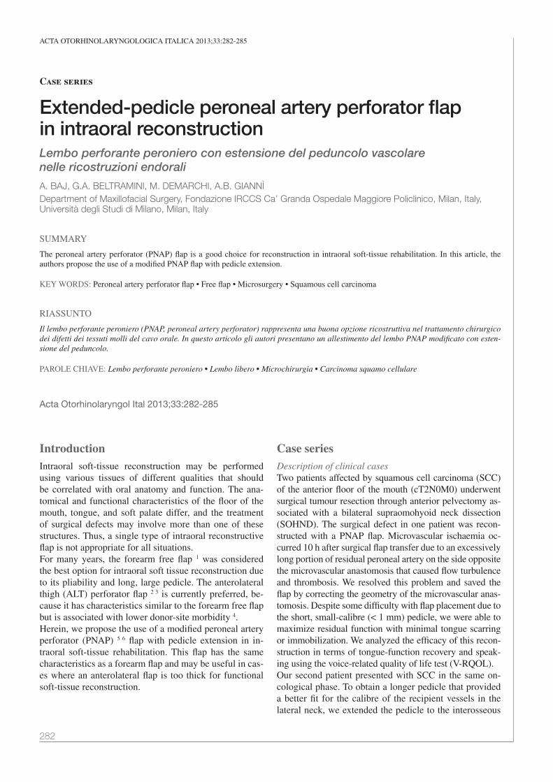
282
ACTA oTorhinolAryngologiCA iTAliCA 2013;33:282-285
Case series
Extended-pedicle peroneal artery perforator flap in intraoral reconstructionLembo perforante peroniero con estensione del peduncolo vascolare nelle ricostruzioni endorali
A. BAj, G.A. BeltrAmini, m. DemArchi, A.B. GiAnnìDepartment of maxillofacial Surgery, Fondazione irccS ca’ Granda Ospedale maggiore Policlinico, milan, italy, Università degli Studi di milano, milan, italy
SummAry
The peroneal artery perforator (PnAP) flap is a good choice for reconstruction in intraoral soft-tissue rehabilitation. in this article, the authors propose the use of a modified PnAP flap with pedicle extension.
Key wordS: Peroneal artery perforator flap • Free flap • Microsurgery • Squamous cell carcinoma
riASSunTo
Il lembo perforante peroniero (PNAP, peroneal artery perforator) rappresenta una buona opzione ricostruttiva nel trattamento chirurgico dei difetti dei tessuti molli del cavo orale. In questo articolo gli autori presentano un allestimento del lembo PNAP modificato con esten-sione del peduncolo.
PArole ChiAve: Lembo perforante peroniero • Lembo libero • Microchirurgia • Carcinoma squamo cellulare
Acta Otorhinolaryngol Ital 2013;33:282-285
IntroductionIntraoral soft-tissue reconstruction may be performed using various tissues of different qualities that should be correlated with oral anatomy and function. The ana-tomical and functional characteristics of the floor of the mouth, tongue, and soft palate differ, and the treatment of surgical defects may involve more than one of these structures. Thus, a single type of intraoral reconstructive flap is not appropriate for all situations.For many years, the forearm free flap 1 was considered the best option for intraoral soft tissue reconstruction due to its pliability and long, large pedicle. The anterolateral thigh (ALT) perforator flap 2 3 is currently preferred, be-cause it has characteristics similar to the forearm free flap but is associated with lower donor-site morbidity 4.Herein, we propose the use of a modified peroneal artery perforator (PNAP) 5 6 flap with pedicle extension in in-traoral soft-tissue rehabilitation. This flap has the same characteristics as a forearm flap and may be useful in cas-es where an anterolateral flap is too thick for functional soft-tissue reconstruction.
Case series Description of clinical casesTwo patients affected by squamous cell carcinoma (SCC) of the anterior floor of the mouth (cT2N0M0) underwent surgical tumour resection through anterior pelvectomy as-sociated with a bilateral supraomohyoid neck dissection (SOHND). The surgical defect in one patient was recon-structed with a PNAP flap. Microvascular ischaemia oc-curred 10 h after surgical flap transfer due to an excessively long portion of residual peroneal artery on the side opposite the microvascular anastomosis that caused flow turbulence and thrombosis. We resolved this problem and saved the flap by correcting the geometry of the microvascular anas-tomosis. Despite some difficulty with flap placement due to the short, small-calibre (< 1 mm) pedicle, we were able to maximize residual function with minimal tongue scarring or immobilization. We analyzed the efficacy of this recon-struction in terms of tongue-function recovery and speak-ing using the voice-related quality of life test (V-RQOL).Our second patient presented with SCC in the same on-cological phase. To obtain a longer pedicle that provided a better fit for the calibre of the recipient vessels in the lateral neck, we extended the pedicle to the interosseous
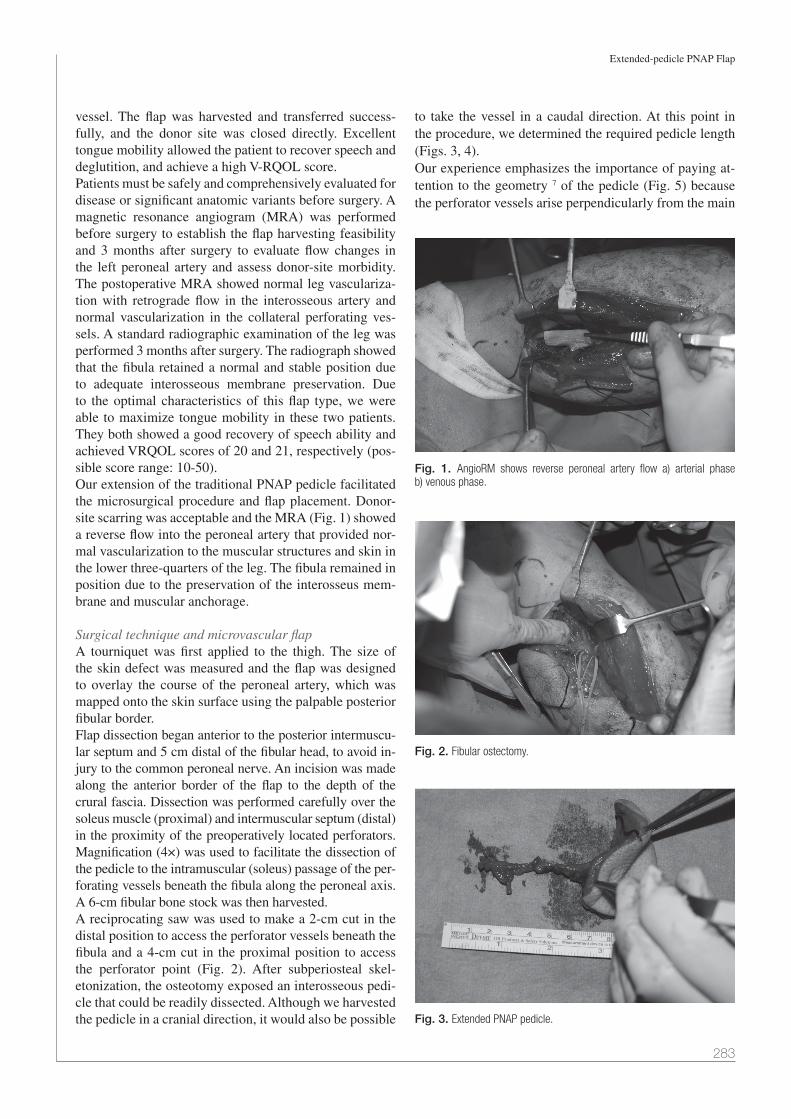
Extended-pedicle PNAP Flap
283
vessel. The flap was harvested and transferred success-fully, and the donor site was closed directly. Excellent tongue mobility allowed the patient to recover speech and deglutition, and achieve a high V-RQOL score.Patients must be safely and comprehensively evaluated for disease or significant anatomic variants before surgery. A magnetic resonance angiogram (MRA) was performed before surgery to establish the flap harvesting feasibility and 3 months after surgery to evaluate flow changes in the left peroneal artery and assess donor-site morbidity. The postoperative MRA showed normal leg vasculariza-tion with retrograde flow in the interosseous artery and normal vascularization in the collateral perforating ves-sels. A standard radiographic examination of the leg was performed 3 months after surgery. The radiograph showed that the fibula retained a normal and stable position due to adequate interosseous membrane preservation. Due to the optimal characteristics of this flap type, we were able to maximize tongue mobility in these two patients. They both showed a good recovery of speech ability and achieved VRQOL scores of 20 and 21, respectively (pos-sible score range: 10-50).Our extension of the traditional PNAP pedicle facilitated the microsurgical procedure and flap placement. Donor-site scarring was acceptable and the MRA (Fig. 1) showed a reverse flow into the peroneal artery that provided nor-mal vascularization to the muscular structures and skin in the lower three-quarters of the leg. The fibula remained in position due to the preservation of the interosseus mem-brane and muscular anchorage.
Surgical technique and microvascular flapA tourniquet was first applied to the thigh. The size of the skin defect was measured and the flap was designed to overlay the course of the peroneal artery, which was mapped onto the skin surface using the palpable posterior fibular border. Flap dissection began anterior to the posterior intermuscu-lar septum and 5 cm distal of the fibular head, to avoid in-jury to the common peroneal nerve. An incision was made along the anterior border of the flap to the depth of the crural fascia. Dissection was performed carefully over the soleus muscle (proximal) and intermuscular septum (distal) in the proximity of the preoperatively located perforators. Magnification (4×) was used to facilitate the dissection of the pedicle to the intramuscular (soleus) passage of the per-forating vessels beneath the fibula along the peroneal axis. A 6-cm fibular bone stock was then harvested.A reciprocating saw was used to make a 2-cm cut in the distal position to access the perforator vessels beneath the fibula and a 4-cm cut in the proximal position to access the perforator point (Fig. 2). After subperiosteal skel-etonization, the osteotomy exposed an interosseous pedi-cle that could be readily dissected. Although we harvested the pedicle in a cranial direction, it would also be possible
to take the vessel in a caudal direction. At this point in the procedure, we determined the required pedicle length (Figs. 3, 4).Our experience emphasizes the importance of paying at-tention to the geometry 7 of the pedicle (Fig. 5) because the perforator vessels arise perpendicularly from the main
Fig. 1. AngioRM shows reverse peroneal artery flow a) arterial phase b) venous phase.
Fig. 2. Fibular ostectomy.
Fig. 3. Extended PNAP pedicle.

A. Baj et al.
284
trunk. For this reason, it is important to close the side op-posite the microvascular anastomosis very close to the origin of the perforator vessels, to avoid turbulence in the main arterial trunk that could cause thrombosis.
DiscussionThe progressive increase in the use of free flaps has fur-thered the development of this reconstructive technique in the last 25 years. Further innovation has occurred recently, following the increased popularity of perforator flaps. The term “perforator flap” was first used by Koshima 8 and defined specifically by Wei 9, although the harvesting of such flaps had been described in the literature about a dec-ade earlier. Hallock 10 recently proposed a more detailed terminology that refers to indirect perforator flaps, indi-cating blood vessels that require intramuscular dissection, and direct perforator flaps, indicating vessels that pass through the intramuscular septa or a more direct path to the skin. All perforator flaps are characterized by the thin-ness of the cutaneous flap segment in comparison with
myocutaneous flaps, a short pedicle length and relatively small vessel calibre, and minimal donor-site morbidity 11 .For many years, the forearm free flap was considered to be the best option for intraoral soft-tissue reconstruction, due to its pliability and the long, large-calibre pedicle 12. However, such flaps are associated with high donor-site morbidity 13-15. Currently, the ALT perforator flap is most commonly used because it has similar characteristics but is associated with lower donor-site morbidity 16 17. How-ever, ALT flaps require thinning, which increases the risk of damage to the perforator vessels. In intraoral recon-struction, the risks of marginal flap necrosis and neck fis-tulization are high 18.Pedicle calibre is also an important consideration. In a conventional axial free-tissue transfer in the head and neck area, the calibre of the flap pedicle 19 frequently fits well with those of recipient vessels, including the thyroid, facial, and lingual arteries. Perforator free flaps provide a less favourable match between the calibre of the pedi-cle (≤ 1 mm) and those of the recipient vessels. In our proposed technique, we sought to improve this match and provide a pedicle of adequate length for use with micro-vascular anastomosis in intraoral reconstruction.For these reasons, we propose a thin and pliable perfora-tor flap with an elongated pedicle, which improves the fit of the pedicle calibre and increases functional reliability in intraoral soft-tissue reconstructions.PNAP flaps have the best characteristics for intraoral re-construction and are associated with reduced donor-site morbidity, but are not widely used in maxillofacial sur-gery because the short and small-calibre pedicle has been assumed to increase complication rates. We have attempt-ed to overcome this limitation by extending the pedicle to the peroneal artery origin. We have demonstrated that this technique is unproblematic and has no negative effect on the blood supply of the fibular donor site. The compro-mise of the fibula free flap for future osseous reconstruc-tions is a disadvantage when preoperative magnetic reso-nance angiogram contraindicates contralateral harvesting.We have thus been able to combine the best flap features with low donor-site morbidity in a user-adjustable, long, large-cal-ibre vessel flap. This technique offers a useful alternative in microvascular free-tissue transfer when a primary ultrathin pliable flap is required to cover intraoral mucosal defects.
References1 Avery CM. Review of the radial free flap: is it still evolving,
or is it facing extinction? Part one: soft-tissue radial flap. Br J Oral Maxillofac Surg 2010; 8:245-52.
2 Wei FC, Jain V, Celik N, et al. Have we found an ideal soft tissue flap? An experience with 672 anterolateral thigh flaps. Plast Reconstr Surg 2002;109:2219-26.
3 Shieh SJ, Chiu HY, Yu JC, et al. Free anterolateral thigh flap for reconstruction of head and neck defects following cancer ablation. Plast Reconstr Surg 2000;105:2349-57.
Fig. 4. Extended PNAP flap harvested: notice flap thickness and the long pedicle.
Fig. 5. Microanastomosis: notice the calibre match between donor and recipient vessels.

Extended-pedicle PNAP Flap
285
4 Kimata Y, Uchiyama K, Ebihara S, et al. Anterolaleral thigh flap donor side complications and morbidity. Plast Reconstr Surg 2000;106:584-9.
5 Ozalp T, Masquelet AC, Begue TC. Septocutaneous perfora-tors of the peroneal artery relative to the fibula: anatomical basis of the use of pedicled fasciocutaneous flap. Surg Radiol Anat 2006;28:54-8.
6 Baj A, Bellocchio G, Marelli S, et al. Reconstruction of the anterior floor of the mouth using a peroneal perfo-rator free flap. A case report. Acta Otorhinolaryngol Ital 2010;30:47-51.
7 Urken ML, Vickery C, Weinberg H, et al. Geometry of the vascular pedicle in free tissue transfers to the head and neck. Arch Otolaryngol Head Neck Surg 1989;115: 954-60.
8 Koshima I, Soeda S, Yamasaki M, et al. The free or pedicled anteromedial thigh flap. Ann Plast Surg 1988;21:480-5.
9 Wei FC, Jain V, Suominen S, et al. Confusion among perfo-rator flaps: What is a true perforator flap? Plast Reconstr Surg 2001;107:874-6.
10 Hallock GG. The complete nomenclature for combined per-forator flaps. Plast Reconstr Surg 2011;127:1720-9.
11 Baj A, Ali Youssef D, Monteverdi R, et al. Recontruction of partial maxillary defects with the double-barrel fibula free flap. Acta Otorhinolaryngol Ital 2010;30:299-302.
12 Soutar DS, McGregor IA. The radial forearm flap in in-
traoral reconstruction: The experience of 60 consecutive cases. Plast Reconstr Surg 1986;78:1-8.
13 Lutz BS, Wei FC, Chang SC, et al. Donor site morbidity af-ter suprafascial elevation of the radial forearm flap: a pro-spective study in 95 consecutive cases. Plast Reconstr Surg 1999;103:132-7.
14 Richardson D, Fisher SE, Vaughan ED, et al. Radial forearm flap donor site complications and morbidity: a prospective study. Plast Reconstr Surg 1997;99:109-15.
15 Pellini R, Mercante G, Spriano G. Step-by-step mandibular reconstruction with free fibula flap modelling. Acta Otorhi-nolaryngol Ital 2012;32:405-9.
16 Camaioni A, Loreti A, Damiani V, et al. Anterolateral thigh cutaneous flap vs. radial forearm free-flap in oral and oro-pharyngeal reconstruction: an analysis of 48 flaps. Acta Otorhinolaryngol Ital 2008;28:7-12.
17 Huang CH, Chen HC, Huang YL, et al. Comparison of the radial forearm flap and the thinned anterolateral thigh cutane-ous flap for reconstruction of tongue defect: an evaluation of donor-site morbidity. Plast Reconstr Surg 2004;114:1704-10.
18 Alkureishi LW, Shaw-Dunn J, Ross GL. Effects of thinning the anterolateral thigh flap on the blood supply to the skin. Br J Plast Surg 2003;56:401-8.
19 Baj A, Ferrari S, Bianchi B, et al. Iliac crest free flap in oro-mandibular reconstruction. 13 cases study. Acta Otorhinola-ryngol Ital 2003;23:102-10.
Address for correspondence: Giada Anna Beltramini, Department of Maxillofacial Surgery, Fondazione IRCCS Ospedale Maggio-re Policlinico, Università degli Studi di Milano, via Francesco Sforza 35, 20122 Milan, Italy. Tel. +39 02 55032532. Fax +39 02 55032745. E-mail: [email protected]
Received: July 28, 2012 - Accepted: March 18, 2013

286
ACTA oTorhinolAryngologiCA iTAliCA 2013;33:286-288
Case report
Mucoepidermoid carcinoma of the tonsil: a very rare presentationCarcinoma mucoepidermoide della tonsilla: una presentazione molto rara
S.J. JarviS1, v. GianGrande2, P.a. Brennan3
1department of Paediatric Otolaryngology, Head and neck Surgery, royal Manchester Children’s Hospital, Oxford road, Manchester, UK; 2 department of Otolaryngology, Clinica San Feliciano, roma, italy; 3 department of Oral and Maxillofacial Surgery, Queen alexandra Hospital, Cosham, Portsmouth, UK
SummAry
mucoepidermoid carcinoma is the most common malignant salivary gland tumour. however, short series or individual case reports have identi-fied this tumour in the maxilla, mandible, breast tissue and thymus. mucoepidermoid carcinoma originates from minor salivary glands, and it is therefore surprising that it is not more commonly seen in the tonsil. To date, we believe there has been only one previously reported case in the world literature of mucoepidermoid carcinoma occurring in the tonsil 1. We present a very rare case of mucoepidermoid carcinoma arising from within the structure of the palatine tonsil, rather from the adjacent pharyngeal wall, together with a short review of the literature.
Key WordS: Palatine tonsil • Mucoepidermoid carcinoma
riASSunTo
Il carcinoma mucoepidermoide rappresenta la neoplasia maligna più comune delle ghiandole salivari. Tuttavia numerosi studi fanno rife-rimento a casi isolati in cui questo tipo di tumore origina dal mascellare, dalla mandibola, dalla ghiandola mammaria e dal timo. Poiché il carcinoma mucoepidermoide origina solitamente nelle ghiandole salivari, l’origine dalla tonsilla palatina è considerata alquanto insolita. Fino ad oggi riteniamo sia stato precedentemente riportato in letteratura solo un caso di carcinoma mucoepidermoide nella tonsilla 1. Presentiamo un caso molto raro di carcinoma muco epidermoide che originante dalla tonsilla palatina piuttosto che dalla adiacente parete faringea, insieme a una revisione dei casi riportati in letteratura.
pArole ChiAve: Tonsilla palatina • Carcinoma mucoepidermoide
Acta Otorhinolaryngol Ital 2013;33:286-288
Introductionmucoepidermoid carcinoma is the most common ma-lignant salivary gland tumour 2-4. it has been reported in all ages with peak incidence at the 4th and 5th decades, with females affected more than males in a 3:1 ratio. it is the most frequent malignant salivary gland neoplasm in children 5. in the major salivary glands, 89.6% of cases present in the parotid 6. mucoepidermoid carcinoma dem-onstrates a broad spectrum of aggressiveness, which can be predicted by microscopic grading.high-grade tumours are highly aggressive and regional lymph node spread is common. The low-grade variant usu-ally demonstrates a favourable outcome, but it is important to note that metastasis may also be present 7. distant me-tastasis is rare, but case reports of metastases to the lungs, brain, ovary and peritoneum have been reported 8. histolog-ically, the tumour is composed of mucous, basaloid, inter-mediate and epidermoid cells. We present an unusual case of mucoepidermoid carcinoma arising in the tonsil.
Case reportA 48-year-old male presented with an asymptomatic lump in the neck at level ii. The lump was progressively increasing in size over 4 weeks. intra-oral examination and flexible nasoendoscopy was normal. A fine-needle aspiration cytology specimen showed malignant cells with no obvious architecture to determine the tissue of origin. ultrasound detected the presence of a suspected necrotic node. A CT scan of the neck and chest con-firmed a right-sided pathologic node at level ii. no other abnormalities were evident. examination under anaes-thesia (euA) was performed, which was suggestive of a bulky mass within the ipsilateral tonsil, and tonsillecto-my was performed. Biopsy confirmed a high-grade mu-coepidermoid carcinoma arising from entirely within the substance of the ipsilateral tonsil, and not from the ad-jacent pharyngeal wall. management involved complete tonsillectomy with ipsilateral neck dissection level ii-iv. histology of the resected specimen confirmed metastatic

Mucoepidermoid carcinoma of the tonsil
287
carcinoma from the tonsil. he underwent postoperative radiotherapy.
Discussionmucoepidermoid carcinoma is the most common malig-nant salivary gland neoplasm 2. its occurrence in the max-illa, mandible, breast and thymus have been reported 9 10.
To date, only one other case of primary mucoepidermoid carcinoma of the tonsil has been documented 1. These tu-mours arise from reserve cells in the salivary duct system and therefore differentiate into mucin-producing cells, or duct-like epidermoid cells. interestingly, both cell types are altered neoplastic cells. mucoepidermoid carcinoma is a malignancy in which histological grading and clinical behaviour correlate well and have a predictive outcome 2. Treatment is therefore based on histological grading of the tumour according primarily to the relative mix of cell types. other factors that contribute are growth pattern and cellular atypia. The incidence of occult regional metasta-sis varies from 6 to 46%. metastasis has been found even in cases with very small tumours.management of the tumour depends on its primary site. low-grade tumours arising in the parotid with no metastasis are treated with superficial parotidectomy, while high-grade tumours are treated by total parotidectomy with or without neck dissection depending on the neck staging. Tumours arising at other sites such as the palate are treated with a minimum of 1 cm excision margin or hemimaxillectomy for high-grade palatal tumours. in the presence of nodal metas-tasis, radiotherapy in addition to surgery is recommended for both the primary site and the neck. The 5-year survival rate has been reported to be as high as 95% in low-grade tumours, and 50% in intermediate/high-grade tumours. Sur-vival falls dramatically with an age over 55 years 11.Tamiolakis et al. concluded that in cases of unknown pri-maries, diagnostic procedures should be aimed at clarify-
ing the histology of the nodal metastases and detecting the primary tumour site 12. in the management of occult pri-mary tumours, bilateral tonsillectomy has been suggested as the minimum investigative procedure at the time of euA 13. our case highlights the need for tonsillar biopsy even in those patients with normal euA findings.
ConclusionsAlthough mucoepidermoid carcinoma is the most com-mon malignant salivary gland tumour, it is a very rare occurrence within the substance of the palatine tonsil. in cases of an unknown primary, diagnostic procedures should be aimed at clarifying the histology of the nodal metastases and detecting the primary tumour site. our case highlights the need to biopsy the tonsil in all cases of unknown primary, even in the presence of normal findings on euA.
References1 li JX. Clinico-pathologic studies on 143 cases of tonsillar
malignancies with special reference to lymphomas. Zhong-hua Zhong liu Za Zhi 1992;14:433-6.
2 loyola Am, Araujo vC, Sousa Som, et al. Minor sali-vary gland tumours. A retrospective study of 164 cases in a Brazilian population. eur J Cancer B oral oncol 1995;31B:197-201.
3 lopes mA, Kowalski lp, Santos g, et al. A clinicopathologic study of 196 intraoral minor salivary gland tumours. J oral pathol med 1999;28:264-7.
4 Jones AS, Beasley nJ, houghton dJ, et al. Tumours of the minor salivary glands. Clin otolaryngol 1998;23:27-33.
5 hicks J, Flaitz C. Mucoepidermoid carcinoma of salivary glands in children and adolescents: assessment of prolifera-tion markers. oral oncol 2000;6:454-60.
6 eversole lr. Mucoepidermoid carcinoma: review of 815 re-ported cases. J oral Surg 1970;28:490-5.
Fig. 2. High-power combined Alcian blue/periodic acid Schiff stain demon-strating mucin (purple).
Fig. 1. High-power haematoxylin and eosin (H&E) staining demonstrating nuclear pleomorphism.

S.J. Jarvis et al.
288
7 goode rK, Auclair pl, ellis gl. Mucoepidermoid carcino-ma of the major salivary glands: clinical and histopathologic analysis of 234 cases with evaluation of grading criteria. Cancer 1998;82:1217-24.
8 Buyukkurt S, Altintas A, gumurdulu d, et al. Mucoepi-dermoid carcinoma of the parotid gland with ovar-ian and peritoneal metastasis. J obstet gynaecol res 2008;34:271-3.
9 hornychova h, ryska A, Betlach J, et al. Mucoepidermoid carcinoma of the breast. neoplasma 2007;54:168-72.
10 noda T, higashiyama m, oda K, et al. Mucoepider-moid carcinoma of the thymus treated by multimodal-
ity therapy: a case report. Ann Thorac Cardiovasc Surg 2006;12:273-8.
11 hiroyuki o, Toshiki T, Koji S, et al. Mucoepidermoid carci-noma of the head and neck: clinical analysis of 43 patients.
Jpn J Clin oncol 2008;38:414-8.12 Tamiolakis d, Thomaidis v, Tsamis i, et al. Malignant
mucoepidermoid tumor arising in the accessory parot-id gland: a case report. Acta medica (hradec Kralove) 2003;46:79-83.
13 Kazi rA. Oncologic rationale for bilateral tonsillectomy in head and neck squamous cell carcinoma of unknown primary source. internet Journal of oncology 2004;2(1).
Address for correspondence: Sara J. Jarvis, department of pa-ediatric otolaryngology, royal manchester Children’s hospi-tal, oxford road, manchester, lancashire, m13 9Wl, uK. Fax +44 1617 015462. e-mail: [email protected]
received: July 28, 2010 - Accepted: April 3, 2011
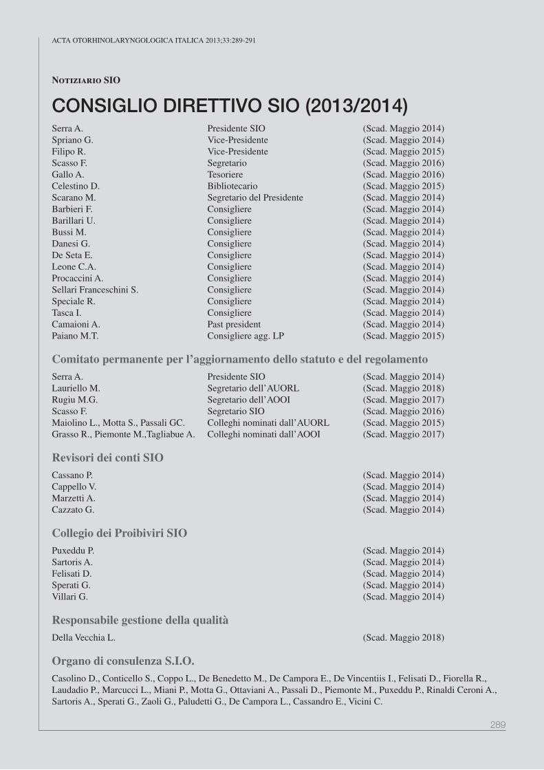
289
ACTA oTorhinolAryngologiCA iTAliCA 2013;33:289-291
Notiziario SIO
CONSIGLIO DIRETTIVO SIO (2013/2014)Serra A. Presidente Sio (Scad. Maggio 2014)Spriano g. Vice-Presidente (Scad. Maggio 2014) Filipo r. Vice-Presidente (Scad. Maggio 2015)Scasso F. Segretario (Scad. Maggio 2016)gallo A. Tesoriere (Scad. Maggio 2016)Celestino D. Bibliotecario (Scad. Maggio 2015)Scarano M. Segretario del Presidente (Scad. Maggio 2014)Barbieri F. Consigliere (Scad. Maggio 2014)Barillari U. Consigliere (Scad. Maggio 2014)Bussi M. Consigliere (Scad. Maggio 2014)Danesi g. Consigliere (Scad. Maggio 2014)De Seta E. Consigliere (Scad. Maggio 2014)leone C.A. Consigliere (Scad. Maggio 2014)Procaccini A. Consigliere (Scad. Maggio 2014)Sellari Franceschini S. Consigliere (Scad. Maggio 2014)Speciale r. Consigliere (Scad. Maggio 2014)Tasca i. Consigliere (Scad. Maggio 2014)Camaioni A. Past president (Scad. Maggio 2014)Paiano M.T. Consigliere agg. lP (Scad. Maggio 2015)
Comitato permanente per l’aggiornamento dello statuto e del regolamento Serra A. Presidente Sio (Scad. Maggio 2014)lauriello M. Segretario dell’AUorl (Scad. Maggio 2018)rugiu M.g. Segretario dell’Aooi (Scad. Maggio 2017)Scasso F. Segretario Sio (Scad. Maggio 2016)Maiolino l., Motta S., Passali gC. Colleghi nominati dall’AUorl (Scad. Maggio 2015)grasso r., Piemonte M.,Tagliabue A. Colleghi nominati dall’Aooi (Scad. Maggio 2017)
Revisori dei conti SIOCassano P. (Scad. Maggio 2014)Cappello V. (Scad. Maggio 2014)Marzetti A. (Scad. Maggio 2014)Cazzato g. (Scad. Maggio 2014)
Collegio dei Proibiviri SIOPuxeddu P. (Scad. Maggio 2014)Sartoris A. (Scad. Maggio 2014)Felisati D. (Scad. Maggio 2014)Sperati g. (Scad. Maggio 2014)Villari g. (Scad. Maggio 2014)
Responsabile gestione della qualitàDella Vecchia l. (Scad. Maggio 2018)
Organo di consulenza S.I.O.Casolino D., Conticello S., Coppo l., De Benedetto M., De Campora E., De Vincentiis i., Felisati D., Fiorella r., laudadio P., Marcucci l., Miani P., Motta g., ottaviani A., Passali D., Piemonte M., Puxeddu P., rinaldi Ceroni A., Sartoris A., Sperati g., Zaoli g., Paludetti g., De Campora l., Cassandro E., Vicini C.

Notiziario SIO
290
DelegatiEUFoS Piantanida r. (Scad. Maggio 2014) De Vincentiis M. (Scad. Maggio 2015 )iFoS Piemonte M. (Scad. Maggio 2014) Barbara M. (Scad. Maggio 2015 )UEMS Piantanida r. (Scad. Maggio 2014) Motta g. (Scad. Maggio 2017 )EhnS Piantanida r. (Scad. Maggio 2014) Motta g. (Scad. Maggio 2017)
CONSIGLIO DIRETTIVO AOOIVicini C. Presidente (Scad. Maggio 2014)iemma M. Vice-Presidente (Scad. Maggio 2014)Pisani P. Vice-Presidente (Scad. Maggio 2014)radici M. Vice-Presidente (Scad. Maggio 2014)rugiu M.g. Segretario-tesoriere (Scad. Maggio 2017)Barbara M. Consigliere (Scad. Maggio 2014)grandi C. Consigliere (Scad. Maggio 2014)Magnani M. Consigliere (Scad. Maggio 2014)Panetti g. Consigliere (Scad. Maggio 2014)Piane C.r. Consigliere (Scad. Maggio 2014)raso n. Consigliere (Scad. Maggio 2014)richichi M. Consigliere (Scad. Maggio 2014)Saetti r. Consigliere (Scad. Maggio 2014)Viti C. Consigliere (Scad. Maggio 2014)
Collegio dei probiviri AOOIDe Benedetto M. (Scad. Maggio 2016)laudadio P. (Scad. Maggio 2016)Sperati g. (Scad. Maggio 2016)Tortoriello g. (Scad. Maggio 2016)Villari g. (Scad. Maggio 2016)
Revisori dei conti AOOICazzato g. (Scad. Maggio 2014)Marzetti A. (Scad. Maggio 2014)
Comitato permanente per l’aggiornamento dello statuto e del regolamento (A.O.O.I.)grasso r. (Scad. Maggio 2017)Piemonte M. (Scad. Maggio 2017)Tagliabue A. (Scad. Maggio 2017)
DelegatiiFoS Piemonte M. (Scad. Maggio 2014)UEMS EUFoS EhnS Piantanida r. (Scad. Maggio 2014)

Notiziario SIO
291
CONSIGLIO DIRETTIVO AUORLCassandro E. Presidente (Scad. Maggio 2015)Bussi M. Vice-Presidente (Scad. Maggio 2015)Pastore A. Vice-Presidente (Scad. Maggio 2015)lauriello M. Segretario-tesoriere (Scad. Maggio 2018)Albera r. Consigliere (Scad. Maggio 2015)De Filippis C. Consigliere (Scad. Maggio 2015)Fiorella M.l. Consigliere (Scad. Maggio 2015)gallina S. Consigliere (Scad. Maggio 2015)Motta g. Consigliere (Scad. Maggio 2015)Paludetti g. Consigliere (Scad. Maggio 2015)Puxeddu r. Consigliere (Scad. Maggio 2015)Filipo r. Past-President (Scad. Maggio 2015)
Collegio dei probiviriBarillari U. (Scad. Maggio 2015)Fusetti M. (Scad. Maggio 2015)Marchiori C. (Scad. Maggio 2015)rinaldi Ceroni A. (Scad. Maggio 2015)Staffieri A. (Scad. Maggio 2015)
Revisori dei contiFerlito S. (Scad. Maggio 2017)Mancini P. (Scad. Maggio 2017)
Comitato permanente per l’aggiornamento dello statuto e del regolamentoBellussi M.l. (Scad. Maggio 2017 )Chiarella g. (Scad. Maggio 2017 )ralli g. (Scad. Maggio 2017 )
DelegatiUEMS Motta g. (Scad. Maggio 2017 )EUFoS De Vincentiis M. (Scad. Maggio 2015)iFoS Barbara M. (Scad. Maggio 2015 )EhnS Motta g. (Scad. Maggio 2017 )
Congresso Nazionale SIO Catania 2014Temi di Comunicazionenovità in oncologia testa e collo (rugiu M.g.)Attualità in otologia (Cuda D.)innovazioni in tema di chirurgia naso-sinusale (Fusetti M.)Tumori laringei, disturbi della deglutizione e patologia cervico-facciale (Fiorella r.)
Tavola rotondagallo A. – Scialoendoscopia
Relazione Ufficialeradici M. – la ricostruzione anatomica, funzionale ed estetica in otorinolaringoiatria
Relazione Ufficiale 2015Serra A. - Tumori del rinofaringe: attualità diagnostiche e terapeutiche
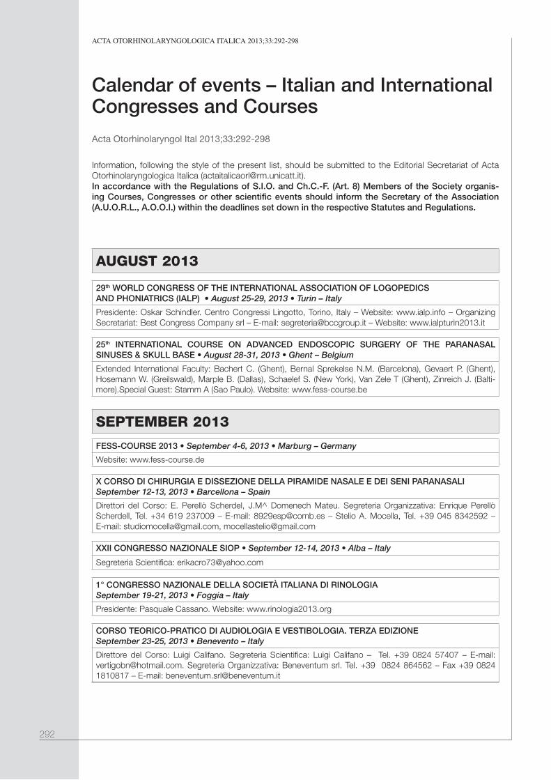
292
ACTA oTorhinolAryngologiCA iTAliCA 2013;33:292-298
Calendar of events – Italian and International Congresses and Courses
Acta Otorhinolaryngol Ital 2013;33:292-298
Information, following the style of the present list, should be submitted to the Editorial Secretariat of Acta Otorhinolaryngologica Italica ([email protected]). In accordance with the Regulations of S.I.O. and Ch.C.-F. (Art. 8) Members of the Society organis-ing Courses, Congresses or other scientific events should inform the Secretary of the Association (A.U.O.R.L., A.O.O.I.) within the deadlines set down in the respective Statutes and Regulations.
AUGUST 2013
29th WORLD CONGRESS OF THE INTERNATIONAL ASSOCIATION OF LOGOpEDICS AND pHONIATRICS (IALP) • August 25-29, 2013 • Turin – Italy
Presidente: Oskar Schindler. Centro Congressi Lingotto, Torino, Italy – Website: www.ialp.info – Organizing Secretariat: Best Congress Company srl – E-mail: [email protected] – Website: www.ialpturin2013.it
25th INTERNATIONAL COURSE ON ADVANCED ENDOSCOpIC SURGERY OF THE pARANASAL SINUSES & SKULL BASE • August 28-31, 2013 • Ghent – Belgium
Extended International Faculty: Bachert C. (Ghent), Bernal Sprekelse N.M. (Barcelona), Gevaert P. (Ghent), Hosemann W. (Greilswald), Marple B. (Dallas), Schaelef S. (New York), Van Zele T (Ghent), Zinreich J. (Balti-more).Special Guest: Stamm A (Sao Paulo). Website: www.fess-course.be
SEPTEMBER 2013
FESS-COURSE 2013 • September 4-6, 2013 • Marburg – Germany
Website: www.fess-course.de
X CORSO DI CHIRURGIA E DISSEZIONE DELLA pIRAMIDE NASALE E DEI SENI pARANASALI September 12-13, 2013 • Barcellona – Spain
Direttori del Corso: E. Perellò Scherdel, J.M^ Domenech Mateu. Segreteria Organizzativa: Enrique Perellò Scherdell, Tel. +34 619 237009 – E-mail: [email protected] – Stelio A. Mocella, Tel. +39 045 8342592 – E-mail: [email protected], [email protected]
XXII CONGRESSO NAZIONALE SIOP • September 12-14, 2013 • Alba – Italy
Segreteria Scientifica: [email protected]
1° CONGRESSO NAZIONALE DELLA SOCIETà ITALIANA DI RINOLOGIASeptember 19-21, 2013 • Foggia – Italy
Presidente: Pasquale Cassano. Website: www.rinologia2013.org
CORSO TEORICO-pRATICO DI AUDIOLOGIA E VESTIbOLOGIA. TERZA EDIZIONESeptember 23-25, 2013 • Benevento – Italy
Direttore del Corso: Luigi Califano. Segreteria Scientifica: Luigi Califano – Tel. +39 0824 57407 – E-mail: [email protected]. Segreteria Organizzativa: Beneventum srl. Tel. +39 0824 864562 – Fax +39 0824 1810817 – E-mail: [email protected]

Cal
end
ar o
f ev
ents
293
LA CHIRURGIA ENDOSCOpICA RINOSINUSALE DALLA TEORIA ALLA pRATICA – Quarta edizioneSeptember 23-25, 2013 • Pavia – Italy
Segreteria Organizzativa: Nadirex International srl, via Riviera 39, 27100 Pavia. Tel. +39 0382 525714 – Fax +39 0382 525736 - E-mail: [email protected] – Website: www.nadirex.com
CHIRURGIA RICOSTRUTTIVA - CORSI PRATICI MONOTEMATICI • September 25-27, 2013 • Rome – Italy
Divisione di Otorinolaringoiatria e Chirurgia Cervico-Facciale dell’Istituto Nazionale Tumori “Regina Elena”, via Elio Chianesi 53, 00144 Roma. Direttore del Corso: Giuseppe Spriano. Segreteria Organizzativa: Andreina Zaccheddu – Tel. +39 06 52666770
12° CONGRESSO NAZIONALE A.I.O.L.p. - LA TECNOLOGIA AL SERVIZIO DELL’AMbULATORIO ORLSeptember 27-28, 2013 • Milan – Italy
Segreteria Scientifica: Marco Capelli – Tel. 333.3753103. Segreteria Organizzativa: Erika Monese – Tel. 339.2168235
AAO-HNSF 2013 ANNUAL MEETING & OTO EXPO • September 29 - October 2, 2013 • Vancouver – Canada
Vancouver Convention Centre, 1055 Canada Place, Vancouver, BC, V6C 0C3 Canada. Website: www.entannualmeeting.org
OcTOBER 2013
VII INTERNATIONAL SYMpOSIUM ON RECENT ADVANCES IN RHINOSINUSITIS AND NASAL pOLYpOSISOctober 4-6, 2013 • Matsue city, Shimane – Japan
Website: www.npcg7.umin.jp – E-mail: [email protected]
FINESSE IN FACIAL pLASTIC SURGERY • October 10-14, 2013 • Regensburg – Germany
Website: www.facial-plastic-surgery.eu
27° CORSO “RINOpLASTICA – FONDAMENTI E TECNICHE ESSENZIALI DI RINOpLASTICA CHIUSA”October 15-19, 2013 • Florence – Italy
Direttore del Corso: Alberto Scattolin, Centro Studi Micheli Pellegrini. Segreteria Organizzativa: Nord Est Con-gressi. E-mail: [email protected] – Website: www.nordestcongressi.it
EUROpEAN UNION OF HEARING AID ACOUSTICIANS (EUHA) 58th INTERNATIONAL CONGRESS OF HEARING AID ACOUSTICIANS (EUHA) • October 16-18, 2013 • Nuremberg – Germany
Website: www.euha.org
XXXIV CONGRESSO NAZIONALE SIAF – DISAbILITà UDITIVA FIGURE pROFESSIONALI E SERVIZI SANITARI • October 16-19, 2013 • Venice – Italy
Website: www.congresso-siaf2013.it
XXXVII CONVEGNO NAZIONALE DI AGGIORNAMENTO AOOI October 18-19, 2013 • Feroleto Antico (CZ) – Italy
Presidente: Raffaele Grasso. Segreteria Organizzativa: L’Orsa Maggiore s.r.l., II Trav. A. De Gasperi 4, 89900 Vibo Valentia. Tel. +39 0963 43538 – Website: www.aooi2013.it
THE 2nd MEDITERRANEAN FESS COURSE - FROM bASICS TO ADVANCED ENDOSCOpIC SINUS SURGERy • October 18-19, 2013 • Malta
Coordinators: Mario Said, Alberto Dragonetti. Contact information: E-mail: [email protected] – Website: www.maltime.com
SECONDO CORSO “LIVE-SURGERY” DI CHIRURGIA ENDOSCOpICA TRANS-NASALE.DALL’ANTROSTOMIA MEDIA ALL’ODONTOIDECTOMIA • October 21-23, 2013 • Brescia – Italy
Direttore del Corso: Piero Nicolai. Segreteria Scientifica: Andrea Bolzoni Villaret, Davide Lombardi. Segreteria Organizzativa: Katia Gissi - E-mail: [email protected] – Website: www.servizicec.it

Cal
end
ar o
f ev
ents
294
8th SURGICAL ANATOMY IN HEAD & NECk CANCERS pROCEDURESOctober 23-25, 2013 • Paris – France
Directors: Marco Benazzo, Department of Otorhinolaryngology, University of Pavia; Fausto Giuseppe Chiesa, Department of Head and Neck Surgery, IEO Milan; Piero Nicolai, Department of Otorhinolaryngology, University of Brescia; Antonio Pastore, Department of Otorhinolaryngology, University of Ferrara – Scientific Secretariat: N. Mevio, F. Mura, D. Scelsi, M. Tagliabue – E-mail: [email protected]. Organizing Secretariat: Bquadro Congressi srl, via S. Giovanni in Borgo 4, 27100 Pavia. Tel. +39 0382 302859 – Fax +39 0382 27697 – E-mail: bolla@bquadro-
2nd bULGARIAN-ITALIAN MEETING ON RHINOLOGY & 6th ENDOSCOpIC SINUS SURGERY COURSEOctober 24-26, 2013 • Trieste - Italy
Directors: Alessandro Varini, ENT Dept, Casa di Cura Salus Trieste, Italy – Dilyana Vicheva, ENT Clinic, Plovdiv, Bulgaria. Scientific Secretariat: A. Varini – Tel. +39 040 3171111 – E-mail: [email protected]. Organizing Secretariat: The Office Trieste – Tel. +39 040 368343 – Fax +39 040 368808 – E-mail: [email protected] – Website: www.theoffice.it
STATE OF THE ART ENDOSCOpIC SkULL bASE SURGERY A HANDS ON COURSEOctober 31- November 3 2013 • Columbus, Ohio – USA
Course Directors: Ricardo L. Carrau – E-mail: [email protected]; Bradley A. Otto; Daniel M. Preve-dello
nOvEMBER 2013
2013 ANNUAL MEETING OF THE ISRAELI SOCIETY OF HEAD AND NECk SURGERY AND ONCOLOGYNovember 6-7, 2013 • Dead Sea – Israel
Website: www.ishnos.com
32nd ISIAN, 15th IRS, 4th pARS, 19th ORL EGYpT • November 6-9, 2013 • Sharm El Sheikh – Egypt
President: R. Kamel. Secretary General: A. Atef. International Coordinator: H. Negm. Website: www.isian.irs-pars2013.org – E-mail: [email protected]
CONVEGNO AOIG: I SENSI E L’INVECCHIAMENTO • November 8, 2013 • Milan – Italy
Presidente: Matteo Richichi. Website: www.aiog.it
1st GLOBAL OTOLOGy RESEARCH FORUM • November 13, 2013 • Antalya – Turkey
Scientific Secretary: Armağan İncesulu, [email protected], [email protected] – Organization Secre-tary: Tuncay Özçelik – E-mail: [email protected] – Website: www.glorf.org
29th WORLD CONGRESS POLITZER SOCIETy MEETING • November 13-17, 2013 • Belek-Antalya – Turkey
Info: Contact Information: [email protected] – Website: www.politzer2013.org
II CORSO “SCUOLA DI DISSEZIONE ANATOMICA CERVICO-FACCIALE”November 18-23, 2013 • Florence – Italy
I sessione • Ringiovanimento non chirurgico del viso – D. Draganic, R. Polselli, A. Rusciani, Y. Saban• Chirurgia endoscopica seni paranasali (base) – P. Bossolesi, E. Emanuelli, F.G. Pagella• Rinoplastica – L. D’Ascanio, G. La Fauci, R. Polselli, A. Scattolin• Chirurgia del collo – A. Camaioni, M. Radici, G. Spriano, L. D’Ascanio• Chirurgia endoscopica orecchio medio – D. Marchionni, L. PresuttiDirettore del Corso: Alberto Scattolin. Segreteria Organizzativa: Nord Est Congressi – E-mail: [email protected] – Website: www.nordestcongressi.it
SVUOTAMENTO DEL COLLO - CORSI PRATICI MONOTEMATICI • November 20-22, 2013 • Rome – Italy
Divisione di Otorinolaringoiatria e Chirurgia Cervico-Facciale dell’Istituto Nazionale Tumori “Regina Elena”, via Elio Chianesi 53, 00144 Roma. Direttore del Corso: Giuseppe Spriano. Segreteria Organizzativa: Andreina Zaccheddu – Tel. +39 06 52666770.

Cal
end
ar o
f ev
ents
295
DEcEMBER 2013
pSO-HNS 57TH ANNUAL CONVENTION – CURRENT OTOLOGICAL CONCEpTS AND FUTURE TRENDSDecember 1-3, 2013 • Manila – Philippines
E-mail: www.pso-hns.org
RHINOFORUM 2013 • December 6-7, 2013 • Warsaw – Poland
President: Antoni Krzeski. Website: www.RhinoForum.pl
TEMPORAL BONE DISSECTION COURSES 2013 • December 10-13, 2013 • Brazil
Website: www.forl.org.br/courses
jAnUARy-DEcEMBER 2013
THE INTERNATIONAL SOCIETy OF SURGICAL ANATOMy • CORSI 2013 • Nizza – France
February 27 - March 1: Chirurgia dei seni paranasali FESS. Direttori: E. Emanuelli, F. Pagella June 26-28: Anterior skull base. Direttori: S. Chibbaro, P. PasquisJune 28-29: Estetica del volto. Direttore: M. SabbaliniDecember 4-6: Chirurgia dei seni paranasali FESS. E. Emanuelli, F. Pagella December 6-7: Rinosettoplastica. Direttori: R. Polselli, Y. Saban
1° CORSO INTERNAZIONALE (2013): ENDOSCOpIA NASO-SINUSALE DI bASEFebruary 28 - March 1: Rome – ItalyMay 2-3: Siena – Italy September 21-22: Foggia – ItalyNovember 14-15: Udine – Italy
Coordinatori: Gaetano Paludetti, Desiderio Passali, Marco Piemonte. Anatomia radiologica e chirurgica naso-sinusale. Clinica della patologia naso-sinusale. Terapia medica e chirurgica della patologia naso-sinusale. Ricerca scientifica in ambito naso-sinusale. Dissezione su testa di agnello. Con il patrocinio della Società Italiana di Rinologia Comitato Educational. Segretria Organizzativa: E-mail: [email protected]
CORSI DI VIDEOCHIRURGIA ENDOSCOPICA NASO-SINUSALE E DEL BASICRANIO • Milano – ItalyMarch 4-8: Corso baseJune 10-15: Corso avanzatoNovember 25-29: Corso intermedio
Direttore: Alberto Dragonetti – E-mail: [email protected]. Segreteria Scientifica: Gabriella Mantini, Val-entina Casoli – Tel. +39 02 64444545 – Fax +39 02 64444003 – E-mail: [email protected] Organizzativa: Eurocompany Srl, via Canova 19, 20145 Milano. Tel. +39 02 315532 – Fax +39 02 33609213 – E-mail: [email protected]
THE MODERN SINONASAL SURGERY: ANATOMY, DIAGNOSTICS AND OpERATIVE TECHNIQUES Varese – ItalyBasic Course • April 8-10 and October 14-16Advanced Course • July 3 and November 18-20
Segreteria Organizzativa: Attingo – Tel. 377 3217150 – E-mail: [email protected]
TEMPORAL BONE SURGICAL DISSECTION COURSE • Barcelona – SpainCourse n. 110 • April 10-12 Course n. 111 • July 3-5 Course n. 112 • November (to be announced)
Information: Instituto de Otología García-Ibáñez, Conchi Castilla, C/ Dr. Roux 91, 08017 Barcelona, Spagna. Tel. +34 93 205 02 04 – Fax +34 93 205 43 67 – E-mail: [email protected], [email protected]
CORSI DI RINOLOGIA “FULL IMMERSION” ANNO 2013 • Imola (BO) – ItalyCorso Basico • June 17-21 Corso Avanzato • October 7-11
Direttore dei Corsi: Ignazio Tasca – E-mail: [email protected] – Website: www.associazionerinologia.it Segreteria del Corso: Filippo Sorace – Tel. +39 051 695 5251 – E-mail: [email protected]. Giacomo Ceroni Compadretti – Tel. +39 051 695 5251 – E-mail: [email protected]. Cristiana Di Lieto – E-mail: [email protected]

Cal
end
ar o
f ev
ents
296
CURSO DE DISECCIÓN ENDOSCÓpICA DE LOS SENOS pARANASALES – ENDOSCOpIC SINUS SURGICAL DISSECTION COURSE • Course n. 47 - Date to be announced • Barcelona – Spain
Instituto de Otología García-Ibáñez, C/ Dr. Roux 91, 08017 Barcelon, Spain. Tel. 93 205 02 04 – Fax 93 205 43 67 - E-mail: [email protected]
CORSI DI DISSEZIONE ANATOMO-CHIRURGICI 2013 • MaltaRhinoseptoplasty, mentoplasty, tip and profile correction • September 23-24Reconstructive laringectomy; CHEP, CHP, SOVRAGLOTTIC • September 24-25FESS dissectional course (English course) • October 18-19
Anatomy Dep. Head: P. Wiesmeyer, University of Malta. Scientific Secretariat: Attilio Denaro, AMSO-Onlus, S.Me.M.C. Websites: www.amso-onlus.it, www.smemc.tumblr.com, www.facebook/amsosmemc, https://twitter.com/amsosmemc. Organizing Secretariat: Kalòs Convegni, via Milano 201, 97019 Vittoria (RG). Tel. e Fax +39 0932 510221 – E-mail: [email protected]
jAnUARy-DEcEMBER 2014
CORSO DI CHIRURGIA OTOLOGICA, OTONEUROLOGICA E IMpLANTOLOGIA UDITIVA – Dissezione dell’osso temporale e del basicranio • January 7-9, 2014 • Paris – France
Direttore del Corso: Olivier Sterkers. Info: Daniele Bernardeschi, Reparto di Otorinolaringoiatria e Chirurgia Cervico-facciale, Ospedale Pitié-Salpétrière e Università Paris Diderot - Paris VII, France. E-mail: [email protected]
II CORSO “SCUOLA DI DISSEZIONE ANATOMICA CERVICO-FACCIALE” January 20-24, 2014 • Florence – Italy
II sessione• Blefaroplastica e otoplastica – P. Persichetti, R. Polselli, A. Rusciani, Y. Saban• Chirurgia endoscopica seni paranasali (avanzato) – P. Bossolesi, E. Emanuelli, F.G. Pagella• Rinoplastica – L. D’Ascanio, G. La Fauci, R. Polselli, A. Scattolin• Anatomia chirurgica della laringe – M. Lucioni, G. Rizzotto, G. Succo, L. D’AscanioDirettore del Corso: Alberto Scattolin. Segreteria Organizzativa: Nord Est Congressi – E-mail: [email protected] – Website: www.nordestcongressi.it
I CORSO – RINGIOVANIMENTO NON CHIRURGICO DEL VISO January 30 - February 1, 2014 • Florence – Italy
Faculty: D. Draganic, A. Rusciani, R. Polselli, Y. Saban. Segreteria Organizzativa: Nord Est Congressi. E-mail: [email protected] – Website: www.nordestcongressi.it
TEMpORAL bONE DISSECTION COURSES 2014February and June 2014 (dates to be announced) • Brazil
Website: www.forl.org.br/courses
3° CORSO TEORICO pRATICO DI LARINGOLOGIA pEDIATRICA • February 3-4, 2014 • Modena – Italy
Segreteria Organizzativa: Meet and Work s.r.l., p.zza del Sole e della Pace 5, 35031 Abano Terme (PD), Italy. Tel. +39 049 8601818 – Fax +39 049 8602389 – E-mail: [email protected]
ATLANTIC OTOLARYNGOLOGY HEAD AND NECk SURGERY UpDATEFebruary 15-17, 2014 • San Juan, Puerto Rico
Website: www.HopkinsCME.edu
11th RHINOCAMP WINTER • March 12-16, 2014 • Saint Moriz – Switzerland
Website: www.rhinocampwinter.org
AMERICAN ACADEMY OF AUDIOLOGY AAA ANNUAL CONVENTIONMarch 26-29, 2014 • Orlando, Fla – USA
Website: www.audiologynow.org
ASOHNS ASM 2014 MODERN AppROACHES TO ENTMarch 29-April 1, 2014 • Brisbane Queensland – Australia
Website: www.asohns.consec.com.au

Cal
end
ar o
f ev
ents
297
10th CONGRESS OF THE EUROpEAN LARYNGOLOGICAL SOCIETY – 2nd JOINT MEETING OF AbEA & ALAApril 9-12, 2014 Antalya, Turkey
Website: www.els2014.org
18th INTERNATIONAL VOICE WORkSHOp 2014 • April 11-12, 2014 • Paris – France
Information to: Jean Abitbol, 1, Rue Largillière – 705016 Paris. E-mail: [email protected]
4th MIDDLE EAST CONGRESS ON RHINOLOGY AND FACIAL pLASTIC SURGERY (MERC2014)April 12-14, 2014 • Tehran – Iran
Website: www.merc2014.com
18th WCbIp/WCbE18th WORLD CONGRESS FOR bRONCHOLOGY AND INTERVENTIONAL pULMONOLOGY18th WORLD CONGRESS FOR bRONCHOESOpHAGOLOGYApril 13-17, 2014 • Kyoto – Japan
Website: www2.convention.co.jp/wcbipwcbe2014/
THE FOURTH MIDDLE EAST CONGRESS ON RHINOLOGY & FACIAL pLASTIC SURGERYApril 23-25, 2014 • Tehran – Iran
Website: www.merc2014.com
3rd INTERNATIONAL SYMpOSIUM ON OTOSCLEROSIS AND STApES SURGERYApril 24-26, 2014 • Siófok – Hungary
President of the Congress: I. Sziklai – Secretary of the Congress: T. Karosi. Website: www.otosclerosis2014.com
2nd ANNOUNCEMENT OTOLOGY JUbILEE: 150 YEARS OF THE ARCHIV FUR OHORENHEILkUNDEpast-present-Future in Otology & Neurology • May 7-10, 2014 • Halle/Saale – Germany
Website: www.otology-jubilee.com
3rd IRANIAN CONGRESS ON COCHLEAR IMpLANT & RELATED SCIENCEMay 10-12, 2014 • Tehran – Iran
IRANCI 2014 Secretariat: Tel. - Fax 098-21-8860-0006 – E-mail: [email protected] – Website: www.iran-cochlear.com
24th CONGRESS OF EUROpEAN RHINOLOGIC SOCIETY (ERS) and 32nd INTERNATIONAL SYMpOSIUM OF INFECTION AND ALLERGY OF THE NOSE. THE NOSE AS INTERFACE June 22-26, 2014 • Amsterdam – The Netherlands
President: W.J. Fokkens. Website: www.ers-isian2014.com – E-mail: [email protected]
5th WORLD CONGRESS OF THE INTERNATIONAL FEDERATION OF HEAD AND NECk ONCOLOGIC SOCIETIES (IFHNOS) – ANNUAL MEETING OF THE AMERICAN HEAD AND NECk SOCIETY (AHNS) July 26-30, 2014 • New York – USA
Congress Chairman: Jatin Shah. Program Chairman: Bevan Yueh. Websites: www.ifhnos2014.org, www.ahns2014.org
BEST EVIDENCE ENT 2014 • August 2-5, 2014 • Wisconsin – USA
Course directors: John S. Rhee, David R. Friedland, Charles J. Harkins. Department of Otolaryngology 9200 West Wisconsin Avenue Milwaukee, WI 53226
40° CONGRESSO CONVENTUS SOCIETAS ORL LATINA • September 1-5, 2014 • Baia de Luanda – Angola
Info: Departamento de ORL da Facultdade de Medicina da Universidade Agostino Neto Hospital Josina Machel- Maria Pia Av. 1° Congresso do MPLA. Tel. 00244-923784901/914381304 – E-mail: [email protected], [email protected]
7th INSTRUCTIONAL WORkSHOp – CONSENSUS IN AUDITORY IMpLANTS “EUpOREAN GUIDELINES IN OTOLOGy AND NEURO-OTOLOGy” • September 13-16, 2014 • Siena – Italy
Website: www.eaono2014.org
EUROpEAN UNION OF HEARING AID ACOUSTICIANS (EUHA) 59th INTERNATIONAL CONGRESS OF HEARING AID ACOUSTICIANS (EUHA) • October 15-17, 2014 • Hanover – Germany
Website: www.euha.org

Cal
end
ar o
f ev
ents
298
5th ASIAN FACIAL PLASTIC SURGERy SOCIETy CONGRESS • October 15-19, 2014 • Cappadocia – Turkey
Website: www.afpss2014.org
jAnUARy-DEcEMBER 2015
3rd CONGRESS OF CE ORL-HNS • June 7-11, 2015 • Prague – Czech Republic
Website: Congress secretariat: GUARANT International Na Pankraci 17, 14021 Prague4, Czech Republic. Website: www.CEorl-hnsprague2015.com
22nd INTERNATIONAL CONGRESS ON THE EDUCATION OF THE DEAF • July 6-9, 2015 • Athens – Greece
Website: www.iced2015.com
WORLD CONGRESS ON LARyNX CANCER 2015 • July 26-30, 2015 • Queensland – Australia
Website: www.wclc2015.org


RIASSUNTO DELLE CARATTERISTICHE DEL PRODOTTO1. DENOMINAZIONE DEL MEDICINALE Unidrox 600 mg compresse rivestite con film. 2. COMPOSIZIONE QUALITATIVA E QUANTITATIVA Prulifloxacina 600 mg. Eccipienti: ogni compressa rivestita con film contiene lattosio 76 mg. Per l’elenco completo degli eccipienti, vedere paragrafo 6.1. 3. FORMA FARMACEUTICA compresse rivestite con film. Compresse oblunghe, di color giallo, rivestite con film. 4. INFORMAZIONI CLINICHE 4.1 Indicazioni terapeutiche Unidrox è indicato per il trattamento di infezioni sostenute da ceppi sensibili, nelle seguenti patologie: infezioni acute non complicate delle basse vie urinarie (cistite semplice); ● infezioni complicate delle basse vie urinarie; ● riacutizzazione di bronchite cronica; ● rinosinusite batterica acuta. La sinusite batterica acuta deve essere adeguatamente diagnosticata in accordo alle linee guida nazionali o locali sul trattamento delle infezioni respiratorie. Per il trattamento della rinosinusite batterica, Unidrox deve essere usato solo in pazienti nei quali la durata dei sintomi sia inferiore a 4 settimane e quando l’impiego di altri antibatterici comunemente raccomandati per il trattamento iniziale di tale infezione venga considerato inappropriato, o nel caso in cui questi siano risultati inefficaci. Nel trattamento di pazienti con malattie infettive, si deve tener conto delle caratteristiche locali relative alla sensibilità agli antibiotici. 4.2 Posologia e modo di somministrazione Limitatamente agli adulti, la posologia indicativa è la seguente: ● pazienti con infezioni acute non complicate delle basse vie urinarie (cistite semplice): è sufficiente una sola compressa da 600 mg. ● pazienti con infezioni complicate delle basse vie urinarie: una compressa da 600 mg una volta al giorno fino ad un massimo di 10 giorni di trattamento. ● pazienti con riacutizzazione di bronchite: una compressa da 600 mg una volta al giorno fino ad un massimo di 10 giorni di trattamento. ● pazienti con rinosinusite batterica acuta: una compressa da 600 mg una volta al giorno fino ad un massimo di 10 giorni di trattamento. In caso di infezioni complicate delle basse vie urinarie e riacutizzazione di bronchite cronica, la durata del trattamento dipende dalla gravità della malattia e dal decorso clinico del paziente e deve comunque proseguire per almeno 48-72 ore dalla remissione/scomparsa dei sintomi. Le compresse di Unidrox devono essere deglutite intere con acqua e devono essere assunte tenendo conto dell’assunzione di cibo (vedere Paragrafo 4.5). Per la mancanza di studi specifici non è possibile determinare la posologia in pazienti con insufficienza renale (pazienti con clearance della creatinina < 60 ml/min) ed in pazienti con insufficienza epatica. Pertanto, in questi pazienti il monitoraggio dei livelli plasmatici del farmaco costituisce il metodo più affidabile per l’adattamento del dosaggio. 4.3 Controindicazioni - Ipersensibilità alla prulifloxacina, ad altri antibatterici chinolonici o ad uno qualsiasi degli eccipienti. - Bambini prima dell’età puberale o ragazzi al di sotto dei 18 anni con incompleto sviluppo scheletrico. - Pazienti con anamnesi di affezioni tendinee correlate alla somministrazione di chinoloni. - Gravidanza e allattamento (vedere paragrafo 4.6). 4.4 Avvertenze speciali e precauzioni di impiego Come per gli altri chinolonici, Unidrox deve essere usato con cautela in pazienti con disturbi del SNC che possano predisporre alle convulsioni o abbassare la soglia convulsiva. Alcune delle altre sostanze appartenenti alla classe dei fluorochinoloni sono state associate a casi di prolungamento dell’intervallo QT. Prulifloxacina ha un potenziale molto basso per induzione di prolungamento dell’intervallo QT. Come a seguito della somministrazione di altri farmaci della stessa classe terapeutica, la tendinite si manifesta raramente. Più frequentemente interessa il tendine di Achille e può portare fino alla sua rottura. Il rischio di tendinite e di rotture tendinee è aumentato nei pazienti anziani e nei pazienti in trattamento con corticosteroidi. I pazienti devono essere informati, in caso di comparsa di segni di infiammazione tendinea, mialgia, dolore o infiammazione a livello articolare, d’interrompere il trattamento e di mantenere a riposo l’arto o gli arti interessati sino a che la diagnosi di tendinite non sia stata esclusa. Il trattamento con antimicrobici, inclusi i chinoloni, può determinare la comparsa di colite pseudomembranosa. Pertanto, in caso di diarrea successiva alla somministrazione di antimicrobici è importante considerare tale possibilità. L’esposizione al sole o a raggi ultravioletti può causare la comparsa di fototossicità in pazienti in trattamento con prulifloxacina, così come con altri chinoloni. Durante il trattamento con Unidrox l’eccessiva esposizione al sole o a raggi ultravioletti deve essere evitata; in caso di comparsa di fototossicità, il trattamento deve essere interrotto. I pazienti con difetti latenti o accertati per l’attività della glucosio-6-fosfato deidrogenasi, sono predisposti a reazioni emolitiche quando vengono trattati con antibatterici della classe dei chinoloni e per tale ragione Unidrox deve essere usato con cautela. Come riportato per altri chinoloni, possono raramente presentarsi fenomeni di rabdomiolisi, caratterizzati da mialgia, astenia, incremento dei valori plasmatici di CPK e mioglobina, e rapido deterioramento della funzionalità renale. In questi casi, il paziente deve essere attentamente controllato e devono essere intraprese adeguate misure correttive, compresa l’eventuale interruzione del trattamento. L’uso dei chinoloni è talvolta correlato alla comparsa di cristalluria; i pazienti in trattamento con questa classe di prodotti devono mantenere un adeguato bilancio idrico al fine di evitare la concentrazione delle urine. La tollerabilità e l’efficacia di Unidrox nei pazienti con insufficienza epatica non è stata valutata. Nel prescrivere una terapia antibiotica dovrebbero essere considerate le linee guida locali e/o nazionali sull’uso appropriato degli antibatterici. Il medicinale contiene lattosio; pertanto i pazienti con rari problemi ereditari d’intolleranza al galattosio, con deficit di Lapp lattasi o da malassorbimento di glucosio-galattosio, non devono assumere questo medicinale. Tenere fuori dalla portata e dalla vista dei bambini. 4.5 Interazioni con altri medicinali ed altre forme di interazione Il trattamento concomitante con cimetidina, antiacidi contenenti Al e Mg o preparazioni contenenti ferro e calcio riduce l’assorbimento di Unidrox; di conseguenza Unidrox dovrebbe essere somministrato 2 ore prima od almeno 4 ore dopo l’assunzione di questi preparati. L’assunzione contemporanea di prulifloxacina e latte determina un decremento dell’area sotto la curva di concentrazione/tempo (AUC) e riduce l’eliminazione urinaria della prulifloxacina, mentre l’ingestione di cibo rallenta e riduce i livelli di picco. L’escrezione urinaria di prulifloxacina diminuisce quando somministrata insieme al probenecid. La somministrazione concomitante di fenbufen con alcuni chinoloni può provocare un aumento del rischio di convulsioni; di conseguenza la somministrazione di Unidrox e fenbufen deve essere attentamente valutata. I chinoloni possono determinare ipoglicemia in pazienti diabetici che assumono farmaci ipoglicemizzanti. La somministrazione concomitante di Unidrox e teofillina può causare una lieve diminuzione della clearance della teofillina che non dovrebbe avere alcuna rilevanza clinica. Tuttavia, come per gli altri chinoloni, è consigliabile il monitoraggio dei livelli plasmatici di teofillina nei pazienti con disordini metabolici o che presentino fattori di rischio. I chinoloni possono incrementare gli effetti degli anticoagulanti orali come il warfarin ed i suoi derivati; qualora questi prodotti siano somministrati insieme ad Unidrox si raccomanda uno stretto monitoraggio con il test di protrombina o con altri affidabili test della coagulazione. Dati preclinici hanno dimostrato che la nicardipina può potenziare la fototossicità della prulifloxacina. Nessuna interazione clinicamente significativa è stata osservata nel corso dello sviluppo clinico di Unidrox a seguito della somministrazione concomitante con gli altri medicinali comunemente impiegati nel trattamento di pazienti affetti dalle patologie riportate al paragrafo 4.1. 4.6 Gravidanza ed allattamento Non sono disponibili dati clinici relativi all’impiego di prulifloxacina durante la gravidanza accertata. Studi sugli animali non hanno indicato teratogenicità. Altri effetti tossici sulla riproduzione sono stati rilevati soltanto in caso di tossicità materna (vedere paragrafo 5.3). Tuttavia, nel ratto, si è osservato che la prulifloxacina attraversa la barriera placentare e passa in gran quantità nel latte materno. Come per altri chinoloni, è stato dimostrato che prulifloxacina determina artropatie negli animali giovani, e pertanto il suo uso durante la gravidanza e l’allattamento è controindicato. 4.7 Effetti sulla capacità di guidare veicoli e sull’uso di macchinari I chinoloni possono causare vertigini e stato di confusione, pertanto, il paziente deve sapere come risponde al trattamento prima di guidare o usare macchinari o iniziare attività che richiedano vigilanza e coordinazione. 4.8 Effetti indesiderati Gli effetti indesiderati di seguito riportati sono riconducibili agli studi clinici effettuati con Unidrox. La maggior parte degli eventi avversi è stata di intensità lieve o moderata. Sono stati utilizzati i seguenti valori di frequenza: Molto comuni (≥ 1/10), Comuni (≥1/100, <1/10), Non comuni (≥ 1/1.000, <1/100), Rari (≥ 1/10.000, < 1/1.000) e Molto Rari (<1/10.000), non nota (la frequenza non può essere definita sulla base dei dati disponibili). Disturbi del metabolismo e della nutrizione – Non comuni: anoressia. Rari: perdita dell’appetito. Disturbi psichiatrici - Rari: disturbi del sonno, sonnolenza, confusione. Patologie del sistema nervoso – Non comuni: cefalea, capogiro. Rari: agitazione psicomotoria, perversione del gusto. Patologie dell’occhio – Rari: iperemia oculare. Patologie dell’orecchio e del labirinto – Rari: sensazione di orecchio chiuso. Patologie vascolari – Rari: vampate di calore. Patologie gastrointestinali - Comuni: epigastralgia. Non comuni: dolore addominale, diarrea, nausea, gastrite, vomito. Rari: feci anormali, patologie gastrointestinali, eruttazione, ulcera della bocca, stomatite angolare, dispepsia, flatulenza, indigestione, fastidio alla cavità orale, moniliasi orale, glossite, dilatazione gastrica. Patologie della cute e del tessuto sottocutaneo – Non comuni: prurito, rash cutaneo, eruzione. Rari: eczema della faccia, eritema della faccia, orticaria. Patologie del sistema muscoloscheletrico e del tessuto connettivo – Rari: dolore articolare diffuso, dolore alla caviglia, patologia muscolare, contrazione muscolare. Patologie sistemiche e condizioni relative alla sede di somministrazione – Rari: febbre. Esami diagnostici – Rari: albumina aumentata, fosfatasi alcalina aumentata, alanina amminotransferasi aumentata, aspartato aminotransferasi aumentata, calcemia aumentata, monociti ematici aumentati, linfociti aumentati, leucociti aumentati, γ GT aumentate, bilirubina aumentata. Le seguenti reazioni avverse sono state segnalate (frequenza non nota): reazione anafilattica/anafilattoide, sindrome di Steven Johnson, ipoglicemia, ipoestesia, dermatite da farmaci, rabdomiolisi, fototossicità. Il trattamento con Unidrox può essere associato a cristalluria asintomatica senza variazione dei livelli di creatinina, ad alterazioni dei parametri di funzionalità epatica ed eosinofilia. Nei casi osservati, tali modificazioni sono state asintomatiche e transitorie. Durante il trattamento con Unidrox non può essere esclusa la comparsa di reazioni avverse e alterazioni dei parametri di laboratorio sopra non citate, ma riportate per gli altri chinoloni. Dati di farmacovigilanza relativi a prulifloxacina e successivi all’immissione in commercio, mostrano sporadiche segnalazioni di tendinopatia (vedere 4.4. Avvertenze speciali e precauzioni di impiego). 4.9 Sovradosaggio La somministrazione orale nel topo, ratto e cane (maschi e femmine) di dosi singole sino a 5000 mg/kg non ha avuto effetti letali. Non sono disponibili informazioni sul sovradosaggio nell’uomo; Unidrox è stato somministrato sino alla dose di 1200 mg/die per 12 giorni in volontari sani mostrando nel complesso una buona tollerabilità. Nel caso di sovradosaggio in acuto, lo stomaco deve essere svuotato provocando vomito o praticando un lavaggio gastrico, il paziente deve essere attentamente seguito e trattato con terapia sintomatica. 5. PROPRIETÀ FARMACOLOGICHE 5.1 Proprietà farmacodinamiche Categoria farmacoterapeutica: fluorochinoloni, codice ATC: J01MA17. Prulifloxacina è un antibatterico appartenente alla classe dei fluorochinoloni dotato di ampio spettro di azione e di elevata efficacia. Dopo somministrazione orale, prulifloxacina viene assorbita dal tratto gastrointestinale ed immediatamente trasformata in ulifloxacina, suo metabolita attivo (vedere paragrafo 5.2). Meccanismo d’azione. Unidrox si è dimostrato attivo in vitro, nei confronti di un’ampia gamma di ceppi Gram-positivi e Gram-negativi. Prulifloxacina esercita la sua azione antibatterica inibendo selettivamente la DNA-girasi, un enzima vitale presente nei batteri, che è coinvolto nella duplicazione, trascrizione e riparazione del DNA. Meccanismo di resistenza. L’insorgenza di antibiotico-resistenza alla prulifloxacina (così come agli altri fluorochinoloni) è generalmente dovuta a mutazioni spontanee nell’ambito della girasi del DNA batterico. In vitro, è stata osservata resistenza crociata con altri fluorochinoloni. Per i particolari meccanismi di insorgenza di resistenza ai fluorochinoloni, non vi è resistenza crociata tra prulifloxacina ed antibiotici di classi diverse, quindi Unidrox può risultare efficace anche in presenza di ceppi batterici resistenti ad aminoglicosidi, penicilline, cefalosporine e tetracicline. Intervalli di inibizione. Sono stati definiti sulla base dei dati di attività antibatterica NCCLS e dei parametri farmacocinetici del prodotto. Si suggeriscono i seguenti intervalli di inibizione: Sensibili: MIC ≤ 1 µg/ml, Intermedi: MIC>1 fino a < 4 µg/ml, Resistenti: MIC ≥ 4 µg/ml. Spettro antibatterico. Occorre considerare che la prevalenza di resistenza acquisita per le specie selezionate può variare geograficamente e con il tempo, pertanto è auspicabile la disponibilità di informazioni locali sulla resistenza, particolarmente quando si trattano infezioni gravi. Se necessario, e qualora la prevalenza locale di resistenze possa rendere discutibile l’utilità del farmaco, si consiglia di richiedere un parere ad un esperto. I dati riportati nella tabella che segue indicano lo spettro antibatterico della prulifloxacina:

Specie comunemente sensibiliAerobi gram-positivi Staphylococcus aureus (meticillino-sensibile)
Staphylococcus epidermidis Streptococcus agalactiae Streptococcus pyogenes Streptococcus pneumoniae *
Aerobi gram-negativi Campylobacter jejuniCitrobacter freundii Citrobacter koseriiEnterobacter aerogenes Enterobacter cloacae Haemophilus influenzae Klebsiella oxytocaKlebsiella pneumoniae Legionella pneumophilaMoraxella catarrhalis Morganella morganii Neisseria gonorrhoeaeProteus mirabilis Proteus vulgaris Providencia rettgeriPseudomonas aeruginosa Salmonella sp.Shigella sp. (comprese S. flexneri e S. sonnei)
Anaerobi Clostridium perfringensPeptostreptococcus sp.Porphyromonas gingivalisPrevotella intermedia
Specie per le quali la resistenza acquisita può rappresentare un problemaAerobi Gram-positivi Enterococcus avium *
Enterococcus faecalis *Enterococcus faecium *
Aerobi Gram-negativi Acinetobacter calcoaceticus *Escherichia coli (compresi ceppi enteroemorragici ed enterotossici) Serratia marcescens *
Organismi intrinsecamente resistentiAerobi Gram-positivi Enterococcus vancomicina-resistente
Staphylococcus aureus meticillino-resistenteAerobi Gram-negativi Providencia stuartiiAnaerobi Bacteroides sp.
Clostridium difficile
* Specie che mostrano una naturale sensibilità intermediaAltre informazioni. Negli studi in vitro l’azione antibatterica di prulifloxacina è stata caratterizzata da una penetrazione batterica migliore e da un effetto post-antibiotico più prolungato rispetto ai fluorochinoloni di riferimento. 5.2 Proprietà farmacocinetiche a) Caratteristiche generali Prulifloxacina è il profarmaco del metabolita attivo, ulifloxacina. Assorbimento - Nell’uomo prulifloxacina è rapidamente assorbita (Tmax = circa 1h) e trasformata in ulifloxacina; dopo somministrazione singola di 600 mg il picco plasmatico medio di ulifloxacina è di 1,6 µg/ml e la AUC è di 7,3 µg*h/ml. Allo steady-state, che si raggiunge entro 2 giorni dall’inizio del trattamento con somministrazione unica giornaliera, il Cmax e l’AUC sono di 2,0 µg/ml e di 7,6 µg*h/ml, rispettivamente. Il cibo ritarda e riduce leggermente la concentrazione al picco plasmatico di ulifloxacina, ma non modifica la AUC. Distribuzione - Nell’uomo, il rapporto polmone/plasma della concentrazione media di Unidrox aumenta nel tempo e, dopo 24 ore, il metabolita attivo ulifloxacina mantiene concentrazioni tissutali medie di 5 volte superiori a quelle del plasma, confermando i risultati ottenuti nell’animale, dove le concentrazioni di ulifloxacina nel polmone e nel rene sono risultate più alte di quelle plasmatiche (1,2 – 2,8 volte e 3 – 8 volte, rispettivamente). Analogamente, dati nell’uomo sulla penetrazione tissutale di ulifloxacina nei seni paranasali hanno mostrato, in termini di AUC, un rapporto tra tessuto e plasma pari a 3,0 nell’etmoide e 2,4 nei turbinati. Il legame proteico nell’uomo, valutato sia in vitro che ex vivo, è pari a circa il 50%, indipendentemente dalla concentrazione del farmaco. La scarsa concentrazione di ulifloxacina riscontrata nel liquido cerebro-spinale dopo somministrazione i.v. nel cane e somministrazione ripetuta p.o. nell’uomo, indica che ulifloxacina difficilmente supera la barriera ematoencefalica. Biotrasformazione - Il profilo metabolico di prulifloxacina nell’animale e nell’uomo è comparabile. Gli studi nell’animale hanno dimostrato che il metabolismo di prulifloxacina inizia durante l’assorbimento intestinale e si completa con il suo passaggio nel fegato. Oltre alla trasformazione in ulifloxacina, sono stati identificati altri metaboliti minori, quali la forma diolica ed alcuni derivati come glucuronide, oxo-derivato e etilen-diammino derivati, la cui concentrazione ed attività è trascurabile rispetto al principio attivo. Negli studi in vitro non sono state osservate interazioni significative con gli isoenzimi del citocromo P-450, a parte una lieve inibizione del CYP1A1/2 che corrisponde ad una debole diminuzione della clearance di teofillina. Poiché le metilxantine, ed in particolare teofillina, costituiscono il substrato principale per l’isoenzima CYP1A1/2, il grado d’interazione con altri substrati dell’isoenzima (vedi warfarin) può considerarsi solo inferiore. Eliminazione - L’emivita del metabolita attivo, ulifloxacina, è di circa 10 ore, sia dopo somministrazione singola che ripetuta allo steady-state nell’uomo, mentre negli animali (ratti, cani e scimmie) varia tra le 2 e le 12 ore. Studi con il prodotto marcato nell’uomo hanno dimostrato che l’eliminazione avviene prevalentemente per via fecale. Dopo somministrazione orale di 600 mg, la radioattività ritrovata nelle urine e nelle feci ammonta in totale approssimativamente al 95%. Tali risultati confermano quanto evidenziato in precedenti studi effettuati sugli animali (ratti, cani e scimmie). La quantità di ulifloxacina escreta con le urine è il 16,7 % della dose somministrata su base molare e la clearance renale di ulifloxacina è di circa 170 ml/min. L’eliminazione renale di ulifloxacina avviene per filtrazione glomerulare e per secrezione attiva. b) Caratteristiche nei pazienti Il profilo farmacocinetico di prulifloxacina negli anziani si è dimostrato simile a quello degli adulti, senza variazioni in funzione dell’età, e pertanto non sono ritenute necessarie modifiche del dosaggio nei pazienti anziani. In pazienti con insufficienza renale lieve o moderata, dopo somministrazione orale di Unidrox 600 mg, il picco plasmatico medio di ulifloxacina raggiunge valori tra 1,30 e 1,62 µg/ml. I valori di AUC variano tra 13,71 e 23,33 µg*h/ml e l’emivita tra 12,3 e 32,4 ore. La clearance renale di ulifloxacina diminuisce rispetto ai volontari sani in funzione del grado di insufficienza. 5.3 Dati preclinici di sicurezza Tossicità ripetuta. In studi di tossicità con somministrazione ripetuta, cartilagini articolari, reni, apparato gastrointestinale e fegato sono risultati i principali organi bersaglio. Con dosi fino a 3 volte più alte rispetto a quelle terapeutiche non sono stati osservati effetti tossici sulle cartilagini articolari (cani giovani); con dosi fino a 6, 10 e 12 volte più alte di quelle terapeutiche non sono stati osservati effetti tossici nel fegato (cani) e rene (cani e ratti) . Il farmaco non prolunga l’intervallo QT in vivo e non dimostra effetti inibenti sulla corrente di potassio a rettificazione ritardata (HERG) in vitro.Tossicità riproduttiva. Gli studi di tossicità riproduttiva non hanno evidenziato teratogenicità. Effetti sulla fertilità o sullo sviluppo embrionale e fetale sono stati osservati soltanto in caso di tossicità materna. Mutagenicità. I saggi standard di genotossicità hanno evidenziato effetti positivi in alcuni test in vitro effettuati con prulifloxacina su colture di cellule di mammifero, ma sono risultati negativi in vivo e nei batteri. Si ritiene che tali effetti siano associati all’inibizione di topoisomerasi II in presenza di alte concentrazioni di prulifloxacina. Potenziale carcinogeno. La prulifloxacina non si è dimostrata cancerogena in un modello sperimentale di iniziazione-promozione a medio termine. Non sono state effettuate prove di carcinogenesi a lungo termine. Antigenicità. La prulifloxacina è risultata priva di effetti antigenici. Fototossicità. La prulifloxacina ha indotto reazioni fototossiche, sebbene in studi comparativi nell’animale abbia mostrato di possedere un’attività fototossica minore rispetto a quella degli altri fluorochinoloni impiegati (ofloxacina, enoxacina, pefloxacina, acido nalidixico e lomefloxacina). Molti chinoloni sono anche fotomutageni/fotocarcinogenici, la possibilità che anche la prulifloxacina abbia tali effetti non può essere esclusa. Nefrotossicità. Dopo somministrazione ripetuta per via orale di 3000 mg/kg/die nel ratto, un dosaggio molto superiore alla dose terapeutica nell’uomo, la prulifloxacina ha causato cristalluria per precipitazione di ulifloxacina. Cardiotossicità. Studi condotti nel cane hanno mostrato che prulifloxacina non provoca modificazioni di rilievo nell’elettrocardiogramma. In particolare, non è stato osservato alcun cambiamento del QTc né dopo singola somministrazione endovenosa nel cane anestetizzato, né dopo somministrazione orale per 6 mesi nel cane conscio, a tutte le dosi somministrate. Studi in vitro hanno confermato l’assenza di effetti inibenti sulle correnti rettificatrici ritardate del potassio (HERG). Tossicità articolare. La prulifloxacina, similmente agli altri fluorochinoloni, ha causato artropatia solo negli animali giovani. Tossicità oculare. Dosi orali di 26,4 o 58,2 mg/kg/die di prulifloxacina una volta al giorno per 52 settimane nella scimmia non hanno causato effetti avversi correlati al trattamento sulla funzionalità o morfologia oculare. Effetto rabdomiolitico. Dosi fino a 10 mg/kg/die di ulifloxacina somministrate per via intravenosa una volta al giorno per 14 giorni consecutivi non hanno indotto rabdomiolisi nel coniglio. 6. INFORMAZIONI FARMACEUTICHE 6.1 Elenco degli eccipienti Nucleo: lattosio monoidrato, cellulosa microcristallina, croscarmellosa sodica, povidone, silice colloidale anidra, magnesio stearato Rivestimento: ipromellosa, glicole propilenico, titanio diossido (E171), talco , ossido ferrico (E172). 6.2 Incompatibilità Non pertinente 6.3 Periodo di validità 3 anni. 6.4 Precauzioni particolari per la conservazione Non conservare a temperatura superiore ai 30°C. Conservare nella confezione originale. 6.5 Natura e contenuto del contenitore Astuccio di cartone contenente 1 blister da 1, 2, 5 compresse rivestite con film o 2 blister da 5 compresse rivestite con film. Blister in materiale accoppiato (Poliammide/alluminio/PVC) termosaldato con materiale ricoprente (alluminio/PVC). E’ possibile che non tutte le confezioni siano commercializzate. 6.6 Precauzioni particolari per lo smaltimento e la manipolazione Nessuna istruzione particolare. Il medicinale non utilizzato ed i rifiuti derivati da tale medicinale devono essere smaltiti in conformità alla normativa locale vigente. 7. TITOLARE DELL’AUTORIZZAZIONE ALL’IMMISSIONE IN COMMERCIO Aziende Chimiche Riunite Angelini Francesco - A.C.R.A.F. S.p.A, Viale Amelia, 70 – 00181 ROMA (Italia). 8. NUMERO DELL’AUTORIZZAZIONE ALL’IMMISSIONE IN COMMERCIO Scatola 1 compressa rivestita con film da 600 mg A.I.C. 035678034, Scatola 2 compresse rivestite con film da 600 mg A.I.C. 035678010, Scatola 5 compresse rivestite con film da 600 mg A.I.C. 035678022, Scatola 10 compresse rivestite con film da 600 mg A.I.C. 035678046 9. DATA DELLA PRIMA AUTORIZZAZIONE/RINNOVO DELL’AUTORIZZAZIONE Data della prima Autorizzazione: 21 Giugno 2004. Data del rinnovo dell’Autorizzazione: 21 Giugno 2009 10. DATA DI REVISIONE DEL TESTO Giugno 2011
INFORMAZIONI AGGIUNTIVEClasse A-RR, 5 compresse rivestite con film da 600 mg: 20,57€Dep. AIFA in data 27/07/12