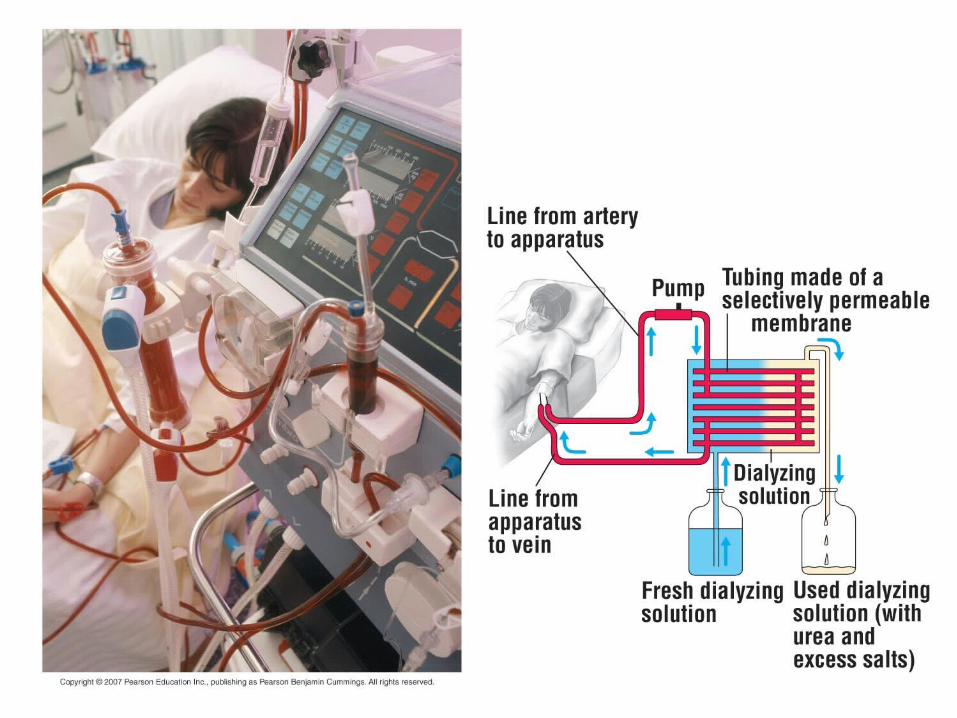
osmoregulation and excretion. osmoregulation: regulation of solute concentrations and water balance...
TRANSCRIPT
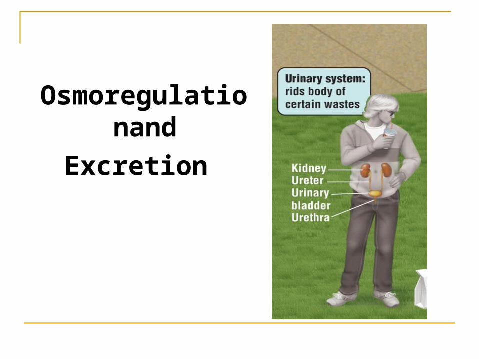
Osmoregulationand
Excretion
Osmoregulation: Regulation of solute concentrations and water balance by a cell or organism.
Osmoregulation balances the uptake and loss of water and solutes.
If cells uptake too much water they will burst If cells uptake too little water, or lose too
much water they will shrivel and die. Osmoregulation is impacted by water
intake/loss and also discharge of metabolic wastes.
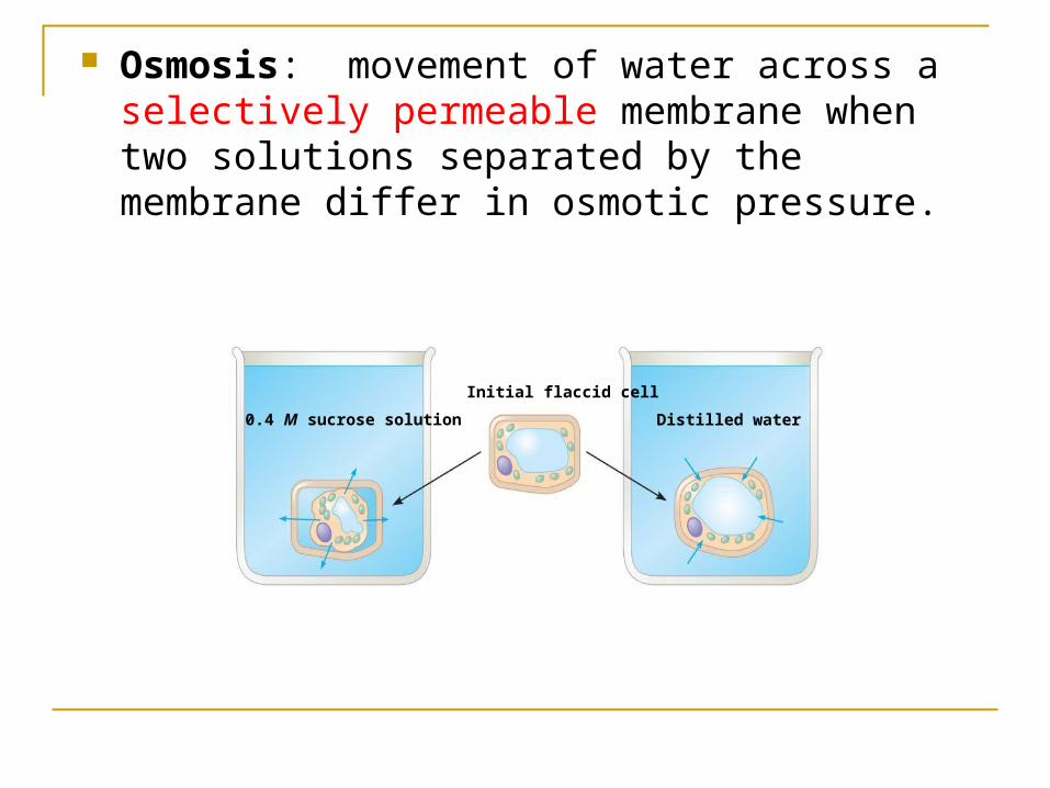
Osmosis: movement of water across a selectively permeable membrane when two solutions separated by the membrane differ in osmotic pressure.
Initial flaccid cell
0.4 M sucrose solution Distilled water
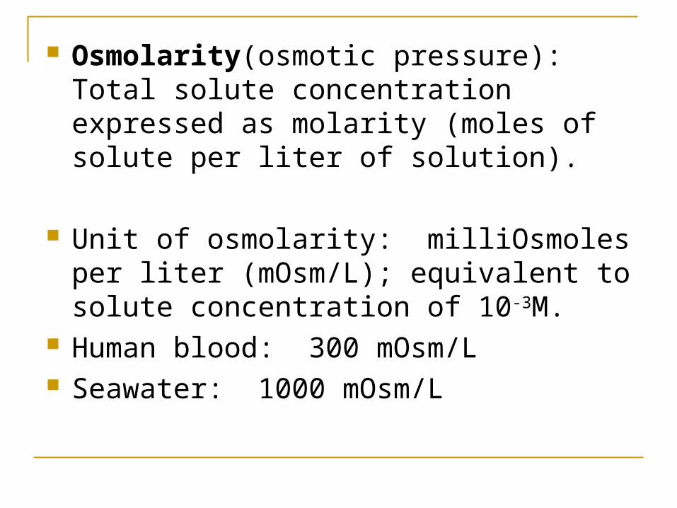
Osmolarity(osmotic pressure): Total solute concentration expressed as molarity (moles of solute per liter of solution).
Unit of osmolarity: milliOsmoles per liter (mOsm/L); equivalent to solute concentration of 10-3M.
Human blood: 300 mOsm/L Seawater: 1000 mOsm/L
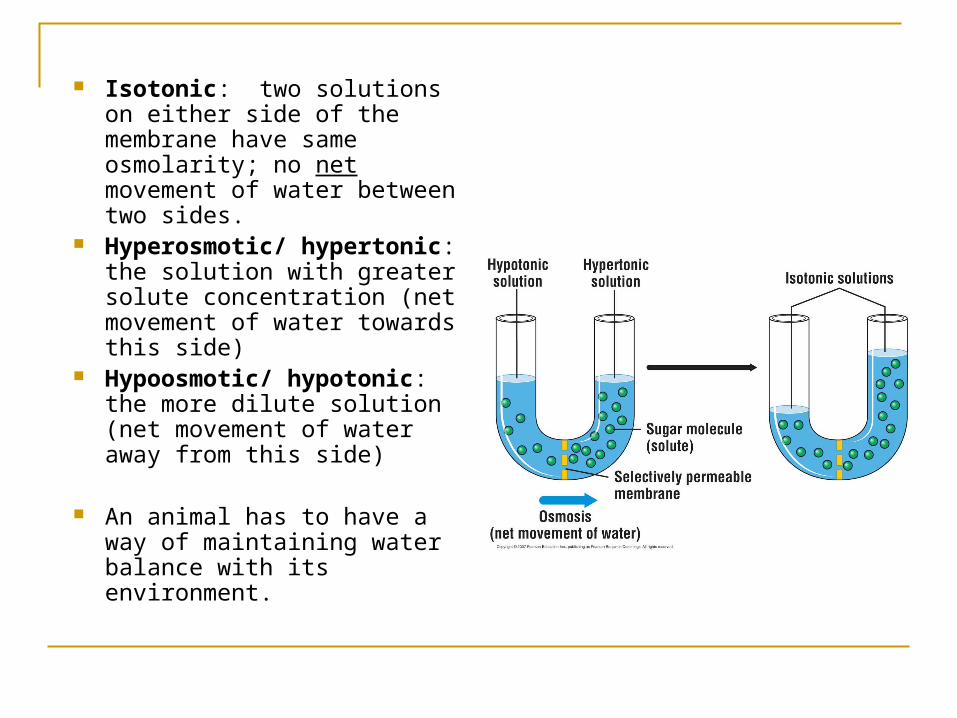
Isotonic: two solutions on either side of the membrane have same osmolarity; no net movement of water between two sides.
Hyperosmotic/ hypertonic: the solution with greater solute concentration (net movement of water towards this side)
Hypoosmotic/ hypotonic: the more dilute solution (net movement of water away from this side)
An animal has to have a way of maintaining water balance with its environment.

Osmoconformer: Isoosmotic with the surroundings Internal osmolarity same as the environment No net gain or loss of water All marine
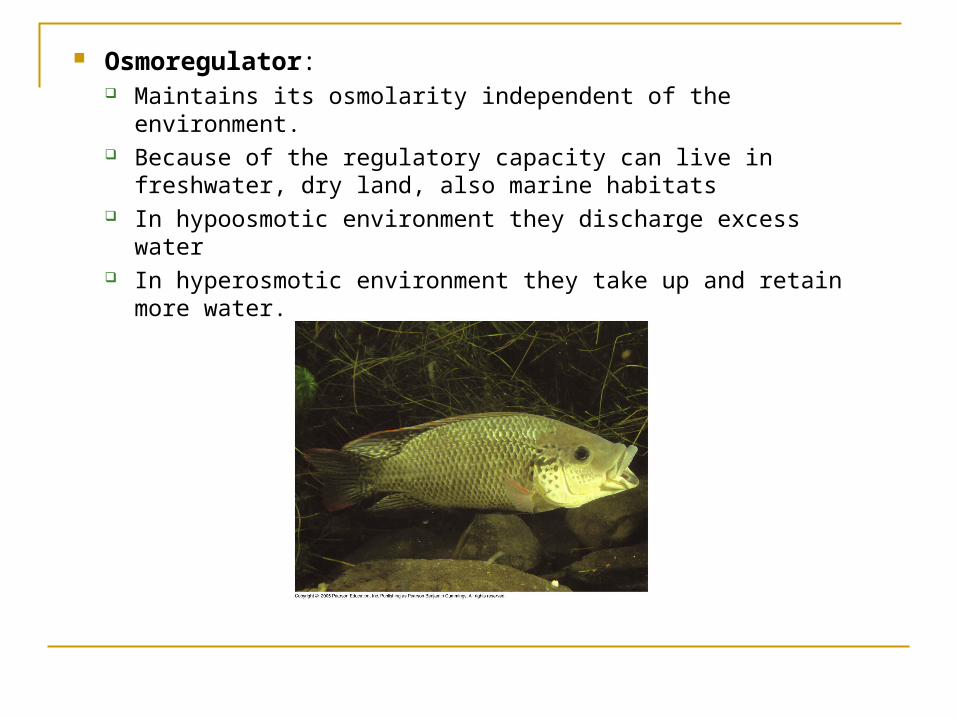
Osmoregulator: Maintains its osmolarity independent of the environment. Because of the regulatory capacity can live in freshwater, dry
land, also marine habitats In hypoosmotic environment they discharge excess water In hyperosmotic environment they take up and retain more
water.

Based on what you know so far about thermoregulation can you make any guesses about osmoregulation in these two groups? Balancing act between control and cost.
Stenohaline: Cannot tolerate substantial changes in external osmolarity
Euryhaline: Can tolerate large fluctuations in osmolarity (organisms in intertidal zones, fishes that migrate between sea water and fresh water)
Osmoregulators and osmoconformers can be found in both these groups.
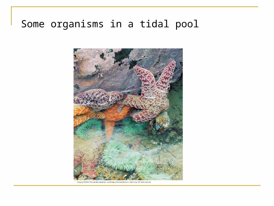
Some organisms in a tidal pool
Marine invertebrates: osmoconformes, need to transport some solutes to maintain homeostatis.
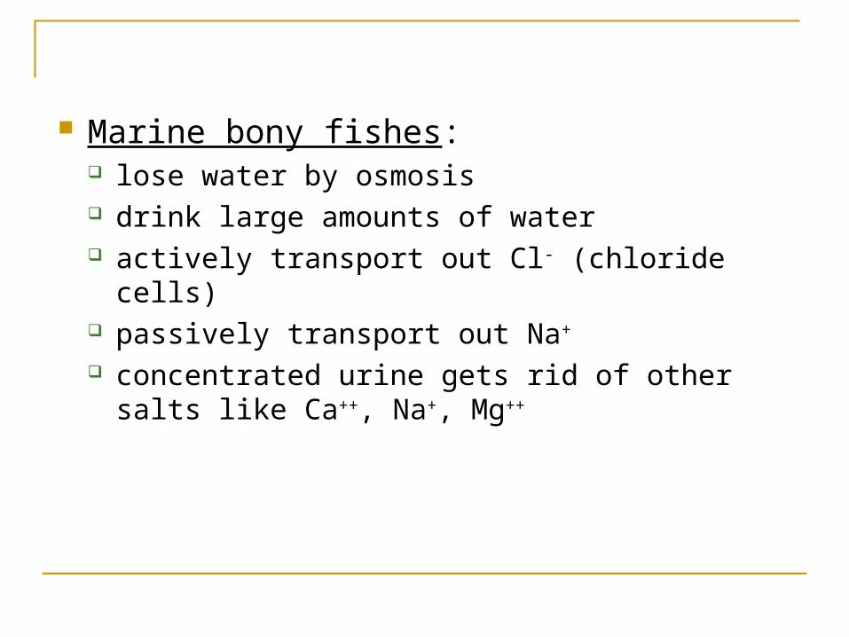
Marine bony fishes: lose water by osmosis drink large amounts of water actively transport out Cl- (chloride cells) passively transport out Na+
concentrated urine gets rid of other salts like Ca++, Na+, Mg++

Gain of water andsalt ions from foodand by drinkingseawater
Osmotic water lossthrough gills and other partsof body surface
Excretion ofsalt ionsfrom gills
Osmoregulation in a saltwater fish
Excretion of salt ions and small amountsof water in scantyurine from kidneys

Marine cartilaginous fishes: Shark tissue contains a high concentration of urea To prevent urea from damaging other organic molecules in
the tissues they have trimethyl amine oxide (TMAO) Because of high solute concentration in tissue water enters
the cells (sharks don’t drink) Produce concentrated urine.
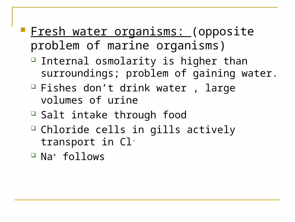
Fresh water organisms: (opposite problem of marine organisms) Internal osmolarity is higher than surroundings;
problem of gaining water. Fishes don’t drink water , large volumes of urine Salt intake through food Chloride cells in gills actively transport in Cl-
Na+ follows
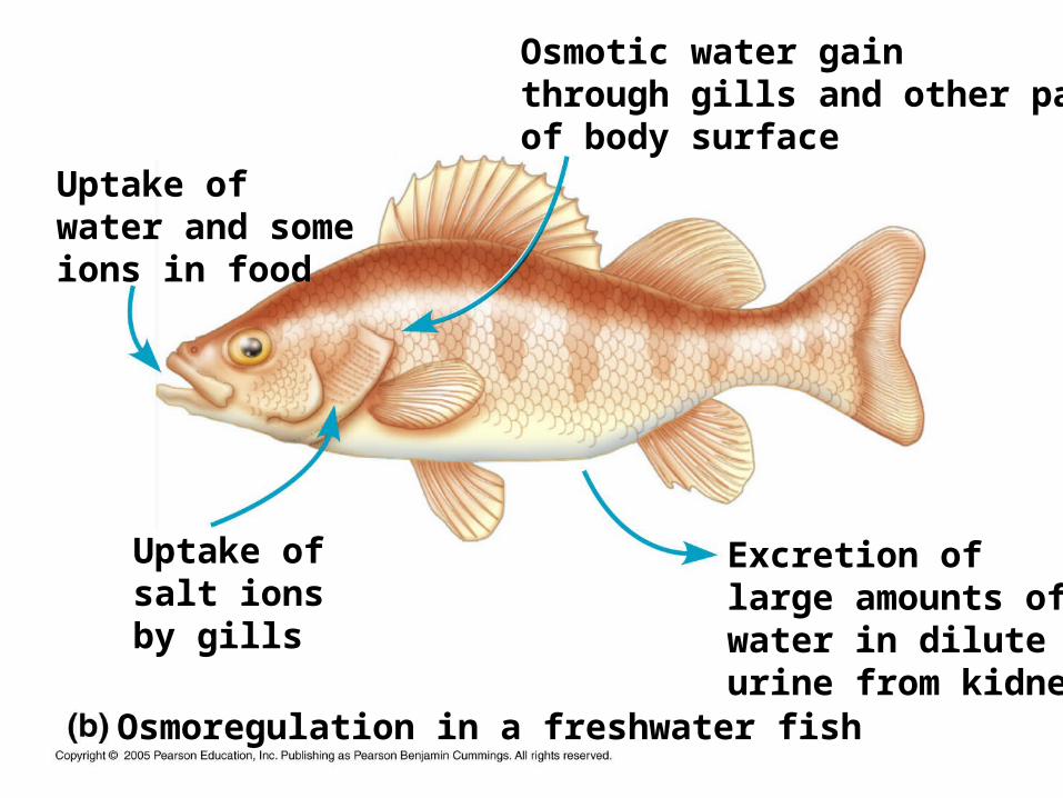
Excretion oflarge amounts ofwater in diluteurine from kidneys
Osmotic water gainthrough gills and other partsof body surface
Osmoregulation in a freshwater fish
Uptake ofsalt ionsby gills
Uptake ofwater and someions in food
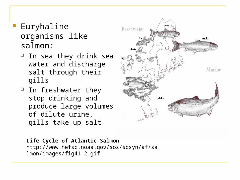
Euryhaline organisms like salmon: In sea they drink sea
water and discharge salt through their gills
In freshwater they stop drinking and produce large volumes of dilute urine, gills take up salt
Life Cycle of Atlantic Salmon http://www.nefsc.noaa.gov/sos/spsyn/af/salmon/images/fig41_2.gif
Anhydrobiosis: dormant state when habitat dries up. 85% to 2% water in water bears. Cell membrane adaptations are poorly understood.
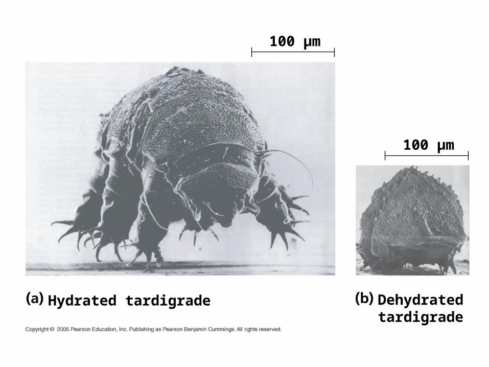
Hydrated tardigrade Dehydrated tardigrade
100 µm
100 µm
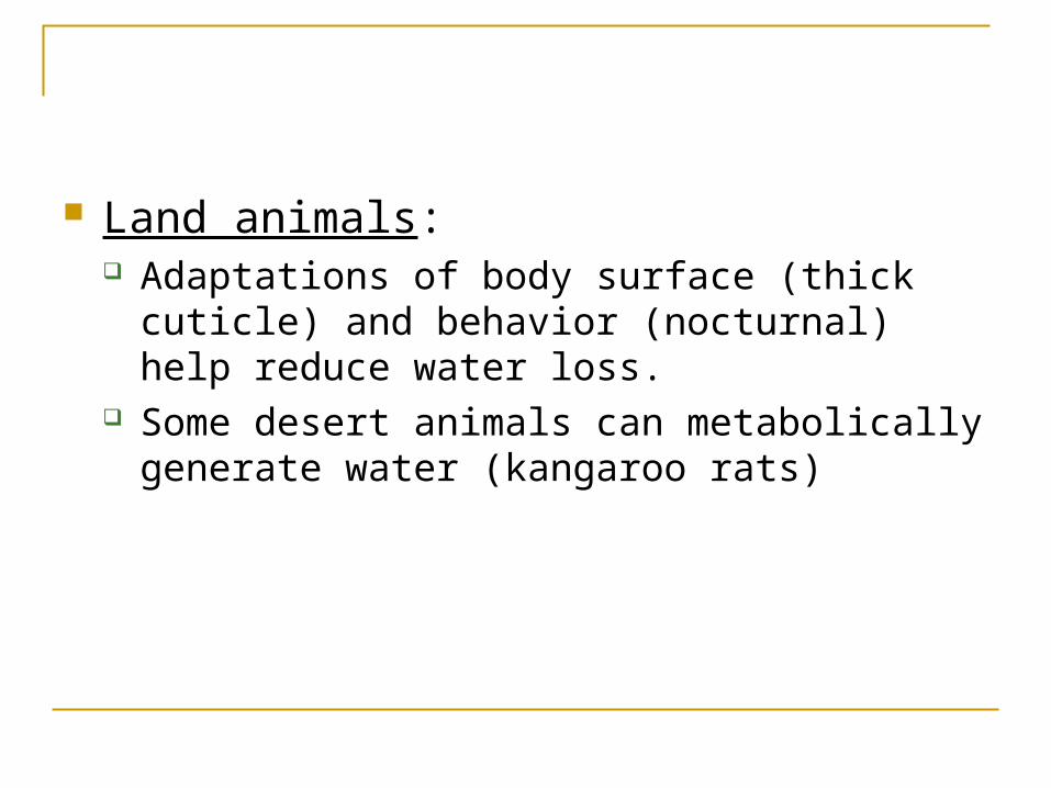
Land animals: Adaptations of body surface (thick cuticle) and
behavior (nocturnal) help reduce water loss. Some desert animals can metabolically generate
water (kangaroo rats)
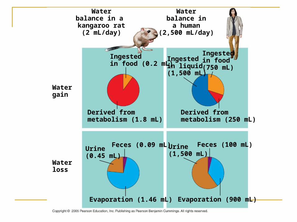
Waterbalance in a kangaroo rat(2 mL/day)
Waterbalance ina human
(2,500 mL/day)
Watergain
Waterloss
Derived frommetabolism (1.8 mL)
Ingestedin food (0.2 mL)
Derived frommetabolism (250 mL)
Ingestedin food (750 mL)
Ingestedin liquid (1,500 mL)
Evaporation (900 mL)
Feces (100 mL)Urine(1,500 mL)
Evaporation (1.46 mL)
Feces (0.09 mL)Urine(0.45 mL)
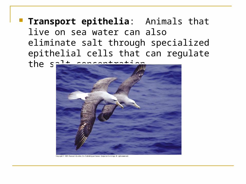
Transport epithelia: Animals that live on sea water can also eliminate salt through specialized epithelial cells that can regulate the salt concentration.
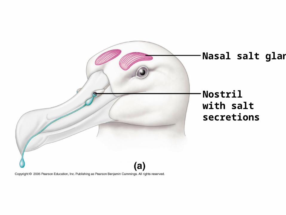
Nostrilwith saltsecretions
Nasal salt gland
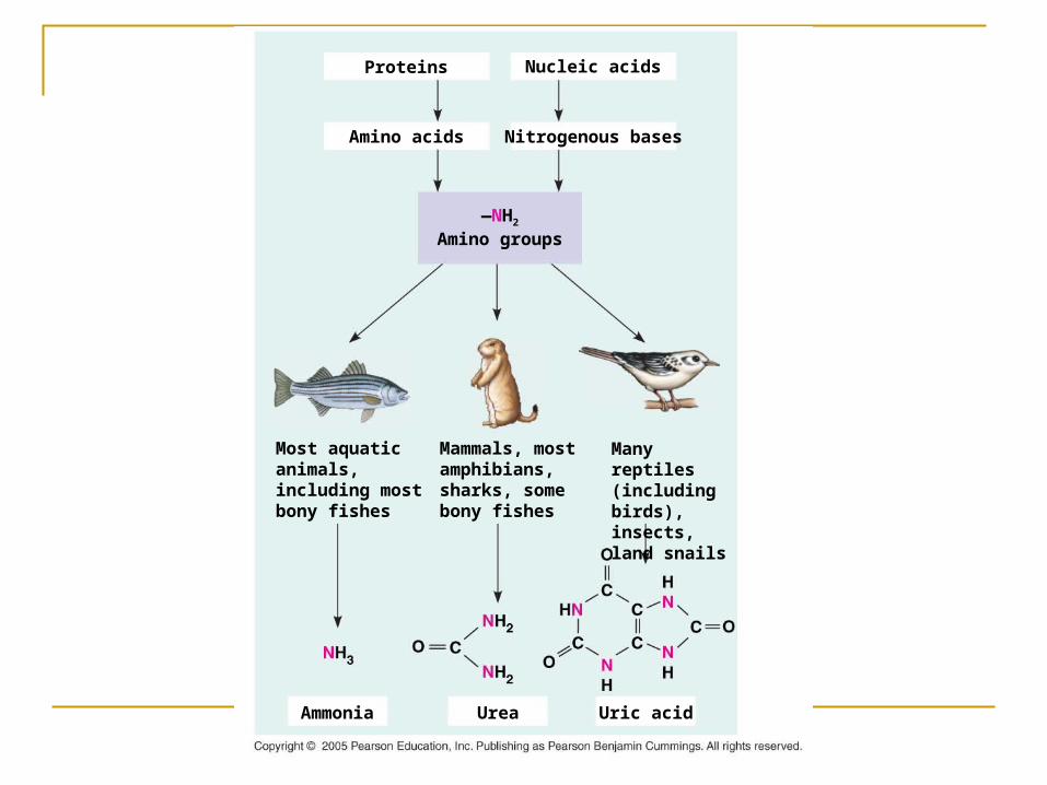
Nitrogenous wastes: As a result of metabolism proteins and amino
acids produce ammonia (NH3). Ammonium ion (NH4+) is highly toxic Animals either get rid of ammonia promptly or
expend energy and convert it to less toxic forms.
Forms of nitrogenous wastes:
ammonia urea uric acid
(differ in cost to convert and toxicity)
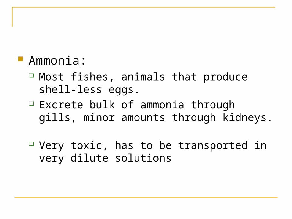
Ammonia: Most fishes, animals that produce shell-less eggs.
Excrete bulk of ammonia through gills, minor
amounts through kidneys. Very toxic, has to be transported in very dilute
solutions
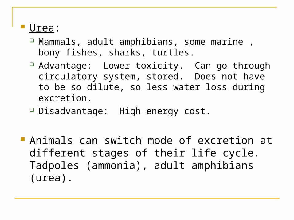
Urea: Mammals, adult amphibians, some marine , bony
fishes, sharks, turtles. Advantage: Lower toxicity. Can go through
circulatory system, stored. Does not have to be so dilute, so less water loss during excretion.
Disadvantage: High energy cost.
Animals can switch mode of excretion at different stages of their life cycle. Tadpoles (ammonia), adult amphibians (urea).
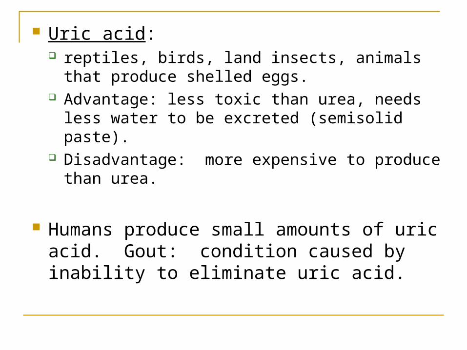
Uric acid: reptiles, birds, land insects, animals that produce
shelled eggs. Advantage: less toxic than urea, needs less water
to be excreted (semisolid paste). Disadvantage: more expensive to produce than
urea.
Humans produce small amounts of uric acid. Gout: condition caused by inability to eliminate uric acid.
Nitrogenous bases
Nucleic acids
Amino acids
Proteins
—NH2
Amino groups
Most aquatic animals, including most bony fishes
Mammals, most amphibians, sharks, some bony fishes
Many reptiles (including birds), insects, land snails
Ammonia Urea Uric acid

Amount of nitrogenous waste: linked to energy budget (higher in endotherms than ectotherms).
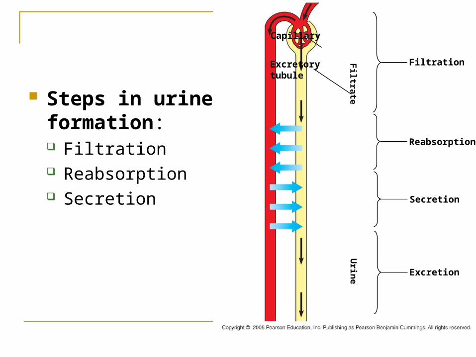
Steps in urine formation: Filtration Reabsorption Secretion
Filtration
Reabsorption
Secretion
Excretion
Excretorytubule
Capillary
Filtrate
Urin
e
Filtration: Cells, large molecules (proteins) stay in the body
fluid Small molecules, (salts, sugars, amino acids,
nitrogenous wastes) and water pass through and form filtrate
Reabsorption : Selective process. Recovery of useful molecules Active transport – reabsorption of certain salts,
vitamins, hormones, amino acids Wastes, nonessential molecules are left behind

Secretion: Selective pumping of various solutes to adjust
osmotic movement of water into and out of the filtrate
Final step – removal of this filtrate from the body – release of urine
Diverse excretory systems:
Excretory system plays a very important role in water balance and homeostasis.
Systems show a lot of variation in different groups
Basic structure – network of tubules that provide a large surface area
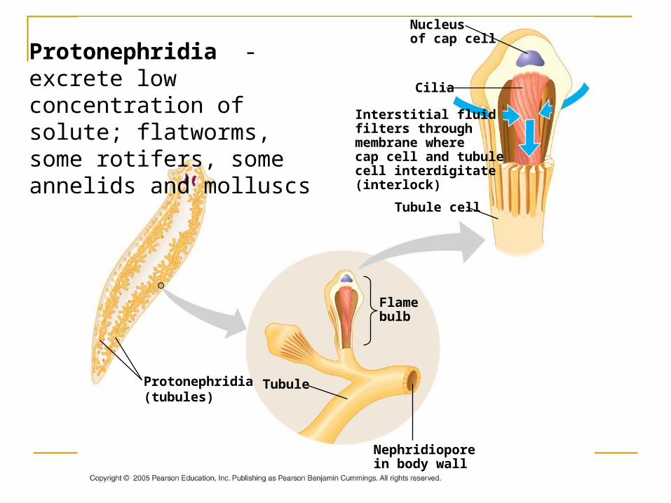
Protonephridia(tubules)
Tubule
Nephridioporein body wall
Flamebulb
Interstitial fluidfilters throughmembrane wherecap cell and tubulecell interdigitate(interlock)
Tubule cell
Cilia
Nucleusof cap cell
Protonephridia - excrete low concentration of solute; flatworms, some rotifers, some annelids and molluscs
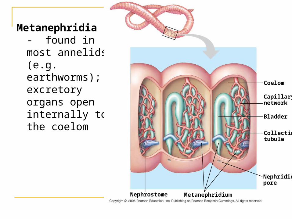
Metanephridia - found in most annelids (e.g. earthworms); excretory organs open internally to the coelom
Collectingtubule
Nephridio-pore
Capillarynetwork
Coelom
Bladder
MetanephridiumNephrostome
Malpighian tubules – extend from hemolymph to digestive tract; cells secrete nitrogenous wastes and other solutes into
hemolymph, these molecules and water move into malpighian tubules, excess water is reabsorbed in the rectum; other essential solutes are reabsorbed; lets the animal conserve water; found in insects, capability to conserve water helps in the
success of this group
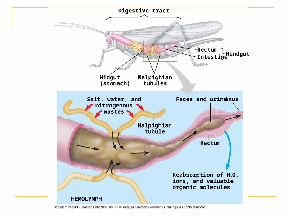
Salt, water, andnitrogenous
wastes
Digestive tract
Midgut(stomach)
Malpighiantubules
RectumIntestine Hindgut
Reabsorption of H2O,ions, and valuableorganic molecules
Malpighiantubule
HEMOLYMPH
Anus
Rectum
Feces and urine
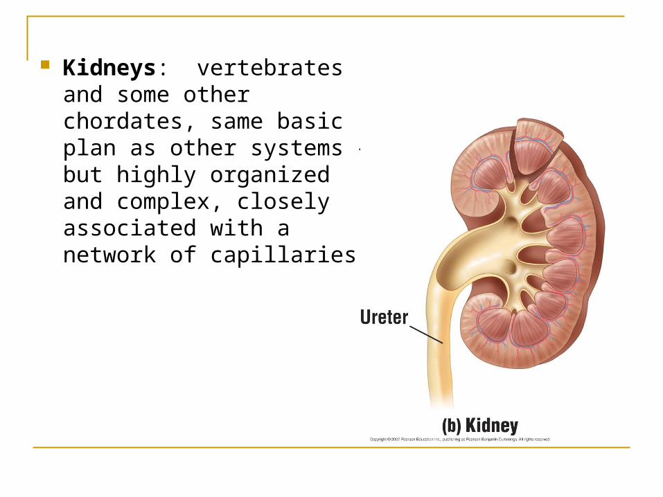
Kidneys: vertebrates and some other chordates, same basic plan as other systems – but highly organized and complex, closely associated with a network of capillaries.

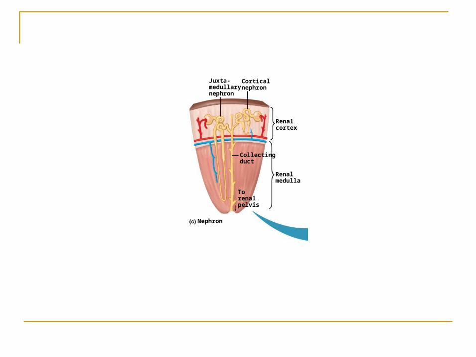
Nephron: functional unit of a kidney
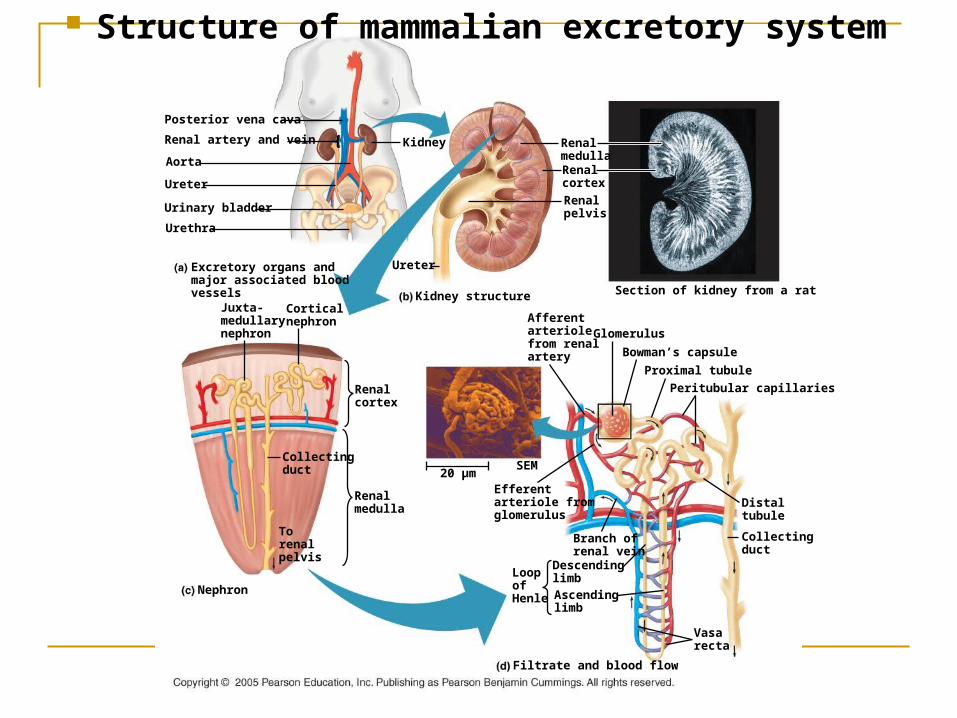
Excretory organs and major associated blood vessels
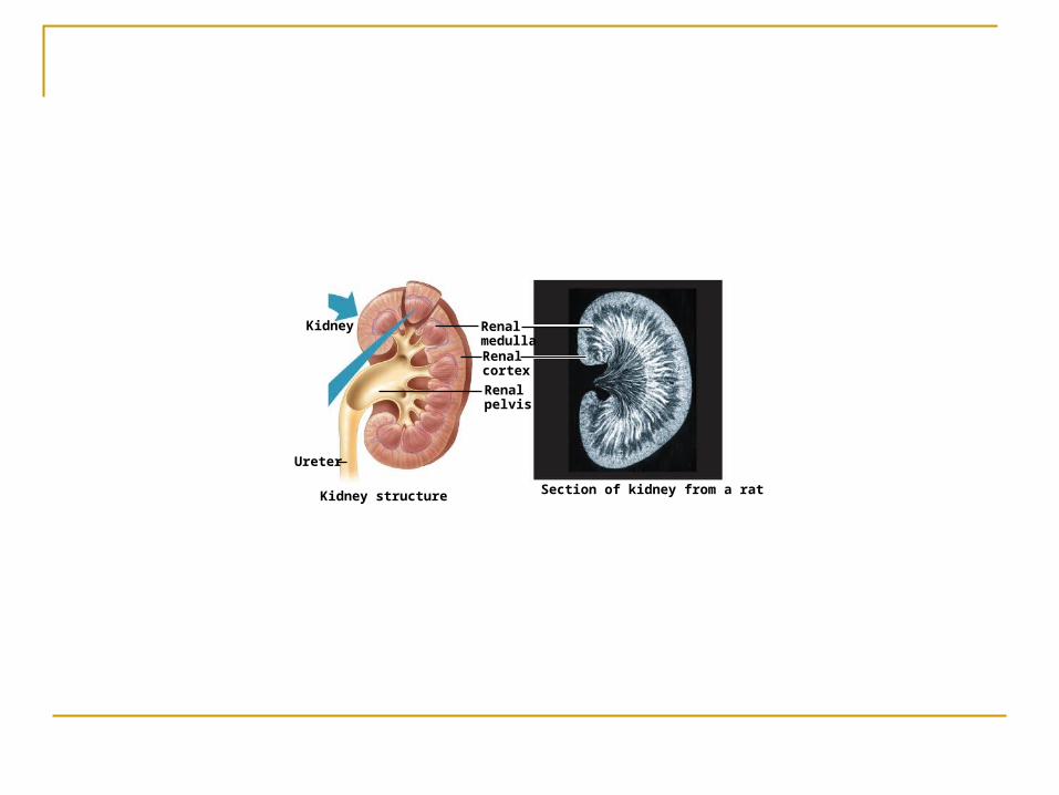
RenalmedullaRenalcortex
Renalpelvis
Section of kidney from a ratKidney structure
Ureter
Kidney
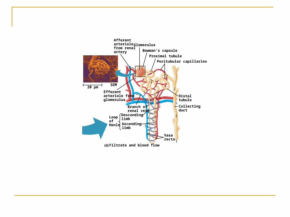
Glomerulus
Bowman’s capsule
Proximal tubule
Peritubular capillaries
Afferentarteriolefrom renalartery
Efferentarteriole from glomerulus
Distaltubule
Collectingduct
SEM20 µm
Branch ofrenal vein
Filtrate and blood flow
Vasarecta
DescendinglimbAscendinglimb
LoopofHenle
Renalmedulla
Nephron
Torenalpelvis
Renalcortex
Collectingduct
Juxta-medullarynephron
Corticalnephron
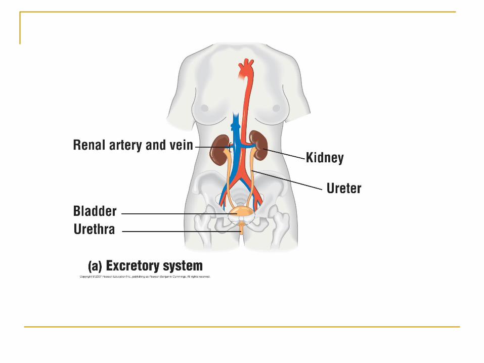
Posterior vena cava
Renal artery and vein
Aorta
Ureter
Urinary bladder
Urethra
Structure of mammalian excretory system
RenalmedullaRenalcortex
Renalpelvis
Section of kidney from a ratKidney structure
Ureter
Kidney
Renalmedulla
Nephron
Torenalpelvis
Renalcortex
Collectingduct
Juxta-medullarynephron
Corticalnephron
Glomerulus
Bowman’s capsule
Proximal tubule
Peritubular capillaries
Afferentarteriolefrom renalartery
Efferentarteriole from glomerulus
Distaltubule
Collectingduct
SEM20 µm
Branch ofrenal vein
Filtrate and blood flow
Vasarecta
DescendinglimbAscendinglimb
LoopofHenle
Detailed look at processing of blood in nephron:
Filtration Afferent arteriole has a bigger diameter than
efferent arteriole, thus pressure builds in the glomerulus
During filtration, blood in glomerulus is forced into Bowman’s capsule (cup shaped swelling at the blind end of the tubule)
Filtration is nonselective, only based on size, caused by the high blood pressure in the capillaries in the Bowman’s capsule.

Microscopic view of Bowman’s capsule
From filtrate to urine:
Filtrate contains water, salts (like NaCl), bicarbonate ions, hydrogen ions, urea, glucose, amino acids, drugs

Filtrate
H2O
Salts (NaCl and others)
HCO3–
H+
Urea
Glucose; amino acids
Some drugs
Key
Active transport
Passive transportINNERMEDULLA
OUTERMEDULLA
NaCl
H2O
CORTEX
Descending limbof loop ofHenle
Proximal tubule
NaCl Nutrients
HCO3–
H+
K+
NH3
H2O
Distal tubule
NaCl HCO3–
H+K+
H2O
Thick segmentof ascendinglimb
NaCl
NaCl
Thin segmentof ascendinglimb
Collectingduct
Urea
H2O
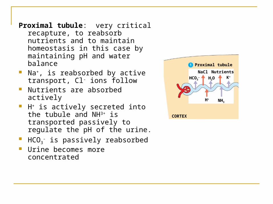
Proximal tubule: very critical recapture, to reabsorb nutrients and to maintain homeostasis in this case by maintaining pH and water balance
Na+, is reabsorbed by active transport, Cl- ions follow
Nutrients are absorbed actively H+ is actively secreted into the
tubule and NH3+ is transported passively to regulate the pH of the urine.
HCO3- is passively reabsorbed
Urine becomes more concentrated
CORTEX
Proximal tubule
NaCl Nutrients
HCO3–
H+
K+
NH3
H2O
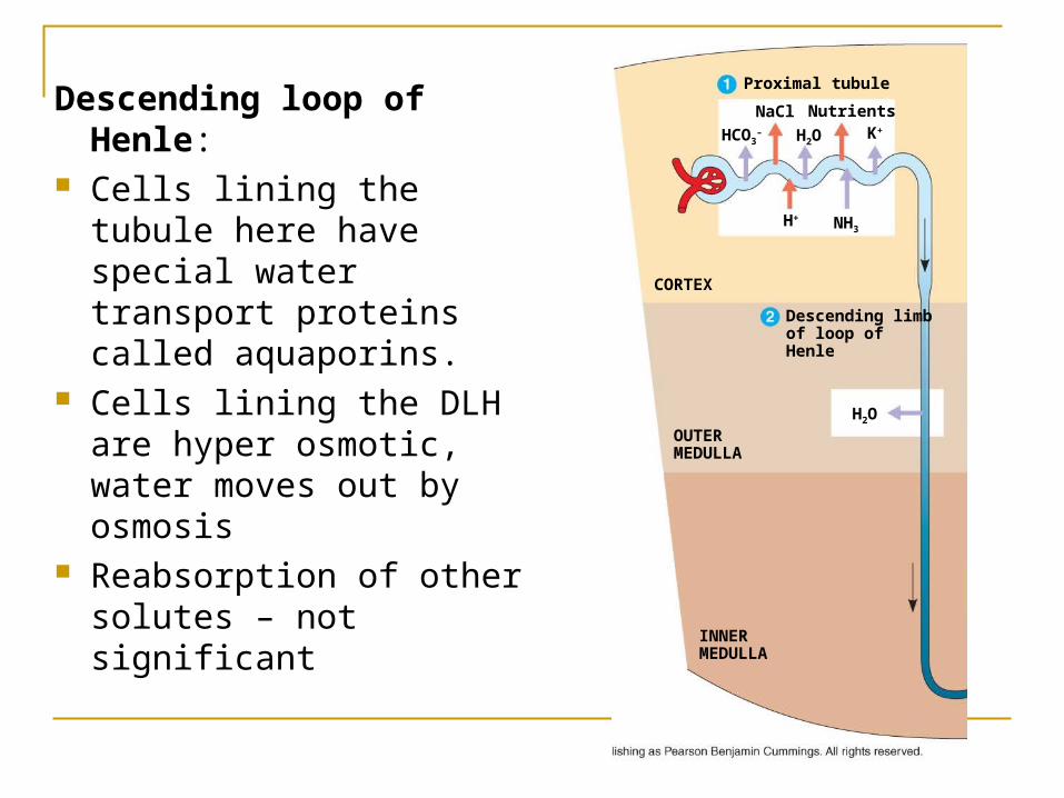
Descending loop of Henle: Cells lining the tubule here
have special water transport proteins called aquaporins.
Cells lining the DLH are hyper osmotic, water moves out by osmosis
Reabsorption of other solutes – not significant
INNERMEDULLA
OUTERMEDULLA
H2O
CORTEX
Descending limbof loop ofHenle
Proximal tubule
NaCl Nutrients
HCO3–
H+
K+
NH3
H2O
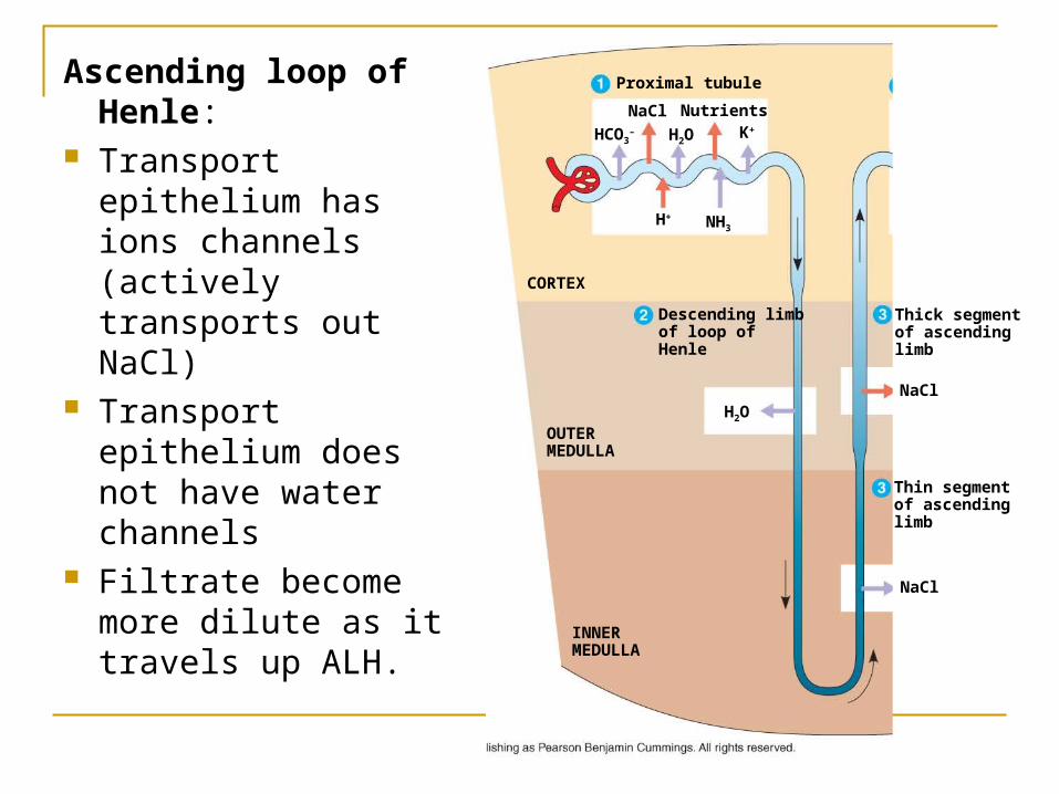
Ascending loop of Henle:
Transport epithelium has ions channels (actively transports out NaCl)
Transport epithelium does not have water channels
Filtrate become more dilute as it travels up ALH.
INNERMEDULLA
OUTERMEDULLA
NaCl
H2O
CORTEX
Descending limbof loop ofHenle
Proximal tubule
NaCl Nutrients
HCO3–
H+
K+
NH3
H2O
Thick segmentof ascendinglimb
NaCl
Thin segmentof ascendinglimb
INNERMEDULLA
OUTERMEDULLA
NaCl
H2O
CORTEX
Descending limbof loop ofHenle
Proximal tubule
NaCl Nutrients
HCO3–
H+
K+
NH3
H2O
Distal tubule
NaCl HCO3–
H+K+
H2O
Thick segmentof ascendinglimb
NaCl
NaCl
Thin segmentof ascendinglimb
Collectingduct
Urea
H2O
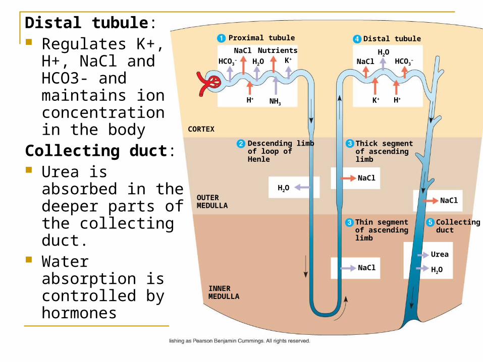
Distal tubule: Regulates K+,
H+, NaCl and HCO3- and maintains ion concentration in the body
Collecting duct: Urea is absorbed
in the deeper parts of the collecting duct.
Water absorption is controlled by hormones
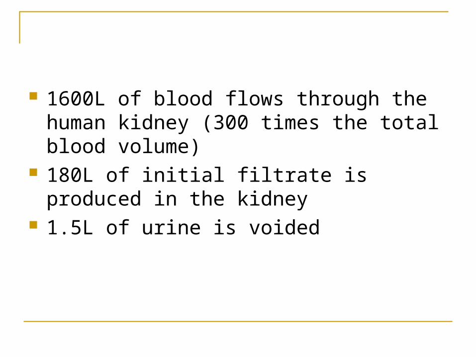
1600L of blood flows through the human kidney (300 times the total blood volume)
180L of initial filtrate is produced in the kidney 1.5L of urine is voided
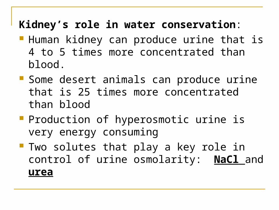
Kidney’s role in water conservation: Human kidney can produce urine that is 4 to
5 times more concentrated than blood. Some desert animals can produce urine that
is 25 times more concentrated than blood Production of hyperosmotic urine is very
energy consuming Two solutes that play a key role in control of
urine osmolarity: NaCl and urea
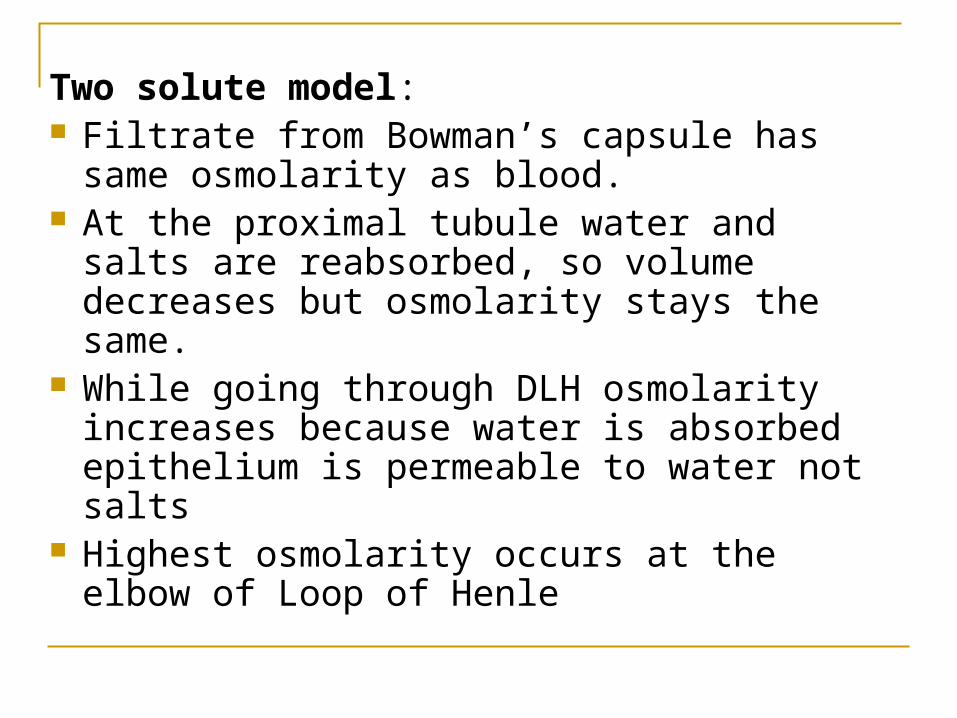
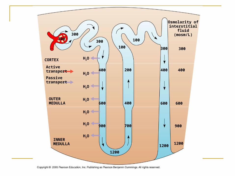
Two solute model: Filtrate from Bowman’s capsule has same
osmolarity as blood. At the proximal tubule water and salts are
reabsorbed, so volume decreases but osmolarity stays the same.
While going through DLH osmolarity increases because water is absorbed epithelium is permeable to water not salts
Highest osmolarity occurs at the elbow of Loop of Henle
INNERMEDULLA
OUTERMEDULLA
CORTEX
Osmolarity ofinterstitial
fluid(mosm/L)
H2O
Activetransport
Passivetransport
300300
300 100
100
400 200H2O
H2O
H2O
H2O
H2O
H2O
600 400
900 700
1200
300
400
600
12001200
600
900
300
400
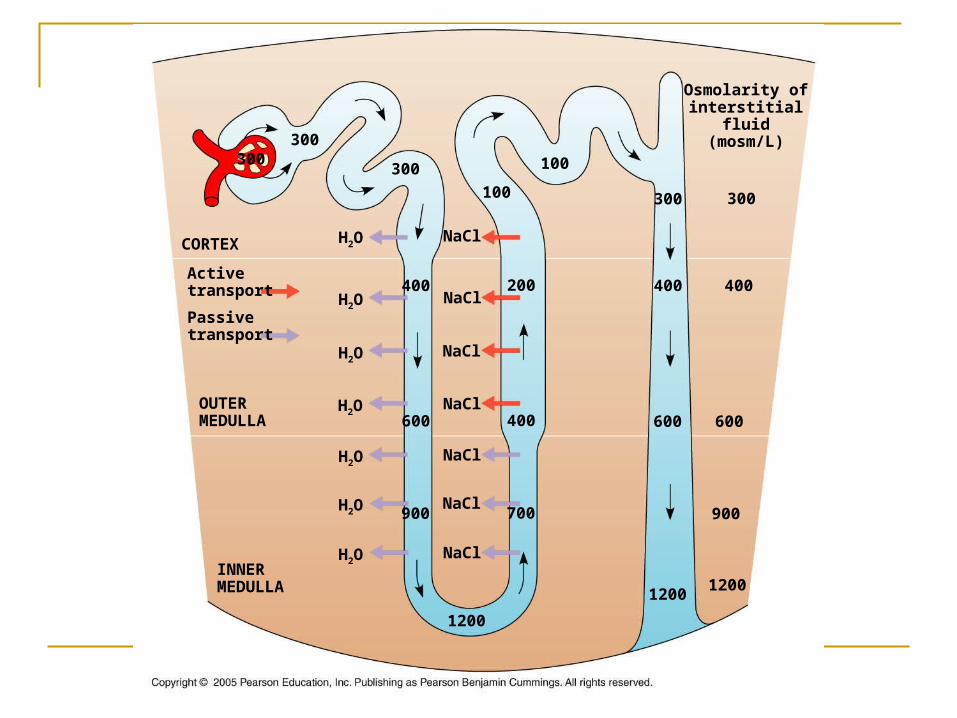
In the Ascending limb (permeable to salts and not water) osmolarity decreases
Loop of Henle creates concentration gradients, by expending energy: countercurrent multiplier system.
INNERMEDULLA
OUTERMEDULLA
CORTEX
Osmolarity ofinterstitial
fluid(mosm/L)
NaClH2O
Activetransport
Passivetransport
300300
300 100
100
400 200H2O
H2O
H2O
H2O
H2O
H2O
600 400
900 700
1200
300
400
600
12001200
600
900
300
400NaCl
NaCl
NaCl
NaCl
NaCl
NaCl

Ascending and descending vessels of vasa recta – blood flows in the opposite direction as the kidney’s osmolarity gradient
As blood flows down towards the inner medulla, blood looses water and gains NaCl
As blood flows away from the medulla towards the cortex, water is gained and NaCl is lost

Urea enters the loop of Henle by diffusion but some leaks out of the collecting duct. This maintains the high osmolarity of the interstitial fluid, draws water out of the filtrate in the collecting duct and keeps urine hyperosmotic.
Urine is isotonic to the interstitial fluid of inner medulla but hyperosmotic to blood

INNERMEDULLA
OUTERMEDULLA
CORTEX
Osmolarity ofinterstitial
fluid(mosm/L)
NaCl
Urea
H2O
Activetransport
Passivetransport
300300
300 100
100
400 200H2O
H2O
H2O
H2O
H2O
H2O
600 400
900 700
1200
300
400
H2O
600
12001200
600
900
300
400NaCl
NaCl
NaCl
NaCl
NaCl
NaCl
UreaH2O
UreaH2O
H2O
H2O
H2O
H2O
Like counter current exchange but this process costs a lot of energy expenditure.
This counter current-like system helps maintain the steep gradient, it is not lost due because the active transport maintains it
For its size energy consumes a lot of ATP

Adaptations of vertebrate kidney to a variety of environments
Mammalian kidney very well adapted to terrestrial life: juxtamedullary nephron is very well adapted to produce hyperosmotic urine.
Bannertail kangaroo rat(Dipodomys spectabilis)
Beaver (Castor canadensis)
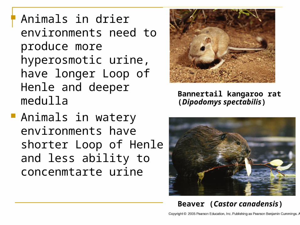
Animals in drier environments need to produce more hyperosmotic urine, have longer Loop of Henle and deeper medulla
Animals in watery environments have shorter Loop of Henle and less ability to concenmtarte urine
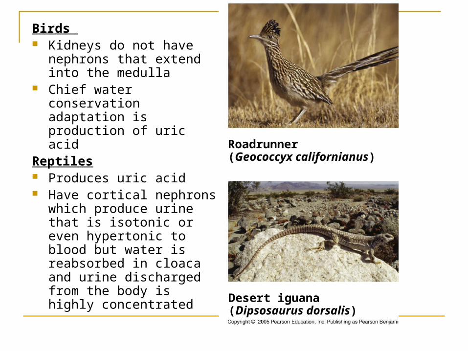
Birds Kidneys do not have
nephrons that extend into the medulla
Chief water conservation adaptation is production of uric acid
Reptiles Produces uric acid Have cortical nephrons
which produce urine that is isotonic or even hypertonic to blood but water is reabsorbed in cloaca and urine discharged from the body is highly concentrated
Roadrunner(Geococcyx californianus)
Desert iguana(Dipsosaurus dorsalis)
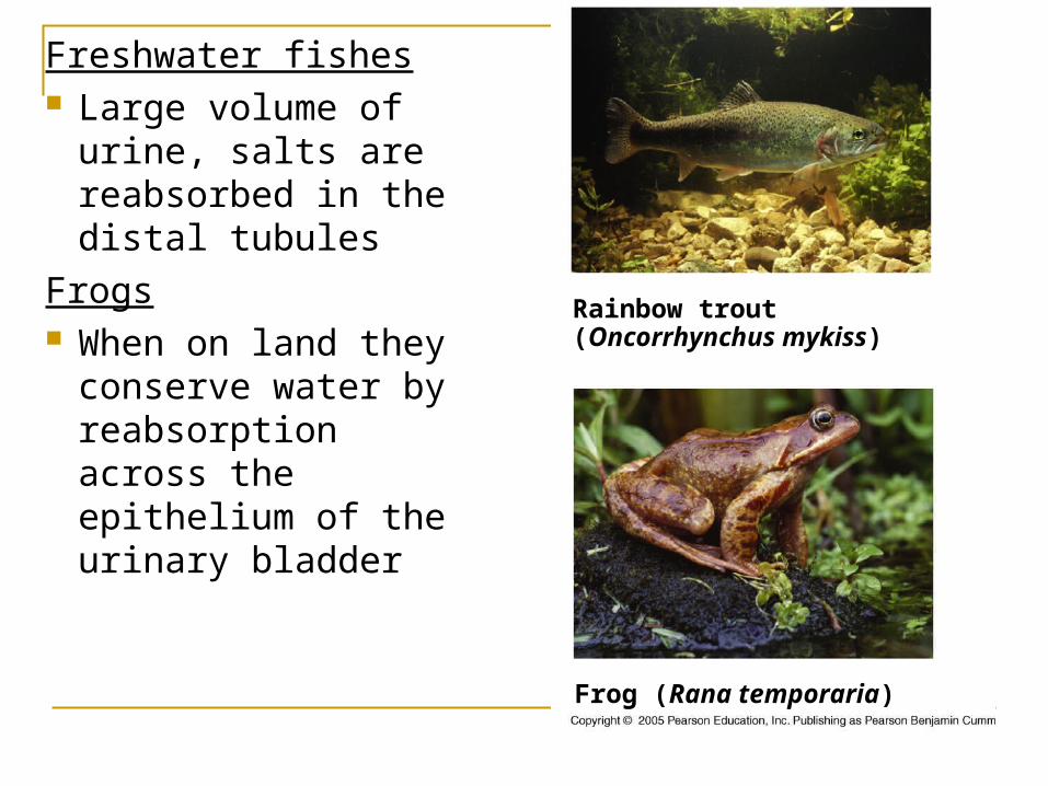
Freshwater fishes Large volume of
urine, salts are reabsorbed in the distal tubules
Frogs When on land they
conserve water by reabsorption across the epithelium of the urinary bladder
Rainbow trout(Oncorrhynchus mykiss)
Frog (Rana temporaria)
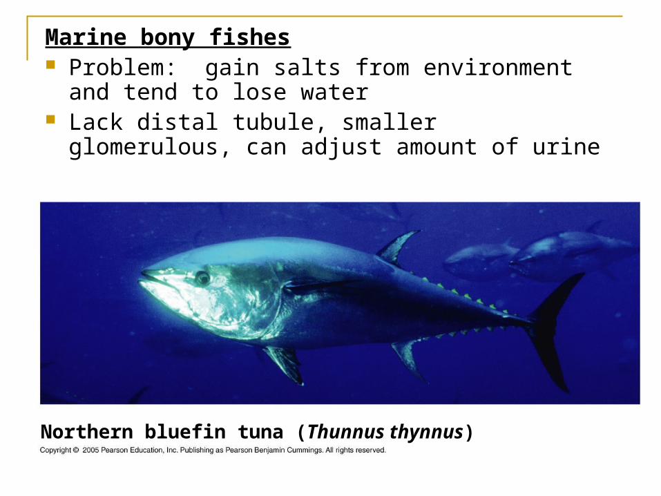
Marine bony fishes Problem: gain salts from environment and tend to
lose water Lack distal tubule, smaller glomerulous, can adjust
amount of urine
Northern bluefin tuna (Thunnus thynnus)
Kidney function, water balance, blood pressure – hormonal control
ADH control & RAAS system

ADH control Osmoregulatory function of
kidney is controlled by nerves and hormones
Antidiuretic hormone (ADH)/vasopressin – key role in osmoregulation
ADH is produced by hypothalamus and is stored in the posterior pituitary
Hypothalamus monitors blood osmolarity using osmoreceptor cells
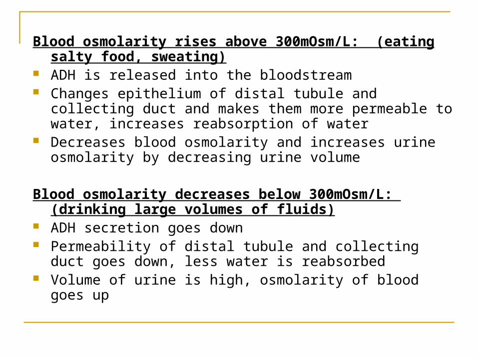
Blood osmolarity rises above 300mOsm/L: (eating salty food, sweating)
ADH is released into the bloodstream Changes epithelium of distal tubule and collecting duct and
makes them more permeable to water, increases reabsorption of water
Decreases blood osmolarity and increases urine osmolarity by decreasing urine volume
Blood osmolarity decreases below 300mOsm/L: (drinking large volumes of fluids)
ADH secretion goes down Permeability of distal tubule and collecting duct goes down, less
water is reabsorbed Volume of urine is high, osmolarity of blood goes up
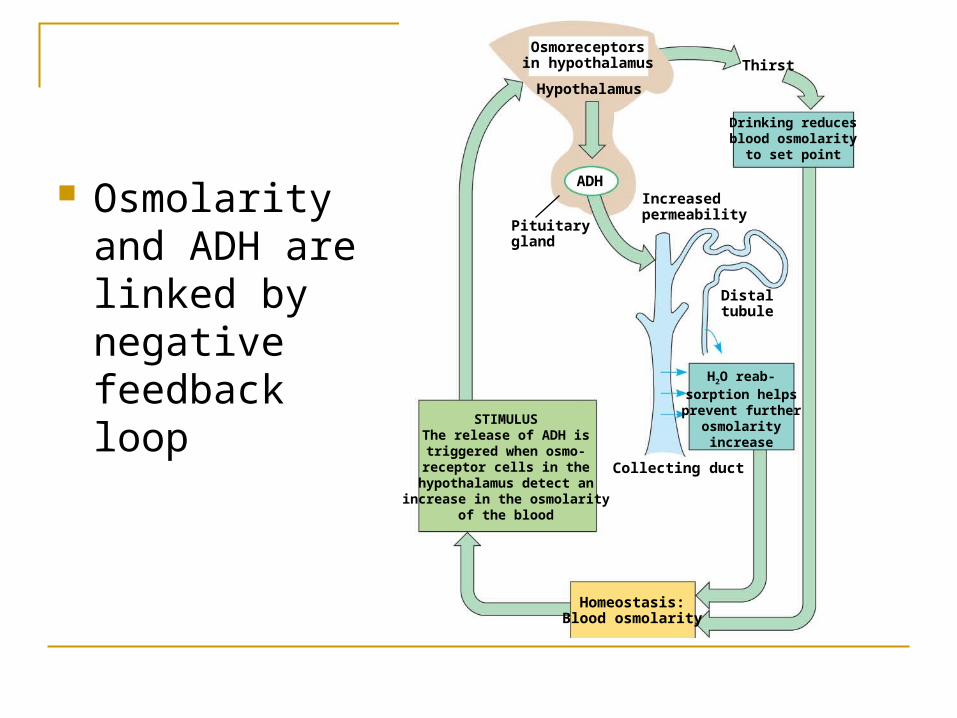
Osmoreceptorsin hypothalamus
Hypothalamus
ADH
Pituitarygland
Increasedpermeability
Distaltubule
Thirst
Drinking reducesblood osmolarity
to set point
Collecting duct
H2O reab-sorption helpsprevent further
osmolarityincrease
Homeostasis:Blood osmolarity
STIMULUSThe release of ADH istriggered when osmo-receptor cells in the
hypothalamus detect anincrease in the osmolarity
of the blood
Osmolarity and ADH are linked by negative feedback loop
Genetic disorders that affect ADH production or ADH receptors can affect osmoregulatory function of kidney, cause severe dehydration by producing very dilute urine: diabetes insipidus
Alcohol inhibits ADH release, can cause excessive water loss, some dehydration and symptoms of hangover
Distaltubule
Aldosterone
Homeostasis:Blood pressure,
volume
STIMULUS:The juxtaglomerular
apparatus (JGA) respondsto low blood volume or
blood pressure (such as due to dehydration or
loss of blood)
Increased Na+
and H2O reab-sorption in
distal tubules
Reninproduction
Arterioleconstriction
Adrenal gland
Angiotensin II
Angiotensin I
JGA
Renin
ACE
An
gio
ten
sin
og
en
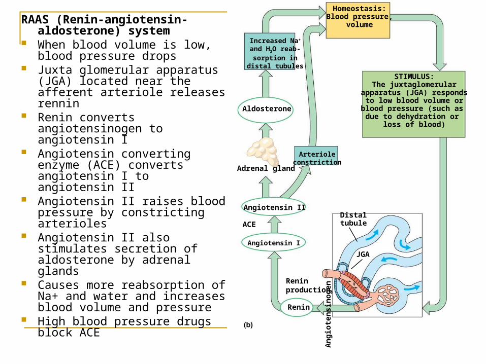
RAAS (Renin-angiotensin-aldosterone) system
When blood volume is low, blood pressure drops
Juxta glomerular apparatus (JGA) located near the afferent arteriole releases rennin
Renin converts angiotensinogen to angiotensin I
Angiotensin converting enzyme (ACE) converts angiotensin I to angiotensin II
Angiotensin II raises blood pressure by constricting arterioles
Angiotensin II also stimulates secretion of aldosterone by adrenal glands
Causes more reabsorption of Na+ and water and increases blood volume and pressure
High blood pressure drugs block ACE