original research the pit-crew model for improving...
TRANSCRIPT

ORIGINAL RESEARCH
The ‘pit-crew’ model for improving door-to-needletimes in endovascular stroke therapy: a Six-SigmaprojectAnsaar T Rai,1 Matthew S Smith,2 SoHyun Boo,3 Abdul R Tarabishy,3
Gerald R Hobbs,4 Jeffrey S Carpenter3
1Department of Radiology,Neurology and Neurosurgery,West Virginia University,Morgantown, West Virginia,USA2Department of Neurology,West Virginia University,Morgantown, West Virginia,USA3Interventional Neuroradiology,West Virginia University,Morgantown, West Virginia,USA4Department of Statistics, WestVirginia University,Morgantown, West Virginia,USA
Correspondence toDr Ansaar T Rai, Departmentof Radiology, Neurology andNeurosurgery, West VirginiaUniversity, Room 2278, HSCS,PO Box 9235, Morgantown,WV 26506, USA;[email protected]
Received 3 December 2015Revised 16 December 2015Accepted 17 December 2015Published Online First11 January 2016
To cite: Rai AT, Smith MS,Boo SH, et al. JNeuroIntervent Surg2016;8:447–452.
ABSTRACTBackground Delays in delivering endovascular stroketherapy adversely affect outcomes. Time-sensitivetreatments such as stroke interventions benefit frommethodically developed protocols. Clearly defined rolesin these protocols allow for parallel processing of tasks,resulting in consistent delivery of care.Objective To present the outcomes of a quality-improvement (QI) process directed at reducing stroketreatment times in a tertiary level academic medicalcenter.Methods A Six-Sigma-based QI process was developedover a 3-month period. After an initial analysis,procedures were implemented and fine-tuned to identifyand address rate-limiting steps in the endovascular carepathway. Prospectively recorded treatment times werethen compared in two groups of patients who weretreated ‘before’ (n=64) or ‘after’ (n=30) the QI process.Three time intervals were measured: emergency room(ER) to arrival for CT scan (ER–CT), CT scan tointerventional laboratory arrival (CT–Lab), andinterventional laboratory arrival to groin puncture (Lab–puncture).Results The ER–CT time was 40 (±29) min in the‘before’ and 26 (±15) min in the ‘after’ group(p=0.008). The CT–Lab time was 87 (±47) min in the‘before’ and 51 (±33) min in the ‘after’ group(p=0.0002). The Lab–puncture time was 24 (±11) minin the ‘before’ and 15 (±4) min in the ‘after’ group(p<0.0001). The overall ER–arrival to groin-puncturetime was reduced from 2 h, 31 min (±51) min in the‘before’ to 1 h, 33 min (±37) min in the ‘after’ group,(p<0.0001). The improved times were seen for bothworking hours and off-hours interventions.Conclusions A protocol-driven process can significantlyimprove efficiency of care in time-sensitive strokeinterventions.
INTRODUCTIONEndovascular treatment of large vessel acute ische-mic strokes in appropriately selected patients hasbeen endorsed as evidence-based care.1 2 The nextsteps in advancing this therapy are to developsystems of care that can be divided into prehospitaland intra-hospital pathways. Other time-dependenttreatments, such as trauma and acute myocardialinfarction, provide a valuable model for endovascu-lar stroke therapy. The value of an initial ‘goldenhour’ in improving outcomes has been shown fortreatment of trauma3 to neonatal resuscitation.4
Early reperfusion for large vessel strokes is a criticaldeterminant of endovascular therapy outcomes.5 6
Emergency medicine publications provide usefulinsight into developing checklists and protocolsgeared towards seamless resuscitation of injured orsick patients.7 These ‘pit-crew’-type protocolsclearly define the role of each team member, allow-ing for synchronized, parallel delivery of care.Our center has performed endovascular stroke
interventions for the past 15 years and participatedin several clinical trials. We had noticed a fall in thevolume of patients in the past 3 years as an aftereffect of negative stroke trials8 9 and also becausewe had decided to offer stroke interventions onlyin the setting of a randomized clinical trial. Thisslowdown in cases adversely affected our treatmenttimes. Towards the end of 2014, we instituted aquality improvement (QI) process to reduce ourdoor-to-needle times and develop a process thatmight consistently reduce times to treatments at allhours of the day and all days of the week. The QIprocess was based on Six-Sigma which, althoughused by corporations for years, has only recentlybeen adopted in medicine.10 This paper providesthe results of those efforts, comparing the treat-ment times before and after implementation of theQI process.
METHODSObjectiveTo determine whether our times for different stepsin endovascular stroke care improved as a result ofthe QI process; the null hypothesis stating thatthere would be no difference in these times.
Patient populationOur setting was a rural, tertiary level, academicmedical center with almost 700 beds, which is alsothe regional level-1 trauma center. Prospectivelyrecorded treatment times before and after the QIimplementation provided the data for this analysis.The pre-QI treatment times included all endovascu-lar patients from 2011 to 2014 who had presentedto our emergency room (ER). Since the project wasspecifically targeted at improving efficiencybetween the ER and interventional neuroradiology(INR), the following patients were excluded:in-house patients undergoing an intervention forstroke, patients undergoing another procedure inthe hospital with a stroke and patients treated withunknown symptom onset. The post-QI treatment
Open AccessScan to access more
free content
Rai AT, et al. J NeuroIntervent Surg 2016;8:447–452. doi:10.1136/neurintsurg-2015-012219 1 of 6
Ischemic stroke on 28 M
ay 2018 by guest. Protected by copyright.
http://jnis.bmj.com
/J N
euroIntervent Surg: first published as 10.1136/neurintsurg-2015-012219 on 11 January 2016. D
ownloaded from

times were those for all patients undergoing endovasculartherapy between January 2015 and October 2015. The follow-ing treatment times were compared:▸ ER to imaging (CT) (ER–CT)▸ imaging to INR lab (CT–Lab)▸ INR lab to groin puncture (Lab–puncture)▸ imaging to needle (groin puncture) (CT–puncture)▸ door to needle (ER–puncture).
The QI processThe QI process was started in the fall of 2014 and incrementallyimplemented over almost 3 months. The impetus to initiate theprocess was treatment delays in stroke interventions, inconsist-encies in times and care between working hours and on-callhours, ad hoc roles of different members, and suboptimal hand-over of patients between different services. The QI team was ledby two Six-Sigma trained engineers and comprised physicians,allied staff, and management from interventional neuroradiol-ogy, neurology, emergency medicine, and anesthesia. Meetingswere held to develop a baseline understanding of the existingpractice, followed by observance of ‘real’ stroke interventionsand patient flow pathways. Subsequent weekly meetings modi-fied the process with each stroke therapy. A final protocol wassigned off towards the end of 2014 and in 2015 all cases fol-lowed the updated protocol.
Important features of the protocol (figure 1) included identifi-cation of key personnel in the team, definition of a clear rolefor every member, and emphasis on parallel processing ofassigned tasks. The protocol contained explicit details, such ashow a nurse enters the hospital, where a technician wouldstand, and the position of the detectors in the INR laboratory.The protocol focused on the three main time-points along thestroke pathway: ER–CT, CT–Lab, and Lab–puncture.
ER–CTThe emergency medical services evaluate stroke patients usingthe Cincinnati Prehospital Stroke Scale11 and alert the stroketeam (neurology, radiology, emergency medicine, and laboratorystaff ) by paging the group. On arrival, the ER staff and on-callneurology residents evaluate the patient to confirm the strokediagnosis. At least one IV line is placed while the patient is inthe ER for the purpose of drawing laboratories and for contrastadministration. After a point-of-care creatinine test (iSTAT;Abbot Laboratories, Abbott Park, Illinois), the stroke patient istransferred to the CT scanner by a dedicated stroke nurse andthe neurology team. The CT technologists already alerted bythe emergency medical services have the scanner ready for thepatient. The radiology resident meets the stroke team in thescanner control room for live review of imaging for all patientspresenting within 6 h with a National Institutes of HealthStroke Scale (NIHSS) score ≥6.
Before implementation of the QI process any CT scanner wasused, but after the QI process a designated scanner(Aquilion-One, Toshiba America Medical Systems, Tustin,California, USA) was used for all stroke patients. The scanner isequipped with 320×0.5 mm detector rows covering 16 cm ofvolume per rotation, allowing for a uniform protocol compris-ing non-contrast CT (NCCT), volumetric CT perfusion (CTP)imaging, and CT angiography (CTA). In addition, the sequenceof scanning was changed from NCCT–CTP–CTA to NCCT–CTA–CTP. This switch was crucial in allowing earlier detectionof a large vessel occlusion (LVO) and alerting the attending neu-rointerventionalist—who could then review the NCCT and CTA(onsite or offsite), while CTP imaging was being carried out and
processed. Another critical component was installation of animage router to simultaneously receive and disseminate imageswhile they were being acquired. This reduced the scanner totransfer time of all images from 12 min to just over 5 min.
The CT stroke scan is performed with a gantry speed of 3rotations/s. After the NCCT, the CTA is initiated from the aorticarch to the cranial vertex with an injection of 40–60 mL ofOptiray 350 (Covidien, Hazelwood, Missouri, USA) through an18–20 g antecubital IV line at a rate of 4–5 mL/s, followed by asimilar volume of saline chaser. Images acquired are automatic-ally sent via the router for immediate review. A volumetricwhole brain, time-resolved CTP sequence follows. The imagingrouter enables transfer of 6080 (19×320) images to a picturearchiving and communication system and the perfusion post-processing software (Vital Images, Minnetonka, Minnesota,USA). IV recombinant tissue plasminogen activator (rt-PA;Activase, Genentech Inc, San Francisco, California, USA) isadministered if indicated after exclusion of hemorrhage on theNCCT.
CT–LabA collaborative decision about endovascular treatment is madebased on clinical presentation, comorbidities, and imaging. Ifthe decision is to treat, the radiology resident places a singleorder set. This was designed to simultaneously trigger multipletasks with one click of the button—namely, anesthesia (in ourview, use of anesthesia makes the procedure safer), intensivecare unit (ICU) bed request, INR order set, Foley placement,peripheral IV line, etc. A single paging system was implementedallowing the resident to contact the neurointerventional team(one nurse, and two technicians) with one phone call. Thepaging system ‘Medcom’ provides the relevant patient informa-tion and location in the ER. If within 5 min the staff has notresponded a repeat page is sent. The system also documents allpaging times, which can be reviewed for QI purposes. The radi-ology resident places a preoperative note in the chart document-ing the decision to treat. The neurology resident’s duty runningin parallel with this is to notify the ICU service of the eventualadmission of the stroke patient. The INR nurse arrives throughthe ER, checks the patient, and alerts the ER staff to initiatetransfer to the INR suite.
Lab–punctureOur setup is illustrated in figure 2. The minimum staffingrequirement for all neurovascular procedures is set at two tech-nicians and one nurse. One technician is scrubbed and onefloats. During working hours, a third technician is available forcomplex elective cases. When two emergent cases occur simul-taneously after hours, the technicians are split and the backupnurse is called in. Improvement in groin puncture times uponpatient arrival in the angiography suite required planning toensure parallel preparation of patient, tray, and anesthesia. TheER stroke nurse hands over the patient to the INR nurse. TheINR technicians prepare the room for patient arrival by openingpre-packaged stroke trays and devices as indicated by the INRattending. A separate stroke cart is stocked with the catheters,wires, thrombectomy devices, and syringes to hold everything inone place for this purpose. The biplane detectors are positionedin such a way as to facilitate patient transfer to the angiographytable. Once the patient is placed on the table, a coordinatedeffort is made between the anesthestist working at the head ofthe patient and left arm (which is positioned out), and one ofthe INR technicians working at the groins for sterile prepar-ation. The second technician continues with catheter, lines, and
2 of 6 Rai AT, et al. J NeuroIntervent Surg 2016;8:447–452. doi:10.1136/neurintsurg-2015-012219
Ischemic stroke on 28 M
ay 2018 by guest. Protected by copyright.
http://jnis.bmj.com
/J N
euroIntervent Surg: first published as 10.1136/neurintsurg-2015-012219 on 11 January 2016. D
ownloaded from

device preparation. The INR nurse assists the anesthesiologist, ifrequired, prepares the flushes, and performs documentation. An8 Fr right common femoral arterial sheath is placed as soon asthe groin is prepped. If by that time a radial arterial line has notbeen established by the anesthesiologist, a second 4 Fr sheath isimmediately placed in the left common femoral artery for inva-sive blood pressure monitoring. The 8 Fr sheath is removed at
the end of the procedure and, if placed, the second 4 Fr sheathis sutured in place to be used in the ICU. An anesthesia cart andventilator is permanently stationed in each of two adjacentbiplane angiography suites, allowing performance of parallelemergent cases. Small coordinated steps in the patient prepar-ation process were designed to emulate defined roles and paral-lel tasks seen in trauma resuscitation.
Figure 1 An overview of the protocol is presented along a timeline from patient arrival to arterial puncture. The role of different team members islisted along the timeline. ER, emergency room; GETA, general endotracheal anesthesia, off hours treatment: stroke intervention performed before7:00 or after 17:00 or at the weekend; ICU, intensive care unit; INR, neurointerventionalist; LCFA, left common femoral artery; LSN, last seen normal;LVO, large vessel occlusion; NIHSS, National Institutes of Health Stroke Scale; onset-ER, time from symptom onset to ER arrival; RCFA, right commonfemoral artery; rt-PA, recombinant tissue plasminogen activator.
Figure 2 INR: neurointerventionalist, T1: technician-1, T2: technician-2, T3: technician-3, N1: nurse-1, N2: nurse-2, A1: anesthesiologist/certifiedregistered nurse anesthetist (CRNA)-1, A2: anesthesiologist/CRNA-2. During the patient preparation stage (A), T1 sets up the procedure trays andprepares the devices and catheters. T2 prepares the patient and helps the attending technician, who punctures the right femoral artery and typicallyplaces an 8 Fr sheath. The patient’s left arm is extended out on an arm board for simultaneous access to anesthesia for placement of lines andadministration of drugs. If there is no radial arterial access by the time the right femoral sheath is placed, the INR punctures the left femoral arteryand places a 4 Fr sheath for invasive blood pressure monitoring. Even though it is possible to obtain arterial tracing via the 8 Fr right femoralsheath, placement of the 4 Fr sheath allows removal of the larger right femoral sheath at the end of the procedure. The patient is transferred to theintensive care unit with the 4 Fr sheath in place for pressure monitoring. The nurse takes a report, prepares the continuous flush lines, assists theanesthesiologist, and charts all times. The A-plane detector is stationed in such a way as to allow easy positioning over the groin in casefluoroscopy is required. For the interventional stage (B), T2 scrubs up and functions as the float. One anesthesiologist (A1) stays to cover the case,assisted by the nurse. This setup with stocked anesthesia cart is duplicated in an immediately adjacent second interventional biplane room. Duringworking hours an additional technician (T3) and nurse (N2) are available. If two simultaneous emergent cases occur after hours, the technicianssplit and the backup nurse (N2) is called in.
Rai AT, et al. J NeuroIntervent Surg 2016;8:447–452. doi:10.1136/neurintsurg-2015-012219 3 of 6
Ischemic stroke on 28 M
ay 2018 by guest. Protected by copyright.
http://jnis.bmj.com
/J N
euroIntervent Surg: first published as 10.1136/neurintsurg-2015-012219 on 11 January 2016. D
ownloaded from
Data analysisThe patient demographics and stroke severity are descriptivelypresented and compared. The significance of simple bivariateassociations was assessed using the Fisher exact test for categor-ical variables. A Shapiro–Wilk W test demonstrated a non-normal distribution of the continuous time-point data.Non-parametric Wilcoxon rank sum tests were thus performedto compare the time-point means ‘before’ and ‘after’ the QIprocess. All statistical analysis was performed using the JMP Pro12.0.1 software package (SAS Institute Inc, Cary, NorthCarolina, USA).
RESULTSA total of 94 patients were divided into a ‘before’ (n=64) and‘after’ (n=30) group based on the implementation of the QIprocess. There were no differences in baseline demographics,stroke severity, symptom onset, comorbidities, and the use of IVrt-PA or general endotracheal anesthesia (GETA) between thetwo groups (table 1). A comparison of the time intervalsshowed a significant reduction in times in the ‘after’ QI imple-mentation group across all parameters (figure 3). The time inter-vals were separately compared for interventions performedduring working hours —that is, 7:00 to 17:00 on weekdays(table 2), and for off-hours and at weekends (table 3). Therewas significant reduction in times for both working hours andoff-hours interventions. We found no correlation between age,NIHSS, onset to ER or any of the comorbidities and thedoor-to-needle time. We also looked at the impact of GETA onthe patient preparation and puncture time after the implementa-tion of the QI process. The mean time from INR room arrivalto groin puncture was 15 (±4) min in patients who receivedGETA versus 15 (±3) min in patients treated without GETA(p=0.87), indicating that GETA caused no delays.
DISCUSSIONThe Six-Sigma process, developed at Motorola in the 1980sbecause quality was lagging behind that of Japanese companies,was adopted by many corporations as a QI practice. An idealhealthcare environment would be efficient and free from errors.However, healthcare is neither of those and preventable errorscause 44 000–98 000 deaths a year.12 Six-Sigma has been
applied in medicine to improve many processes from catheter-related10 and surgical-site infections13 to ER,14 surgery,15 andbehavioral health systems.16 Six-Sigma achieves higher levels ofquality by understanding and improving a process and decreas-ing, if not eliminating, variability. This is achieved using theDMAIC framework for ‘define’, ‘measure’, ‘analyze’, ‘improve’,and ‘control’.17 Each step in the DMAIC chain is fundamentalto the success of the QI process.18
In our setting, the ‘process’ of endovascular stroke therapyrequired improvement of treatment times. The first step was todefine specific goals —for example, door-to-needle time of nomore than 120 min, split into key intervals such as ER–CT, CT–Lab, and Lab–puncture. Different members of the team wereclearly identified for each stage and unambiguous roles assignedto each of them. The process was refined over a period ofalmost 3 months at weekly meetings, where all stroke interven-tions performed during that week were dissected, with iterationsbased on feedback from all involved. The performance of thedifferent subprocesses and of the overall process was measuredand incrementally improved. The new processes were thenimplemented, tested, and further enhanced. Our protocols man-dated that processes should be performed simultaneously andnot sequentially. For example, once it is decided to proceedwith an intervention, the radiology and neurology residentsperform their tasks independently and concurrently. Likewise,once the patient is in the INR suite, the nurse, the technicians,the attending physician, and the anesthesia team move at thesame time. This has resulted in a 62% reduction in time topuncture from room arrival. Furthermore, this reduction in timeis consistent regardless of the type of anesthesia used.
Our overall door-to-needle times are now at just over 90 minand the reduction in times is significant for both working hours(7:00–17:00 on weekdays) and off-hours interventions (tables 2and 3). Our goal is to reduce this to 60 min. This may requirethe presence of an in-house interventional team, which couldbring down the technicians/nurses’ response time from 30 minto <5 min. That alone will allow treatment to start within anhour of patient arrival. The other rate-limiting step is inimaging. We have significantly reduced our times for imageacquisition, processing, and dissemination through a separateinformatics project and it now takes about 10 min from placingthe patient on the table to display of images; this includesNCCT, CTA, and CTP. A NCCT is the minimum imaging assess-ment that is required in stroke patients. By eliminating CTA andCTP we will gain, at most, 5–8 min. We do not think it is worthlosing the large amount of information obtained from CTA/CTPmerely to reduce the time by an additional 5–8 min.Information about clot location and burden, vascular anatomy,tandem lesions, access, and state of collaterals can make the sub-sequent intervention safer and faster.
Endovascular stroke therapy is entering its next phase ofgrowth, which will require streamlining of both prehospitaland intra-hospital care processes. Delivery of endovascularstroke therapy is resource intensive and costly,19 requiringround-the-clock readiness. Similar processes have been devel-oped for acute coronary syndromes, specifically ST segmentelevation myocardial infarctions (STEMIs).20 However, thenumber of current and projected future endovascular strokeinterventions is much lower than STEMI interventions,21
owing to a higher prevalence of ischemic heart disease thanstroke.22 This means that while both STEMI and LVO inter-ventions require similar resources and investments to operate around-the-clock service, the cost of LVO interventions will belower owing to the smaller number of procedures performed.
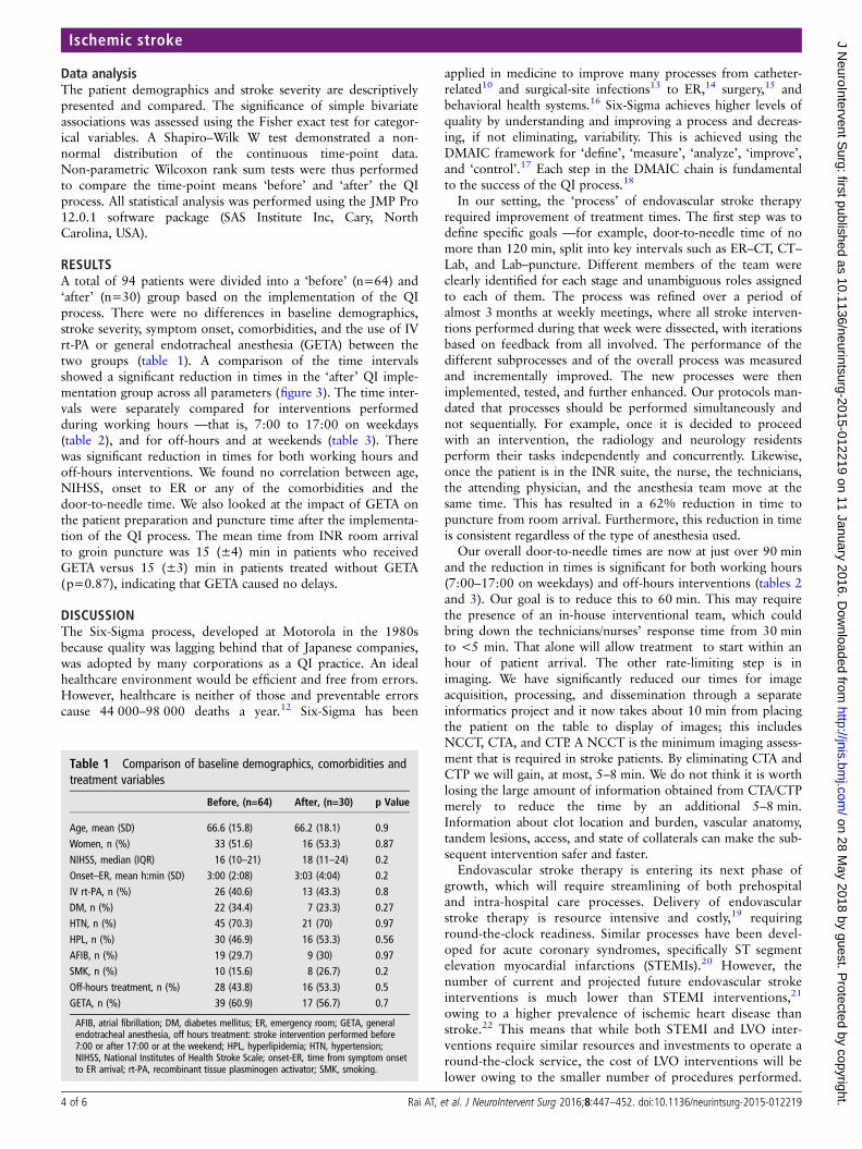
Table 1 Comparison of baseline demographics, comorbidities andtreatment variables
Before, (n=64) After, (n=30) p Value
Age, mean (SD) 66.6 (15.8) 66.2 (18.1) 0.9Women, n (%) 33 (51.6) 16 (53.3) 0.87NIHSS, median (IQR) 16 (10–21) 18 (11–24) 0.2Onset–ER, mean h:min (SD) 3:00 (2:08) 3:03 (4:04) 0.2IV rt-PA, n (%) 26 (40.6) 13 (43.3) 0.8DM, n (%) 22 (34.4) 7 (23.3) 0.27HTN, n (%) 45 (70.3) 21 (70) 0.97HPL, n (%) 30 (46.9) 16 (53.3) 0.56AFIB, n (%) 19 (29.7) 9 (30) 0.97SMK, n (%) 10 (15.6) 8 (26.7) 0.2Off-hours treatment, n (%) 28 (43.8) 16 (53.3) 0.5GETA, n (%) 39 (60.9) 17 (56.7) 0.7
AFIB, atrial fibrillation; DM, diabetes mellitus; ER, emergency room; GETA, generalendotracheal anesthesia, off hours treatment: stroke intervention performed before7:00 or after 17:00 or at the weekend; HPL, hyperlipidemia; HTN, hypertension;NIHSS, National Institutes of Health Stroke Scale; onset-ER, time from symptom onsetto ER arrival; rt-PA, recombinant tissue plasminogen activator; SMK, smoking.
4 of 6 Rai AT, et al. J NeuroIntervent Surg 2016;8:447–452. doi:10.1136/neurintsurg-2015-012219
Ischemic stroke on 28 M
ay 2018 by guest. Protected by copyright.
http://jnis.bmj.com
/J N
euroIntervent Surg: first published as 10.1136/neurintsurg-2015-012219 on 11 January 2016. D
ownloaded from

This is an important factor to consider when defining regionalstroke care. It may be beyond the capability of smaller hospi-tals (defined as ≤300 beds) to invest in high-qualityround-the-clock stroke therapy but the temptation could bethere from a marketing perspective and a perceived financialbenefit. Add to that an oversupply of physicians21 and wemight find small regional hospitals with one to two physiciansoffering endovascular stroke care, not realizing that the post-operative care by dedicated neurospecialists and allied staff isas important as the procedure itself. The consequences of sucha model could be dire with patients scattered among compet-ing hospitals with variable standards of care and inconsistentquality metrics. Additionally, the comparatively lower preva-lence of ischemic stroke will result in multiple centers carryingout a small volume of procedures instead of a regional high-volume center to which all such patients could be efficientlytransferred and treated.
LimitationsThe process at our institution was developed based on the man-power and resources within our system—a tertiary level aca-demic medical center with in-house resident teams. We realizethat our specific methodology may not be applicable to othersystems with different resources.
CONCLUSIONTime-critical interventions such as endovascular stroke therapycan benefit from a systemically implemented protocol-drivenapproach. While the methodology can be different in differentplaces based on available resources, its key elements includeclear definition of roles and parallel processing of tasks. It alsorequires constant monitoring and process improvements forconsistent performance. As the field grows it is important toregionalize systems of care based on population densities, facilitycapabilities, and health system networks.
Figure 3 Graphic comparison of the treatment times ‘before’ and ‘after’ implementation of the quality-improvement process.
Table 2 Comparison of time parameters during working hours onweekdays
Working hours—7:00–17:00 (n=50)
Before QI (n=36) After QI (n=14) p Value
ER–CT, min 42 (±28) 27 (±17) 0.011CT–Lab, min 67 (±41) 33 (±9) 0.0008Lab–puncture, min 24 (±16) 16 (±3) 0.0006CT–puncture, min 90 (±45) 49 (±10) 0.0002Door–puncture, min 132 (±53) 75 (±18) <0.0001
ER, emergency room; QI, quality improvement.
Table 3 Comparison of time parameters after hours or onweekends
Off hours and weekends (Sat–Sun) (n=44)
Before QI (n=28) After QI (n=16) p Value
ER–CT, min 38 (±30) 26 (±13) 0.35CT–lab, min 113 (±40) 67 (±38) 0.0025Lab–puncture, min 24 (±13) 15 (±4) 0.0035CT–puncture, min 138 (±40) 82 (±39) 0.0002Door–puncture, min 175 (±36) 108 (±42) <0.0001
ER, emergency room; QI, quality improvement.
Rai AT, et al. J NeuroIntervent Surg 2016;8:447–452. doi:10.1136/neurintsurg-2015-012219 5 of 6
Ischemic stroke on 28 M
ay 2018 by guest. Protected by copyright.
http://jnis.bmj.com
/J N
euroIntervent Surg: first published as 10.1136/neurintsurg-2015-012219 on 11 January 2016. D
ownloaded from

Contributors ATR: study design, data analysis, manuscript preparation. MSS, SHB,ART, JSC: manuscript preparation. GRH:data analysis.
Competing interests None declared.
Ethics approval Institutional review board.
Provenance and peer review Not commissioned; externally peer reviewed.
Open Access This is an Open Access article distributed in accordance with theCreative Commons Attribution Non Commercial (CC BY-NC 4.0) license, whichpermits others to distribute, remix, adapt, build upon this work non-commercially,and license their derivative works on different terms, provided the original work isproperly cited and the use is non-commercial. See: http://creativecommons.org/licenses/by-nc/4.0/
REFERENCES1 Powers WJ, Derdeyn CP, Biller J, et al. 2015 American Heart Association/American
Stroke Association focused update of the 2013 guidelines for the earlymanagement of patients with acute ischemic stroke regarding endovasculartreatment: a guideline for healthcare professionals from the American HeartAssociation/American Stroke Association. Stroke 2015;46:3020–35.
2 Fiorella DJ, Fargen KM, Mocco J, et al. Thrombectomy for acute ischemic stroke: anevidence-based treatment. J Neurointerv Surg 2015;7:314–15.
3 Kotwal RS, Howard JT, Orman JA, et al. The effect of a golden hour policy on themorbidity and mortality of combat casualties. JAMA Surg Published Online First:30 Sep 2015. doi:10.1001/jamasurg.2015.3104
4 Ashmeade TL, Haubner L, Collins S, et al. Outcomes of a neonatal golden hourimplementation project. Am J Med Qual Published Online First: 5 Sep 2014.pii: 1062860614548888.
5 Mazighi M, Chaudhry SA, Ribo M, et al. Impact of onset-to-reperfusion time onstroke mortality: a collaborative pooled analysis. Circulation 2013;127:1980–5.
6 Berkhemer OA, Fransen PS, Beumer D, et al. A randomized trial of intraarterialtreatment for acute ischemic stroke. N Engl J Med 2015;372:11–20.
7 Kelleher DC, Carter EA, Waterhouse LJ, et al. Effect of a checklist on advancedtrauma life support task performance during pediatric trauma resuscitation. AcadEmerg Med 2014;21:1129–34.
8 Broderick JP, Palesch YY, Demchuk AM, et al. Endovascular therapy after intravenoust-PA versus t-PA alone for stroke. N Engl J Med 2013;368:893–903.
9 Kidwell CS, Jahan R, Gornbein J, et al. A trial of imaging selection andendovascular treatment for ischemic stroke. N Engl J Med 2013;368:914–23.
10 Frankel HL, Crede WB, Topal JE, et al. Use of corporate Six Sigmaperformance-improvement strategies to reduce incidence ofcatheter-related bloodstream infections in a surgical ICU. J Am Coll Surg2005;201:349–58.
11 Kothari RU, Pancioli A, Liu T, et al. Cincinnati prehospital strokescale: reproducibility and validity. Ann Emerg Med 1999;33:373–8.
12 Kohn LT, Corrigan J, Donaldson MS. To err is human: building a safer healthsystem. Washington DC: National Academy Press, 2000.
13 Kles CL, Murrah CP, Smith K, et al. Achieving and sustaining zero: preventingsurgical site infections after isolated coronary artery bypass with saphenous veinharvest site through implementation of a staff-driven quality improvement process.Dimens Crit Care Nurs 2015;34:265–72.
14 Sanders JH, Karr T. Improving ED specimen TAT using lean Six Sigma. Int J HealthCare Qual Assur 2015;28:428–40.
15 Mason SE, Nicolay CR, Darzi A. The use of lean and Six Sigma methodologies insurgery: a systematic review. Surgeon 2015;13:91–100.
16 Lucas AG, Primus K, Kovach JV, et al. Rethinking behavioral health processes byusing design for Six Sigma. Psychiatr Serv 2015;66:112–14.
17 Pyzdek T, Keller PA. The Six Sigma handbook. New York: McGraw-Hill Education,2014.
18 Kwak YH, Anbari FT. Benefits, obstacles, and future of Six Sigma approach.Technovation 2006;26:708–15.
19 Rai AT, Evans K. Hospital-based financial analysis of endovascular therapy andintravenous thrombolysis for large vessel acute ischemic strokes: the ‘bottom line’.J Neurointerv Surg 2015;7:150–6.
20 Ahmed B, Lischke S, Straight F, et al. Consistent door-to-balloon times of less than90 minutes for STEMI patients transferred for primary PCI. J Invasive Cardiol2009;21:429–33.
21 Rai AT. Red pill, blue pill: reflections on the emerging large vessel stroke ‘market’.J Neurointerv Surg 2015;7:623–5.
22 Mozaffarian D, Benjamin EJ, Go AS, et al. Heart disease and stroke statistics—2015 update: a report from the American Heart Association. Circulation 2015;131:e29–322.
6 of 6 Rai AT, et al. J NeuroIntervent Surg 2016;8:447–452. doi:10.1136/neurintsurg-2015-012219
Ischemic stroke on 28 M
ay 2018 by guest. Protected by copyright.
http://jnis.bmj.com
/J N
euroIntervent Surg: first published as 10.1136/neurintsurg-2015-012219 on 11 January 2016. D
ownloaded from