original contribution comparative influence of
TRANSCRIPT

30 Trakia Journal of Sciences, Vol. 15, № 1, 2017
Trakia Journal of Sciences, No 1, pp 30-41, 2017
Copyright © 2017 Trakia University
Available online at:
http://www.uni-sz.bg
ISSN 1313-7050 (print)
ISSN 1313-3551 (online) doi:10.15547/tjs.2017.01.006
Original Contribution
COMPARATIVE INFLUENCE OF IMMOBILIZATION MEDIUM AND
MUTATION ON EPS –PRODUCTION BY L. PLANTARUM MK O2 ISOLATED
FROM FERMENTED MILK
B. Adebayo-Tayo*, O. Agidigbi, S. Alao
Department of Microbiology, University of Ibadan, Ibadan, Oyo State, Nigeria
ABSTRACT PURPOSE: Comparative effect of mutation and immobilization on EPS-production L. plantarum MKO2
was investigated. METHODS: L. plantarum strain and EPS produced by wild, mutants, immobilized and
unimmobilized strains was characterized. RESULTS: The EPS production by the Wild type L. plantarum
MK O2 and the Mutant L. plantarum Muv 11 and Muv 12 ranged from 209.89 – 268.19 mg/l in which the
highest value was produced by Mutant L. plantarum Muv 12. EPS production on immobilization in sodium
alginate ranged from 225.00 – 263.44mg/l. Mutant L. plantarum Muv 12 had the highest while Wild type L.
plantarum MK O2 had the least. EPS – production by un –immobilized Wild type L. plantarum MK O2
and Mutant L. plantarum Muv 11 and Muv 12 ranged from 215.00 – 255.00 mg/l. Immobilization in
sodium alginate favoured EPS-production by wild type L. plantarum MK O2 (225.00 mg/l) and Mutant L.
plantarum Muv 12 (272.00 mg/l) while immobilization in agar matrix favoured EPS –production by
Mutant L. plantarum Muv 11 (265.67 mg/l). FT-IR spectroscopy of the EPS showed the presence of the
varying degrees of functional groups which are usually associated with polysaccharides, thus confirming
the EPS. CONCLUSION: Mutation and immobilization increased EPS production by the L. plantarum
strains.
Key words: EPS - production, Mutation, Immobilization, Sodium alginate, Agar matrix.
INTRODUCTION
Microbial exopolysaccharides (EPS) are a broad
group of polymers secreted by microorganisms,
which may be associated with the cell surface as
capsular polysaccharides, or excreted into the
surroundings of the cell as extracellular slime (1-
2). Lactic acid bacteria (LAB) are a very
important group of bacteria normally utilized in
the food industry. They are involved in the
natural fermentation of a variety of foods
including milk, vegetables, beverages and meat.
The activities of LAB in these foods result in the
production of finished products with improved
properties such as enhanced aroma and flavour,
extended shelf life and improved functional
properties. These properties are ensured through
the production of useful metabolites such as
______________________________ *Correspondence to: B. Adebayo-Tayo, Department
of Microbiology, University of Ibadan, Ibadan, Oyo
State, Nigeria, E-mail address:
lactic acid, diacetyl, hydrogen peroxide and
bacteriocins. In addition to these
metabolites,some LAB also LAB redirect a
small proportion of fermentable sugars in their
environment towards the biosynthesis of
exopolysaccharides (3). A wide variety of EPS
are known to be produced by several genera of
LAB, including Lactobacillus, Leuconostoc,
Pediococcus and Streptococcus (4).
Approximately thirty species of lactobacilli are
described as EPS producers. Among them, the
best known are L. casei, L. acidophilus,
L.brevis, L. curvatus, L. delbrueckii, L.
helveticus, L. rhamnosus, L plantarum and L.
johnsonii (5). Dextran (synthesized by certain
LAB such as Leuconostoc mesenteroides) was
the first microbial polysaccharide to be
commercially exploited and to receive
authorization for use in foods (6).
Two major groups of EPS have been identified:
homopolysaccharides and heteropolysaccha-
rides. Homopolysaccharides are made up of a
ADEBAYO-TAYO, B., et al.
Trakia Journal of Sciences, Vol. 15, № 1, 2017 31
single type of monosaccharide, usually glucose
or fructose which occurs as repeated units of
three to eight monosaccharides. Thus, they are
referred to as α-glucans (dextrans) or fructans.
Heteropolysaccharides consist of at least two
different sugars out of D-glucose, D-galactose
and L-rhamnose in different ratios. Some other
residues such as n-glycerol-3-phosphate, N-
acetyl-amino sugars, phosphate and acetyl
groups may also be found in EPS produced by
LAB (7, 4). EPS produced by LAB have been
are mostly composed of repeated units of a
certain number of various sugar residues or
sugar derivatives (8-9) joined by α- and β-
linkages (10). The carbohydrate composition of
EPS is unique to different strains of bacteria and
may vary depending on the growth conditions;
however, they usually consist of similar
monomer subunits, with D-galactose, D-glucose
and L-rhamnose occurring almost always,
though in different ratios (11-14).
LABs are currently used in the production of
many fermented dairy products due to their
contribution to the textural characteristics of the
food product through the production of EPS. For
example, several strains of Lactobacillus
helveticus used for mozzarella production
produce EPS and contribute to water retention in
the product (12-15). EPS may act both as a
texturizer by improving the rheology (viscosity
and elasticity) of a product, and as physical
stabilizers by binding hydration water and
interacting with other milk constituents (ions
and proteins) thus limiting syneresis (16). EPS
also contribute to human health as prebiotics or
due to antitumor, immunomodulating or
cholesterol-lowering activities (4). It helps
probiotics to survive the gastric acid and bile
salts in the gastrointestinal tract (17). The
increased viscosity of foods containing EPS is
thought to increase the residence time of
ingested fermented milk in the gastrointestinal
tract, thereby supporting transient colonization
by probiotic bacteria (18).
Random mutagenesis, a classical method of
strain improvement, has been widely used in the
food industry to improve microbial strains with
desirable qualities (19 – 20). Since the genome
of an organism controls its potential
productivity, consequently, the genome may be
modified to increase the potential yield.
Ultraviolet (UV) light has been shown to be
lethal and mutagenic in a variety of organisms,
including bacteria. Mutagenic lesions are formed
in the DNA as a result of exposure of cells to
UV radiation. Two of the most frequent lesions
are the cyclobutane pyrimidine dimers and the 6-
4 pyrimidine-pyrimidone photoproducts formed
at adjacent pyrimidines (21). Other types of
DNA lesions include pyrimidine hydrates,
purine photoproducts, strand brakes, and DNA
cross-links. These however occur at a much
lower frequency (22).
Immobilisation involves restricting the mobility
of microbial cells, enzymes or other proteins
inside or on the surface of a carrier such that
their catalytic activity is preserved (23).
Currently, different immobilization techniques
have found wide applications not only in the
field of biotechnology, but also in
pharmaceutical, environmental, food and
biosensor industries (24). It is an important
means of increasing the performance of
microbial cultures (25) and it has been used to
carry out high cell density fermentations for both
cell and metabolite production. The main
advantage of immobilization is easy separation
of biological material from the reaction medium
containing the desired product. This
significantly accelerates the production process
of a variety of compounds (26). Another
advantage offered by immobilization is cost-
effectiveness of the reaction. Immobilized cell
biomass may be re-used several times without
necessarily re-culturing them. Other advantages
offered by immobilization cell technology are
protection against shear damage, high biological
stability during long-term continuous
fermentation (27 – 29) improved resistance to
contamination and bacteriophage attack,
enhancement of plasmid stability and prevention
from washing-out during continuous cultures
(30). This research aimed at investigating the
comparative influence of immobilization
medium on EPS – production by L. plantarum
MK O2 isolated from fermented milk.
MATERIALS AND METHODS
Bacterial Strains
The Lactobacillus plantarum strain used in this
study was isolated from milk obtained from
Sabo market in Ibadan, Oyo State, Nigeria.
Isolation of Organisms
The milk sample was allowed to ferment in the
laboratory for 48 hours after which isolation of
the LAB was carried out according to the
method of Sobrun et al (30). Serial dilution of
the sample was carried out and it was plated on
de Man, Rogosa and Sharpe (MRS) agar
supplemented with sucrose (2% w/v), sodium
azide (0.02% w/v) and bromocresol purple
ADEBAYO-TAYO, B., et al.
32 Trakia Journal of Sciences, Vol. 15, № 1, 2017
(0.012% w/v). The cultures were incubated at
35°C for 48 hours under anaerobic conditions.
Mucoid and ropy colonies exhibiting yellow
zones were selected and re-streaked on MRS
agar repeatedly to obtain pure cultures. The
isolates were maintained on MRS agar slants
4°C and in a maintenance medium consisting of
MRS broth with 20% glycerol.
Quantification of EPS produced by the
strains
The amount of EPS produced was determined
according to the method of Dubois et al. (31)
using the exopolysaccharide selection medium
(ESM) described by Van den Berg (32) and
modified by Ludbrook et al. (33). The sterile
ESM was inoculated with 24 hour broth culture
of the LAB isolates and incubated at 35°C for 24
hours. The cultures were heated in a water bath
at 80°C for 15 minutes and left to cool at room
temperature. They were then treated with 5%
(w/v) trichloroacetic acid (TCA) and left at 4°C
overnight followed by centrifugation at 15000g
for 20 minutes at 4°C to remove cells and
precipitated proteins. The crude EPS was
precipitated out by the addition of two volumes
of chilled absolute ethanol and kept at 4°C
overnight. The samples were centrifuged again
at 15000g for 10 minutes and the resulting
precipitate re-dissolved in 10ml of water. Phenol
(1ml, 5% w/v) was added to 1ml of the
dissolved precipitate in a test tube and allowed
to stand for 1 minute. 5ml of concentrated
sulphuric acid was added and allowed to stand
for 30 minutes. The absorbance was measured
using a spectrophotometer at 490 nm against a
blank (distilled water) and compared to a graph
generated from the results obtained for a series
of D-glucose standards to determine the sugar
concentration. The L. plantarum strain used in
this study was selected because of its high EPS
production capacity.
Identification of Isolate
Identification of the isolate was carried out using
morphological, physiological and biochemical
characteristics such as gram staining, casein
hydrolysis, starch hydrolysis, gelatin hydrolysis,
oxidase test, homofermentative and
heterofermentative test and carbohydrate
fermentation.
Confirmation of the identity was done using
molecular characterization. DNA extraction was
carried out according to the method of De et al.
(34). The isolated DNA was then used as a
template for selective amplification by the
Polymerase Chain reaction (PCR). DNA sample
from the isolate obtained by this protocol were
assayed by restriction enzyme digestion.
Sequencing of the fragments was carried out
using the automatic Big Dye (dideoxy chain
terminator) sequencer ABIPRISM 3730xl. For
identification of the closest relatives, newly
determined sequences were compared to those
available in the V2-V3 region of the 16S rRNA
sequences using the GenBank DNA databases
and the standard nucleotide-nucleotide BLAST
algorithm.
Molecular Identification of the Highest EPS-
Producing Strains
The highest EPS –producing L. plantarum (MK
02) was identified using molecular technique
based on the 16SrRNA gene amplification by
polymerase chain reaction (PCR). Purification of
the PCR products and the determination of
sequences were performed by Macrogen USA
(9700 Great Seneca Highway, Rockville, MD
20850, USA). The amplified gene was sequence
and the sequence compared with the NCBI
database
(http://blast.ncbi.nlm.nih.gov/Blast.cgi).
Mutagenesis
Ultraviolet radiation was used to induce
mutagenesis in the L. plantarum strain. Dilutions
of the bacterial culture were plated on MRS agar
and irradiated with UV light of wavelength 254
nm at a distance of 30 cm for 5, 10, 15, 20, 25,
30, 60, 90 and 120 seconds. Cultures which
were not exposed to UV radiation served as
control. The cultures were wrapped in
aluminium foil to prevent the penetration of light
which induces photoreactivation and incubated
at 35˚C for 24 hours. Colony count of all the
cultures was carried out and colonies were
selected from cultures having 2% survival rate.
Immobilisation of Cells
In Sodium alginate: The entrapment method was
used to immobilise the cells in sodium alginate
according to the method of Pasha et al. (35).
Two gram (2g) of sodium alginate was dissolved
in 100 ml 0.9% NaCl. The solution was then
autoclaved at 121ºC for 15 min and 2 ml
inoculums of wild type whole cells and mutants
each were added to 10 ml of the solution
prepared. The sodium alginate-cell solution was
then added drop wise with stirring to a 0.1 M
CaCl2 solution. The gel beads formed were left
in solution for 1 hour before being filtered off.
The beads were then washed in a 0.9 % NaCl
solution for 20 min.

ADEBAYO-TAYO, B., et al.
Trakia Journal of Sciences, Vol. 15, № 1, 2017 33
In Agar: The method of Manohar and
Karegoudar (36) was used to immobilize both
wild type and mutant cells in agar matrix. The
LAB strains were cultured in MRS broth for 12
hours and centrifuged at 5000 rpm for 10
minutes to obtain cell pellets. The cell pellets
were suspended in sterile saline solution and
mixed with sterile 4% (w/v) agar-saline solution,
allowed to set and cut with a sterile cork borer to
obtain cell-entrapped beads. The agar beads
were washed successively with distilled water
and saline.
Production of EPS by Un-immobilized Wild
type and Mutant Strains
The bacterial cells were grown in MRS broth at
35°C for 18 hours after which the broth cultures
were used to inoculate the exopolysaccharide
selection medium (10% v/v) and incubated at
35°C for 48 hours. The EPS produced was then
isolated and purified according to the method of
Cerning et al. (37). Quantification of the EPS
was carried out as described by Dubois et al.
(31).
Production of EPS by Immobilised Wild type
and Mutant Strains The exopolysaccharide selection medium was
inoculated with 5% (w/v) of the immobilised
cells according to the method of Karigar et al.
(38) and incubated at 35°C for 48 hours. The
EPS produced was isolated and purified
according to the method of Cerning et al. (39)
while its quantification was carried out using the
method described by Dubois et al. (31).
Fourier Transform Infrared Spectroscopy
(FT-IR) of EPS
The EPS were prepared for infrared analysis by
grinding a mixture of 2 mg of the EPS with 200
mg dry KBr, followed by pressing the mixture
into a 16 mm diameter mold. The Fourier
transform-infrared (FT-IR) spectra were
recorded on a Shimadzu IR Affinity 1S
instrument with a resolution of 4 cm-1
in the
4000-400 cm-1
region.
RESULTS AND DISCUSSION
The isolate was characterized genotypically and
the EPS produced by the strains was
characterized. A BLAST (Basic Local
Alignment Search Tool) analyses of the
16SrRNA gene nucleotide sequence of strain
MKO2 amplified product showed a 97%
similarity to L. plantarum. A phylogenetic tree
was constructed between it and similar
sequences found in GenBank.
Molecular identification of the LAB strain MK
02 as Lactobacillus plantarum follows the trend
of previous works which have identified this
organism as a potent EPS producer. Talon et al.
(40) and Francois et al. (41) reported the
production of both capsular and extracellular
EPS by L. plantarum strains. Remus et al. (42)
reported the presence of four clusters of genes in
the genome of L. plantarum which are
associated with surface polysaccharide
production, indicating the capacity of this strain
for EPS production.
The wild type L. plantarum strain (MK 02) used
in this study produced an EPS concentration of
218.67 mg/l after 18 hours of fermentation in a
medium containing skim milk. EPS produced by
wild type L. plantarum MK 02 is shown in
Figure 1.
Figure 1. EPS produced by wild type L. plantarum MK 02
ADEBAYO-TAYO, B., et al.
34 Trakia Journal of Sciences, Vol. 15, № 1, 2017
This study was carried out with the aim of
increasing the EPS production capacity of the
isolate using mutagenesis and immobilization
techniques.
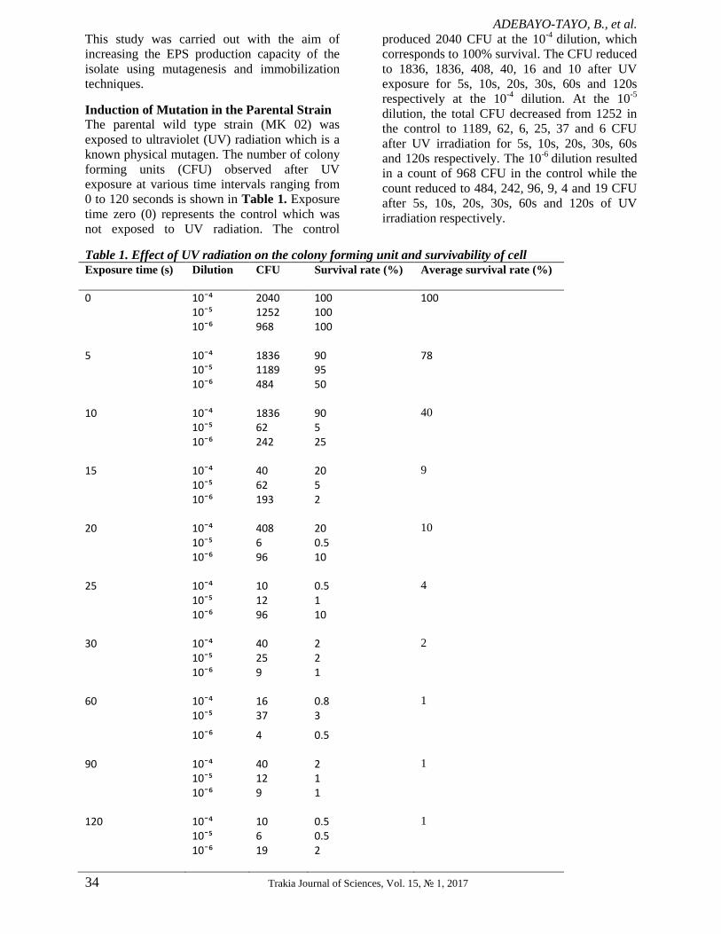
Induction of Mutation in the Parental Strain
The parental wild type strain (MK 02) was
exposed to ultraviolet (UV) radiation which is a
known physical mutagen. The number of colony
forming units (CFU) observed after UV
exposure at various time intervals ranging from
0 to 120 seconds is shown in Table 1. Exposure
time zero (0) represents the control which was
not exposed to UV radiation. The control
produced 2040 CFU at the 10-4
dilution, which
corresponds to 100% survival. The CFU reduced
to 1836, 1836, 408, 40, 16 and 10 after UV
exposure for 5s, 10s, 20s, 30s, 60s and 120s
respectively at the 10-4
dilution. At the 10-5
dilution, the total CFU decreased from 1252 in
the control to 1189, 62, 6, 25, 37 and 6 CFU
after UV irradiation for 5s, 10s, 20s, 30s, 60s
and 120s respectively. The 10-6
dilution resulted
in a count of 968 CFU in the control while the
count reduced to 484, 242, 96, 9, 4 and 19 CFU
after 5s, 10s, 20s, 30s, 60s and 120s of UV
irradiation respectively.
Table 1. Effect of UV radiation on the colony forming unit and survivability of cell Exposure time (s) Dilution CFU Survival rate (%) Average survival rate (%)
0 10¯⁴ 2040 100 100 10¯⁵ 1252 100 10¯⁶ 968 100 5 10¯⁴ 1836 90 78 10¯⁵ 1189 95 10¯⁶ 484 50
10 10¯⁴ 1836 90 40
10¯⁵ 62 5
10¯⁶ 242 25
15 10¯⁴ 40 20 9
10¯⁵ 62 5
10¯⁶ 193 2
20 10¯⁴ 408 20 10
10¯⁵ 6 0.5
10¯⁶ 96 10
25 10¯⁴ 10 0.5 4
10¯⁵ 12 1
10¯⁶ 96 10
30 10¯⁴ 40 2 2
10¯⁵ 25 2
10¯⁶ 9 1
60 10¯⁴ 16 0.8 1
10¯⁵ 37 3
10¯⁶ 4 0.5
90 10¯⁴ 40 2 1
10¯⁵ 12 1
10¯⁶ 9 1
120 10¯⁴ 10 0.5 1
10¯⁵ 6 0.5
10¯⁶ 19 2

ADEBAYO-TAYO, B., et al.
Trakia Journal of Sciences, Vol. 15, № 1, 2017 35
The survival rate after UV exposure as seen in
Figure 2 shows that after 5s of UV irradiation,
78% of the cells were found to have survived,
10% survived 20s of UV irradiation while only
1% were able to grow after 120s of radiation. In
general, the percentage of organisms which
survived the UV irradiation reduced as the
exposure time increased.
Figure 2. Survival rate of L. plantarum cells after exposure to UV radiation
Following the mutagenesis procedure, 12
mutants were selected and further screened for
EPS production. The EPS production rate of the
mutant strains (Muv 1- Muv 12) as compared to
the wild type (MK 02) is shown in Figure 3.
The wild type L. plantarum MK O2 and the
selected mutants have the ability to produce
EPS. The EPS-production ranged from 98.00 –
240.00mg/l. Four mutants (Muv7, Muv 10, Muv
11 and Muv 12 produced higher EPS than the
wild type while the rest 8 mutants produced EPS
lower than the wild type L. plantarum MK O2.
The two highest EPS-producing mutant L.
plantarum Muv 11 and Muv 12 were selected
for further study.
Figure 3. Comparism of EPS production by wild type L. plantarum MK O2 and Mutant L.
plantarum Muv 11 and Muv 12
The results of ultraviolet mutagenesis in the L.
plantarum strain (MK 02) indicate that the rate
of survival of the organism decreased as the time
of exposure to UV radiation increased. The
number of cells which survived UV irradiation
reduced drastically from 2.04 x 10³
CFU to
about 10 CFU after 120s of UV
exposure,thereby achieving a survival rate of 1%
at 120s of UV irradiation. Four, out of the
twelve mutants isolated showed more yields of
EPS than the wild type strain. This implies that
the genetic modifications that occurred in the
LAB as a result of random mutagenesis had led
to the development of strains with improved
characteristics. Increase in EPS production from
218.67 mg/l in the wild type strain MK 02 to
252.0 mg/l and 253.22 mg/l in its mutant strains

ADEBAYO-TAYO, B., et al.
36 Trakia Journal of Sciences, Vol. 15, № 1, 2017
Muv 11 and Muv 12 respectively represent
about 14% increase in EPS production. Previous
studies have shown UV radiation to be a potent
mutagenic agent and it has been used as a strain
improvement technique for the development of
microbial strains capable of producing higher
yields of desired metabolites. Increased lactic
acid production was observed by Sobrun et al.
(30) in a mutant strain of LAB which produced a
maximum concentration of 29.0 g/l, compared to
the wild type strain which produced about 19.0
g/l of lactic acid. Pasha (43) also reported higher
yields of glutamic acid from UV-induced
mutants of Corynebacterium glutamicum.
Cell Immobilization
Immobilization of wild type L. plantarum MK
O2 and the mutant L. plantarum Muv 11 and
Muv 12 on agar matrix and sodium alginate is
shown in Figure 4. The EPS production by the
wild type L. plantarum MK O2 and the Mutant
L. plantarum Muv 11 and Muv 12 when
immobilized on agar matrix ranged from 209.89
– 268.19mg/l in which the highest value was
produced by Mutant L. plantarum Muv 12 and
the least was produced by wild type L.
plantarum MK O2.
The EPS – production by wild type L. plantarum
MK O2 and the Mutant L. plantarum Muv 11
and Muv 12 when immobilized on sodium
alginate ranged from 225.00 – 263.44mg/l.
Figure 4. EPS production by wild type L. plantarum MK O2 and Mutant L. plantarum Muv 11 and Muv 12
immobilized in agar and sodium alginate.
Mutant L. plantarum Muv 12 had the highest
value while wild type L. plantarum MK O2 had
the least.
Increased EPS production resulted from
immobilization of both wild type and mutant
strains. Immobilization in sodium alginate
favoured EPS-production by wild type L.
plantarum MK O2 (225.00mg/l) and Mutant L.
plantarum Muv 12 (272.00mg/l).
Immobilization in agar matrix favoured EPS –
production by Mutant L. plantarum Muv 11
(265.67mg/l).
Figure 5 shows EPS production by the un-
immobilized cells. EPS – production by un –
immobilized wild type L. plantarum MK O2 and
mutant L. plantarum Muv 11 and Muv 12
ranged from 215.00 – 255.00 mg/l. Mutant L.
plantarum Muv 12 had the least production.
Figure 5. EPS production by un-immobilized wild type L. plantarum MK O2 and Mutant L. plantarum Muv 11 and
Muv 12

ADEBAYO-TAYO, B., et al.
Trakia Journal of Sciences, Vol. 15, № 1, 2017 37
Immobilization of wild type and mutant strains
of LAB was carried out to improve the EPS
production capacity of the strains. Immobilized
cell technology has been applied in a wide
variety of research applications. For instance,
the high potential of Lactobacillus rhamnosus
RW-9595M for EPS production and the
importance of immobilized cell technology were
emphasized in the studies of Bergmaier et al.
(44) and Bergmaier et al. (45).
From this study, higher EPS concentrations were
obtained from immobilized cells as compared to
un-immobilized cells, while sodium alginate was
a more favourable matrix for immobilizing cells
for enhanced EPS production. From these
investigations, immobilization in sodium
alginate produced up to 7% increase in EPS
production in the mutant Muv 12 while agar
immobilization produced about 5% increase in
EPS yield as observed in Muv 11. The increase
in EPS production by immobilized cells could be
attributed to the fact that immobilization enables
the concentration of high density of cells in the
immobilization matrix, thus making more cells
available for EPS production. Competition
between cells may be reduced when
immobilized than when they are free in the
medium as fewer cells are encapsulated within
each unit of the immobilization matrix. Also,
prevention of feedback inhibition could account
for higher yields of EPS by immobilized cells
because as the EPS is being produced, the
immobilization matrix separates the cells from
the products formed. Thus, inhibition of further
EPS synthesis is prevented. These observations
are in agreement with the work of El-Gizawy et
al. (46) where the production of EPS was found
to have been enhanced by microencapsulation
due to an increase in the viable count of the used
strain of Lactobacillus bulgaricus. Ismail and
Nampothiri (47) also studied EPS production by
encapsulated L. bulgaricus and found that
encapsulated cells gave higher EPS production
than free cells.
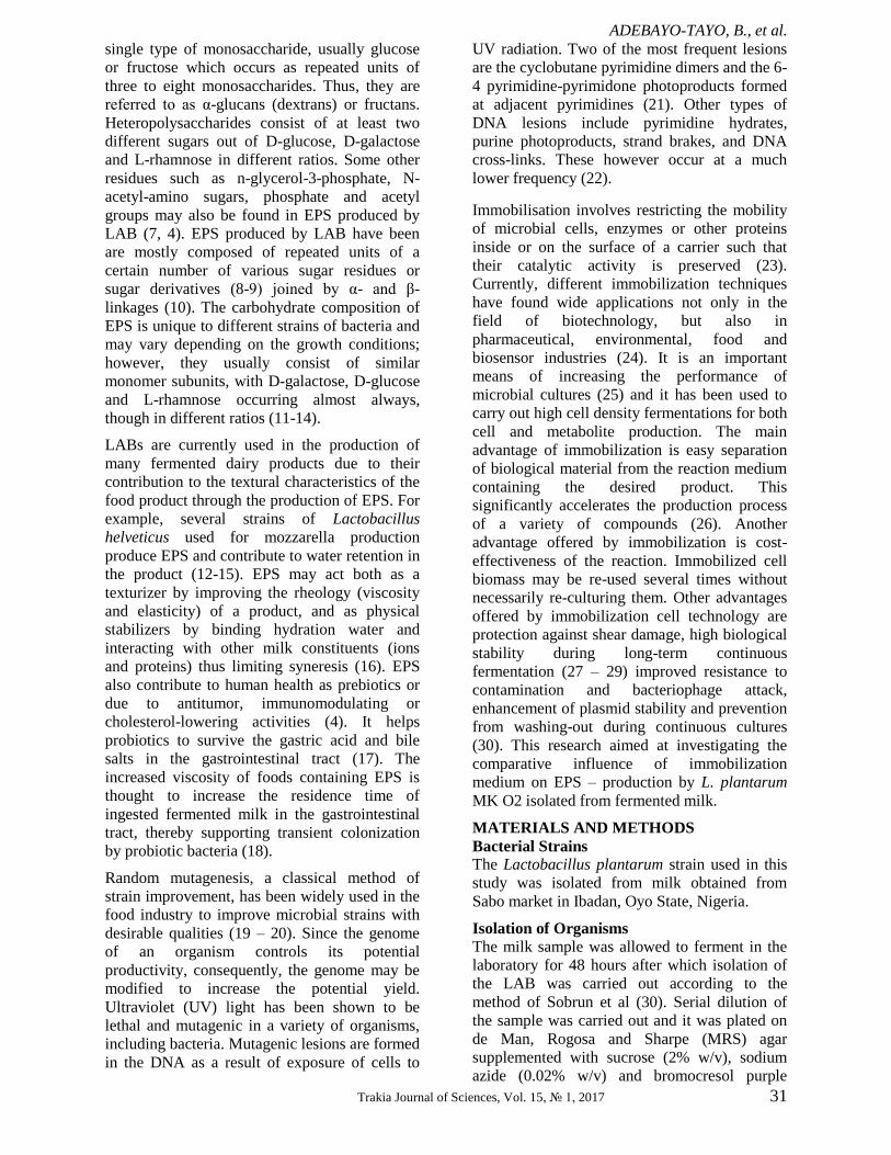
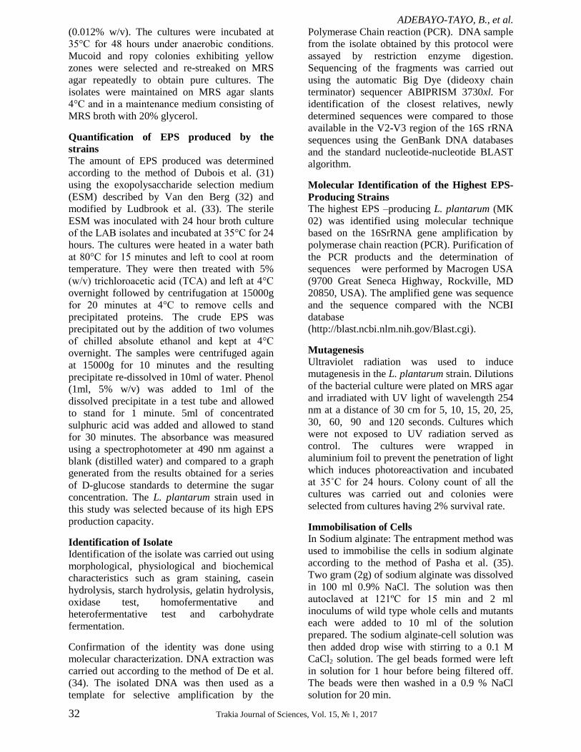
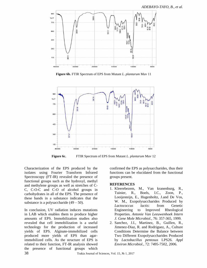
Characterization of the EPS produced by the
strains using Fourier Transform Infrared
(FT-IR) Spectroscopy The EPS produced by the bacterial strains was
analysed using FT-IR to determine the
functional groups present in them. The spectra
produced by the EPS of both wild type (Figure
6a) and mutant strains (Figure 6b, Figure 6c)
showed similarities in having absorption peaks
around 3000cm-1
(characteristic of carbon- and
hydrogen-containing species), 2928 cm-1
(corresponding to methyne C-H stretch),
2070cm-1
( CO group), 1654 cm-1
(indicating the
presence of olefinic un-saturation C=C), 1456-
1236cm-1
(representing the phenol or tertiary
alcohol bend), 1153-1101 cm-1
(tertiary alcohol
C-O stretch), 1051cm-1
(C-H out-of-plane
bending) and 908-455 cm-1
(alcohol OH out-of-
plane bend). Absorptions in the region of
3300cm-1
indicate that the compound is likely to
be unsaturated or aromatic while peaks at
1654cm-1
further highlight the presence of un-
saturation in the EPS. The absorption peak at
2063.90cm-1
shows the presence of nitrogen
compounds having multiple bonds such as
cyanides (nitriles), cyanates, isocyanates,
thiocyanates and diazo compounds (48). Also,
the presence of multiply-bonded CO group is
indicated by absorption bands around 2000cm-1
.
However, the EPS produced by both mutants
differ from that of the wild type strain in having
one peak less in the regions 1456.30-1236.41cm-1
and 908.50-455.22 cm-1
. This implies lesser OH
bonding in the mutants’ EPS as these two
regions are characteristic of alcohol bends.
Figure 6a. FTIR Spectrum of EPS from wild type L. plantarum MK O2
ADEBAYO-TAYO, B., et al.
38 Trakia Journal of Sciences, Vol. 15, № 1, 2017
Figure 6b. FTIR Spectrum of EPS from Mutant L. plantarum Muv 11
Figure 6c. FTIR Spectrum of EPS from Mutant L. plantarum Muv 12
Characterization of the EPS produced by the
isolates using Fourier Transform Infrared
Spectroscopy (FT-IR) revealed the presence of
functional groups such as the hydroxyl, methyl
and methylene groups as well as stretches of C-
C, C-O-C and C-O of alcohol groups in
carbohydrates in all of the EPS. The presence of
these bands in a substance indicates that the
substance is a polysaccharide (49 – 50).
In conclusion, UV radiation induces mutations
in LAB which enables them to produce higher
amounts of EPS. Immobilization studies also
revealed that cell immobilization is a useful
technology for the production of increased
yields of EPS. Alginate-immobilized cells
produced more yields of EPS than agar-
immobilized cells. As the structure of EPS is
related to their function, FT-IR analysis showed
the presence of functional groups which
confirmed the EPS as polysaccharides, thus their
functions can be elucidated from the functional
groups present.
REFERENCES
1. Kleerebezem, M., Van kranenburg, R.,
Tuinier, R., Boels, I.C., Zoon, P.,
Looijesteijn, E., Hugenholtz, J.and De Vos,
W. M., Exopolysaccharides Produced by
Lactococcus lactis: from Genetic
Engineering to Improved Rheological
Properties. Antonie Van Leeuwenhoek Intern
J. Gene Mole Microbiol., 76: 357-365, 1999.
2. Sanchez, J.I., Martinez, B., Guillen, R.,
Jimenez-Diaz, R. and Rodriguez, A., Culture
Conditions Determine the Balance between
Two Different Exopolysaccharides Produced
by Lactobacillus pentosus LPS26. Appl
Environ Microbiol., 72: 7495-7502, 2006.

ADEBAYO-TAYO, B., et al.
Trakia Journal of Sciences, Vol. 15, № 1, 2017 39
3. Sutherland, I.W., Physiology and Industrial
Production. In I.W. Sutherland (ed.),
Biotechnology of Microbial
Exopolysaccharides. Cambridge University
Press, Cambridge England. 1990.
4. De Vuyst, L. and Degeest, B.,
Heteropolysaccharides from Lactic Acid
Bacteria. FEMS Microbiol Rev., 23: 153-177,
1999.
5. Jaiswal, P., Sharma, R., Sanodiya, B.S. and
Bisen, P.S., Microbial Exopolysaccharides:
Natural Modulators of Dairy Products. J Appl
Pharmaceu Sci., 4 (10): 105-109, 2014.
6. Sarwat, F., Qader, U., Aman, A. and Ahmed,
N., Production and Characterization of a
Unique Dextran from an Indigenous
Leuconostoc mesenteroides CMG713. Intern
J Biol Sci., 4:379–386, 2008.
7. Landersjö, C., Yang, Z., Huttunen, E. and
Widmalm, G., Structural Studies of the
Exopolysaccharide Produced by
Lactobacillus rhamnosus strain GG (ATCC
53103). Biomacromole., 3: 880-884, 2002.
8. Górska, S., Jachymek, W. and Rybka, J.,
Structural and Immunochemical Studies of
Neutral Exopolysaccharides Produced by
Lactobacillus johnsonii 142. Carbohyd Res.
345:108114, 2010.
9. Margolles, A. and Sanchez, B., Selection of a
Bifidobacterium animalis subsp. lactis Strain
with a Decreased Ability to Produce Acetic
Acid. Appl Environ Microbiol. 78:3338-
3342, 2012.
10. Nakajima, H., Suzuki, Y. and Hirota, T.,
Cholesterol Lowering Activity of Ropy
Fermented Milk. J Fd Sci., 57: 327-1329,
1992.
11. Gruter, M., Leeflang, B.R., Kuiper, J.,
Kamerling, J.P. and Vliegenthart, J.F.G.,
Structural Characterisation of the
Exopolysaccharide Produced by
Lactobacillus delbrueckii subspecies
bulgaricus grown in Skimmed Milk.
Carbohydr Res., 239: 209-226, 1993.
12. Yamamoto, Y., Nunome, T., Yamauchi, R.,
Kato, K. and Sone, Y., Structure of an
Exocellular Polysaccharide of Lactobacillus
helveticus TN-4, a Spontaneous Mutant
Strain of Lactobacillus helveticus TY1-2.
Carbohydr Res., 275: 319-332, 1995.
13. Lemoine, J., Chirat, F., Wie uszeski, J.M.
Strecker, G., Favre, N. and Neeser, J.R.,
Structural Characterization of the Exocellular
Polysaccharides Produced by Streptococcus
thermophilus Sfi39 and Sfi12. Appl. Environ
Microbiol., 63:3512-3518, 1997.
14. Kojic, M., Vujcic, M., Banina, A.,
Cocconcelli, P., Cerning, J. and Topisirovic,
L., Analysis of Exopolysaccharide
Production by Lactobacillus casei CG11
Isolated from Cheese. Appl Environ
Microbiol., 58: 4086-4088, 1992.
15. Low, D., Ahlgren, J.A., Horne, D.,
McMahon, D.J., Oberg, C.J. and Broadbent,
J.R., Role of Streptococcus thermophilus
MR- 1C Capsular Exopolysaccharide in
Cheese Moisture Retention. Appl. Environ
Microbiol., 64: 2147–2151, 1998.
16. De Vuyst, L., De Vin, F., Vaningelgem, F.
and Degeest, B., Recent Developments in
the Biosynthesis and Applications of
Heteropolysaccharides from Lactic Acid
Bacteria. Intern Diar J., 11: 687-707, 2001.
17. Sengül, N., Aslím, B. and Uçar, G., Effects
of Exopolysaccharide-Producing Probiotic
Strains on Experimental Colitis in Rats. Dis
Col Rect., 49: 250-258, 2006.
18. German, B., Schiffrin, E., Reniero, R.,
Mollet, B., Pfeife, R.A. and Neeser, J.R., The
Development of Functional Foods: Lessons
from the Gut. Trends Biotechnol., 17: 492–
499, 1999.
19. Margolles, A. and Sanchez, B., Selection of a
Bifidobacterium animalis subsp. lactis Strain
with a Decreased Ability to Produce Acetic
Acid. Appl Environ Microbiol., 78:3338-
3342, 2012.
20. Saarela, M., Alakomi, H.L., Matto, J.,
Ahonen, A.M., Puhakka, A. and Tynkkynen,
S., Improving the Storage Stability of
Bifidobacterium breve in Low pH Fruit Juice.
Inter J Fd Microbiol., 149:106-110, 2011.
21. Friedberg, E.C., Walker, G.C., Seide, W.,
DNA Repair and Mutagenesis. ASM Press,
Washington, D.C. Pp. 28–29, 1995.
22. Chandrasekhar, D. and Houten, B.V., In Vivo
Formation and Repair of Cyclobutane
Pyrimidine Dimers and 6-4 Photoproducts
Measured at the Gene and Nucleotide Level
in Escherichia coli. Mutation Res., 450: 19-
40, 2000.
23. Jack, T.R., Zajic, J.E., The Immobilization of
Whole Cells. Advanced Biochemical
Engineering and Biotechnology, Springer
Berlin 125-145, 2006.
24. Peinado, R.A., Moreno, J.J., Maestre, O. and
Mauricio, J.C. Use of a Novel
Immobilization Yeast System for
Winemaking. Biotechnol Let., 27: 1421-
1424, 2005.
25. Groboillot, A.F., Boadi, D.K., Poncelet, D.
and Neufeld, R.J., Immobilization of Cells

ADEBAYO-TAYO, B., et al.
40 Trakia Journal of Sciences, Vol. 15, № 1, 2017
for Application in the Food Industry. Critical
Rev Biotechnol., 14: 75–108, 1994.
26. Martynenko, N.N. and Gracheva, I.M.,
Physiological and Biochemical
Characteristics of Immobilized Champagne
Yeasts and their Participation in
Champagnizing Processes: A Review. Appl
Biochem Microbiol., 39 (5): 439-445, 2003.
27. Denkova, Z., Krastanov, A.C.P. and Murgov,
I., Immobilized Lactic Acid Bacteria for
Application as Dairy Starters and Prebiotic
Preparation. J Gen Appl Microbiol., 50: 107-
114, 2004.
28. Shene, C. and Bravo, S., Whey Fermentation
by Lactobacillus delbruckii subsp. bulgaricus
for Exopolysaccharides Production in
Continuous Culture. Enz Microb Technol.,
40: 1578-1584, 2007.
29. Lacroix, C., Grattepanche, F., Doleyres, Y.
and Bergmaier, D., Immobilized Cell
Technologies for Dairy Industry.
Applications of Cell Immobilisation
Biotechnology. Springer-Verlag: Focus on
Biotechnol., 8B: 295-319, 2005.
30. Sobrun, Y., Bhaw-Luximon, A., Jhurry, D.
and Puchooa, D., Isolation of Lactic Acid
Bacteria from Sugar Cane Juice and
Production of Lactic Acid from Selected
Improved Strains. Adv Biosci Biotechnol., 3:
398-407, 2012.
31. Dubois, M., Gilles, K.A., Hamilton, J.K.,
Rebers, P.A. and Smith, F., Colorimetric
Method for Determination of Sugars and
Related Substances. Analyt Chemist., 28:
350-356, 1956.
32. Van Den Berg, D.J.C., Smits, A., Pot, B.,
Ledeboer, A.M., Kersters, K., Verbakel,
J.M.A and Verrips, C.T., Isolation, Screening
and Identification of Lactic Acid Bacteria
from Traditional Food Fermentation Process
and Culture Collections. Fd Biotechnol.,
7:189–205, 1993.
33. Ludbrook, K.A., Russel, C.M. and Greig, R.I.
Exopolysaccharide Production from Lactic
Acid Bacteria Isolated from Fermented Food.
J Fd Sci., 62: 597–600, 1997.
34. De, S., Kaur, G., Roy, A., Dogra, G..,
Kaushik, R., Yadav, P., Singh, R., Datta,
T.K. and Goswami, S.L. A Simple Method
for the Efficient Isolation of Genomic DNA
from Lactobacilli Isolated from Traditional
Indian Fermented Milk (dahi). Ind J
Microbiol., 50(4): 412–418, 2011.
35. Pasha, Y.S., Ali, M.N., Tabassum, H. and
Mohammed, M.K., Comparative Studies on
Production of Glutamic Acid using Wild
Type, Mutants, Immobilized Cells and
Immobilized Mutants of Corynebacterium
glutamicum. Intern J Engineer Sci Technol.,
3 (5): 3941-3949, 2011.
36. Manohar, S. and Karegoudar, T.B.,
Degradation of Naphthalene by Cells of
Pseudomonas sp. Strain NGK1 Immobilized
in Alginate, Agar and Polyacrylamide. Appl
Microbiol Biotechnol. 49: 785–792, 1998.
37. Cerning, J., Production of
Exopolysaccharides by Lactic Acid Bacteria
and Dairy Propionibacteria. Lait, 75: 463-
472, 1995.
38. Karigar, C., Mahesh, A., Nagenahalli, M. and
Jin Yun, D., Phenol Degradation by
Immobilized Cells of Arthrobacter citreus.
Biodegrad., 17: 47–55, 2006.
39. Coates, J., Interpretation of Infrared Spectra,
A Practical Approach. In: Meyers, R.A. (Ed.)
Encyclopedia of Analytical Chemistry. John
Wiley and Sons, Chichester pp. 10815-
10837, 2000.
40. Tallon, R., Bressollier, P. and Urdaci, M.C.,
Isolation and Characterization of Two
Exopolysaccharides Produced by
Lactobaciilus plantarum EP56. Res
Microbiol., 154: 705-712, 2003.
41. Francois, Z., Ahmed, N., Fe´licite, M.,
Elsoda, M., Effect of Ropy and Capsular
Exopolysaccharides Producing Strain of
Lactobacillus plantarum 162RM on
Characteristics and Functionality of
Fermented Milk and Soft Karish type
Cheese. Afri J Biotechnol., 3: 512–518, 2004.
42. Remus, D.M., van Kranenburg, R., van
Swam, H., Taverne, N., Bongers, R.S., Wels,
M., Wells, J.M., Bron, P.A. and
Kleerebezem, M., Impact of 4 Lactobacillus
plantarum Capsular Polysaccharide Clusters
on Surface Glycan Composition and Host
Cell Signaling. Microbial Cell Fact., 11: 149,
2012.
43. Pasha, Y.S., Ali, M.N., Tabassum, H. and
Mohammed, M.K., Comparative Studies on
Production of Glutamic Acid using Wild
Type, Mutants, Immobilized Cells and
Immobilized Mutants of Corynebacterium
glutamicum. Intern J Engineer Sci Technol.,
3 (5): 3941-3949, 2011.
44. Bergmaier, D., Champagene, C.P. and
Laeroix, C., Exopolysaccharide Production
during Batch Cultures with Free and
Immobilized Lactobacillus Rhamnosus RW-
9595M. J Appl Microbiol., 95: 1049-1057,
2003.
ADEBAYO-TAYO, B., et al.
Trakia Journal of Sciences, Vol. 15, № 1, 2017 41
45. Bergmaier, D., Champagene, C.P. and
Laeroix, C., Growth and
ExopolysaccharidesProduction during Free
and Immobilized Cell Chemostat Culture of
Lactobacillus Rhamnosus RW-9595M. J
Appl Microbiol., 98: 272-284, 2005.
46. El-Gizawy, S.A., Barakat, O.S., Sharaf,
O.M., EL-Shafei, K., Fathy, F.A. and EL-
Sayed, H.S., Effect of Growth Conditions on
the Production of Exopolysaccharides by
Microencapsulated Lactobacillus bulgaricus
and Use it to Improve Quality of Kareish
Cheese. J Appl Sci Res., 9 (2): 1097-1109,
2013.
47. Ismail, B. and Nampoothiri, K.M.,
Exopolysaccharide Production and
Prevention of Syneresis in Starch using
Encapsulated Probiotic Lactobacillus
plantarum. Fd Technol Biotechnol., 48(4):
484–489, 2010
48. Coates, J., Interpretation of Infrared Spectra,
A Practical Approach. In: Meyers, R.A. (Ed.)
Encyclopedia of Analytical Chemistry. John
Wiley and Sons, Chichester, pp. 10815-
10837, 2000.
49. Cho, J., Amy, G., Pellegrino, J. and Yoon,
Y., Characterization of Clean and Natural
Organic Matter (NOM) Fouled NF and UF
Membranes and Foulants Characterization.
Desalination 118, 101–108, 1998.
50. Nataraj, S., Schomacker, R., Kraume, M.,
Mishra, M.I. and Drews, A., Analyses of
Polysaccharide Fouling Mechanisms during
Crossflow Membrane Filtration. J Membrane
Sci., 308: 152–161, 2008.

ADEBAYO-TAYO, B., et al.
42 Trakia Journal of Sciences, Vol. 15, № 1, 2017