original article xiangqing anodyne spray (xqas): a ... article xiangqing anodyne spray (xqas): a...
TRANSCRIPT

Int J Clin Exp Med 2015;8(8):12716-12725www.ijcem.com /ISSN:1940-5901/IJCEM0010218
Original Article Xiangqing anodyne spray (XQAS): a combination of ethanol extracts of Cynanchum paniculatum and Illicium henryi for treating soft-tissue injury
Shudong Wang1*, Wei Qu2*, Tao Li2, Keke Guo2, Wenya Liu1, Zheng Wang1, Jihua Fu2
1Department of Pharmaceutics, Jinling Hospital, Nanjing University School of Medicine, China; 2Department of Physiology, China Pharmaceutical University, China. *Equal contributors.
Received May 14, 2015; Accepted July 3, 2015; Epub August 15, 2015; Published August 30, 2015
Abstract: Purpose: To compare the pharmacodynamic effects of an anodyne spray (XQAS) containing extracts of two herbs, Cynanchum paniculatum (CP) and Illicium henryi (IH), with those of spray containing the vehicle alone, CP alone (CPS) or IH alone (IHS), when applied topically acute soft tissue injury (STI) in an animal model. Methods: Acute closed STI was modeled by hammer blow in the hind leg muscle of rat. In the acute test, XQAS, vehicle and normal saline (NS) were applied topically with instantly and repeatedly every 2 h for 8 h after modeling. In the chronic test, XQAS, vehicle, NS, CPS and IHS were applied topically respectively with instantly and repeatedly every 8 h for 96 h after modeling. Results: XQAS (150 μl/time) rapidly suppressed STI-caused muscle swelling, high con-tents of inflammatory mediators such as prostaglandin-E2, interleukin-lβ, nitric oxide and so on. XQAS (100 and 250 μl/time) also showed chronic effects with dose-dependent suppressions of muscle swelling, up-regulated mRNA expressions of nuclear factor-κB p65 (NF-κB p65), cyclooxygenase-2 and interleukin-lβ, high contents of inflamma-tory mediators, and muscle cells impairment and necrosis induced by STI, while XQAS was more effective than CPS or HIS on treating STI. Conclusion: XQAS can suppress STI-caused increased gene expressions of NF-κB p65 and its downstream genes which mediate biosyntheses of inflammatory mediators, resulting in suppressed swelling, inflammatory reaction and cell impairment in the injured muscle. There is a synergistic effect between CPS and IHS on curing STI.
Keywords: Xiangqing anodyne spray (XQAS), soft-tissue injury (STI), inflammation, synergistic effect, rapid effect
Introduction
Closed or open soft-tissue injury (STI) is a near-ly invariable consequence of musculoskeletal trauma and the severity of STI is one of the most decisive prognostic determinants of the outcome of complex injury to the extremities. Owing to the high frequency of high-velocity or high-energy trauma and sports-related injuries, the prevalence of severe soft-tissue trauma continues to increase [1]. The occurrence of STI is presumed to have a very complicated mech-anism. Inflammation can be conceptualized as a response of the immune system to the injury. This phase is intimately related with the subse-quent phases of regeneration and fibrosis [2]. It is believed that, since there is a consistent and lasting response to inflammation, inflammation is the most important reaction when STI occurs. The inflammatory response is dependent on
two factors, namely the extent of actual physi-cal damage and the degree of muscle visualiza-tion at the time of injury [3, 4]. STI care can be traced back to early civilizations, and many of these treatments were based on the use of herbal remedies. Approximately one-third of all traditional medicines in use are for the treat-ment of wounds and soft tissue disorders, com-pared to only 1%-3% of modern drugs [5]. Many traditional remedies are based on systematic observations and methodologies have been time-tested. However, many of them are still lacking scientific evidence. There are only a few prospective randomized, controlled trials that have proved the clinical efficacy of these tradi-tional healing agents for STI [6, 7].
Xiangqing anodyne spray (XQAS), a spray formu-lation applied topically, is composed of the eth-anol extracts of Cynanchum paniculatum (CP)
Xiangqing acesodyne sprays for soft-tissue injury
12717 Int J Clin Exp Med 2015;8(8):12716-12725
and Illicium henryi (IH) with additional penetra-tion enhancers. In traditional Chinese medi-cine, the ethanol or water-soluble extract of CP has been used for relieving pain such as rheu-matic arthralgia, lumbago, pain due to traumat-ic injuries, abdominal pain, toothache etc, as well as skin diseases such as eczema, rubella and neurodermatitis [8]. Paeonol (2’-hydroxy-4’-methoxyacetophenone), a main active com-pound in the radix of Cynanchum paniculatum, has been reported as an analgesic and anti-inflammatory drug with a beneficial effect in prevention and treatment of thromboembolic diseases [9]. The water-soluble extract of root bark of IH has also been applied to clinical as an analgesic agent by intramuscular injection in China [10]. The flavonoids such as quercetin, quercitrin and other quercetin glycosides are main active compounds in the IH and exhibit- ed pronounced antinociceptive properties [11, 12].
XQAS is expected to have a comprehensive property of both CP and IH in treating STI. Using the mass-drop injury model introduced by Stratton SA, Heckmann R, Francis RS [13], we tried to look at whether XQAS can rapidly relieve swelling and inflammation; Further, the long-term effects of XQAS and the advantage com-pared with the extract of CP and IH were inves-tigated with a special focus on the formation of certain important inflammatory mediators such as cytokines interleukin-lβ (IL-1β), tumor necro-sis factor α (TNF-α), nitric oxide (NO) and pros-taglandin E-2 (PGE-2).
Material and methods
Xiangqing anodyne spray (XQAS) preparation
The XQAS was prepared as following: Firstly, weigh 50 g dried roots of the plant Cynanchum paniculatum (Family-Asclepiadaceae) and Illi- cium henryi (Family-Magnoliaceae) respectively and crush through 20 mesh sieve, add eight-fold 75% ethanol as solvent and dip for 48 h, then filtrate extract by percolation and collect percolate to be concentrated; Secondly, the penetration enhancers were added in XQAS, which contain 1% Azone, 2% peppermint oil, 1% and 2% PEG 400 borneol; Finally, XQAS con-tains 0.50 g/ml Cynanchum paniculatum (cru- de drug) and 0.50 g/ml Illicium henryi (crude drug), containing 0.858 mg/ml paeonol and 7.33 mg/ml quercetin analyzed by high perfor-mance liquid chromatography. With the same
method, the Cynanchum paniculatum spray (CPS) and Illicium henryi spray (IHS) were also prepared respectively, containing 1.0 g/ml cru- de drug respectively with equal penetration en- hancers with XQAS.
Animals
All animal experiments were performed in ac- cordance with China state regulations on ani-mal experimentation and approved by Animal Experimental Ethical Center of Southeast Uni- versity (protocol No. 20120023). Male Sprague-Dawley rats (250 to 300 g) supplied by Suzhou Industrial Park, Matt Ireland Ltd, were random-ized by bodyweight and housed in polypropyl-ene cages. The animals were maintained on a standard 12 h light-12 h dark cycle, in a tem-perature-controlled environment (24 ± 2°C), with free access to water and chow. They were acclimatized for at least 1 week before starting the experiments.
Closed soft-tissue injury (STI) modeled by ham-mer blow
As previously reported [13], the rats were anes-thetized by ethyl ether, the inside of hind leg was epilated with depilatory Na2S solvent in advance and fixed in lateral, the thigh muscle were hit with a cylindrical hammer (200 g in weight, 1.0 cm in top diameter and 1.5 cm in bottom diameter) by free falling vertically from inside of a hard smooth plastic tube (75 cm in length and 1.5 cm in inner diameter), only to result in closed STI but not to make femoral fracture.
Acute treatment with XQAS on the STI
The rats were randomly divided into four groups (n = 8 per group): control (Ctrl), model (Mod), vehicle treatment (Veh) and XQAS treatment (XQAS). Except for the control group, rats were made by two-time blows to hind leg muscle. After modeling, each rat was instantly applied topically with smearing corresponding fluid dr- ug on the surface of hit hind leg with normal saline (NS, 150 μl/time) in the Ctrl and Mod groups, penetration enhancers (150 μl/time) in the Veh group, and XQAS (150 μl/time) in the XQAS group, respectively, and during the 8 h after modeling, each group was administrated repeatedly every 2 h. The circumference locat-ing in the damaged leg muscle was measured at 0 h (Before making the model), 1 h, 2 h, 3 h,
Xiangqing acesodyne sprays for soft-tissue injury
12718 Int J Clin Exp Med 2015;8(8):12716-12725
Table 1. Primer used for RT-PCRGene Forward primer (5’-3’) Reverse primer (5’-3’)NF-κB P65 GGGACTATGACTTGAATGCG CAGGCTAGGGTCAGCGTATCOX-2 CAATGAGTACCGCAAACGC CTCCGAAGGTGCTAGGTTTIL-1β GATGACGACCTGCTAGTGT CTTCTTCTTTGGGTATTGTTGAPDH ATGTATCCGTTGTGGATCTG GATGGTATTCGAGAGAAGGG
4 h, 6 h and 8 h, muscle swelling rate was then calculated (MSR (%) = (D/C-1) × 100%, where D represents the circumference at 0 h, while C represents the circumference at the time after hitting) [14]. At the end of the experiment, the rat were sacrificed after anesthetization with urethane (1.0 g/kg), then the leg muscle was dissected and stored at -80°C until further examination.
Repeated treatment with XQAS on the STI
The rats were randomly divided into seven groups (n = 6 per group): control (Ctrl), model (Mod), vehicle treatment (Veh), high-dose XQAS treatment (HXQAS), low-dose XQAS treatment (LXQAS), CPS treatment (CPS) and IHS treat-ment (IHS). Except for the control group, rats were subject to three-time blows to hind leg muscle. After modeling, the rats in each group were instantly applied topically with NS (100 μl/time) in the Ctrl and Mod groups, penetration enhancers (100 μl/time) in the Veh group, XQAS in the HXQAS (250 μl/time) and LXQAS (100 μl/time) groups, CPS (100 μl/time) in the CPS group, or IHS (100 μl/time) in the IHS group, respectively. Topical application was repeated every 8 h for 96 h after modeling, and the cir-cumference locating in the damaged leg mus-cle was measured at 0 h (Before making the model), 4 h, 24 h, 48 h, 72 h and 96 h to calcu-late MSR. At the end of experiment, rats were processed like acute trial, then the leg muscle each rat was divided into two parts: one was stored at -80°C to be use to measure biochemi-cal parameters and detect relational mRNA expressions; the other was fixed in neutral buff-ered formalin to be used to observe the mor-phological change.
Biochemical analysis of the muscle tissue
The muscle was homogenized (Jinda Bioche- mistry Company, Shanghai, China) in 0.02 M phosphate buffered solution (PBS pH 7.4). The parameter was measured according to the pro-tocols of respective kits. The levels of interleu-
tometer kits respectively, provided by Nanjing Jiancheng Bioengineering Institute.
Determinations of mRNA expressions in the muscle tissue
The muscle tissues of six rats of each group were used for polymerase chain reaction (PCR) examination via performing reverse transcrip-tion-PCR (RT-PCR) assay. Total RNA was isolat-ed from tissues using RNAiso plus Isolation Reagent (TAKARA, Otsu, Shiga, Japan). Total RNA solution was first reserve transcribes and then immediately amplified in a GeneAmp PCR system (Eppendorf). RT-PCR for Nuclear factor-κB p65 (NF-κB p65), Cyclooxygenase -2 (COX-2) and IL-1β were carried out in the same system (Eppendorf). Primers used in the RT-PCR are shown in Table 1. The gel was photographed by GeneGenius automatic gel imaging and analy-sis system (Syngene, Cambridge, UK) and the bands on the film were scanned by densitome-try for quantitation. To exclude variations due to RNA quantity and quality, the data for all genes were adjusted to GAPDH.
Histological observation of muscle tissue
After fixation in neutral buffered formalin, the muscle was sectioned and processed routinely for hematoxylin-eosin (HE) staining for histo-pathological examination. All histopathological examinations were performed by a trained pathologist. The muscle injury, including mainly muscle fibers degeneration and necrosis, inter-stitial ecchymosis and inflammatory cell infil-trates, in each sample was assessed using the method reported by Jonathan R. Bunn, John Canning, George Burke, et al [15].
Statistical analysis
Results were expressed as mean ± standard deviation (SD). For statistical significance, ex- cept MRS differences between groups were tested using analysis of covariance (the base-line value in the control as a covariate), the
kin-1 beta (IL-1β), tumor necrosis factor-alpha (TNF-α) and prostaglan-din-E2 (PGE-2) in the muscle tissue were measured by ELISA kits provid-ed by Abcam (HK) Ltd. The content of melondialdehyde (MDA), nitric oxide (NO) and lactate dehydrogenase (LDH) were measured by spectropho-

Xiangqing acesodyne sprays for soft-tissue injury
12719 Int J Clin Exp Med 2015;8(8):12716-12725
other data were tested using one-way analysis of variance (ANOVA) and both followed by LSD’s multiple comparison test. P<0.05 was consid-ered significant.
Results
Acute treatment with XQAS markedly de-pressed hammers blow-induced STI
As shown in Figure 1A, significantly MSR in the injured hind leg of model rats increased during 8 h after hitting (all P<0.01 vs. Ctrl); while XQAS markedly reduced MSR beginning from 1 h after hitting (P<0.05 or P<0.01 vs. Veh), sug-gesting a rapid and markedly inhibition of mus-cle swelling by XQAS.
Levels of PGE-2, IL-1β and NO (Figure 1B-D), biomarkers of inflammation in the injured mus-cle of model rat were notably elevated at 8 h after the hitting (P<0.01 vs. Ctrl, respectively) while they were markedly reduced in the XQAS-
treated rats (P<0.01 vs. Veh, respectively). Levels of MDA and LDH, biomarkers of cellular impairment in the injured muscle (Figure 1E, 1F) were also elevated significantly in the model (P<0.01 vs. Ctrl, respectively); while XQAS treat-ment significantly lowered them (P<0.01 vs. Veh, respectively). The data showed that the marked inflammation and cellular impairment in the muscle induced by hammer blow could be suppressed rapidly by XQAS treatment, sug-gesting a rapid protective effect of XQAS on injured muscle cells.
Repeated treatment with XQAS showed a marked protection on muscle against hitting injury with a dose-dependent manner and ad-vantage of combination of CPS and IHS
Repeated treatment with XQAS could constant-ly reduce MSR in the injured muscle induced by hammer blow. Elevated MSR in the injured hind leg of model rats was significant during 96 h after hitting (all P<0.01 vs. Ctrl), while signifi-
Figure 1. Data of MSR (A), PGE-2 (B), IL-1β (C), NO (D), MDA (E) and LDH (F) in the acute injured hind leg muscle of rats with the control (Ctrl), model (Mod), vehicle treatment (Veh) and Xiangqing anodyne spray treatment (XQAS). The data were shown as mean ± SD (n = 8 per group). **P<0.01 vs. Veh; $$P<0.01 vs. Ctrl.

Xiangqing acesodyne sprays for soft-tissue injury
12720 Int J Clin Exp Med 2015;8(8):12716-12725
cantly lower MRS exhibited in the HXQAS group beginning from 4 h after hitting (all P<0.01 vs. Veh) and the LXQAS group beginning from 48 h after hitting (all P<0.05 vs. Veh). Moreover, the depressing effect of high-dose XQAS treatment on MRS was more significant than low-dose XQAS (P<0.05 or P<0.01, LXQAS vs. HXQAS), suggesting a dose-dependent manner. CPS or IHS alone didn’t show marked depressing effect on MRS (all P>0.05 vs. Veh, respective-ly), indicating a synergetic effect between CPS
and IHS in the XQAS. The results are shown in Figure 2A.
Repeated treatment with XQAS inhibited productions of inflammatory cytokines with protecting muscle cells from hammer blow-induced impairment
Levels of PGE-2, IL-1β, TNF-α and LDH in the injured hind leg muscle of model rats were sig-nificantly raised on day 4 after hitting (P<0.01 vs. Ctrl, respectively); vehicle treatment did not
Figure 2. Data of MSR (A), PGE-2 (B), IL-1β (C), TNF-α (D) and LDH (E) in the injured hind leg muscle of rats with the control (Ctrl), model (Mod), vehicle treatment (Veh), high-dose Xiangqing anodyne spray (XQAS) treatment (HXQAS), low-dose XQAS treatment (LXQAS), Cynanchum paniculatum spray treatment (CPS) and Illicium henryi spray treat-ment (IHS). The data were shown as mean ± SD (n = 6 per group). $$P<0.01 vs. Ctrl; *P<0.05, **P<0.01 vs. Veh; &P<0.05, &&P<0.01 vs. LXQAS.

Xiangqing acesodyne sprays for soft-tissue injury
12721 Int J Clin Exp Med 2015;8(8):12716-12725
change this status (P>0.05 vs. Mod, respec-tively), while XQAS treatment in both the HXQAS and LXQAS groups markedly decreased the lev-els of aforementioned biomarkers (P<0.05 or P<0.01 vs. Veh, respectively) and showed more marked decrease in the HXQAS group than LXQAS group (P<0.01, respectively), in a dose-dependant manner. CPS or IHS treatment de- creased level of IL-1β in the CPS group (P<0.01 vs. Veh) and decreased levels of PGE-2 and IL-1β in the IHS group (P<0.05 vs. Veh, respec-tively) but without decreased levels of LDH and TNF-α in both groups (P>0.05 vs. Vet, respec-tively). The results are shown in Figure 2B-E.
Repeated treatment with XQAS suppressed hammer blow-induced elevations of mRNA ex-pressions of NF-κB p65, COX-2 and IL-1β in the hind leg muscle of rats
The NF-κB p65, COX-2 and IL-1β mRNA expres-sions were significantly up-regulated in the in-
alone (both P<0.05 or P<0.01, respectively). The results are shown in Figure 3.
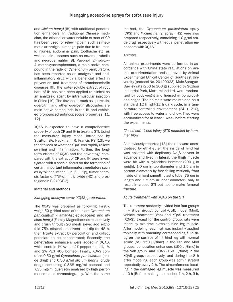
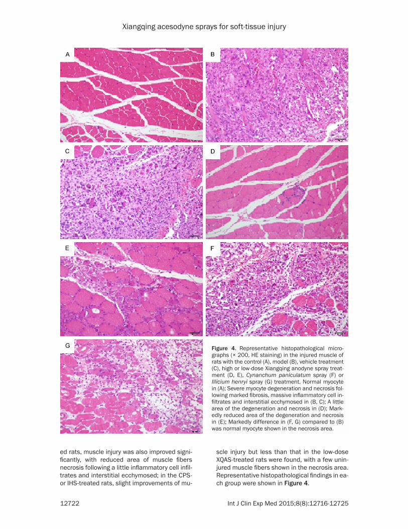
Long-term treatment with XQAS lowered blow-induced muscle degeneration and necrosis detected by histopathological finding
In the control rats, no injury was found in the hind leg muscle with the normal muscle cells; in the injured muscle of model rats, severe mus-cle injury with severe muscle fibers degenera-tion and collagen fiber hyperplasia, muscle cells necrosis, massive inflammatory cell infil-trates and interstitial ecchymosed in the necro-sis area were found; in the vehicle-treated rats, severe muscle injury was similar with the model rats; in the high-dose XQAS-treated rats, mus-cle injury was mild with greatly reduced area of muscle fibers degeneration and necrosis following little inflammatory cell infiltrates and interstitial ecchymosed; in the low XQAS-treat-
Figure 3. Relative expressions of NF-κB p65, COX-2 and IL-1β mRNA in the injured hind leg muscle of rats with the control (Ctrl), model (Mod), vehicle treatment (Veh), high-dose Xiangqing anodyne spray (XQAS) treatment (HX-QAS), low-dose XQAS treatment (LXQAS), Cynanchum paniculatum spray treatment (CPS) and Illicium henryi spray treatment (IHS). The mRNA expres-sions were detected by reverse transcription-PCR assay. The data are shown as mean ± SD (n = 6 per group) and a representative photo for PCR assay was shown here. $$P<0.01, vs. Ctrl; *P<0.05, **P<0.01 vs. Veh; &P<0.05, &&P<0.01 vs. LXQAS.
jured muscle of model rats detected on day 4 post hitting (P<0.01 vs. Ctrl, respectively), while high- or low-dose XQAS treatment both markedly do- wn-regulated mRNA expres-sions of them (P<0.05 or P<0.01 vs. Veh, respectively), and HXQAS group showed stronger down-regulation on the COX-2 mRNA expression (P<0.05) and stronger down-regulated tendency on the mRNA expressions of NF- κB p65 and IL-1β (P>0.05, respectively) than LXQAS. CPS treatment only displayed a slight depressing effects, with decreased mRNA expres-sion of IL-1β (P<0.05 vs. Veh) and decreased tendency on the mRNA expressions of NF-κB p65 and COX-2 (P>0.05 vs. Veh, respecitively), while IHS treatment merely showed de- creased tendency on the mR- NA expressions of NF-κB p65, COX-2 and IL-1β (P>0.05 vs. Veh, respectively). The depres- sant effects of low-dose XQAS treatment on the mRNA ex- pressions of NF-κB p65 and COX-2 were more significant than CPS or IHS treatment
Xiangqing acesodyne sprays for soft-tissue injury
12722 Int J Clin Exp Med 2015;8(8):12716-12725
ed rats, muscle injury was also improved signi- ficantly, with reduced area of muscle fibers necrosis following a little inflammatory cell infil-trates and interstitial ecchymosed; in the CPS- or IHS-treated rats, slight improvements of mu-
scle injury but less than that in the low-dose XQAS-treated rats were found, with a few unin-jured muscle fibers shown in the necrosis area. Representative histopathological findings in ea- ch group were shown in Figure 4.
Figure 4. Representative histopathological micro-graphs (× 200, HE staining) in the injured muscle of rats with the control (A), model (B), vehicle treatment (C), high or low-dose Xiangqing anodyne spray treat-ment (D, E), Cynanchum paniculatum spray (F) or Illicium henryi spray (G) treatment. Normal myocyte in (A); Severe myocyte degeneration and necrosis fol-lowing marked fibrosis, massive inflammatory cell in-filtrates and interstitial ecchymosed in (B, C); A little area of the degeneration and necrosis in (D); Mark-edly reduced area of the degeneration and necrosis in (E); Markedly difference in (F, G) compared to (B) was normal myocyte shown in the necrosis area.
Xiangqing acesodyne sprays for soft-tissue injury
12723 Int J Clin Exp Med 2015;8(8):12716-12725
Discussion
Closed soft tissue injury (STI) frequently occur- red in daily life, and effective treatment of STI is important to avoid sequelae. As the muscle tis-sue is damaged, local tissue mediators are released, including histamines, kinin, prosta-glandins, and leukotrienes etc, which result in inflammation, edema and pain. Inflammation starts from migration of leukocytes and dam-aged soft tissue cells [16, 17]. Therefore, early diagnosis and treatment can improve function-al results and attenuate structural damage in the early and acute period of skeletal muscle injury [15]. As the extension of injury time, severe muscle fibers degeneration and necro-sis following marked collagen fiber hyperplasia, massive inflammatory cell infiltrates and inter-stitial ecchymosed will emerge in the site of damaged tissue to create irreversible structural changes [18]. Hence, rapid attenuating tissue swelling and inhibiting inflammation by prevent-ing injured cells necrosis are necessary for treatment of STI. Our results showed that XQAS had a rapid effect on treating acute closed STI by rapidly attenuating muscle swelling and myo-cyte impairment and markedly depressing pro-ductions of pro inflammatory factors in the injured muscle. In particular, we found that in the muscle subjected to severe acute closed STI, long-term treatment with repeated admin-istration of XQAS for four days could effectively prevent muscle cells necrosis and fibrosis, with marked reduction in myocyte necrosis and inhi-bition in collagen fiber hyperplasia and inflam-matory cell infiltrates in the injured muscle. Moreover, XQAS showed a dose-dependent effect on STI, which comes from its extractive components getting from Cynanchum panicu-latum (CP) and Illicium henryi (IH) and exhibited synergistic effects between two extracts.
Nuclear Factor κB (NF-κB) is thought as a ge- netic switch to control the expressions of many target genes and directly participates in inflam-mation [19]. A key component of NF-κB is het-erodimer transcription factor p50/p65 in the cytoplasm, an inactive state combined with in- hibitory proteins (inhibitor kappa B, IκBα) [20]. When the cells receive various stimuli, such as angiogenesis, oxidative stress and various cy- tokines, NF-κB is released rapidly from IκB to activate the downstream gene expressions of multiple cytokines, chemotactic cytokine and
matrix proteins involved in inflammation, immu-nological responses and/or proliferation [21], such as IL-1β and Cyclooxygenase (COX). COX, an enzyme that converts arachidonic acid to PGs, has been found to have two isoforms namely COX-1 and COX-2. COX-2 is responsible for production of large amounts of pro inflam-matory PGs, especially PGE-2, at the inflamma-tory site [22]. PGE-2, IL-1β, NO and TNF-α are important inflammatory mediators. PGE-2 has an effect on dilating blood vessels, resulting in fever and pain. It can also work synergistically with other inflammatory mediators to exacer-bate inflammatory response [23]; IL-1β is wide-ly involved in many pathological processes, such as tissue damage and edema formation [15]; NO modulates vascular diameter, causing vasodilatation and increasing perfusion, which is essential for remodeling the damaged mus-cle but resulting in tissue edema [24, 25]; TNF-α, a polypeptide cytokine with a wide range of biological activity, involves in inflammatory re- action of ischemic tissue injury and plays an important role in immune and inflammatory response [26]. In addition, MDA, as a product of free radical damage, is an important compo-nent of pathological inflammation injury, and its content can indirectly represent the degree of free radicals injury [27]; LDH, as a key enzyme in the cell, can directly mark to muscle injury and oxidative stress [28]. The results suggest-ed that XQAS can suppress over-expressed NF-κB p65 mRNA in the injured muscle, as well as its downstream inflammatory mediators such as COX-2 and IL-1β mRNA, resulting in rap-idly inhibition on releases of inflammatory me- diates such as PGE-2, IL-1β, NO and TNF-α, and blunting inflammatory reaction, cell injury and tissue edema.
Paeonol and quercetin are considered as major active compounds in the CP and IH. Paeonol exerts its anti-inflammatory and analgesic ef- fects associated with decreased production of pro inflammatory cytokines, such as NO and PGE-2, accompanied by down-regulated protein expressions of iNOS and COX-2 which were ele-vated in the inflammatory tissue [29]. It was showed that quercetin can suppress genera-tion of proinflammatory cytokine TNF-α in a dose-dependent manner via modulation to NF- κB and IκBα [30]. Therefore, the effect of XQAS on damaged tissue could be due to paeonol and quercetin. But XQAS, a combination of CPS

Xiangqing acesodyne sprays for soft-tissue injury
12724 Int J Clin Exp Med 2015;8(8):12716-12725
and IHS, showed more effective than CPS or IHS treatment alone, suggesting that there could be a synergism between quercetin and paeonol on curing STI.
Taken together, XQAS treatment produced rapid effects on STI with the muscle swelling, inflam-mation and cell injury being suppressed quickly in the injured muscle; XQAS also showed signifi-cant effects on closed STI-induced muscle necrosis and fibrosis to prevent muscle cells impairment and degeneration. The mechanism of action of XQAS relates with suppressed expression of NF-kB p65 in the damaged tissue and with attenuated expressions of its down-stream genes which mediate syntheses of inflammatory mediators. Moreover, XQAS treat-ment also exhibits significantly greater effec-tiveness than CPS or IHS treatment alone, sug-gestion synergistic effect of combination of the extracts of Cynanchum paniculatum and Illicium henryi on soft-tissue injury.
Acknowledgements
The authors are grateful to Professor Rong Hu (China Pharmaceutical University) for her con-tribution in manuscript revision, and Wenxia Bai, an associate researcher (Jiangsu center for safety evaluation of drugs, China) for her contribution to histopathological examination.
Disclosure of conflict of interest
None.
Address correspondence to: Jihua Fu, Department of Physiology, China Pharmaceutical University, 639 Longmian Avenue, Nanjing 211198, China. Tel: +86 25 8618 52 26; Fax: +86 25 8618 52 26; E-mail: [email protected]
References
[1] Gierer P, Mittlmeier T, Bordel R, Schaser KD, Gradl G and Vollmar B. Selective cyclooxygen-ase-2 inhibition reverses microcirculatory and inflammatory sequelae of closed soft-tissue trauma in an animal model. J Bone Joint Surg Am 2005; 87: 153-160.
[2] Gates C and Huard J. Management of skeletal muscle injuries in military personnel. Operative Techniques in Sports Medicine 2005; 13: 247-256.
[3] Kearns SR, Daly AE, Sheehan K, Murray P, Kelly C and Bouchier-Hayes D. Oral vitamin C reduces the injury to skeletal muscle caused
by compartment syndrome. J Bon Joint Surg Br 2004; 86: 906-911.
[4] Beiner JM, Jokl P, Cholewicki J and Panjabi MM. The effect of anabolic steroids and corti-costeroids on healing of muscle contusion in-jury. Am J Sports Med 1999; 27: 2-9.
[5] Mantle D, Gok MA and Lennard TW. Adverse and beneficial effects of plant extracts on skin and skin disorders. Adverse Drug React Toxicol Rev 2001; 20: 89-103.
[6] Liu JM, Wang L, Geng YP, Wang QB, Luo LJ and Zhong Y. Genetic diversity and population structure of Lamiophlomis rotata (Lamiaceae), an endemic species of Qinghai-Tibet Plateau. Genetica 2006; 128: 385-394.
[7] Li MZ, Jia ZP, Shen T, Zhang RX, Zhang HX and Li ZY. Effect of Herba Lamiophlomis Rotata ex-tract on rats blood conglomeration parameters by oral administration. Zhong Yao Cai 2006; 29: 160-163.
[8] Deng C, Yao N, Wang B and Zhang X. Deve- lopment of microwave-assisted extraction fol-lowed by headspace single-drop microextrac-tion for fast determination of paeonol in tradi-tional Chinese medicines. J Chromatogr A 2006; 1103: 15-21.
[9] Hirai A, Terano T, Hamazaki T, Sajiki J, Saito H, Tahara K, Tamura Y and Kumagai A. Studies on the mechanism of antiaggregatory effect of Moutan Cortex. Thromb Res 1983; 31: 29-40.
[10] Liu JS and Zhou QR. [The toxic principle of Illicium henryi Diels and structure of 6-deoxyp-seudoanisatin]. Yao Xue Xue Bao 1988; 23: 221-223.
[11] Filho AW, Filho VC, Olinger L and de Souza MM. Quercetin: further investigation of its antinoci-ceptive properties and mechanisms of action. Arch Pharm Res 2008; 31: 713-721.
[12] Gadotti VM, Schmeling LO, Machado C, Liz FH, Filho VC, Meyre-Silva C and Santos AR. Anti- nociceptive action of the extract and the flavo-noid quercitrin isolated from Bauhinia micro-stachya leaves. J Pharm Pharmacol 2005; 57: 1345-1351.
[13] Stratton SA, Heckmann R and Francis RS. Therapeutic ultrasound: its effects on the ln-tegrity of a nonpenetrating wound*. J Orthop Sports Phys Ther 1984; 5: 278-281.
[14] Suleyman H, Demirezer LO, Kuruuzum A, Banoglu ZN, Gocer F, Ozbakir G and Gep- diremen A. Antiinflammatory effect of the aqueous extract from Rumex patientia L-roots. J Ethnopharmacol 1999; 65: 141-148.
[15] Bunn JR, Canning J, Burke G, Mushipe M, Marsh DR and Li G. Production of consistent crush lesions in murine quadriceps muscle--a biomechanical, histomorphological and immu-nohistochemical study. J Orthop Res 2004; 22: 1336-1344.

Xiangqing acesodyne sprays for soft-tissue injury
12725 Int J Clin Exp Med 2015;8(8):12716-12725
[16] Barlow Y and Willoughby J. Pathophysiology of soft-tissue repair. Br Med Bull 1992; 48: 698-711.
[17] Noble KA. Pathophysiology corner: inflamma-tion I. J Perianesth Nurs 2005; 20: 56-58.
[18] Schaser KD, Vollmar B, Menger MD, Schewior L, Kroppenstedt SN, Raschke M, Lubbe AS, Haas NP and Mittlmeier T. In vivo analysis of microcirculation following closed soft-tissue in-jury. J Orthop Res 1999; 17: 678-685.
[19] Perkins ND. The Rel/NF-κB family: friend and foe. Trends Biochem Sci 2000; 25: 434-440.
[20] Henkel T, Machleidt T, Alkalay I, Kronke M, Ben-Neriah Y and Baeuerle PA. Rapid proteoly-sis of I-kappa-B-alpha is necessary for activa-tion of transcription factor NF-kappa-B. Nature 1993; 365: 182-185.
[21] Zhang YW, Wu CY and Cheng JT. Merit of Astragalus polysaccharide in the improvement of early diabetic nephropathy with an effect on mRNA expressions of NF-kappaB and IkappaB in renal cortex of streptozotoxin-induced dia-betic rats. J Ethnopharmacol 2007; 114: 387-392.
[22] Lee SH, Soyoola E, Chanmugam P, Hart S, Sun W, Zhong H, Liou S, Simmons D and Hwang D. Selective expression of mitogen-inducible cy-clooxygenase in macrophages stimulated with lipopolysaccharide. J Biol Chem 1992; 267: 25934-25938.
[23] Choi JH, Jung BH, Kang OH, Choi HJ, Park PS, Cho SH, Kim YC, Sohn DH, Park H, Lee JH and Kwon DY. The anti-inflammatory and anti-noci-ceptive effects of ethyl acetate fraction of cy-nanchi paniculati radix. Biol Pharm Bull 2006; 29: 971-975.
[24] Rubinstein I, Abassi Z, Coleman R, Milman F, Winaver J and Better OS. Involvement of nitric oxide system in experimental muscle crush in-jury. J Clin Invest 1998; 101: 1325-1333.
[25] Moncada S and Higgs EA. Endogenous nitric oxide: physiology, pathology and clinical rele-vance. Eur J Invest 1991; 21: 361-374.
[26] Tracey KJ, Beutler B, Lowry SF, Merryweather J, Wolpe S, Milsark IW, Hariri RJ, Fahey TJ 3rd, Zentella A, Albert JD, et al. Shock and tissue injury induced by recombinant human cachec-tin. Science 1986; 234: 470-474.
[27] Venditti P and DiMeo S. Antioxidants, tissue damage, and endurance in trained and un-trained young male rats. Arch Biochem Biophys 1996; 331: 63-68.
[28] Childs A, Jacobs C, Kaminski T, Halliwell B and Leeuwenburgh C. Supplementation with vita-min C and N-acetyl-cysteine increases oxida-tive stress in humans after an acute muscle injury induced by eccentric exercise. Free Radic Biol Med 2001; 31: 745-753.
[29] Chou TC. Anti-inflammatory and analgesic ef-fects of paeonol in carrageenan-evoked ther-mal hyperalgesia. Br J Pharmacol 2003; 139: 1146-1152.
[30] Nair MP, Mahajan S, Reynolds JL, Aalinkeel R, Nair H, Schwartz SA and Kandaswami C. The flavonoid quercetin inhibits proinflammatory cytokine (tumor necrosis factor alpha) gene ex-pression in normal peripheral blood mononu-clear cells via modulation of the NF-kappa beta system. Clin Vaccine Immunol 2006; 13: 319-328.