original article the effects of 2 µg and 4 µg doses of...
TRANSCRIPT

Pain Res Manag Vol 19 No 2 March/April 2014 75
The effects of 2 µg and 4 µg doses of dexmedetomidine in combination with intrathecal
hyperbaric bupivacaine on spinal anesthesia and its postoperative analgesic characteristics
Abdulkadir Yektaş MD, Enver Belli MD
Department of Anesthesiology and Reanimation, Bagcilar Training and Research Hospital, Istanbul, TurkeyCorrespondence and reprints: Abdulkadir Yektaş, Bağcılar Training and Research Hospital, Merkez Mahallesi Mimar Sinan Caddesi 6. Sokak,
Bagcilar, Istanbul, Turkey. Telephone 90-212-488-6963, fax 90-212-488-6963, e-mail [email protected], [email protected]
Despite developments in postoperative pain management, many patients continue to experience postoperative pain. It is difficult
to provide effective postoperative pain management with manageable side effects. Although intrathecal opioids are effectively and widely used, their use is limited due to side effects (1,2). One way to reduce the intensity and frequency of side effects in analgesic treatment is the use of two or more drugs of different classes in a low-dose combination. For this purpose, multimodal or balanced analgesia has acquired a crucial role in postoperative pain management in recent years (3,4). Recent studies have focused on nonopioid receptors with additional analgesic effects. Previous studies have revealed that intrathecal dex-medetomidine, an α2-adrenoceptor agonist, provides analgesia in sev-eral species, including humans, and that combinations with local anesthetics decrease the side effects of local anesthetics and pos-toperative analgesic use (5-8). Tamagaki et al (9) showed that intrathecal dexmedetomidine increased the analgesic effects of sys-temic daily morphine by means of upregulation of the adrenergic receptor subtypes (α2A, α2B and α2C) on the dorsal horn and
the lumbar dorsal root ganglia (9). In the present study, we aimed to compare the postoperative analgesic characteristics and side effects of different doses of intrathecal dexmedetomidine added to hyperbaric bupivacaine, and to evaluate the effects of these combinations on spinal anesthesia.
METHODSAfter the study was approved by the local ethics committee of Fırat University Medical Faculty (Turkey) (approval date and number: 25.09.2003; 7/3), 60 male patients scheduled for inguinal surgery under spinal anesthesia, classified as American Society of Anesthesiologists physical status class I and between 20 and 30 years of age, were included in the study. The present study was conducted in 2003 in a military hospital with a capacity of 100 beds. After the patients were informed about the study and the study procedures, informed consent was obtained.
The criteria for exclusion from the study were: patients with drug addiction; patients who had required additional sedation and analgesia
originAl ArTicle
©2014 Pulsus Group Inc. All rights reserved
A Yektas, E Belli. The effects of 2 µg and 4 µg doses of dexmedetomidine in combination with intrathecal hyperbaric bupivacaine on spinal anesthesia and its postoperative analgesic characteristics. Pain Res Manag 2014;19(2):75-81.
OBJECTIVE: To compare the postoperative analgesic characteristics and side effects of two different doses of intrathecal dexmedetomidine in com-bination with hyperbaric bupivacaine, and to evaluate the effects of these combinations on spinal anesthesia.METHODS: After obtaining approval from the local ethics committee, 60 male patients who were undergoing inguinal surgery and were classified as American Society of Anesthesiologists physical status class I were included in the study. The present study was conducted in 2003 in a mili-tary hospital with a capacity of 100 beds. The patients were randomly assigned to three groups of 20 patients: group 1, 0.5 mL saline added to 3 mL (15 mg) hyperbaric bupivacaine; and groups 2 and 3, 2 µg dexme-detomidine and 4 µg dexmedetomidine added to 3 mL (15 mg) hyperbaric bupivacaine, respectively. Medications were administered by intrathecal injection in a total volume of 3.5 mL. The postoperative analgesic charac-teristics, effects on spinal anesthesia and side effects were recorded.RESulTS: Demographic characteristics were similar among the groups. The mean (± SD) time to onset of pain was 220.75±112.7 min in group 1, 371.5±223.5 min in group 2 and 1042.50±366.78 min in group 3. Time to first pain sensation in group 3 was significantly longer than that in groups 1 and 2 (P<0.001).CONCluSION: Two different doses of dexmedetomidine, an α2-adrenoceptor agonist with analgesic effects, resulted in an increased duration of analgesia and efficacy, decreased postoperative analgesic use and was associated with no notable adverse effects.
Key Words: Adjuvants; Anesthesia; Anesthetics; Bupivacaine; Dexmedetomidine; Local; Spinal
les effets de doses de 2 µg et de 4 µg de dexmédétomidine administrées avec de la bupivacaïne hyperbare par voie intrathécale sur l’anesthésie rachidienne et leurs caractéristiques analgésiques postopératoires
OBJECTIF : Comparer les caractéristiques analgésiques postopératoires et les effets secondaires de deux doses différentes de dexmédétomidine administrées avec de la bupivacaïne hyperbare par voie intrathécale et en évaluer les effets sur l’anesthésie rachidienne.MÉTHODOlOGIE : Après avoir obtenu l’approbation du comité d’éthique local, les chercheurs ont inclus dans l’étude 60 patients de sexe masculin qui subissaient une opération inguinale et dont l’état physique était de classe I selon l’American Society of Anesthesiologists. La présente étude a été menée en 2003 dans un hôpital militaire de 100 lits. Les patients ont été répartis au hasard en trois groupes de 20 patients : le groupe 1 a reçu 0,5 mL de soluté physiologique ajouté à 3 mL (15 mg) de bupivacaïne hyperbare, et les groupes 2 et 3 se sont fait administrer 2 µg et 4 µg de dexmédétomidine ajoutés à 3 mL (15 mg) de bupivacaïne hyper-bare, respectivement. Les médicaments ont été administrés par voie intrathécale, pour un volume total de 3,5 mL. Les caractéristiques analgé-siques postopératoires, les effets sur l’anesthésie rachidienne et les effets secondaires ont été consignés.RÉSulTATS : Les caractéristiques démographiques étaient similaires entre les groupes. Le délai moyen (± ÉT) avant l’apparition de la douleur était de 220,75±112,7 min dans le groupe 1, de 371,5±223,5 min dans le groupe 2 et de 1 042,50±366,78 min dans le groupe 3. Le délai jusqu’à la première sensation de douleur dans le groupe 3 était considérablement plus long que dans les groupes 1 et 2 (P<0,001).CONCluSION : Deux doses différentes de dexmédétomidine, un ago-niste α2-adrénergique aux effets analgésiques, augmentaient la durée et l’efficacité de l’analgésie, réduisaient l’utilisation d’analgésiques après l’opération et n’avaient pas d’effets indésirables importants.
Yektaş and Belli
Pain Res Manag Vol 19 No 2 March/April 201476
during previous surgical procedures; patients who had undergone previous surgical procedures and recalled experiencing pain; patients in whom cerebrospinal fluid (CSF) could not be obtained after three attempts during spinal anesthesia; and patients whose levels of educa-tion were below graduation from primary school.
Patients were not premedicated. Before being taken to the operat-ing room, the spinal anesthesia procedure was explained to the patients and, during the postoperative period, they were requested to state the time of onset of pain. They were informed about the digital pain scale and were trained on the use of patient-controlled analgesia (500 mg tramadol in 100 mL of saline). A loading dose was given only to patients who needed analgesics.
In the operating room, all patients were monitored using electro-cardiography, pulse oximetry and noninvasive arterial blood pressure. A 20-gauge intravenous cannula was inserted. After spinal anesthesia was administered, 0.9% saline infusion was initiated at a rate of 10 mL/kg/h. The patients were placed in a seated position and, follow-ing the sterilization of the lumbar regions using polyvinyl prolidone iodine, 2 mL of 2% prilocaine was administered to the cutaneous and the subcutaneous tissues. A 25-gauge Quincke spinal needle was inserted into L4–L5 disc space. When the CSF flow was observed, the prepared medical solution was administered.
The patients were randomly divided into three groups according to the medical mixture administered into the CSF: group 1: 15 mg of 0.5% hyperbaric bupivacaine plus 0.5 mL saline; group 2: 15 mg of 0.5% hyperbaric bupivacaine plus 2 µg dexmedetomidine; and group 3: 15 mg of 0.5% hyperbaric bupivacaine plus 4 µg dexmedetomidine.
Following intrathecal administration of the medication, the patients were placed in the supine position. The sensory block level was assessed by the pin-prick test, and the motor block level was evaluated by Bromage scoring (0, lifting leg; 1, bending knee; 2, moving foot; 3, no movement) at 5 min and 20 min. The mean arterial pressure, heart rate (HR) and peripheral oxygen saturation were recorded at baseline (before spinal anesthesia), at the first minute of spinal anesthesia and at 5 min intervals until the regression of sensory block and the recovery of motor block. The end of the sensory block duration was recorded as the moment when the sensory block duration dropped by two levels after it reached the highest level as of the moment spinal anesthesia was per-formed. The end of the motor block duration was recorded as the moment when the Bromage score returned to 0 after the spinal anes-thesia was performed. Surgery commenced 20 min after intrathecal administration of the medication.
In the intraoperative and postoperative period, side effects, includ-ing bradycardia, hypotension, nausea, vomiting and sedation, were recorded. The side effects were treated symptomatically. A decrease in mean arterial pressure <80% of initial values was considered to repre-sent hypotension and was treated with intravenous 5 mg ephedrine and fluid infusion. An HR <50 beats/min was considered to be bradycardia, and was managed by intravenous atropine (0.5 mg). In case of nausea and vomiting (without hypotension), 8 mg of ondansetron was admin-istered intravenously. In cases of shivering, the patients were warmed using a warming unit. Those who complained of itching received 45.5 mg of intravenous pheniramine. The intraoperative sedation level of the patients was assessed using the Ramsay sedation scale (RSS) (10). The patient was considered to be sedated if the RSS score was ≥2.
Hot-pack application was used in patients with urinary retention; if urinary retention became persistent, the urine was collected using a Foley catheter and removed. The patients were not mobilized for 24 h following the intrathecal medications. Following mobilization, post-spinal headache (PSH) was managed by intravenous fluid infusion and oral acetaminophen 250 mg + propyphenazone 150 mg + caffeine 50 mg. The patients who continued to experience headache in spite of the three-day treatment received an epidural blood patch. None of the patients developed persistent PSH. The modified Aldrete scoring sys-tem (11), a discharge scoring tool, was used to assess the sedation levels of the patients. According to the Aldrete scoring system, patients who achieved a total score ≥9 were considered to be fit for discharge. After the patients completely recovered from the sensory and motor block-ade, patients with a modified Aldrete’s score of ≥9 were referred to the department of general surgery and their ensuing follow-ups were per-formed in this department. The patients were discharged after a follow-up period of one week. The pain onset time in the postoperative period was recorded. A numerical pain rating scale was used for the assessment of pain. Each patient was asked to rate his pain from 0 (no pain) to 10 (unbearable pain). Following the recording of pain, a patient-controlled analgesia pump was connected. Following the administra-tion of intrathecal medications, intravenous medication infusion continued for up to 24 h. At the end of 24 h, the total amount of tramadol used was recorded in milligrams. After discharge, the patients received a neurological examination on an annual basis. Lumbar and thoracic-spinal magnetic resonance imaging (MRI) images of the patients were performed 10 years after the administration of intrathecal dexmedetomidine. The lower-extremities radiculopathy protocol and electromyography (EMG) studies were performed at year 10.
Statistical analysisThe sample size was calculated according to the time to initiation of pain, and it was estimated that a group sample size of 20 patients for each group would be sufficient to detect a difference of 140 min between group 1 and group 2, with a significance level of 0.05 and β=0.2. The study was planned as a randomized and double-blind study. All data were assessed using SPSS version 11.5 (IBM Corporation, USA) for Windows (Microsoft Corporation, USA). Mean (± SD) values were used for the complementary statistics of the data. Data distribution was assessed using the Kolmogorov-Smirnov test. The c2 test was used for the assessment of the demographic data and occurring side effects. The time to initiation of pain and the numerical pain rat-ing scale score at that moment, the amount of tramadol used until 24 h after administration of intrathecal medication, and the motor and sensory block levels at 5 min and 20 min were compared among the groups using the Kruskal-Wallis test. The Mann-Whitney U test with Bonferroni corrections was used in the subgroup analysis. P<0.05 was considered to be statistically significant.
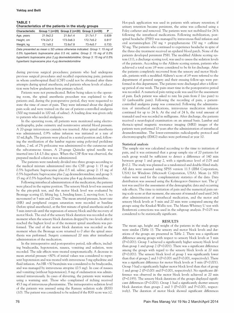
RESulTSThe mean age, height and weight of the patients in the study groups were similar (Table 1). The sensory and motor block levels and dur-ations of the groups are presented in Table 2. There was a significant difference among groups with respect to sensory block levels at 5 min (P=0.001). Group 3 achieved a significantly higher sensory block level than group 1 and group 2 (P<0.001). There was a significant difference among the groups with regard to the sensory block levels at 20 min (P<0.001). The sensory block level of group 1 was significantly lower than that of groups 2 and 3 (P<0.001 and P<0.001, respectively). There was a significant difference for motor block levels at 5 min (P<0.001). Group 3 had a significantly higher motor block level than that of group 1 and group 2 (P<0.001 and P<0.001, respectively). No significant dif-ference was observed in the motor block levels achieved at 20 min (P=0.195). The sensory block durations of the groups displayed signifi-cant differences (P<0.001). Group 1 had a significantly shorter sensory block duration than groups 2 and 3 (P=0.001 and P<0.001, respect-ively). The duration of motor block showed significant differences
TAble 1Characteristics of the patients in the study groupsCharacteristic Group 1 (n=20) Group 2 (n=20) Group 3 (n=20) PAge, years 21.9±2.2 21.8±1.4 21.7±1.7 0.839Height, cm 174.2±7.2 173.0±6.3 172.7±5.2 0.817Weight, kg 72.1±9.2 72.6±7.8 73.4±5.7 0.733
Data presented as mean ± SD unless otherwise indicated. Group 1: 15 mg of 0.5% hyperbaric bupivacaine plus 0.5 mL saline; Group 2: 15 mg of 0.5% hyperbaric bupivacaine plus 2 µg dexmedetomidine; Group 3: 15 mg of 0.5% hyperbaric bupivacaine plus 4 µg dexmedetomidine

Postoperative analgesic effects of bupivacaine plus dexmedetomidine
Pain Res Manag Vol 19 No 2 March/April 2014 77
among the groups (P<0.001). Group 1 and group 2 had shorter motor block durations than group 3 (P=0.001 and P<0.001, respectively).
The time of pain onset and numerical pain rating scale scores of the groups are presented in Table 3. The time to initiation of pain was significantly different among the groups (P<0.001). The time to initia-tion of pain was significantly shorter in group 1 and group 2 compared with group 3 (P=0.001 and P<0.001, respectively). There was a signifi-cant difference among the groups with respect to the numerical pain rating scale scores at pain onset (P<0.001). The pain rating score of group 1 was significantly higher than that of group 2 and group 3 (P=0.001 and P<0.001, respectively).
The amount of tramadol used in the first 2 h (after the onset of pain) and during 24 h following spinal anesthesia are presented in Table 4. The tramadol requirement in the first 2 h after initiation of pain and the total tramadol requirement during 24 h following spinal anesthesia were significantly different among the groups (P<0.001 for each). Tramadol requirement in the first 2 h and during 24 h in group 1 was significantly higher than that of group 2 and group 3 (P=0.001 and P<0.001, respectively).
There was no significant difference between group 2 and group 3 regarding tramadol requirement in the first 2 h (P=0.277); however, the total amount of tramadol requirement during 24 h was signifi-cantly higher in group 2 than in group 3 (P<0.001).
The intraoperative and postoperative side effects are summarized in Table 5. Itching, sweating, dizziness and respiratory depression were not observed in any of the patients.
There was a significant difference among the groups with respect to the frequency of hypotension (P<0.001). Hypotension was more fre-quent in group 1 than in the other groups (P<0.05 for each).
There were no statistically significant differences among groups in terms of bradycardia (P=0.626), nausea/vomiting (P=0.459), shivering (P=0.459), urinary retention (P=0.449) and PSH (P=0.349). After spinal anesthesia, one patient in group 1 and one in group 3 received epidural blood patches on day 5 due to PSH.
The annual neurological examinations of the patients in the groups were assessed and judged to be normal. The thoracic and lum-bar MRI images in year 10 were assessed and judged to be normal. No findings that would support radiculopathy in the lower extremities of patients were encountered in the EMG study.
There were significant differences among the groups in occurrence of sedation (P<0.05). Sedation was only observed in group 3.
DISCuSSIONIn the present study, we evaluated whether different doses of intrathecal dexmedetomidine had any effects on the properties of hyperbaric bupivacaine used for spinal anesthesia. Our aim was to provide quality anesthesia and prolonged postoperative analgesia, while reducing the requirement for postoperative analgesics. We observed that inthrathecal dexmedetomidine at 2 µg and 4 µg doses provided prolonged postoperative analgesia and reduced the need for postoperative analgesics.
TAble 2The mean sensory and motor block levels and durations of the study groups
Group 1 (n=20) Group 2 (n=20) Group 3 (n=20) PSensory block (pin-prick), thoracic segment 5 min 7.05±2.78 5.15±3.39 3.50±2.43* 0.001 20 min 5.50±1.84 5.55±1.43 3.00±2.53† <0.001Motor block (Bromage score) 5 min 1.80±0.61 1.75±0.78 3.00±0.00‡ <0.001 20 min 2.75±0.63 2.80±0.52 3.00±0.00 0.195Sensory block duration, min 66±28.03§ 98.75±34.40¶ 147.50±13.72 <0.001Motor block duration, min 130.50±42.73 151.50±29.96 226.50±65.47** <0.001
Data presented as mean ± SD unless otherwise indicated. *When sensory block levels at 5 min were compared for group 3 (15 mg of 0.5% hyperbaric bupivacaine plus 4 µg dexmedetomidine) and groups 1 (15 mg of 0.5% hyperbaric bupivacaine plus 0.5 mL saline) and 2 (15 mg of 0.5% hyperbaric bupivacaine plus 2 µg dexmedetomidine), the sensory block level in group 3 was higher than in groups 1 and 2 (P<0.001). When sensory block levels were compared at 20 min, there was a statistically significant difference among the groups (P<0.001); †When sensory block levels of group 3 and groups 1 and 2 were compared at 20 min, the block level in group 3 was higher than that in groups 1 and 2 (P<0.001 and P<0.001, respectively); ‡Motor block levels in group 3 were significantly higher than in groups 1 and 2 at 5 min (P<0.001 and P<0.001, respectively); §Sensory block duration in group 1 was lower than that in groups 2 and 3 (P=0.001 and P<0.001, respectively); ¶Sensory block duration in group 2 was lower than that in group 3 (P<0.001); **Motor block duration in group 3 was higher than that in groups 1 and 2 (P=0.001 and P<0.001, respectively)
TAble 3Time to first pain onset and numerical pain rating scale scores of the study groups
Group 1 (n=20) Group 2 (n=20) Group 3 (n=20) PTime to first pain onset, min 220.7±112.7 371.5±223.5 1042.50±366.78* <0.001Numerical rating scale scores 9.35±0.67† 1.55±0.68 1.30±0.57 <0.001
Data presented as mean ± SD unless otherwise indicated. *The time to pain onset in group 3 (15 mg of 0.5% hyperbaric bupivacaine plus 4 µg dexmedetomidine) was longer than that in groups 1 (15 mg of 0.5% hyperbaric bupivacaine plus 0.5 mL saline) and 2 (15 mg of 0.5% hyperbaric bupivacaine plus 2 µg dexmedetomidine) (P=0.001 and P<0.001, respectively); †Numerical rating scale scores in group 1 were higher than in groups 2 and 3 (P=0.001 and P<0.001, respectively)
TAble 4The amount of tramadol consumption from the time of pain onset up to 2 h and at 24 hTramadol consumption Group 1 (n=20) Group 2 (n=20) Group 3 (n=20) PFrom the time of pain onset up to 2 h, mg 69.00±5.03* 45.00±0.00 20.00±0.00 <0.001During 24 h following spinal anesthesia, mg 107.25±14.64† 69.75±13.13‡ 47.06±60.22 <0.001
Data presented as mean ± SD unless otherwise specified. *Group 1 (15 mg of 0.5% hyperbaric bupivacaine plus 0.5 mL saline) consumed more tramadol than groups 2 (15 mg of 0.5% hyperbaric bupivacaine plus 2 µg dexmedetomidine) and 3 (15 mg of 0.5% hyperbaric bupivacaine plus 4 µg dexmedetomidine) during the first 2 h after the time of pain onset (P=0.001 and P<0.001, respectively); †Consumed tramadol in 24 h was higher in group 1 than that in groups 2 and 3 (P=0.001 and P<0.001, respectively); ‡Consumed tramadol in 24 h was higher in group 2 than in group 3 (P=0.002)

Yektaş and Belli
Pain Res Manag Vol 19 No 2 March/April 201478
Dexmedetomidine is a highly specific, potent and selective α2-adrenoceptor agonist (7,8). It provides anxiolysis, analgesia and cooperative sedation without suppressing respiration (12,13).
In radioligand-binding studies, α2-adrenoceptors have been shown to have α2A, α2B, α2C and α2D subtypes (14). α2A-receptors play a crucial role in most principle pharmacological and therapeutic effects of dexmedetomidine. Functional α2A-receptor-deficient mice were shown to lack sedative, anesthetic and analgesic effects of dex-medetomidine, while mice with inactivated α2B and α2C receptors displayed normal responses (15).
A meta-analysis by Abdallah and Brull (16) showed that dex-medetomidine is a potential local anesthesic adjuvant that can exhibit a facilitatory effect when administered intrathecally as part of spinal anesthesia. However, there are presently insufficient safety data to sup-port the use of perineural dexmedetomidine in the clinical setting (16).
Many animal studies have been performed on the effects of intrathecal dexmedetomidine. Konakci et al (17) studied the effects of epidural dexmedetomidine in 21 New Zealand rabbits. They div-ided the rabbits into three groups and administered 5 µg or 10 µg dexmedetomidine, and lidocaine only, respectively, to the groups. There was a significant difference in demyelination of the oligoden-drocytes in the white matter between the lidocaine-only and 10 µg dexmedetomidine groups; however, there was no difference between the lidocaine-only and 5 µg dexmedetomidine groups in this regard. Brummett et al (18) performed a study involving 31 Sprague Dawley rats. The sciatic nerve of the rats was blocked with bupivacaine, or dexmedetomidine and bupivacaine; the contralateral sciatic nerve of the rats were blocked with saline or dexmedetomidine. The sci-atic nerves blocked with bupivacaine and dexmedetomidine showed significantly lower levels of perineural inflammation than the sciatic nerves blocked with only bupivacaine. The researchers used 0.005% dexmedetomidine in this study. Their results were in contradiction with the results of Konakci et al (17). Schoeler et al (19) exposed organotypic hippocampal slices to dexmedetomidine and created a focal mechanical trauma. As a result, they demonstrated that dex-medetomidine provided neuroprotection in an in vitro traumatic brain injury model. Sanders et al (20) also showed that dexmedetomidine prevented cortical apopitozis in in vitro and in vivo conditions. Chrysostomou and Schmitt (21) stated that dexmedetomidine is a
neuroprotective, cardioprotective and renoprotective agent. Kimura et al (22) demonstrated that intraspinal dexmedetomidine was beneficial in neuropathic pain in rats. To our knowledge, there is no in vitro or in vivo study reporting any toxic effects of dexmedetomidine, except for the study of Konakci et al (17), in which they evaluated the physio-logical and histological toxicological effects of dexmedetomidine.
In our study, the patients were assessed to be normal during their yearly neurological examinations and the thoracic and lumbar spines were assessed as normal during the MRI studies performed in year 10 following anesthesia. Similarly, no radiculopathy findings were detected in lower extremities of patients in their EMG studies con-ducted in year 10.
Dexmedetomidine exerts its analgesic effects via binding to α2-adrenoceptors in the spinal cord (23,24). Similar to these studies, we observed that sensory block levels at 5 min increased with dex-medetomidine, and the highest levels were observed in group 3. This finding shows that spinal anesthesia starts earlier with increasing doses of dexmedetomidine. Sensory block levels at 20 min also increased with increased doses of dexmedetomidine, showing that increasing doses of dexmedetomidine improves the quality of anesthesia. The level of motor blockade at 5 min also increased with dexmedetomidine, yielding the same level at 20 min (Table 2).
In a study by Ishii et al (7), the effects of dexmedetomidine on postsynaptic transmission in substantia gelatinosa were investigated using the whole-cell patch technique in rat spinal cord sections. Dexmedetomidine hyperpolarized the membrane potantials of sub-stantia gelatinosa neurons by G-protein mediated activation of K+ channels through α2A and α2C adrenoceptors. This action of dex-medetomidine may contribute, at least in part, to its antinociceptive action in the spinal cord. It was reported that this mechanism of action partially contributed to the antinociceptive action of dexmedetomidine in the spinal cord. Although the time to initiation of pain was similar in the groups receiving physiological saline and 2 µg of dex-medetomidine, the numerical pain rating scale scores in the group receiving 2 µg dexmedetomidine was significantly lower than that of the group receiving physiological saline.
In a study by Marc et al (8) involving dogs, it was demonstrated that following intrathecal, epidural and intravenous dexmedetomidine administration, there was an increase in twitching of the skin against painful stimuli and stimulation of withdrawal by mechanical pressure on the rear paw. Maximum medication effect was observed at 3 min of intravenous administration and at 15 min of intrathecal and epi-dural applications. The duration of action was 90 min by intravenous and intrathecal routes, and 240 min by the epidural route follow-ing maximum effective dose. Maximum antinociceptive effects were observed around the dermatomes proximal to the catheter tip following intrathecal administration. Following epidural or intrathecal applica-tion, the time to maximum distribution of the block was assessed and withdrawal of the rear paw was suppressed, but no suppression was seen in the front paw. When compared with the respiratory rates before admin-istration of the medication, an intravenous dose of 10 µg/kg, an epidural dose of 50 µg and an intracisternal dose of 15 µg dexmedetomidine led to a significant decrease in the respiratory rates at rest; however, no change was observed in end-tidal carbon dioxide at rest. Reversal of the decrease in respiratory rate was dependent on atipamezole (300 µg/kg) but independent of naloxone (30 µg/kg). HR significantly decreased in a dose-dependent manner after intravenous application. The notable decrease in HR following epidural administration was only observed at 50 µg, which was the highest dose administered. Following an intrathecal 10 µg and an intracisternal 15 µg dose, the HR decreased slightly, although not significantly. When the highest dose of medica-tion was administered to the animals through intrathecal, epidural and intracisternal routes, only minor depression was observed. Additionally, a dose-dependent decrease was observed in the core temperature follow-ing intravenous, intracisternal and epidural dexmedetomidine (lower dose) application; however, no change was observed in the core tem-perature following intrathecal application. Similarly, the level of sensory
TAble 5Side effects according to group
Side effectGroup
P1 (n=20) 2 (n=20) 3 (n=20)Hypotension 14 6 7 0.022*Bradycardia 11 8 9 0.626Vomiting/nausea 2 4 5 0.459Tremor 2 4 5 0.459Urinary retention 3 6 6 0.449Postspinal headache 0 2 1 0.349Sedation 0 0 8 <0.001†
Data presented as n unless otherwise indicated. *Based on a comparison of groups in terms of hypotension, statistically significant differences were observed among groups (P=0.022). When the groups were compared in pairs, group 1 (15 mg of 0.5% hyperbaric bupivacaine plus 0.5 mL saline) and group 2 (15 mg of 0.5% hyperbaric bupivacaine plus 2 µg dexmedetomidine) were sig-nificantly differen, with a higher hypotension rate in group 1 (P=0.012). There were statistically significant differences between group 1 and group 3 (15 mg of 0.5% hyperbaric bupivacaine plus 4 µg dexmedetomidine), with a higher hypo-tension rate in group 1 (P=0.029); †When the groups were compared in terms of sedation, statistically significant differences were observed among the groups (P<0.001). When the groups were compared in pairs, there were statistically significant differences between group 1 and group 3; group 3 had a higher rate of sedation (P=0.002). There were statistically significant differences between group 2 and group 3; group 3 had a higher rate of sedation (P=0.002)

Postoperative analgesic effects of bupivacaine plus dexmedetomidine
Pain Res Manag Vol 19 No 2 March/April 2014 79
and motor block at 5 min significantly increased in a dose-dependent manner in our study. Following maximum effective dose, the duration of action was found to be 90 min by the intrathecal route in the above-mentioned study, and the duration of action was 371.5±223.5 min and 1042.50±366.78 min in groups 2 and 3 in the present study, respectively; however, instead of dexmedetomidine, we used a combination of dex-medetomidine and bupivacaine in our study.
In a study by Calasans-Maia et al (6) involving guinea pigs using α2-adrenoceptor agonists together with local anesthetics, the duration of spinal anesthesia was found to be prolonged. Intrathecal clonidine extended the duration of motor block produced by local anesthetics (6). The affinity of dexmedetomidine for α2- adrenoceptors was found to be more than eight times higher than that of clonidine (6). This study investigated whether intrathecal or intraperitoneal dexmedetomidine with 0.5% levobupivacaine caused motor nerve block. Seventy-two guinea pigs were randomly divided into 12 groups, and intrathecal levobupivacaine (50 µL dose) was administered to all animals. One-half of the animals received intrathecal dexmedetomidine, while the remaining received intraperitoneal dexmedetomidine. Intrathecal dex-medetomidine (0.1 µg to 0.2 µg and 0.4 µg) prolonged motor block produced by levobupivacaine and extended the duration of motor block from 48 min to 84.5 min to 101.5 min and 105 min, respectively. Similarly, intraperitoneal dexmedetomidine prolonged motor block duration from 48.5 min to 88 min to 114.5 min, respectively. When yohimbine was added before intrathecal or intraperitoneal
dexmedetomidine application, motor block duration decreased from 101.5 min to 76.5 min and from 114.5 min to 90 min, respectively. The duration of motor block following spinal injection of levobupivacaine was prolonged by intrathecal and systemic dexmedetomidine. This effect is partially dependent on α2-adrenoceptor activation (6). Similarly, the duration of motor and sensory block prolonged with increasing doses of dexmedetomidine in our study (Table 2).
When a certain amount of clonidine is systemically administered, it causes hypotension; however, this effect is lower in the case of dex-medetomidine, which is more selective (25). We observed hypoten-sion in seven patients in the 4 µg dexmedetomidine group (Table 5).
In a study performed by Shukla et al (26), the patients were divided into three groups as follows: the dexmedetomidine group (15 mg hyper-baric bupivacaine plus 10 µg dexmedetomidine), the magnesium sul-phate group (15 mg hyperbaric bupivacaine plus 50 mg magnesium sulphate) and the control group (15 mg hyperbaric bupivacaine plus 0.1 mL saline), and the medications were administered via the intrathecal route. In the dexmedetomidine group, the onset of anesthe-sia was found to be faster and the duration of anesthesia was longer. In the magnesium group, there was a delay in the onset of block, but the duration of anesthesia was significantly prolonged compared with that of the control group. However, the duration of anesthesia in the magne-sium group was shorter than that of the dexmedetomidine group. There was no significant difference among the groups with regard to the fre-quency of side effects and hemodynamic changes (26) (Table 6).
TAble 6Comparing the results of our findings with trial outcomes
Author (reference), year Study groups
bupivacaine or ropivacaine,
mg
Sensory block duration (time of
regression to S1), min
Sensory block duration (time to
segment regression from highest sensory
level), minMotor block
duration, minTime to first
pain onset, min Side effects (n)Shukla et al (26),
201110 µg dexmedetomidine (n=30) 15 352±45 331±35 0.1 mL saline (n=30) 15 194±55 140±340.1 mL (50 mg) magnesium
sulfate (n=30)15 265±65 251±51
Gupta et al (27), 2011
0.1 mL saline + ropivacaine (n=30)
15 239.33±16.8 62.7±8.3 241.67±21.67 Nausea (2)Hypotension (1)
5 µg dexmedetomidine + ropivacaine (n=30)
15 468.3±36.78 125.6±16.5 478.4±20.9 Nausea (1)Bradycardia (2)Hypotension (2)
Gupta et al (28), 2011
5 µg dexmedetomidine (n=30) 12.5 187±10 120±22.225 µg fentanyl (n=30) 12.5 476±20 76±20
Kanazi et al (29), 2006
0.5 mL saline (n=19) 12 190±48 80±28 163±47 3 µg dexmedetomidine (n=16) 12 303±75 122±37 250±7630 µg clonidine (n=16) 12 272±38 101±37 216±38
Al-Mustafa et al (30), 2009
10 µg dexmedetomidine (n=22) 12.5 338.9±44.8 302.9±36.75 µg dexmedetomidine (n=22) 12.5 277.1±23.2 246.4±25.70.5 mL saline (n=22) 12.5 165.5±32.9 140.1±32.3
Eid et al (31), 2011
0.5 mL saline (n=16) 15 238± 57 76.9±26.8 202±41.8 20010 µg dexmedetomidine (n=15) 15 320±65.8 103±28.7 280±46 45015 µg dexmedetomidine (n=16) 15 408.7±68 200.6±30.9 336±58 650
Yektas and Belli (current study), 2014
0.5 mL saline (n=20) 15 66±28.03 130.50±42.73 220.7±112.7 Vomiting (2)Bradycardia (11)Hypotension (14)
2 µg dexmedetomidine (n=20) 15 98.75±34.40 151.50±29.96 371.5±223.5 Nausea (4)Bradycardia (8)Hypotension (6)
4 µg dexmedetomidine (n=20) 15 147.50±13.72 226.50±65.47 1042.50±366.78 Sedation (8)Vomiting (5)Bradycardia (9)Hypotension (7)
Data presented as mean ± SD unless otherwise specified
Yektaş and Belli
Pain Res Manag Vol 19 No 2 March/April 201480
In a study by Gupta et al (27), dexmedetomidine added to intrathecal ropivacaine led to a prolongation in the motor and sensory block durations. The results of our study are similar to the above-mentioned studies (Table 6).
In another study by Gupta et al (28), it was demonstrated that intrathecal dexmedetomidine prolonged hemodynamic stability and the duration of motor and sensory block; when compared with fentanyl, it reduced the analgesic requirement in 24 h. We also observed that the requirement for analgesics in the postoperative 24 h decreased with increasing doses of dexmedetomidine (Table 6).
In a study by Kanazi et al (29), it was demonstrated that when added to 3 µg dexmedetomidine or 30 µg clonidine, intrathecal bupivacaine retained hemodynamic stability and extended motor and sensory block durations, requiring no sedation. The results of our study were similar (Table 6).
In a study by Al-Mustafa et al (30), dexmedetomidine had a dose-dependant effect on the onset and regression of sensory and motor block when used as an adjuvant to bupivacaine in spinal anesthesia. The results of our study were similar (Table 6).
In a study by Eid et al (31), 10 µg of dexmedetomidine adminis-tered intrathecally provided significant increase in the sensory and motor block of spinal anesthesia in addition to prolonged postopera-tive analgesia. The longer sensory and motor blockade produced by a 15 µg dose and the desirable sedation level may be beneficial in patients undergoing lengthy complex surgery as an alternative to epi-dural or prolonged general anesthetics, and may preclude the use of intravenous sedatives. It may be prudent to compare the anesthetic and postoperative effects of dexmedetomidine with long-acting intrathecal opioids. The results of our study were similar (Table 6).
In the present study, dexmedetomidine accelerated the onset of sensory and motor blocks (at 5 min) compared with bupivacaine alone. Dexmedetomidine significantly increased the sensory block
levels at 20 min, but motor block levels at 20 min were similar in all groups. Dexmedetomidine significantly prolonged the sensory and motor block durations and, as the dose of dexmedetomidine was increased, block duration was also increased.
Dexmedetomidine prolonged the time to initiation of pain in a dose-dependent manner. The time to initiation of pain was signifi-cantly longer in the 4 µg dexmedetomidine group than that of the other groups. The numerical pain rating scores at pain onset was sig-nificantly lower in the dexmedetomidine-added groups, and as dex-medetomidine dose increased, the numerical pain rating scores decreased. Tramadol consumption in 2 h after the onset of pain was significantly lower in the dexmedetomidine-added groups and as dex-medetomidine dose increased, tramadol consumption decreased. The amount of tramadol required during 24 h after spinal anesthesia was significantly decreased in the dexmedetomidine groups, and as the dexmedetomidine dose increased, the level of significance decreased. When the frequency of the side effects were compared, hypotension was found to be significantly more frequent in the 0.5 mL saline group (n=14) (Table 5). Sedation was only observed in the 4 µg dex-medetomidine group (n=8) (Table 5).
CONCluSIONSA 4 µg dose of dexmedetomidine, an intrathecal adjuvant, leads to hemodynamic stability and remarkable effects on spinal anesthesia, and can be safely used to decrease tramadol requirement in the pos-toperative period with intrathecal hyperbaric bupivacaine. Because it significantly prolongs the sensory and motor block duration, we con-clude that it can be used for long operations that require spinal anes-thesia. On the other hand, 40% of the patients in the 4 µg dexmedetomidine group developed sedation in the range of RSS = 2 to 4. Because the sensorial block level rose above the level of T4, the dose of the hyperbaric bupivacaine needs to be adjusted.
REFERENCES1. Abouleish E. Apnoea associated with the intrathecal administration
of morphine in obstetrics. Br J Anaesth 1988;60:592-4.2. Cousins MJ, Mather LE. Intrathecal and epidural administration of
opioids. Anesthesiology 1984;61:276-310.3. Klamt JG, Garcia IV, Prado WA. Analgesic and adverse effects of a
low dose of intrathecally administered hyperbaric neostigmine alone or combined with morphine in patients submitted to spinal anesthesia: Pilot studies. Anesthesia 1999;54:27-31.
4. Murali Krishna T, Panda NB, Batra YK, Rajeev S. Combination of low doses of intrathecal ketamine and midazolam with bupivacaine improves postoperative analgesia in orthopaedic surgery. Eur J Anaesthesiol 2008;25:299-306.
5. Al Mustafa MM, Bardan IZ, Abu Ali HM, Al Barazangi BA, Massad IM, Al-Ghanem SM. Intravenous dexmedetomidine prolongs bupivacaine spinal analgesia. Middle East J Anesthesiol 2009;20:225-31.
6. Calasans-Maia JA, Zapata-Sudo G, Sudo RT. Dexmedetomidine prolongs spinal anesthesia induced by levobupivacaine 0.5% in guinea-pigs. J Pharm Pharmacol 2005;57:1415-20.
7. Ishii H, Kohno T, Yamakura T, Ikoma M, Baba H. Action of dexmedetomidine on the substantia gelatinosa neurons of the rat spinal cord. Eur J Neurosci 2008;27:182-90.
8. Sabbe MB, Penning JP, Ozaki GT, Yaksh TL. Spinal and systemic action of the alpha 2 receptor agonist dexmedetomidine in dogs. Antinociception and carbon dioxide response. Anesthesiology 1994;80:1057-72.
9. Tamagaki S, Suziki T, Hagihira S, Hayashi Y, Mashimo T. Systemic daily morphine enhances the analgesic effect of intrathecal deksmedetomidine via up-regulation of alpha 2 adrenergic receptor subtypes A, B and C in dorsal root ganglion and dorsal horn. J Pharm Pharmacol 2010;62:1760-7.
10. Martin E, Ramsay G, Mants J, Sum-Ping ST. The role of alpha 2-adrenoceptor agonist dexmedetomidine in postsurgical sedation in the intensive care unit. J Intensive Care Med 2003;18:29-41.
11. Fredman B, Lahav M, Zohar E, Golod M, Paruta I, Jedeikin R. The effect of midazolam premedication on mental and psychomotor
recovery in geriatric patients undergoing brief surgical procedures. Anesth Analg 1999;89:1161-6.
12. Bhana N, Goa KL, Mc Clellan KJ. Dexmedetomidine. Drugs 2000;59:263-8.
13. Virtanen R, Savola JM, Saano V, Nyman L. Characterization of the selectivity, specificity and potency of medetomidine as an alpha 2-adrenoreceptor agonist. Eur J Pharmacol 1988;150:9-14.
14. Lomasney JW, Cotecchia S, Lefkowitz RJ, Caron MG. Molecular biology of alpha adrenergic receptors: implications for receptor classification and for structure-function relationships. Biochim Biophys Acta 1991;1095:127-39.
15. Scheinin M, Lomasney JW, Hayden-Hixson DM, et al. Distribution of alpha-2 adrenergic receptor subtype gene expression in rat brain. Brain Res Mol Brain Res 1994;21:133-49.
16. Abdallah FW, Brull R. Facilitatory effects of perineural dexmedetomidine on neuraxial and peripheral nerve block: A systematic review and meta-analysis. Br J Anaesth 2013;110:915-25.
17. Konakci S, Adanir T, Yilmaz G, Rezonko T. The efficacy and neurotoxicity of dexmedetomidine administered via the epidural route. Eur J Anaesthesiol 2008;25:403-9.
18. Brummett CM, Norat MA, Palmisano JM, Lydic R. Perineural administration of dexmedetomidine in combination with bupivacaine enhances sensory and motor blockade in sciatic nerve block without inducing neurotoxicity in rat. Anesthesiology 2008;109:502-11.
19. Schoeler M, Loetscher PD, Rossaint R, et al. Dexmedetomidine is neuroprotective in an in vitro model for traumatic brain injury. BMC Neurol 2012;11:12-20.
20. Sanders RD, Sun P, Patel S, Li M, Maze M, Ma D. Dexmedetomidine provides cortical neuroprotection: Impact on anaesthetic-induced neuroapoptosis in the rat developing brain. Acta Anaesthesiol Scand 2010;54:710-6.
21. Chrysostomou C, Schmitt CG. Dexmedetomidine: Sedation, analgesia and beyond. Expert Opin Drug Metab Toxicol 2008;4:619-27.
22. Kimura M, Saito S, Obata H. Dexmedetomidine decreases hyperalgesia in neuropathic pain by increasing acetylcholine in the spinal cord. Neurosci Lett 2012;529:70-4.

Postoperative analgesic effects of bupivacaine plus dexmedetomidine
Pain Res Manag Vol 19 No 2 March/April 2014 81
23. Eisenach JC, Shafer SL, Bucklin BA, Jackson C, Kallio A. Pharmacokinetics and pharmacodynamics of intraspinal dexmedetomidine in sheep. Anesthesiology 1994;80:1349-59.
24. Kauppila T, Jyväsjärvi E, Hämäläinen MM, Pertovaara A. The effect of a selective alpha2-adrenoceptor antagonist on pain behavior of the rat varies, depending on experimental parameters. Pharmacol Biochem Behav 1998;59:477-85.
25. Carr DB, Cousins MJ. Spinal route of analgesia: Opioid and future options for spinal analgesic chemotherapy. In: Cousins MJ, Bridenbaugh PO, Carr DB, Horlocker TT, eds. Cousins and Bridenbaugh’s Neural Blockade in Clinical Anesthesia and Pain Medicine, 4th edn. Philadelphia; Lippincott Williams & Wilkins, 1998:886-947.
26. Shukla D, Verma A, Agarwal A, Pandey HD, Tyagi C. Comparative study of intrathecal dexmedetomidine with intrathecal magnesium sulfate used as adjuvants to bupivacaine. J Anaesthesiol Clin Pharmacol 2011;27:495-9.
27. Gupta R, Bogra J, Verma R, Kohli M, Kushwaha JK, Kumar S. Dexmedetomidine as an intrathecal adjuvant for postoperative analgesia. Indian J Anaesth 2011;55:347-51.
28. Gupta R, Verma R, Bogra J, Kohli M, Raman R, Kushwaha JK. A comparative study of intrathecal dexmedetomidine and fentanyl as adjuvants to bupivacaine. J Anaesthesiol Clin Pharmacol 2011;27:339-43.
29. Kanazi GE, Aouad MT, Jabbour-Khoury SI, et al. Effect of low-dose dexmedetomidine or clonidine on the characteristics of bupivacaine spinal block. Acta Anaesthesiol Scand 2006;50:222-7.
30. Al-Mustafa MM, Abu-Halaweh SA, Aloweidi AS, et al. Effect of dexmedetomidine added to spinal bupivacaine for urological procedures. Saudi Med J 2009;30:365-70.
31. Eid H, Shafie M, Youssef H. Dose related prolongation of hyperbaric bupivacaine spinal anesthesia by dexmedetomidine. ASJA 2011;4:83-95

Submit your manuscripts athttp://www.hindawi.com
Stem CellsInternational
Hindawi Publishing Corporationhttp://www.hindawi.com Volume 2014
Hindawi Publishing Corporationhttp://www.hindawi.com Volume 2014
MEDIATORSINFLAMMATION
of
Hindawi Publishing Corporationhttp://www.hindawi.com Volume 2014
Behavioural Neurology
EndocrinologyInternational Journal of
Hindawi Publishing Corporationhttp://www.hindawi.com Volume 2014
Hindawi Publishing Corporationhttp://www.hindawi.com Volume 2014
Disease Markers
Hindawi Publishing Corporationhttp://www.hindawi.com Volume 2014
BioMed Research International
OncologyJournal of
Hindawi Publishing Corporationhttp://www.hindawi.com Volume 2014
Hindawi Publishing Corporationhttp://www.hindawi.com Volume 2014
Oxidative Medicine and Cellular Longevity
Hindawi Publishing Corporationhttp://www.hindawi.com Volume 2014
PPAR Research
The Scientific World JournalHindawi Publishing Corporation http://www.hindawi.com Volume 2014
Immunology ResearchHindawi Publishing Corporationhttp://www.hindawi.com Volume 2014
Journal of
ObesityJournal of
Hindawi Publishing Corporationhttp://www.hindawi.com Volume 2014
Hindawi Publishing Corporationhttp://www.hindawi.com Volume 2014
Computational and Mathematical Methods in Medicine
OphthalmologyJournal of
Hindawi Publishing Corporationhttp://www.hindawi.com Volume 2014
Diabetes ResearchJournal of
Hindawi Publishing Corporationhttp://www.hindawi.com Volume 2014
Hindawi Publishing Corporationhttp://www.hindawi.com Volume 2014
Research and TreatmentAIDS
Hindawi Publishing Corporationhttp://www.hindawi.com Volume 2014
Gastroenterology Research and Practice
Hindawi Publishing Corporationhttp://www.hindawi.com Volume 2014
Parkinson’s Disease
Evidence-Based Complementary and Alternative Medicine
Volume 2014Hindawi Publishing Corporationhttp://www.hindawi.com