original article ros attenuates the antitumor effect … · original article ros attenuates the...
TRANSCRIPT
Int J Clin Exp Pathol 2017;10(8):8292-8302www.ijcep.com /ISSN:1936-2625/IJCEP0057307
Original Article ROS attenuates the antitumor effect of Raddeanin on ovarian cancer cells Skov3
Fan Zhao3*, Yuxia Gao2*, Xiuming Chu1,4, Junyu Chen1, Liandi Huang1, Jing Zhao5, Jiayue Zhang5, Shuhua Zhao1
1The Second Hospital, Jilin University, Changchun 130041, China; 2School of Nursing, Jilin University, Changchun 130021, China; 3China Japan Union Hospital, Jilin University, Changchun 130041, China; 4The Fourth Hospital, Jilin University, Changchun 130041, China; 5Medical Department, Yanbian University, Yanji 133000, China. *Equally contributors.
Received May 12, 2017; Accepted June 20, 2017; Epub August 1, 2017; Published August 15, 2017
Abstract: Epithelial ovarian cancer is the fourth commonest cause of female cancer death, but no proper evidence had proved that surgery could prolong the survival time. Hence, new effective chemotherapy is necessary to improve the survival. Raddeanin A (RA), anoleanane-type triterpenoid sponin, is isolated from Anemone raddeana. Previ-ous study had proved that RA exerted antitumor activity through inhibiting proliferation and promoting apoptosis of some kinds of cancer cell. ROS is a double-edged sword for tumors and might contribute to therapy resistant. In this study, we discuss at the first time whether ROS was involved in the antitumor effect of RA on Skov3 cells, and analysis the mechanism. The results showed that after be treated by RA, the proliferation of Skov3 was inhibited, and this effect can be enhanced by ROS inhibitor NAC. Pretreated with NAC can enhance the cell cycle block but not apoptosis induced by RA. Moreover, as a by-production of RA, ROS induced autophagy can attenuate RA’s antitumor activity, and autophagy inhibitor 3-MA could recover RA’s antitumor effect. These results demonstrated that ROS and autophagy could be considered as two pro-tumor factors in some conditions. The combination of RA and ROS inhibitor or autophagy inhibitor or both of them may be the novel strategies at least in ovarian cancer therapy.
Keywords: ROS, Raddeanin A (RA), epithelial ovarian cancer, Skov3 cells, autophagy, cell cycle
Introduction
Epithelial ovarian cancer is the fourth common-est cause of female cancer death in the devel-oped country. One of the reasons for the high death rate is the recurrence, especially in the patients with platinum-resistant [1]. The me- dian survival time of those recurrent ovarian cancer (ROC) patients was only 12 months. Unfortunately, there is no proper evidence to prove that surgery could prolong the survival time of ROC patients [2]. Hence, it is neces- sary to found new effective chemotherapy for improving survival.
Anemone raddeana Regel, which has been included in Chinese Pharmacopeia, is a tradi-tional Chinese medical herb for the treatment of cancer. Among all of the identified oleanane-typed saponins from it, Raddeanin A (RA) have been reported with the antitumor activity for
some kinds of cancer cells [3, 4]. Previous stu- dies had reported the significant effect of RA on the growth of the tumor cells such as liver cancer, lung cancer and gastric cancer cells [4]. What’s more, Ying-Yun Guan has found that RA can suppress the angiogenesis and growth of human colorectal tumor by inhibiting VEGFR signaling pathway [5]. Our previous study has proven the antitumor activity of RA on Skov3, an ovarian cancer cell line, but the factors influ-encing its biological activity are uncertain.
ROS, a group of molecules including superoxide anion (O2•
-), hydroxyl radical (OH•) and hydro-gen peroxide (H2O2), can be involved in oxida-tive stress, leading to proteins, lipids and DNA damage [6]. Interestingly, research has also reported that ROS couldpromote tumor cells to survival [7], which indicated that ROS might induce the drug resistance.

ROS attenuates the effect of Raddeanin on Skov3
8293 Int J Clin Exp Pathol 2017;10(8):8292-8302
As a tumor regulator, ROS can mediate not only cell proliferation, but also autophagy [8]. Autophagy is the process by which cells recycle cytoplasm and dispose of excess or defective organelles [9]. Autophagy can act as a positive or negative role for the proliferation of tumor, which seems to be correlated to autophagy lev-els induced by ROS accumulation at different stages of tumor growth [10, 11]. On one hand, autophagy protects against the production of ROS that induces the cell damage and DNA mutation described to induce tumorigenesis [12, 13]. However, during the later stages of tumor process, autophagy is required for pro-moting cell tolerance to stress caused by che- mical drugs [6]. The role of autophagy in can- cer chemotherapy is complex. The relation of RA resistance and ROS-induced autophagy is unclear.
In this study, NAC and 3-MA were used re- presentatively to test whether ROS and ROS-induced autophagy could influence the che- motherapeutic effect of RA via comparing the regulation of autophagy proteins and the ef- fects of cell proliferation, cell apoptosis, and cell cycle of Skov3 in vitro.
Materials and methods
Ovarian cancer cell line skove3
Skov3 cells were obtained from the American Type Culture Collection (Bethesda, BD). The cell lines were cultured in IMDM medium supple-mented with 10% fetal bovine serum (Gibicol, MD) and maintained in a humidified cell incu- bator with 5% CO2 at 37°C.
Reagents and chemicals
RA was purchased from Yuanye bio-technology (Shanghai, China), NAC was purchased from Biyuntian bio-Technology (Shanghai, China), 3-MA were purchased from Selleck chemicals (Houston, TX, USA).
MTT assay
In vitro cytotoxicity of RA in ovarian cancer cell Skov3 were measured by MTT (3-[4,5-diethyl, as previously-thiazol-2-yl]-2,5-diphenyltetrazoli-um-bromide, Sigma-Aldrich, St Louis, MO, USA) reagent, as previously described [14]. Briefly, ovarian cancer cells were cultured in 96-well
plates with 3 × 10³ per well and incubated at 37°C in the presence of variable concentra-tions of RA with 0 μmol/L, 2.5 μmol/L and 5 μmol/L. After 48 h, MTT was added to a final concentration of 1 mM. After 4 hours, forma-zan crystals were dissolved by the addition of DMSO. Optical densities were measured using a visible light microplate reader at 490 nm. The data for the cell lines are presented as mean ± standard error from 3 independent experiments.
Analysis of apoptosis and cell cycle
Ovarian cancer cells were treated with the va- riable concentrations of RA for 3 and 6 hours. The cells were harvested and fixed with ice- cold 70% (v/v) alcohol for 24 h. The cells were pelleted, washed with PBS, and resuspended in PBS containing 50 μg/mL propidium iodide (PI), 0.1% Triton X-100 (v/v), and 1 μg/mL DNase-free RNase. DNA content was deter-mined by flow cytometry analysis using a FA- CScan flow cytometer (Becton Dickinson, San Jose, CA, USA) as previously described [14]. Cell cycle analysis was performed using Mul- ticycle software (Phoenix Flow Systems, Inc., San Diego, CA, USA).
To detect apoptosis, Skov3 cells treated with RA were harvested and centrifuged at 1000 g for 10 min. The cell pellets were suspended into 500 μl binding buffer, and incubated with FITC-conjugated annexin V (Sanjian, Tianjin, China) and propidium iodide (PI) at room tem-perature in dark for 15 min. Then, the samples were analyzed by flow cytometry. Early apopto-sis cells with PI-negative staining and annexin V-positive staining were enumerated [15].
DHE staining
Ovarian cancer cells were treated with the indi-cated drugs for 3 hours or 6 hours at 37°C.After were washed with PBS twice, the cells were incubated with DHE at 37°C for 30 min-utes and then visualized with fluorescence microscope.
DCHF-DA staining
Skov3 cells were seeded at a density of 3 × 10⁵ cells per well and then were treated with vari-ous indicated treatments. All the cells were har-vested in full serum media, spun down for 5

ROS attenuates the effect of Raddeanin on Skov3
8294 Int J Clin Exp Pathol 2017;10(8):8292-8302
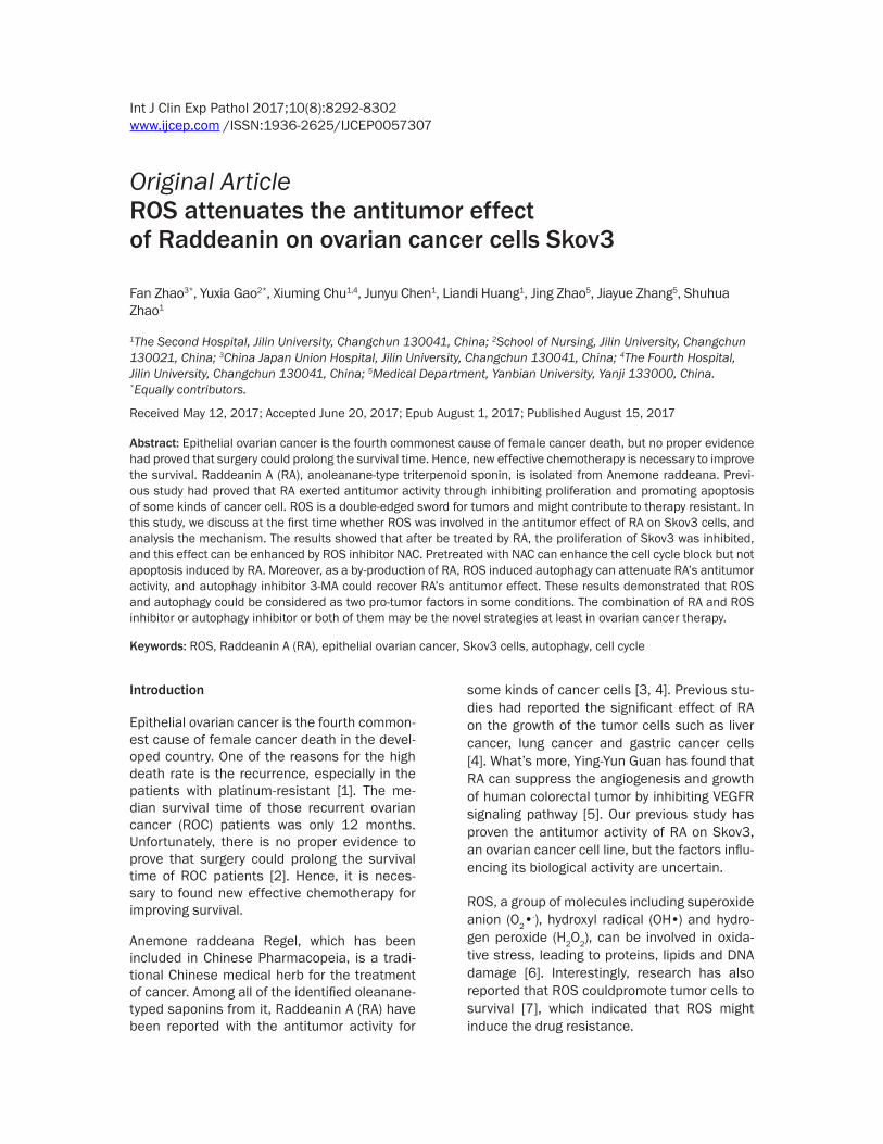
Figure 1. The inhibition rate and ROS level in Skov3 cells after exposing to RA for 3 h and 6 h. A. MTT assay of Skov3 cells treated with RA (0, 1, 2 and 4 μM for 3 h and 6 h). Either 3 hours or 6 hours after treatment, the inhibition rates of Skov3 at distinct treatment groups (1 μM, 2 μM and 4 μM) were all significantly increased compared to the control group (0 μM). B and C. Flow cytometry data with DCFH-DA staining for ROS level in the cells treated with RA (4 μM for 3 h and 6 h). The production of ROS in RA groups was significantly increased compared to that of control group, and the 6 h group showed significant reduction in ROS lever increase as compared to 3 h group.
minutes at 1000 rpm and resuspended in PBS containing 10 μM DCHF-DA. Cells were main-tained at 37°C in dark for 30 minutes. Then cells were pelleted and resuspended in PBS and fnally analyzed by flow cytometry (Becton Dickinson model FACS Calibur) [16].
AO staining
The Skov3 cells were treated with indicated different treatments for indicated time points followed by staining with 1 μM acridine orange for 15 min. Cells were then washed with PBS and visualized with fluorescence microscope [17].
Western blot analysis
Soluble proteins were extracted (in the pres-ence of complete protease and phosSTOP phosphatase inhibitors, Roche Applied Scien- ces, Indianapolis, IN, USA) and subjected to SDS-polyacrylamide gel electrophoresis. Sepa- rated proteins were electrophoretically trans-ferred onto polyvinylidene difluoride (PVDF)
membranes (Thermo Fisher Inc., Rockford, IL, USA) and immunoblotted with anti-p53, -p21, -ATR, -CDK4, -cyclin B1, -ChK1 or -beta-actin antibody (protentech group, USA), as previously described [14]. Immunoreactive proteins were visualized using the Odyssey Infrared Imaging System (Li-Cor, Lincoln, NE, USA), as described by the manufacturer.
Statistical analysis
Data are presented as the mean ± SE and represent three independent experiments. Statistical analyses were performed with SP- SS17.0, difference among the groups were compared with one-factor analysis of variance (ANOVA). Values of P<0.05 were considered statistically significant.
Results
The inhibition rate of RA may be regulated by the ROS level of Skov3 in vitro
Our previous study proved the inhibition acti-vity of RA on Skov3, to further identify the sen-
ROS attenuates the effect of Raddeanin on Skov3
8295 Int J Clin Exp Pathol 2017;10(8):8292-8302
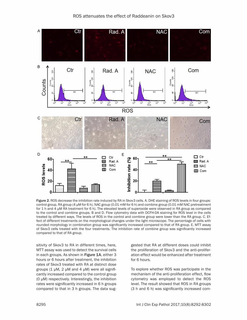
Figure 2. ROS decrease the inhibition rate induced by RA in Skov3 cells. A. DHE staining of ROS levels in four groups: control group, RA group (4 μM for 6 h), NAC group (0.01 mM for 6 h) and combine group (0.01 mM NAC pretreatment for 1 h and 4 μM RA treatment for 6 h). The elevated levels of superoxide were observed in RA group as compared to the control and combine groups. B and D. Flow cytometry data with DCFH-DA staining for ROS level in the cells treated by different ways. The levels of ROS in the control and combine group were lower than the RA group. C. Ef-fect of different treatments on the morphological changes under the light microscope. The percentage of cells with rounded morphology in combination group was significantly increased compared to that of RA group. E. MTT assay of Skov3 cells treated with the four treatments. The inhibition rate of combine group was significantly increased compared to that of RA group.
sitivity of Skov3 to RA in different times, here, MTT assay was used to detect the survival cells in each groups. As shown in Figure 1A, either 3 hours or 6 hours after treatment, the inhibition rates of Skov3 treated with RA at distinct dose groups (1 μM, 2 μM and 4 μM) were all signifi-cantly increased compared to the control group (0 μM) respectively. Interestingly, the inhibition rates were significantly increased in 6 h groups compared to that in 3 h groups. The data sug-
gested that RA at different doses could inhibit the proliferation of Skov3 and the anti-prolifer-ation effect would be enhanced after treatment for 6 hours.
To explore whether ROS was participate in the mechanism of the anti-proliferation effect, flow cytometry was employed to detect the ROS level. The result showed that ROS in RA groups (3 h and 6 h) was significantly increased com-
ROS attenuates the effect of Raddeanin on Skov3
8296 Int J Clin Exp Pathol 2017;10(8):8292-8302
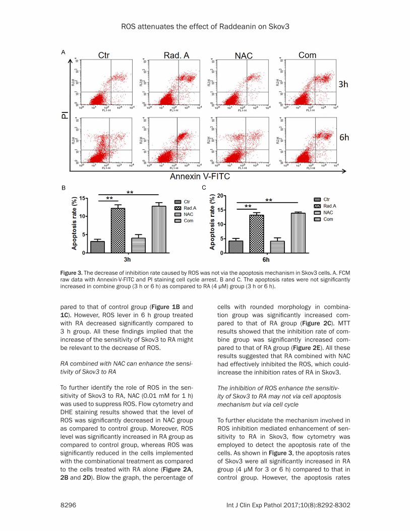
Figure 3. The decrease of inhibition rate caused by ROS was not via the apoptosis mechanism in Skov3 cells. A. FCM raw data with Annexin-V-FITC and PI staining cell cycle arrest. B and C. The apoptosis rates were not significantly increased in combine group (3 h or 6 h) as compared to RA (4 μM) group (3 h or 6 h).
pared to that of control group (Figure 1B and 1C). However, ROS lever in 6 h group treated with RA decreased significantly compared to 3 h group. All these findings implied that the increase of the sensitivity of Skov3 to RA might be relevant to the decrease of ROS.
RA combined with NAC can enhance the sensi-tivity of Skov3 to RA
To further identify the role of ROS in the sen- sitivity of Skov3 to RA, NAC (0.01 mM for 1 h) was used to suppress ROS. Flow cytometry and DHE staining results showed that the level of ROS was significantly decreased in NAC group as compared to control group. Moreover, ROS level was significantly increased in RA group as compared to control group, whereas ROS was significantly reduced in the cells implemented with the combinational treatment as compared to the cells treated with RA alone (Figure 2A, 2B and 2D). Blow the graph, the percentage of
cells with rounded morphology in combina- tion group was significantly increased com-pared to that of RA group (Figure 2C). MTT results showed that the inhibition rate of com-bine group was significantly increased com-pared to that of RA group (Figure 2E). All these results suggested that RA combined with NAC had effectively inhibited the ROS, which could-increase the inhibition rates of RA in Skov3.
The inhibition of ROS enhance the sensitiv-ity of Skov3 to RA may not via cell apoptosis mechanism but via cell cycle
To further elucidate the mechanism involved in ROS inhibition mediated enhancement of sen-sitivity to RA in Skov3, flow cytometry was employed to detect the apoptosis rate of the cells. As shown in Figure 3, the apoptosis rates of Skov3 were all significantly increased in RA group (4 μM for 3 or 6 h) compared to that in control group. However, the apoptosis rates

ROS attenuates the effect of Raddeanin on Skov3
8297 Int J Clin Exp Pathol 2017;10(8):8292-8302
Figure 4. The decrease of inhibition rate caused by ROS was related to the cell cycle arrest mechanism in Skov3 cells. A. FCM raw data with PI staining for cell cycle arrest. B. Values represent the percentage of the cells in each cell cycle phase in 3 h group. There was no significantly difference among the groups after treated for 3 h. C. Values represent the percentage of the cells in each cell cycle phase in 6 h group. After treated by NAC (0.01 mM for 1 h) combined with RA (4 μM for 6 h), population of the cells in G1 phase significantly decreased but that in G2 phase significantly increased as compared to the cells treated with RA alone. D. Western blot assay for protein expression of p53, p21, CDK4, ATR, Chk1 and cyclin B1. E. Relative optical density (OD) value from each group was showed as average from Western blot analysis. p53 and p21 were up-regulated but ATR, cyclin B1, CDK4 and Chk1 were down-regulated in combination group compared to that of RA group.

ROS attenuates the effect of Raddeanin on Skov3
8298 Int J Clin Exp Pathol 2017;10(8):8292-8302
Figure 5. RA increased autophagy level through producing low level ROS. A. AO staining of autophagy levels in four treatments in 6 h. The elevated levels of autophagy were observed in RA (4 μM for 6 h) group as compared to the NAC (0.01 mM for 1 h) and combine groups. B. Western blot assay for protein expression of LC3. C. Relative optical density (OD) value from each group was showed as average from Western blot analysis. The cells implemented with the combination showed lower expression of LC3 protein than the cells only treated with RA. D. MTT assay of Skov3 cells treated with four groups: control group, RA group (4 μM for 6 h), 3-MA group (1 mM for 6 h) and combine group (1 mM 3-MA pretreatment for 1 h and 4 μM treatment for 6 h). The inhibition rate was significantly increased in the cells treated by RA combined with 3-MA compared to that of the cells in RA group.
were not significantly increased in the combine group (3 h or 6 h) as compared to RA group (4 μM for 3 or 6 h). These results indicated that the apoptosis process might not involve in the mechanism that mediated the enhancement of sensitivity to RA in Skov3 via inhibiting ROS.
To testify whether the mechanism is related to cell cycle block, flow cytometry was employed to detect the population of cells in G0/G1, S and G2/M phases. The results showed that after treating by NAC (0.01 mM for 1 h) com-bined with RA (4 μM for 3 or 6 h), population of the cells in G1 phase significantly decreased but that in G2 phase significantly increased as compared to the cells treated with RA alone in 6 h group (Figure 4A and 4C). However, there was no significantly difference in 3 h group (Figure 4A and 4B). Additionally, Western blot was employed to detect the expressions of p53, p21, CDK4, ATR, Chk1 and cyclin B1, which are cell cycle regulatory proteins. The results showed that p53 and p21 were up-
regulated but ATR, cyclin B1, CDK4 and Chk1 were down-regulated in combination group compared to that of RA group, which indi- cated that the inhibition of ROS could down-regulate the expressions of ATR, cyclin B1, CDK4 and Chk1 but up-regulate the expres-sions of p53 and p21 (Figure 4D and 4E). The- se implied that the inhibition of ROS could inhibit Skov3 going through the G2/M check-point and then induce cell cycle arrest. So, we can draw a conclusion that the decreasing of ROS induced by RA could block cell cycle but has no remarkable effect of apoptosis. In other words, the inhibition of ROS could increase the sensitivity of Skov3 to RA through blocking cell cycle.
RA combined with NAC can increase the inhibi-tion rate through inhibiting autophagy
To illuminate another ROS-related mechanism that reinforced the chemosensitivity of RA in
ROS attenuates the effect of Raddeanin on Skov3
8299 Int J Clin Exp Pathol 2017;10(8):8292-8302
Skov3, AO staining, MTT and Western blot we- re employed to detect the level of autophagy, the inhibition rate of RA and the expres- sion of LC3, a representative protein of au- tophagy. The cells implemented with the com- bination of NAC (0.01 mM for 1 h) and RA (4 μM for 6 h) showed lower expression of LC3 protein than the cells only treated with RA and the ele-vated levels of superoxide were observed in the RA group as compared to the NAC and the com-bine groups. (Figure 5A-C), which was accor-dant with the changes of ROS level (Figure 2A and 2B). Furthermore, the inhibition rate was significantly increased in the cells treated by RA combined with 3-MA (1 mM for 1 h pretreat-ment), an autophagy suppression, compared to that of the cells in RA group. Taken together, these results suggested that ROS could im- pact the chemosenstivity of RA in Skov3 th- rough regulating autophagy mechanism.
Discussion
The progress of tumor is directly related to the proliferation of cells. This study found that RA could inhibit the proliferation of Skov3 in vitro.ROS is caused by an imbalance in the redox status of the body. The increasing of ROS can lead to tissue damage, and thus stimulate the uncontrolled growth of cells which initiate the process of carcinogenesis [18]. But ROS is a double-edged sword for tumors and might con-tribute to therapy resistant [19]. In many kinds of tumors, the high level ROS production had been considered as a critical factor in the me- chanism of anti-tumor effect of chemotherapy or radiotherapy [20-22]. However, low level ROS mediated cellular proliferation process, including acceleration of cell cycle, induction of autophagy and metastasis, improvement of tumor cells survive. And ROS was reported to play a key role in the initiation and progres-sion of various types of cancers [7, 23-25].
The sensitivity of tumor to chemotherapy is based on the inhibition rate of proliferation. In this study, the sensitivity of Skov3 to RA was enhanced after be treated for 6 hours com-pared to 3 hours, whereas the level of ROS was simultaneously decreased (Figure 1A-C).Moreover, the increasing of inhibition rate of RA in Skov3 was definitely related to the decreasing of ROS (Figure 2). Which indicate that although RA could inhibit the develop- mentof Skov3, its by-product such as low level
ROS can promote the proliferation of Skov3, which might be one of the therapy resistance mechanism of RA. Moreover, pre-treating with NAC enhanced the anti-proliferation effect of RA, indicated that the sensitivity of Skov3 to RA was potentiated when the cells lost the pro-tection of ROS. This also highlights the advan-tage that a positive impact in inhibition rate of NAC-pretreatment which directly decrease the ROS level in Skov3 cells.
The concept that programmed cell death by apoptosis serves as a natural barrier to cancer development has been established by many researchers over the last two decades [26]. The induction of apoptosis has been regarded as the key mechanism of anti-tumor chemo-therapy [27]. High level ROS caused by most chemotherapy lead to cell apoptosis [28]. However, no significant decrease of apoptosis rates was detected in the combination groups compared to the RA group, after be treated for either 3 h or 6 h in this study (Figure 3), imply-ing that the increasing of apoptosis rate in Skov3 induced by RA was not through produc-ing high level ROS. Moreover, low level ROS could promote the cell division [28], which was accordant to our research (Figure 2). Nevertheless, the apoptosis rates were not increased in NAC-pretreatment groups (Figu- re 3), which indicated that the inhibition of ROS enhanced the sensitivity of Skov3 to RA was not via cell apoptosis mechanism.
Although the regulation mechanism of ROS in tumors is not completely clear, more and mo- re studies stressed that ROS’s function as a tumor regulator seemed to occur at multiple mechanisms, such as apoptosis, cell cycle and autophagy. Thus, we supposed whether ROS inhibition enhanced the anti-tumor effect of RA in Skov3 via block cell cycle. Normal cell progression from G1-S-G2-M-G0 during the division cycle is tightly controlled by the cyclins, the cyclin-dependent kinase inhibitor (CKI) and the cyclin-dependent kinases (CDKs). This stu- dy proved that RA could induce G2/M arrest of Skov3 cells by down-regulating the level of Cyclin B1, and the protein complex of cyclin B1 and CDK1 urges cells through G2/M phase [29]. P21 and P53 are two major members of CKI family. P21 is an efficient inhibitor of CDK4, and it can arrest cell in G2 when overexpressed. The G2/M checkpoint, an important site that prevents cells from entering mitosis when DNA

ROS attenuates the effect of Raddeanin on Skov3
8300 Int J Clin Exp Pathol 2017;10(8):8292-8302
is damaged, is mediated by the activation and accumulation of p53, which then up-regulates the p21 to maintain G2 arrest in response to DNA damage [30]. Recently, it had been re- ported that the production of low level ROS could promote the cell cycle process through inducing the transcription and translation of the cyclins and CDKs [31]. In this study, west-ern blot results showed that the level of cyclin B1 was significantly decreased in combined group compared to RA group and the expres-sion of p53/p21 was significantly up-regulated in combined group compared with RA group (Figure 4D and 4E), which was correspond to the G2/M arrest results (Figure 4A and 4C). These findings suggested that while RA was effective for holding the Skov3 cells at the G2 phase, its effect had been weakened by the low level ROS. In other words, it was well in line with our previous idea that ROS inhibition enhanced the anti-tumor effect of RA in Skov3 via cell cycle block mechanism.
The checkpoint kinase 1 (Chk1) is an essential kinase that is phosphorylated by ataxia-telan- giectasia-mutated and Rad-3 related (ATR) and required for the G2/M DNA damage che- ckpoint, and it was regarded as a tumor sup-press or for many years [32]. However, increas-ing evidence suggests that Chk1 may actually promote tumor growth, and the enhanced acti-vation of Chk1 led to resistance of cancer cells, including prostate and lung cancer, to che- motherapy [33]. In this study, the increasing of Chk1/ATR expression (Figure 4D and 4E) directly or indirectly caused by the ROS, by-product of RA, possessed survival advanta- ges over Skov3 cells, because the more Chk1 protein Skov3 had, the better they were to facil-itate the DNA damage in G2 phase caused by the RA chemotherapy [30, 33]. All these ob- servations implied that low level ROS promoted Skov3 survive and proliferation through nega-tively impacting the sensitive of Skov3 to RA via cell cycle mechanism. This model provided a strong support to target ROS in human ovari-an cancer therapy.
The another regulation mechanism of ROS in tumor is autophagy which is regulated by nu- merous stresses such as hypoxia, ROS level and chemotherapies [6]. Alterations in ROS lev-els and autophagy play a crucial role in cancer initiation and progression, and both are recog-
nized as the potential targets for cancer treat-ment. It had been reported that Ciclopiro- xolamine (CPX) could induce autophagy in human rhabdomyosarcoma cells by ROS [8]. Hahm et al found that the use of antioxidants in prostate cancer inhibited autophagy levels [34]. In the present study, for the first time, we found that RA induced LC3 expression, which was representative for autophagy, in Skov3 cells. Interestingly, low level ROS was simulta-neously increased by RA as previous describe. Thus, NAC, the ROS scavenger, was used to identified the relation between ROS and au- tophagy. Our results that NAC remarkably at- tenuated RA-induced LC3 expression sugge- sted that RA triggered autophagy by induction of low level ROS (Figure 5A-C). In order to fur-ther discuss the RA resistance mechanism caused by low level ROS in Skov3, 3-MA, an autophagy inhibitor was employed to pretreat the Skov3 cells. The present observations th- at the disruption of autophagy in the presence of 3-MA increased the RA-induced inhibition rate of Skov3 cells clearly indicated that RA- induced autophagy was a drug resistance mechanismat least in ovarian cancer cells (Figure 5D). All these findings identified that the autophagy induced by low level ROS pro-duced by RA was a potential drug resistance mechanism in the RA therapy of ovarian can-cer, and we also provided a promising strategy for ovarian cancer therapy that the combina- tion of RA and an autophagy inhibitor.
In conclusion, this study showed that low level ROS induced autophagy and then led to the decrease of inhibition rates of RA in human ovarian cancer Skov3 cells. Disruption of ROS using NAC increased RA-induced inhibition ra- tes, indicating that RA-induced low level ROS was a pro-proliferation mechanism in Skov3 cells. Further, we found that the mechanism was not related to the apoptosis pathway, but the dysregulation of cell-cycle arrest in G2 phase. Additionally, the ROS-mediated autoph-agy was involved in our study. 3-MA, an inhibi-tor of autophagy, induced the inhibition rates of RA, suggesting that ROS-mediated auto- phagy was a pro-survival mechanism in the cells. Our findings suggested that the combi- nation of RA and ROS inhibitor or autophagy inhibitor or both of them may be the promising strategies at least in ovarian cancer therapy.
ROS attenuates the effect of Raddeanin on Skov3
8301 Int J Clin Exp Pathol 2017;10(8):8292-8302
Acknowledgements
This work was financially supported by the Science & Technology Development Planning Project of Jilin Province (20140203013YY and 2014220101001742) and the Project of Jilin Province Development and Reform Commission (2041G073). The authors would like to thank Na Li and Lijing Zhao for their advice to improve the experiment and manuscript.
Disclosure of conflict of interest
None.
Address correspondence to: Dr. Shuhua Zhao, Department of Gynaecology and Obstetrics, The Second Hospital, Jilin University, Changchun 1300- 41, China. Tel: 86-0431-88796567; Fax: 86-0431-85619580; E-mail: [email protected]
References
[1] Jayson GC, Kohn EC, Kitchener HC and Leder-mann JA. Ovarian cancer. Lancet 2014; 384: 1376-1388.
[2] Suh DH, Kim HS, Chang SJ and Bristow RE. Surgical management of recurrent ovarian cancer. Gynecol Oncol 2016; 142: 357-367.
[3] Luan X, Guan YY, Wang C, Zhao M, Lu Q, Tang YB, Liu YR, Yu DH, Wang XL and Qi H. Determi-nation of Raddeanin A in rat plasma by liquid chromatography-tandem mass spectrometry: application to a pharmacokinetic study. J Chro-matogr B Analyt Technol Biomed Life Sci 2013; 923-924: 43-47.
[4] Qian S, Chen QL, Guan JL, Wu Y and Wang ZY. ChemInform abstract: synthesis and biological evaluation of Raddeanin A, a triterpene sapo-nin isolated from anemone raddeana. Chem Pharm Bull (Tokyo) 2014; 62: 779-785.
[5] Guan YY, Liu HJ, Luan X, Xu JR, Lu Q, Liu YR, Gao YG, Zhao M, Chen HZ and Fang C. Radd-eanin A, a triterpenoid saponin isolated from anemone raddeana, suppresses the angiogen-esis and growth of human colorectal tumor by inhibiting VEGFR2 signaling. Phytomedicine 2015; 22: 103-110.
[6] Poillet-Perez L, Despouy G, Delage-Mourroux R and Boyer-Guittaut M. Interplay between ROS and autophagy in cancer cells, from tumor ini-tiation to cancer therapy. Redox Biol 2015; 4: 184-192.
[7] Weinberg F and Chandel NS. Reactive oxygen species-dependent signaling regulates cancer. Cell Mol Life Sci 2009; 66: 3663.
[8] Zhou H, Shen T, Shang C, Luo Y, Liu L, Yan J, Li Y and Huang S. Ciclopirox induces autophagy through reactive oxygen species-mediated ac-
tivation of JNK signaling pathway. Oncotarget 2014; 5: 10140-10150.
[9] Shintani T and Klionsky DJ. Autophagy in health and disease: a double-edged sword. Science 2004; 306: 990-5.
[10] White E. Deconvoluting the context-dependent role for autophagy in cancer. Nat Rev Cancer 2012; 12: 401-410.
[11] Ávalos Y, Canales J, Bravosagua R, Criollo A, Lavandero S and Quest AF. Tumor suppression and promotion by autophagy. Biomed Res Int 2014; 2014: 603980.
[12] Debnath J. The multifaceted roles of autopha-gy in tumors--implications for breast cancer. J Mammary Gland Biol Neoplasia 2011; 16: 173-187.
[13] Morselli E, Galluzzi L, Kepp O, Vicencio JM, Criollo A, Maiuri MC and Kroemer G. Anti- and pro-tumor functions of autophagy. Biochim Biophys Acta 2009; 1793: 1524-1532.
[14] Wang G, Niu X, Zhang W, Caldwell JT, Edwards H, Chen W, Taub JW, Zhao L and Ge Y. Synergis-tic antitumor interactions between MK-1775 and panobinostat in preclinical models of pan-creatic cancer. Cancer Lett 2015; 356: 656-668.
[15] Liu YB, Zhang L, Guo YX, Gao LF, Liu XC, Zhao LJ, Guo BF, Zhao LJ, Zhao XJ, Xu DQ. Plasmid-based survivin shRNA and GRIM-19 carried by attenuated salmonella suppresses tumor cell growth. Asian J Androl 2012; 14: 536-545.
[16] Maleki J, Nourbakhsh M, Shabani M, Korani M, Nourazarian SM, Dahaghi MR and Moghadasi MH. 17β-estradiol stimulates generation of re-active species oxygen and nitric oxide in ovari-an adenocarcinoma cells (OVCAR3). Iran J Can-cer Prev 2015; 8: e2332.
[17] Bahrami F, Pourgholami MH, Mekkawy AH, Rufener L and Morris DL. Monepantel induces autophagy in human ovarian cancer cells through disruption of the mTOR/p70S6K sig-nalling pathway. Am J Cancer Res 2014; 4: 558-571.
[18] Nourazarian AR, Kangari P and Salmaninejad A. Roles of oxidative stress in the development and progression of breast cancer. Asian Pac J Cancer Prev 2014; 15: 4745-4751.
[19] Okon IS and Zou MH. Mitochondrial ROS and cancer drug resistance: implications for thera-py. Pharmacol Res 2015; 100: 170.
[20] Wondrak GT. Redox-directed cancer therapeu-tics: molecular mechanisms and opportuni-ties. Antioxid Redox Signal 2009; 11: 3013-3069.
[21] Kaufmann SH and Earnshaw WC. Induction of apoptosis by cancer chemotherapy. Exp Cell Res 2000; 256: 42-49.
[22] Conklin KA. Chemotherapy-associated oxida-tive stress: impact on chemotherapeutic effec-

ROS attenuates the effect of Raddeanin on Skov3
8302 Int J Clin Exp Pathol 2017;10(8):8292-8302
tiveness. Integr Cancer Ther 2004; 3: 294-300.
[23] Friedl P and Alexander S. Cancer invasion and the microenvironment: plasticity and reciproci-ty. Cell 2011; 147: 992-1009.
[24] Djavaherimergny M, Amelotti M, Mathieu J, Be-sançon F, Bauvy C, Souquère S, Pierron G and Codogno P. NF-κB activation represses tumor necrosis factor-α-induced autophagy. J Biol Chem 2006; 281: 30373-30382.
[25] Hurd TR, Degennaro M and Lehmann R. Redox regulation of cell migration and adhesion. Trends Cell Biol 2012; 22: 107-115.
[26] Douglas H and Weinberg RA. Hallmarks of can-cer: the next generation. Cell 2011; 144: 646-674.
[27] Wang T, Lv JH, Zhang XF, Li CJ, Han X and Sun YJ. Tissue inhibitor of metalloproteinase-1 protects MCF-7 breast cancer cells from pacli-taxel-induced apoptosis by decreasing the sta-bility of cyclin B1. Int J Cancer 2010; 126: 362-370.
[28] Holbrook NJ and Ikeyama S. Age-related de-cline in cellular response to oxidative stress: links to growth factor signaling pathways with common defects. Biochem Pharmacol 2002; 64: 999.
[29] Norbury C and Nurse P. Animal cell cycles and their control. Ann Rev Biochem 1992; 61: 441-470.
[30] Stark GR and Taylor WR. Control of the G2/M transition. Mol Biotechnol 2006; 32: 227-248.
[31] Boonstra J and Post JA. Molecular events as-sociated with reactive oxygen species and cell cycle progression in mammalian cells. Gene 2004; 337: 1-13.
[32] Liu Q, Guntuku S, Cui XS, Matsuoka S, Cortez D, Tamai K, Luo G, Carattini-Rivera S, Demayo F and Bradley A. Chk1 is an essential kinase that is regulated by Atr and required for the G(2)/M DNA damage checkpoint. Genes Dev 2000; 14: 1448-59.
[33] Zhang Y and Hunter T. Roles of Chk1 in cell bi-ology and cancer therapy. Int J Cancer 2014; 134: 1013-1023.
[34] Hahm ER, Sakao K and Singh SV. Honokiol ac-tivates reactive oxygen species-mediated cyto-protective autophagy in human prostate can-cer cells. Prostate 2014; 74: 1209-1221.