original article role of pdgfs/pdgfrs signaling pathway in … · 2016-08-09 · role of...
TRANSCRIPT

Int J Clin Exp Pathol 2014;7(1):16-27www.ijcep.com /ISSN:1936-2625/IJCEP1311044
Original ArticleRole of PDGFs/PDGFRs signaling pathway in myocardial fibrosis of DOCA/salt hypertensive rats
Bin Fan*, Likun Ma*, Qian Li, Lin Wang, Junling Zhou, Jiawei Wu
Department of Cardiology, Affiliated Provincial Hospital, Anhui Medical University, Hefei, Anhui, 230001, China. *Equal contributors.
Received November 20, 2013; Accepted December 13, 2013; Epub December 15, 2013; Published January 1, 2014
Abstract: This study aimed to investigate the role of PDGF/PDGFR signaling pathway in myocardial fibrosis of desoxy-corticosterone (DOCA) induced salt-sensitive hypertensive rats and explore the influence of PDGF/PDGFR signaling pathway on fibroblasts and myofibroblasts in the heart. 60 male SD rats underwent right nephrectomy and bred with 1% sodium chloride and 0.1% potassium chloride for 4 weeks, and then randomly divided into 3 groups (CON group, DOCA group and DOCA+IMA group). Results showed that: 1) 14 and 28 days after intervention, the SBP in DOCA and DOCA+IMA group was significantly higher than that in CON group. At days 28, the severity of myocardial fibro-sis and PVCA/VA ratio in DOCA group were significantly increased when compared with CON group. The severity of myocardial fibrosis and PVCA/VA ratio in DOCA+IMA group were markedly lower than those in DOCA group although they were higher than those in CON group. 2) At days 14, the mRNA expressions of PDGFRα and PDGFRβ in DOCA group were significantly higher than CON and DOCA+IMA group. At days 28, the mRNA expressions of PDGFRβ, FSP-1, α-SMA, procollagen I and procollagen III in DOCA group were significantly higher than those in CON group. In addition, in a specific group, the PDGFRβ mRNA expression was higher than the PDGFRα mRNA expression. In DOCA+IMA group, the mRNA expressions of PDGFRβ, FSP-1, α-SMA, procollagen I and procollagen III were markedly reduced when compared with DOCA group. 3) At 14 days, the protein expressions of PDGFRα and PDGFRβ in DOCA group were significantly higher than those in CON group. The PDGFRα protein expression in DOCA+IMA group was markedly lower than that in DOCA group. At days 28, the protein expressions of PDGFRα and PDGFRβ in DOCA group were significantly increased when compared with CON group. The protein expressions of PDGFRα and PDGFRβ in DOCA+IMA group were significantly lower than those in DOCA group. At day 28, the cardiac interstitium mainly contained vimentin positive fibroblasts, and α-SMA positive cells were less identified in CON group. In DOCA group, α-SMA positive fibroblasts (spindle-shaped) increased significantly, but the myofibroblasts reduced significantly in DOCA+IMA group when compared with DOCA group. 4) PDGFRα protein expression was observed in fibroblasts and myofibroblasts, but not in VSMCs. PDGFRβ protein expression was noted in not only fibroblasts and myofibroblasts but also VSMCs. Thus, During myocardial fibrosis of DOCA induced salt-sensitive hypertensive rats, PDGFRα acts at early stage, but PDGFRβ functions in the whole process. PDGFRα and PDGFRβ expressions increase in fibroblasts and myofibroblasts, suggesting that PDGF/PDGFR signaling pathway is involved in the myocardial fibrosis via stimu-lating fibroblasts to proliferate and transform into myofibroblasts.
Keywords: Platelet-derived growth factor, platelet-derived growth factor receptor, fibroblasts, myofibroblasts, des-oxycorticosterone, imatinib, myocardial fibrosis
Introduction
Myocardial fibrosis (MF) is a pathological pro-cess with excessive proliferation of fibroblasts and imbalance between deposition and degra-dation of extracellular matrix (ECM) in myocar-dial interstitium and a terminal pathological manifestation of multiple cardiovascular dis-eases [1, 2]. Currently, the pathogenesis of
myocardial fibrosis is still poorly understood. Previous work on myocardial fibrosis of desoxy-corticosterone (DOCA) induced salt-sensitive hypertensive rats demonstrated that PDGF/PDGFR signaling pathway was involved in the myocardial fibrosis [3]. However, the ways, which bridge the PDGF/PDGFR signaling path-way and myocardial fibrosis, are still unclear. There is evidence showing that the fibroblasts

PDGFs/PDGFRs in myocardial fibrosis
17 Int J Clin Exp Pathol 2014;7(1):16-27
in the heart have the potential to differentiate into myofibroblasts [4]. In study on myocardial fibrosis after myocardial infarction, investiga-tors proposed that PDGF/PDGFR signaling pathway is involved in the pathogenesis of myo-cardial fibrosis via stimulating fibroblasts to proliferate and transform into myofibroblasts and to secret massive collagens [5]. However, this has not been confirmed in vivo. In the pres-ent study, the cells with PDGFR expression and the changes in fibroblasts and myofibroblasts of the heart were investigated in DOCA induced salt-sensitive hypertensive rats, aiming to explore the role of PDGF/PDGFR signaling path-way in the myocardial fibrosis.
Materials and methods
Animal model and grouping
Specific pathogen free male SD rats (n=60) weighing 200-250 g were purchased from the Experimental Animal Center of Affiliated Provincial Hospital of Anhui Medical University. Animals were anesthetized intraperitoneally with 10% chloral hydrate at 400 mg/kg and then received nephrectomy. At 1 week after surgery, animals were bred with 1% sodium chloride and 0.2% potassium chloride and ran-domly assigned into 3 groups (n=20 per group): 1) CON group: animals were subcutaneously treated with soybean oil once every 4 days and intragastrically treated with distilled water once daily; 2) DOCA group: animals were subcutane-ously treated with DOCA at 60 mg/kg/4d and intragastrically treated with distilled water once daily; 3) DOCA+IMA group: animals were subcu-
taneously treated with DOCA at 60 mg/kg/4d and intragastrically treated with imatinib at 60 mg/kg/d once daily.
In CON group, the volume of intragastrical-dis-tilled water was equal to that of intragastrical drugs in other two groups; in CON group, the volume of subcutaneous soybean oil was iden-tical to that of subcutaneous drugs dissolved in soybean oil. Treatment was done for 28 days. Before and at 14 and 28 days after treatment, systolic blood pressure (SBP) was measured. At 14 and 28 days after treatment, animals were anesthetized with 10% chloral hydrate at 400 mg/kg and then sacrificed (n=10 at each time point). The heart was harvested and the atri-ums, major vessels and connective tissues were removed. The remaining ventricular tis-sues were divided into 3 parts: one was stored at -80°C for real-time fluorescence quantitative PCR; one was fixed in 4% paraformaldehyde for 24 h and embedded in paraffin followed by HE staining and immunohistochemistry; one was fixed in 4% paraformaldehyde for 24 h and dehydrated in 30% sucrose for frozen section-ing and subsequent immunofluorescence staining.
Main reagents
The following reagents were used in the pres-ent study: DOCA (Sigma, USA), imatinib (LC, labs; USA), sirius red dye (Beijing Haide Biotech Co., Ltd), phosphate buffer solution (PBS), poly-lysine coated slides (Wuhan Boster Biotech Co., Ltd), SP kit for immunohistochemistry, DAB, FITC conjugated goat anti-mouse second-ary antibody, rhodamine red conjugated goat anti-rabbit secondary antibody (Beijing Zhongshan Golden Bridge Biotech Co., Ltd), rabbit anti-rat PDGFRα, rabbit anti-rat PDGFRβ, mouse anti-rat Vimentin and mouse anti-rat α-SMA primary antibodies (Abcam UK), rabbit anti-rat p-PDGFRβ primary antibody (Santa Cruz, USA), DAPI (Sigma, USA), citrate acid solu-tion for antigen retrieval, anti-quencher (Beyotime Institute of Biotechnology), Jung embedding reagent for frozen sections (Leica, Germany), sodium chloride, potassium chloride and chloral hydrate (Sinapharm Chemical Reagent Co., Ltd).
The primers for FSP-1, α-SMA, PDGFR-α, PDGFR-β, procollagen I, procollagen III and GADPH (Table 1) were designed with Primer-Blast software of the National Center for Biotechnology Information (http://www.ncbi.
Table 1. Primers used in RT-PCRGene Primer sequencesPDGFRα Sense GAGACCCTCCTTCTACCACCT
Anti-sense GTTGTCAGAGTCCACACGCATPDGFRβ Sense GCACCGAAACAAACACACCTT
Anti-sense ATGTAACCACCGTCGCTCTCFSP-1 Sense ACCTCTCTGTTCAGCACTTCC
Anti-sense GAACTTGTCACCCTCGTTGCα-SMA Sense CCAGTCGCCATCAGGAACC
Anti-sense AGCAAAGCCCGCCTTACAGprocollagen I Sense ACGCATGAGCCGAAGCTAAC
Anti-sense AGGGACCCTTAGGCCATTGTprocollagen III Sense ATAGACCTCAAGGCCCCAAG
Anti-sense CCACCCATTCCTCCGACTGADPH Sense TGGGAAGCTGGTCATCAAC
Anti-sense GCATCACCCCATTTGATGTT
PDGFs/PDGFRs in myocardial fibrosis
18 Int J Clin Exp Pathol 2014;7(1):16-27
nlm.nih.gov/tools/primer-blast) and synthe-sized in Shanghai Sangon Biotech. RNeasy Mini Kit for mRNA extraction (Qiagen Germany), PrimeScript™ RT reagent Kit with gDNA Eraser for reverse transcription, and SYBR® Premix Ex Taq™ II for real time quantitative PCR (Takara, Japan) were used for PCR. Sirius red dye, hema-toxylin - eosin, formaldehyde, microtome (Leica), Cryostat for sectioning (Leica), and microscope camera system (Nikon eclipse 80i, Japan) were provided by the comprehensive laboratory of Basic Medicine of Anhui Medical University. Thermal cycler (Applied Biosystems Step One Plus System) was provided by the Department of Parasitology of Basic Medicine of Anhui Medical University.
Detection of myocardial fibrosis
The left ventricle was fixed in 4% paraformalde-hyde for 24 h and then dehydrated. After embedded in paraffin, the left ventricle was sectioned (4 μm in thickness) onto polylysine-coated slides followed by heating. At 28 days, paraffin embedded sections were also pre-pared and stained with picric acid and Sirius red for collagen staining. Photographs were captured via the microscope camera system and analyzed with Image Pro plus 6.0. The severity of myocardial interstitial fibrosis was determined (area of myocardial interstitial col-lagen/area of total field and perivascular colla-gen volume area [PVCA]/vascular area [VA]).
Detection of mRNA expression of PDGFR, FSP-1, α-SMA, procollagen I and procollagen III by real time fluorescence quantitative PCR
mRNA expressions of target genes were mea-sured by real time fluorescence quantitative PCR according to manufacturer’s instructions. In brief, the heart tissues were homogenized, followed by extraction of mRNA with RNeasy Mini Kit. Then, mRNA was mixed with genomic DNA free gDNA Eraser followed by reverse tran-scription into cDNA with PrimeScript™ RT reagent Kit with gDNA Eraser kit. Amplification of cDNA was done on thermal cycler (Applied
Biosystems Step One Plus System). The reac-tion mixture was 20 μl in volume, and amplifica-tion was performed according to manufactur-er’s instructions (SYBR® Premix Ex Taq™ II PCR kit and Applied Biosystems Step One Plus System). The reaction conditions were as fol-lows: pre-denaturation at 95°C for 30 s and 40 cycles of 95°C for 5 s and 60°C for 30 s. The melt curve was employed to determine the specificity of products. The supporting software was used to analyze the Ct value of products. According to the following formula: ΔCt=Cttarget
gene - Ctinternal reference, the ΔCt was calculated in two groups. The Ct value of target gene is nega-tively proportional to the copies of this gene, and thus, the larger the ΔCt, the lower the expression of a gene is. Then, 2-ΔΔCt method was employed to calculate the relative mRNA expression of target genes.
Detection of protein expressions of PDGFRs, p-PDGFRβ, vimentin and α-SMA in heart by immunohistochemistry
The paraffin embedded sections were prepared as above mentioned and then deparaffinized. After antigen retrieval in citrate acid at 95°C for 10 min, sections were blocked in 3% H2O2 for 10 min at 37°C and then treated with 10% normal goat serum for 30 min at 37°C. Subsequently, these sections were inde-pendently treated with PDGFRα (1:400), PDGFRβ (1:200), p-PDGFRβ (1:200), vimentin (1:2500) and α-SMA (1:100) at 4°C overnight and then with biotinylated secondary antibody and HRP conjugated streptavidin at 37°C for 25 min followed by visualization with DAB. Washing was performed between procedures (5 min in each) and a final counterstaining was done with hematoxylin. After dehydration and transparentization, mounting was done. In the negative control group, the primary antibody was replaced with PBS. Under a light micro-scope, cells with brown cytoplasm were regard-ed as positive. Analysis was performed with Image Pro Plus 6.0, and the integrated optical density (OD) of PDGFRα, PDGFRβ and p-PDGFRβ was determined. The α-SMA positive spindle-shaped cells (myofibroblasts) were counted in each section and the average was calculated.
Detection of cells positive for PDGFRα and PDGFRβ by immunofluorescence staining
The heart tissues were fixed in 4% paraformal-dehyde for 24 h and then in 30% sucrose at
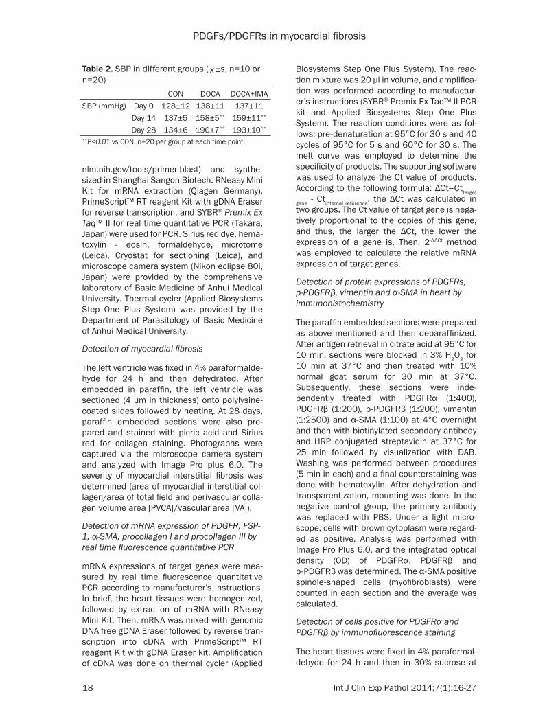
Table 2. SBP in different groups (_x±s, n=10 or n=20)
CON DOCA DOCA+IMASBP (mmHg) Day 0 128±12 138±11 137±11
Day 14 137±5 158±5** 159±11**
Day 28 134±6 190±7** 193±10**
**P<0.01 vs CON. n=20 per group at each time point.

PDGFs/PDGFRs in myocardial fibrosis
19 Int J Clin Exp Pathol 2014;7(1):16-27
4°C until these tissues sank. After embedded in embedding reagent for frozen sections, 6-μm sections were obtained onto polylysine-coated
1:200; rhodamine red conjugated goat anti-rabbit antibody: 1:200; DAPI: 1:2000) at 37°C for 30 min. After washing in PBS 5 times (5 min
Figure 1. Sirius red staining of myocardial interstitium in different groups on day 28 (× 200). A: CON; B: DOCA; C: DOCA+IMA. Myocardial fibrosis was the most severe in DOCA group, but attenuated in DOCA+IMA group.
Figure 2. Sirius red staining of perivascular interstitium in different groups on day 28 (× 200). A: CON; B: DOCA; C: DOCA+IMA. Perivascular fibrosis was the most severe in DOCA group, but attenuated in DOCA+IMA group.
Figure 3. mRNA expressions of PDGFRα and PDGFRβ in different groups. _x±s,
n=10. **P<0.01 vs CON. ##P<0.01 vs DOCA.
slides. These slides were treated with acetone at 4°C for 15 min and then with PBS. After treatment with 0.5% Triton X-100 at 37°C for 30 min, sections were incubated with 10% normal goat serum at 37°C for 45 min. Subsequently, these sections were treat-ed with primary antibody (α-SMA: 1:100; vimentin: 1:2500; PDGFRα: 1:200; PDGFRβ: 1:100) at 4°C overnight and secondary antibody (FITC conjugated goat anti-mouse antibody:
PDGFs/PDGFRs in myocardial fibrosis
20 Int J Clin Exp Pathol 2014;7(1):16-27
for each), mounting was done with anti-quench-er, and sections were observed under a fluores-cence microscope and photographed (× 200). Image J image analysis software was employed to analyze and merge these photographs.
Statistical analysis
Statistical analysis was done with SPSS version 19.0. Data were expressed as mean ± stan-dard deviation (x±s). Comparisons of means between two groups were done with indepen-dent t test, and those of rates were performed with chi square test. A value of P<0.05 was con-sidered statistically significant.
Results
Change in blood pressure in different groups
Before interventions, the SBP was comparable among groups (P>0.05). At 14 and 28 days after intervention, the SBP in DOCA group and DOCA+IMA group were markedly higher than those in CON group (P<0.01), but there was no significant difference between DOCA group and DOCA+IMA group (P>0.05) (Table 2).
Myocardial interstitial and perivascular fibrosis
Sirius red staining showed the collagens in myocardial interstitium were red and the myo-cardium was yellow. Results showed the myo-cardial fibrosis was the most severe, and the amount of collagens in myocardial interstitium was the highest in DOCA group. In addition, the ratio of myocardial interstitial collagen area to total field area was 27.23% and PVCA/VA ratio
lagen III in heart
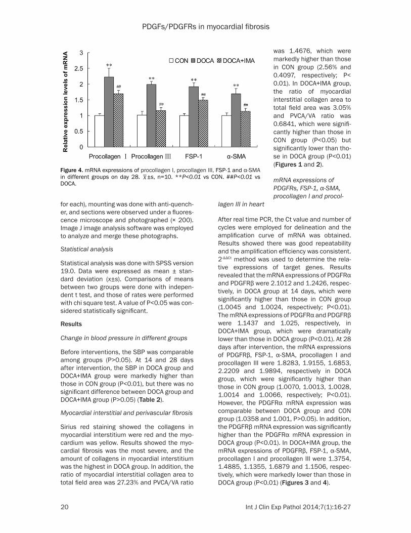
After real time PCR, the Ct value and number of cycles were employed for delineation and the amplification curve of mRNA was obtained. Results showed there was good repeatability and the amplification efficiency was consistent. 2-ΔΔCt method was used to determine the rela-tive expressions of target genes. Results revealed that the mRNA expressions of PDGFRα and PDGFRβ were 2.1012 and 1.2426, respec-tively, in DOCA group at 14 days, which were significantly higher than those in CON group (1.0045 and 1.0024, respectively; P<0.01). The mRNA expressions of PDGFRα and PDGFRβ were 1.1437 and 1.025, respectively, in DOCA+IMA group, which were dramatically lower than those in DOCA group (P<0.01). At 28 days after intervention, the mRNA expressions of PDGFRβ, FSP-1, α-SMA, procollagen I and procollagen III were 1.8283, 1.9155, 1.6853, 2.2209 and 1.9894, respectively in DOCA group, which were significantly higher than those in CON group (1.0070, 1.0013, 1.0028, 1.0014 and 1.0066, respectively; P<0.01). However, the PDGFRα mRNA expression was comparable between DOCA group and CON group (1.0358 and 1.001, P>0.05). In addition, the PDGFRβ mRNA expression was significantly higher than the PDGFRα mRNA expression in DOCA group (P<0.01). In DOCA+IMA group, the mRNA expressions of PDGFRβ, FSP-1, α-SMA, procollagen I and procollagen III were 1.3754, 1.4885, 1.1355, 1.6879 and 1.1506, respec-tively, which were markedly lower than those in DOCA group (P<0.01) (Figures 3 and 4).
Figure 4. mRNA expressions of procollagen I, procollagen III, FSP-1 and α-SMA in different groups on day 28.
_x±s, n=10. **P<0.01 vs CON. ##P<0.01 vs
DOCA.
was 1.4676, which were markedly higher than those in CON group (2.56% and 0.4097, respectively; P< 0.01). In DOCA+IMA group, the ratio of myocardial interstitial collagen area to total field area was 3.05% and PVCA/VA ratio was 0.6841, which were signifi-cantly higher than those in CON group (P<0.05) but significantly lower than tho- se in DOCA group (P<0.01) (Figures 1 and 2).
mRNA expressions of PDGFRs, FSP-1, α-SMA, procollagen I and procol-
PDGFs/PDGFRs in myocardial fibrosis
21 Int J Clin Exp Pathol 2014;7(1):16-27
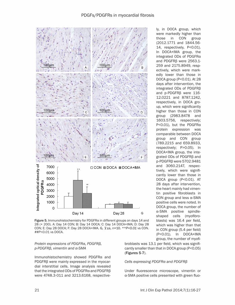
Protein expressions of PDGFRα, PDGFRβ, p-PDGFRβ, vimentin and α-SMA
Immunohistochemistry showed PDGFRα and PDGFRβ were mainly expressed in the myocar-dial interstitial cells. Image analysis revealed that the integrated ODs of PDGFRα and PDGFRβ were 4748.3-011 and 3213.6168, respective-
broblasts was 13.1 per field, which was signifi-cantly smaller than that in DOCA group (P<0.05) (Figures 5-7).
Cells expressing PDGFRα and PDGFRβ
Under fluorescence microscope, vimentin or α-SMA positive cells presented with green fluo-
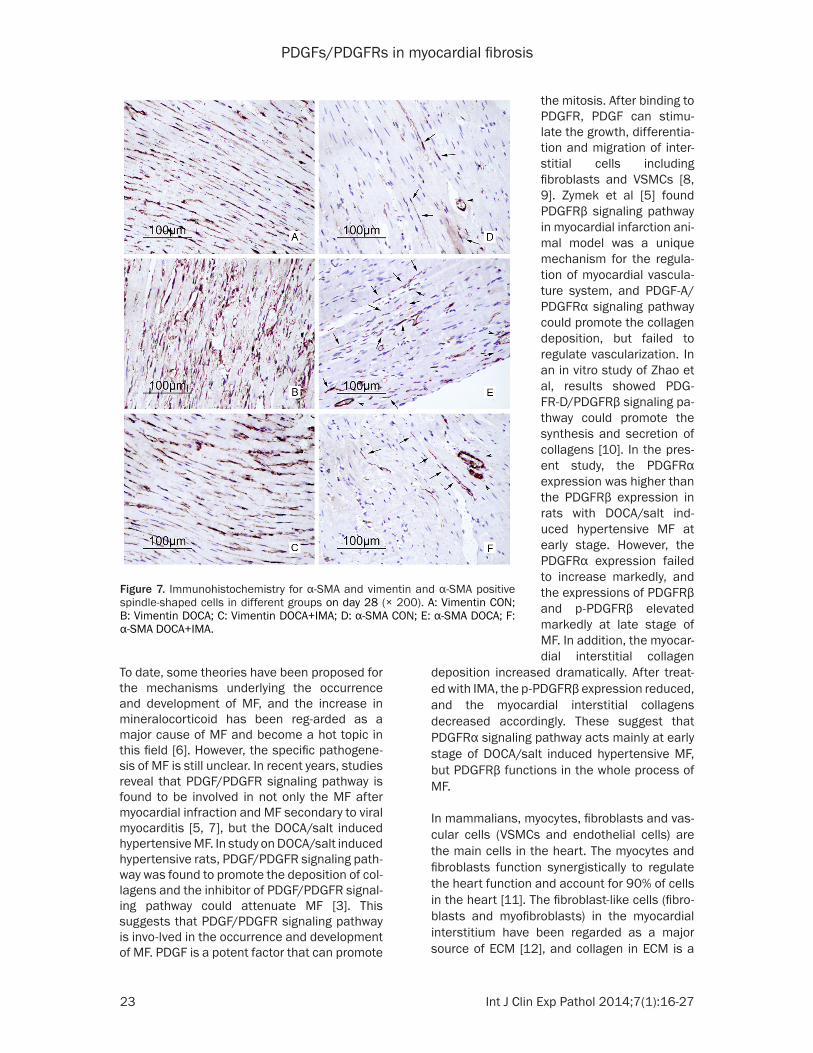
ly, in DOCA group, which were markedly higher than those in CON group (2012.1771 and 1844.56-14, respectively, P<0.01). In DOCA+IMA group, the integrated ODs of PDGFRα and PDGFRβ were 2563.1-259 and 2175.8949, resp-ectively, which were mark-edly lower than those in DOCA group (P<0.01). At 28 days after intervention, the integrated ODs of PDGFRβ and p-PDGFRβ were 116- 12.0221 and 8787.1242, respectively, in DOCA gro- up, which were significantly higher than those in CON group (2983.8478 and 1603.5756, respectively; P<0.01), but the PDGFRα protein expression was comparable between DOCA group and CON group (789.2215 and 659.8933, respectively; P>0.05). In DOCA+IMA group, the inte-grated ODs of PDGFRβ and p-PDGFRβ were 5702.9481 and 3060.2147, respec-tively, which were signifi-cantly lower than those in DOCA group (P<0.01). AT 28 days after intervention, the heart mainly had vimen-tin positive fibroblasts in CON group and less α-SMA positive cells were noted. In DOCA group, the number of α-SMA positive spindle-shaped cells (myofibro-blasts) was 16.4 per field, which was higher than that in CON group (5.4 per field) (P<0.01). In DOCA+IMA group, the number of myofi-
Figure 5. Immunohistochemistry for PDGFRα in different groups on days 14 and 28 (× 200). A: Day 14 CON; B: Day 14 DOCA; C: Day 14 DOCA+IMA; D: Day 28 CON; E: Day 28 DOCA; F: Day 28 DOCA+IMA. G,
_x±s, n=10. **P<0.01 vs CON.
##P<0.01 vs DOCA.

PDGFs/PDGFRs in myocardial fibrosis
22 Int J Clin Exp Pathol 2014;7(1):16-27
rescence mainly in the cytoplasm; PDGFRα or PDGFRα positive cells presented with red fluo-rescence mainly in the cytoplasm. The merged fluorescence was orange. DAPI positive nuclei presented with blue fluorescence. Results showed fibroblasts and myofibroblasts were positive for PDGFRα, which was mainly, found in the cytoplasm. In VSMCs, no PDGFRα expres-sion was observed. In addition, PDGFRβ expres-
Figure 6. Immunohistochemistry for PDGFRβ and p-PDGFRβ in different groups on days 14 and 28 (× 200). A-F: PDGFRβ, A: Day 14 CON; B: Day 14 DOCA; C: Day 14 DOCA+IMA; D: Day 28 CON; E: Day 28 DOCA; F: Day 28 DOCA+IMA. G-I: p-PDGFRβ, G: Day 28 CON; H: Day 28 DOCA; I: Day 28 DOCA+IMA.
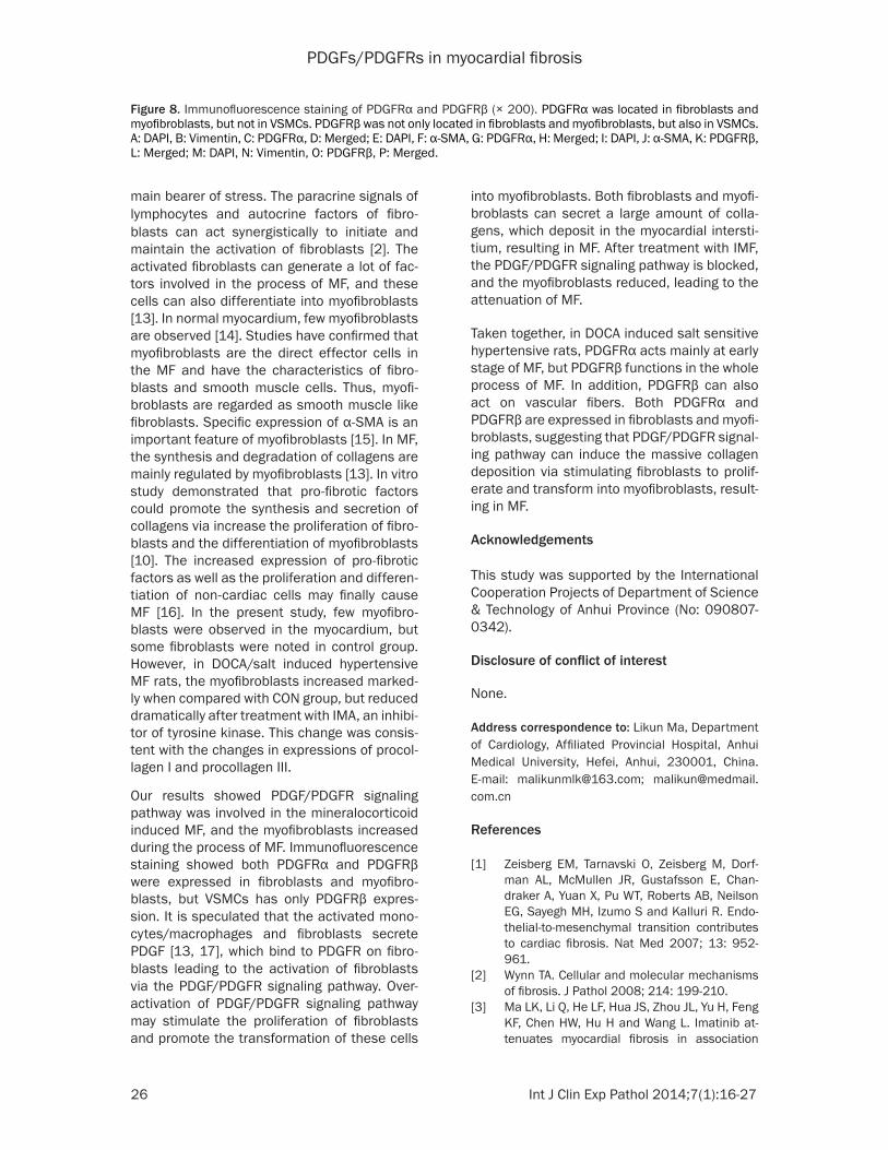
sion was observed in not only fibroblasts and myofibroblasts but also VSMCs (Figure 8).
Discussion
MF is a pathological process of excessive depo-sition of collages due to their abnormal metab-olism and a major cause of heart failure. In mul-tiple cardiovascular diseases at end stage, MF is a major pathological feature of my-ocardium.
PDGFs/PDGFRs in myocardial fibrosis
23 Int J Clin Exp Pathol 2014;7(1):16-27
To date, some theories have been proposed for the mechanisms underlying the occurrence and development of MF, and the increase in mineralocorticoid has been reg-arded as a major cause of MF and become a hot topic in this field [6]. However, the specific pathogene-sis of MF is still unclear. In recent years, studies reveal that PDGF/PDGFR signaling pathway is found to be involved in not only the MF after myocardial infraction and MF secondary to viral myocarditis [5, 7], but the DOCA/salt induced hypertensive MF. In study on DOCA/salt induced hypertensive rats, PDGF/PDGFR signaling path-way was found to promote the deposition of col-lagens and the inhibitor of PDGF/PDGFR signal-ing pathway could attenuate MF [3]. This suggests that PDGF/PDGFR signaling pathway is invo-lved in the occurrence and development of MF. PDGF is a potent factor that can promote
deposition increased dramatically. After treat-ed with IMA, the p-PDGFRβ expression reduced, and the myocardial interstitial collagens decreased accordingly. These suggest that PDGFRα signaling pathway acts mainly at early stage of DOCA/salt induced hypertensive MF, but PDGFRβ functions in the whole process of MF.
In mammalians, myocytes, fibroblasts and vas-cular cells (VSMCs and endothelial cells) are the main cells in the heart. The myocytes and fibroblasts function synergistically to regulate the heart function and account for 90% of cells in the heart [11]. The fibroblast-like cells (fibro-blasts and myofibroblasts) in the myocardial interstitium have been regarded as a major source of ECM [12], and collagen in ECM is a
the mitosis. After binding to PDGFR, PDGF can stimu-late the growth, differentia-tion and migration of inter-stitial cells including fibroblasts and VSMCs [8, 9]. Zymek et al [5] found PDGFRβ signaling pathway in myocardial infarction ani-mal model was a unique mechanism for the regula-tion of myocardial vascula-ture system, and PDGF-A/PDGFRα signaling pathway could promote the collagen deposition, but failed to regulate vascularization. In an in vitro study of Zhao et al, results showed PDG- FR-D/PDGFRβ signaling pa- thway could promote the synthesis and secretion of collagens [10]. In the pres-ent study, the PDGFRα expression was higher than the PDGFRβ expression in rats with DOCA/salt ind- uced hypertensive MF at early stage. However, the PDGFRα expression failed to increase markedly, and the expressions of PDGFRβ and p-PDGFRβ elevated markedly at late stage of MF. In addition, the myocar-dial interstitial collagen
Figure 7. Immunohistochemistry for α-SMA and vimentin and α-SMA positive spindle-shaped cells in different groups on day 28 (× 200). A: Vimentin CON; B: Vimentin DOCA; C: Vimentin DOCA+IMA; D: α-SMA CON; E: α-SMA DOCA; F: α-SMA DOCA+IMA.

PDGFs/PDGFRs in myocardial fibrosis
24 Int J Clin Exp Pathol 2014;7(1):16-27

PDGFs/PDGFRs in myocardial fibrosis
25 Int J Clin Exp Pathol 2014;7(1):16-27
PDGFs/PDGFRs in myocardial fibrosis
26 Int J Clin Exp Pathol 2014;7(1):16-27
main bearer of stress. The paracrine signals of lymphocytes and autocrine factors of fibro-blasts can act synergistically to initiate and maintain the activation of fibroblasts [2]. The activated fibroblasts can generate a lot of fac-tors involved in the process of MF, and these cells can also differentiate into myofibroblasts [13]. In normal myocardium, few myofibroblasts are observed [14]. Studies have confirmed that myofibroblasts are the direct effector cells in the MF and have the characteristics of fibro-blasts and smooth muscle cells. Thus, myofi-broblasts are regarded as smooth muscle like fibroblasts. Specific expression of α-SMA is an important feature of myofibroblasts [15]. In MF, the synthesis and degradation of collagens are mainly regulated by myofibroblasts [13]. In vitro study demonstrated that pro-fibrotic factors could promote the synthesis and secretion of collagens via increase the proliferation of fibro-blasts and the differentiation of myofibroblasts [10]. The increased expression of pro-fibrotic factors as well as the proliferation and differen-tiation of non-cardiac cells may finally cause MF [16]. In the present study, few myofibro-blasts were observed in the myocardium, but some fibroblasts were noted in control group. However, in DOCA/salt induced hypertensive MF rats, the myofibroblasts increased marked-ly when compared with CON group, but reduced dramatically after treatment with IMA, an inhibi-tor of tyrosine kinase. This change was consis-tent with the changes in expressions of procol-lagen I and procollagen III.
Our results showed PDGF/PDGFR signaling pathway was involved in the mineralocorticoid induced MF, and the myofibroblasts increased during the process of MF. Immunofluorescence staining showed both PDGFRα and PDGFRβ were expressed in fibroblasts and myofibro-blasts, but VSMCs has only PDGFRβ expres-sion. It is speculated that the activated mono-cytes/macrophages and fibroblasts secrete PDGF [13, 17], which bind to PDGFR on fibro-blasts leading to the activation of fibroblasts via the PDGF/PDGFR signaling pathway. Over-activation of PDGF/PDGFR signaling pathway may stimulate the proliferation of fibroblasts and promote the transformation of these cells
into myofibroblasts. Both fibroblasts and myofi-broblasts can secret a large amount of colla-gens, which deposit in the myocardial intersti-tium, resulting in MF. After treatment with IMF, the PDGF/PDGFR signaling pathway is blocked, and the myofibroblasts reduced, leading to the attenuation of MF.
Taken together, in DOCA induced salt sensitive hypertensive rats, PDGFRα acts mainly at early stage of MF, but PDGFRβ functions in the whole process of MF. In addition, PDGFRβ can also act on vascular fibers. Both PDGFRα and PDGFRβ are expressed in fibroblasts and myofi-broblasts, suggesting that PDGF/PDGFR signal-ing pathway can induce the massive collagen deposition via stimulating fibroblasts to prolif-erate and transform into myofibroblasts, result-ing in MF.
Acknowledgements
This study was supported by the International Cooperation Projects of Department of Science & Technology of Anhui Province (No: 090807- 0342).
Disclosure of conflict of interest
None.
Address correspondence to: Likun Ma, Department of Cardiology, Affiliated Provincial Hospital, Anhui Medical University, Hefei, Anhui, 230001, China. E-mail: [email protected]; [email protected]
References
[1] Zeisberg EM, Tarnavski O, Zeisberg M, Dorf-man AL, McMullen JR, Gustafsson E, Chan-draker A, Yuan X, Pu WT, Roberts AB, Neilson EG, Sayegh MH, Izumo S and Kalluri R. Endo-thelial-to-mesenchymal transition contributes to cardiac fibrosis. Nat Med 2007; 13: 952-961.
[2] Wynn TA. Cellular and molecular mechanisms of fibrosis. J Pathol 2008; 214: 199-210.
[3] Ma LK, Li Q, He LF, Hua JS, Zhou JL, Yu H, Feng KF, Chen HW, Hu H and Wang L. Imatinib at-tenuates myocardial fibrosis in association
Figure 8. Immunofluorescence staining of PDGFRα and PDGFRβ (× 200). PDGFRα was located in fibroblasts and myofibroblasts, but not in VSMCs. PDGFRβ was not only located in fibroblasts and myofibroblasts, but also in VSMCs. A: DAPI, B: Vimentin, C: PDGFRα, D: Merged; E: DAPI, F: α-SMA, G: PDGFRα, H: Merged; I: DAPI, J: α-SMA, K: PDGFRβ, L: Merged; M: DAPI, N: Vimentin, O: PDGFRβ, P: Merged.

PDGFs/PDGFRs in myocardial fibrosis
27 Int J Clin Exp Pathol 2014;7(1):16-27
with inhibition of the PDGFRalpha activity. Arq Bras Cardiol 2012; 99: 1082-1091.
[4] Yu M, Ishibashi-Ueda H, Ohta-Ogo K, Gabbiani G, Yamagishi M, Hayashi K, Hirota S, Bocha-ton-Piallat ML and Hao H. Transient expression of cellular retinol-binding protein-1 during car-diac repair after myocardial infarction. Pathol Int 2012; 62: 246-253.
[5] Zymek P, Bujak M, Chatila K, Cieslak A, Thak-ker G, Entman ML and Frangogiannis NG. The role of platelet-derived growth factor signaling in healing myocardial infarcts. J Am Coll Cardi-ol 2006; 48: 2315-2323.
[6] Ishimaru K, Ueno H, Kagitani S, Takabayashi D, Takata M and Inoue H. Fasudil attenuates myocardial fibrosis in association with inhibi-tion of monocyte/macrophage infiltration in the heart of DOCA/salt hypertensive rats. J Cardiovasc Pharmacol 2007; 50: 187-194.
[7] Leipner C, Grun K, Muller A, Buchdunger E, Borsi L, Kosmehl H, Berndt A, Janik T, Uecker A, Kiehntopf M and Bohmer FD. Imatinib me-sylate attenuates fibrosis in coxsackievirus b3-induced chronic myocarditis. Cardiovasc Res 2008; 79: 118-126.
[8] Andrae J, Gallini R and Betsholtz C. Role of platelet-derived growth factors in physiology and medicine. Genes Dev 2008; 22: 1276-1312.
[9] Donovan J, Abraham D and Norman J. Platelet-derived growth factor signaling in mesenchy-mal cells. Front Biosci (Landmark Ed) 2013; 18: 106-119.
[10] Zhao T, Zhao W, Chen Y, Li VS, Meng W and Sun Y. Platelet-derived growth factor-D pro-motes fibrogenesis of cardiac fibroblasts. Am J Physiol Heart Circ Physiol 2013; 304: H1719-1726.
[11] Porter KE and Turner NA. Cardiac fibroblasts: at the heart of myocardial remodeling. Phar-macol Ther 2009; 123: 255-278.
[12] Yue L, Xie J and Nattel S. Molecular determi-nants of cardiac fibroblast electrical function and therapeutic implications for atrial fibrilla-tion. Cardiovasc Res 2011; 89: 744-753.
[13] Camelliti P, Borg TK and Kohl P. Structural and functional characterisation of cardiac fibro-blasts. Cardiovasc Res 2005; 65: 40-51.
[14] Zhao W, Zhao T, Huang V, Chen Y, Ahokas RA and Sun Y. Platelet-derived growth factor in-volvement in myocardial remodeling following infarction. J Mol Cell Cardiol 2011; 51: 830-838.
[15] Sun Y and Weber KT. Infarct scar: a dynamic tissue. Cardiovasc Res 2000; 46: 250-256.
[16] Teekakirikul P, Eminaga S, Toka O, Alcalai R, Wang L, Wakimoto H, Nayor M, Konno T, Gor-ham JM, Wolf CM, Kim JB, Schmitt JP, Molken-tin JD, Norris RA, Tager AM, Hoffman SR, Mark-wald RR, Seidman CE and Seidman JG. Cardiac fibrosis in mice with hypertrophic cardiomyopa-thy is mediated by non-myocyte proliferation and requires Tgf-beta. J Clin Invest 2010; 120: 3520-3529.
[17] Raines EW. PDGF and cardiovascular disease. Cytokine Growth Factor Rev 2004; 15: 237-254.