original article osteogenesis of bone marrow mesenchymal ... · pdf fileon...
TRANSCRIPT

Int J Clin Exp Pathol 2016;9(10):9775-9785www.ijcep.com /ISSN:1936-2625/IJCEP0034380
Original Article Osteogenesis of bone marrow mesenchymal stem cells on nano-hydroxyapatite/bacterial cellulose coposite scaffolds in rats
Tong-Qu Song1, Bao-Jian Ge2, Hung-Liang Chen2, Xiao-Wei Yang3, Feng Yuan2
1Department of Spinal Surgery, Xuzhou Central Hospital, Xuzhou 221000, Jiangsu, China; 2Department of Spinal Surgery, The Affiliated Hospital of Xuzhou Medical University, Xuzhou 221000, Jiangsu, China; 3Department of Spinal Surgery, The Third People’s Hospital of Xuzhou, Xuzhou 221000, Jiangsu, China
Received June 24, 2016; Accepted August 30, 2016; Epub October 1, 2016; Published October 15, 2016
Abstract: The composite scaffolds are extensively employed to repair and reconstruct damaged bone tissue func-tion. The present study was designed to explore the nano-hydroxyapatite/bacterial cellulose (nHA/BC) on the pro-liferation and osteogenic differentiation of rat bone marrow mesenchymal stem cells (BMSCs). The osteoblast in-duced BMSCs were cultured on nHA/BC scaffolds. The proliferation of BMSCs was determined by the cell counting kit-8 (CCK-8) assay. The osteogenic response of BMSCs to nHA/BC scaffolds was examined by alizarin red staining. The mice were divided into three groups: BMSCs + βnHA/BC, osteoblastic BMSCs + nHA/BC and nHA/BC without cells. The scaffolds of nHA/BC exhibited a good compatibility to BMSCs, and significantly promoted the proliferation of BMSCs, and obviously improved calcium deposition. The osteoblastic BMSCs on nHA/BC scaffolds stimulated the phosphorylation of p38. In addition, the recombination of osteoblastic BMSCs with nHA/BC composite significantly boosted the bone formation in vivo. The scaffolds of nHA/BC may enhance the proliferation and osteoblastic dif-ferentiation of BMSCs via modulation of p38 signal transduction pathway. The recombination of nHA/BC scaffolds with BMSCs exerts good ability of ectopic osteoblast, and they may be served as a kind of bone substitute for bone regeneration.
Keywords: Osteogenesis, bone mesenchymal stem cells, nHA/BC
Introduction
The goal of bone tissue engineering is to re- place defect area or damaged tissue by implant-ing bone substitutes [1, 2]. Bone tissue engi-neering emerges as a promising way for bone regeneration to recover the body’s normal phy- siological structure and function [3]. Seed cells, growth factors and scaffolds are essential components during produces for bone tissue engineering [4]. Numerous investigations have confirmed the feasibility of composite scaffolds for bone tissue engineering [5]. Tissue engi-neered scaffolds are widely used to repair and reconstruct damaged bone tissue function [6]. The weight ratio of compound, pore size, poros-ity and mechanical properties are crucial fac-tors for influencing clinical application of the composite scaffolds [7].
Bone marrow-derived mesenchymal stem cells (BMSCs) are recognized as a valuable cell type
for bone regeneration, and are widely used in cytotherapy for musculoskeletal diseases [8]. BMSCs are usually considered as seed cells in cytotherapy due to their differentiation ability into osteogenic and chondrogenic lineages [8, 9]. Furthermore, BMSCs are extensively used for tissue engineering for their low immunoge-nicity, abundant material source and multi-dire- ctional differentiation potency [10]. To date, the approaches based on the seeding of BMSCs into biodegradable polymeric scaffolds have been greatly developed by many researchers [11]. Previous study showed that the composite scaffolds of poly (ε-caprolactone), thermoplas-tic zein and hydroxyapatite (PCL/TZ-HA) signifi-cantly promoted the proliferation and osteogen-ic differentiation of rabbit BMCSs in vitro [12]. Another study proved that BMSCs on hydroxy-apatite scaffolds exhibited a faster and better healing of bone segmental defects [13]. Nano-hydroxyapatite (nHA) is demonstrated to a ho-
Osteogenesis of bone marrow mesenchymal stem cells
9776 Int J Clin Exp Pathol 2016;9(10):9775-9785
peful biomaterial for bone repair due to its bio-activity, biocompatibility and osteoconductivity [14].
Bacterial cellulose (BC) is also used for bone tissue regeneration in tissue engineering, due to its high biocompatibility, high crystallinity, hi- gh tensile strength and elastic modulus, and good biodegradability [15]. However, it is still unclear whether nHA/BC composite scaffolds could have excellent bioactivity of attachment, proliferation and osteoblastic differentiation for BMSCs. The present study was designed to in- vestigate the role of nHA/BC composite scaf-folds on the proliferation, osteogenic differen-tiation and osteogenesis of rat BMSCs in vitro and in vivo.
Materials and methods
Animals
The experiments were conformed to the recom-mendations in the Guide for the Care and Use of Laboratory Animals of the National Institutes of Health. The procedures were reviewed and approved by the Committee on the Ethics of Animal Experiments of Xuzhou Medical Univer- sity. The male rats were purchased from Labor- atory Animal Center, Xuzhou Medical University and caged in temperature-controlled and hu- midity-controlled room with a 12 h light and 12 h dark cycle, and were provided standard chow and tap water ad libitum.
Preparation of scaffolds
The nHA/BC scaffolds were synthesized by a biomimetic technique [16-18] and kindly pro-vided by Material Institute of Tianjing University. Before BMSCs seeding, the nHA/BC scaffolds were pre-wetted in DMEM-F12 medium for 4 h and dried in an incubator overnight under ster-ile conditions.
Isolation and expansion of BMSCs
BMSCs were isolated and cultured as previous-ly described [19]. In brief, the Spraque-Dawley (SD) rats aged 3 months old were sacrificed by an overdose of pentobarbital sodium (150 mg/kg, iv). The femurs, muscle and connective tis-sue of each rat were aseptically excised, and the bone marrow was extracted by flushing the
medullar cavities under sterile conditions. The collected marrow was flushed with essential culture media (DMEM supplemented with 10% FBS, 100 U/ml penicillin, and 100 μg/ml strep-tomycin) by a 27-gauge needle attached to a 10 ml syringe. The cell suspension was centrifuged at 1000 rpm for 10 min, and then the whole cells were re-suspended in fresh primary media and added into Percoll separating medium (GIBCOBRL company), and were seeded into a 25 cm2 flask for incubation in a 5% CO2 incuba-tor at 37°C with 95% humidity. The non-adher-ent cells were washed away after 48 h by re- placing the medium with fresh complete medi-um. Then the medium was exchanged every three days and the cells were passaged when the cells grew on reaching a confluence of 80-90% in the flasks.
Flow cytometry
BMSCs were characterized and confirmed by analysis of cell surface markers (CD29, CD34, CD44 and CD90) with flow cytometry as previ-ous report [20]. BMSCs at passage 3 were incu-bated with 0.25% trypsin-EDTA (ethylene diami-ne tetraacetic acid) for 2 min at 37°C and sub-jected to centrifugation at 1000 r for 5 min. The obtained cells were washed with phosphate-buffered saline (PBS), and the cells were then subsequently cultured with rat monoclonal anti-CD29-FITC (BD Biosciences, San Jose, CA, USA), rat polyclonal anti-CD34-FITC (Bioss Biosynthesis Biotechnology Co., Ltd., Beijing, China), rat polyclonal anti-CD44-PE (R&D Systems, Inc., Minneapolis, MN, USA), and rat polyclonal anti- CD90-PE (Bioss Biosynthesis Biotechnology Co., Ltd., Beijing, China) for 30 min incubation in the dark at 4°C. The cells were washed by PBS and analyzed using a flow cytometer (Gallios; Beckman Coulter, Brea, CA, USA).
Morphology and proliferation of BMSCs on nHA/BC scaffolds
The confluent BMSCs at passage 3 were digest-ed with 0.25% trypsin-EDTA, and seeded onto nHA/BC scaffolds-placed 24 wells with 105 cells per ml under a 5% CO2 incubator at 37°C with 95% humidity. The cells on nHA/BC cop- osite scaffolds were changed with fresh DMEM medium supplemented with 10% FBS after 24 h. Inverted phase contrast microscope was

Osteogenesis of bone marrow mesenchymal stem cells
9777 Int J Clin Exp Pathol 2016;9(10):9775-9785
used to investigate the cellular morphology and attachment of BMSCs on nHA/BC coposite scaffolds [20]. CCK-8 assay was applied to detect the proliferation of the BMSCs on nHA/BC scaffolds [21].
Osteogenic induction of BMSCs
The BMSCs at passage 3 were trypsinized and seeded at a density of 105/ml into nHA/BC scaffolds-placed 6 well plates or non-scaffolds-placed 6 well plates and then incubated over-night at 37°C and 5% CO2 to permit cell attach-ment. The medium was replaced with osteo-genic induction medium after 24 h. The osteo-genic induction medium contained 10-7 mol/L dexamethasone (Sigma-Aldrich), 10 mmol/L β-glycerophosphate (China Pharmaceutical Sh- anghai Chemical Reagent Co., Ltd.), 50 μg/ml ascorbic acid (Sigma-Aldrich), 1% penicillin/streptomycin and 10% FBS in DMEM [22]. BMSCs were planted on nHA/BC support and cultured by osteogenic induction medium (Gr- oup A); BMSCs were cultured by osteogenic induction medium (Group B); BMSCs were planted on nHA/BC support and cultured by DMEM medium (Group C); BMSCs were cul-tured by DMEM medium (Group D). The calcium nodules deposited in the extracellular matrix of BMSCs in four groups were detected with aliza-rin red staining after the three-week induction, and the calcium nodule staining was captured by Canon DSLR camera.
Western blot
After induction in osteogenic medium or DMEM medium for one week in four groups, the BM- SCs were harvested and were lysed using 100 μl of radioimmunoprecipitation assay (RIPA) ly- sis buffer (Beyotime Institute of Biotechnology, Shanghai, China). The samples were then cen-trifuged and total protein concentration was measured using the Bicinchoninic Acid kit. Equal quantities of total protein in each group were subjected to sodium dodecyl sulfate poly-acrylamide gel electrophoresis and transferred onto polyvinylidene fluoride membrane. The membrane was blocked with 5% non-fat milk for 60 min at room temperature, following by overnight incubation with rabbit anti-p38 (Cell Signaling Technology, Beverly, MA, USA), rabbit anti-phosphorylated p38 (Cell Signaling Tech-
nology, Beverly, MA, USA) and rabbit anti-GAP-DH (Santa Cruz, CA, USA) at 4°C. The mem-brane was incubated with a secondary antibody conjugated to horseradish peroxidase, and visualized by the enhanced chemiluminescent reagent (Merck Millipore, Billerica, Massachu- setts, USA). GAPDH was served as an internal control [23].
Ectopic bone formation by implantation and degradation test in vivo
The approval of the surgery of animals was the same with those stated in the isolation section of BMSCs. All rats were anaesthetized with sodium pentobarbital (50 mg/kg, i.p.) and placed in a lateral position. The adequacy of anesthesia was determined by the absence of corneal reflexes and paw withdrawal response to a noxious pinch and surgical manipulation [24]. The rats were divided into three groups: BMSCs + β-nHA/BC, osteoblastic BMSCs + nHA/BC and nHA/BC without cells. After prepa-ration of the skin, a long incision was made along the bilateral back axis, and the muscle was bluntly dissected, and intramuscular po- uches including one muscle pouch on the left and two muscle pouch on the right were creat-ed in this model. The BMSCs/scaffold compos-ites or scaffolds were implanted into the spati-um intermusculare. The left muscle pouch was implanted with osteoblastic BMSCs + nHA/BC scaffolds, the nHA/BC without cells was im- planted into upper right side muscle pouch, and BMSCs + nHA/BC were implanted into upper lower side muscle pouch. All the disk scaffolds seeded with 8×10⁵ cells as previously described. The incisions were sutured and the rats were fed at the condition of cleaned cages with water and food ad libitum. The rats were anesthetized at 12 weeks after implantation via intraperitoneal injection with a 10% chloral hydrate solution (0.4 ml/100 g), and were then scanned along the back axial plane by comput-erized tomography (CT, Light Speed 64 slice spiral CT, General Electric, USA, 2.5 mm slice thickness). The position and bone formation in the implants were observed in the CT images, and the implanting materials were taken out and observed by visual [25]. In addition, the rats were sacrificed and implants with the sur-rounding tissues were harvested. Paraffin 5 μm-thick sections were prepared and stained
Osteogenesis of bone marrow mesenchymal stem cells
9778 Int J Clin Exp Pathol 2016;9(10):9775-9785
with hematoxylin and eosin (HE) to observe the ectopic bone formation in rat [26].
Statistics analysis
All data were expressed as mean ± SE. All ex- periments were repeated at least three times. The statistical analysis was conducted by SPSS 19.0 software (SPSS Inc., Chicago, IL, USA). Comparisons between two groups were made by Student’s t test. Differences within groups in multiple assays were tested by ANOVA and Dunnett’s t-test. A value of P<0.05 was consid-ered statistically significant.
Results
Identification of rat BMSCs
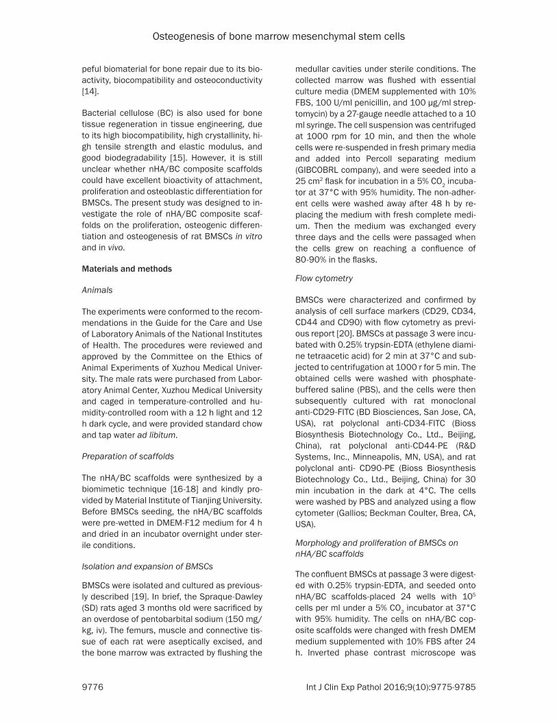
The primary BMSCs of rats presented attach-ment to the wall and characterized as circle or oval shape with strong refraction after 36 h cul-ture. The primary BMSCs showed to be short fusiform, triangular or polygonal shape after three days’ culture (Figure 1A). The BMSCs
were purified and passaged upon 90% conflu-ence at 14 d, and the BMSCs were uniform long fusiform at the first passage (Figure 1B). The BMSCs at the second passage were grown to totally confluence about 5~7 d culture with uni-form morphology (Figure 1C). The BMSCs at the third passage were in swirling and paralleled growth and presented as long fusiform (Figure 1D).
Detection of surface antigen of rat BMSCs at the third passage with flow cytometry
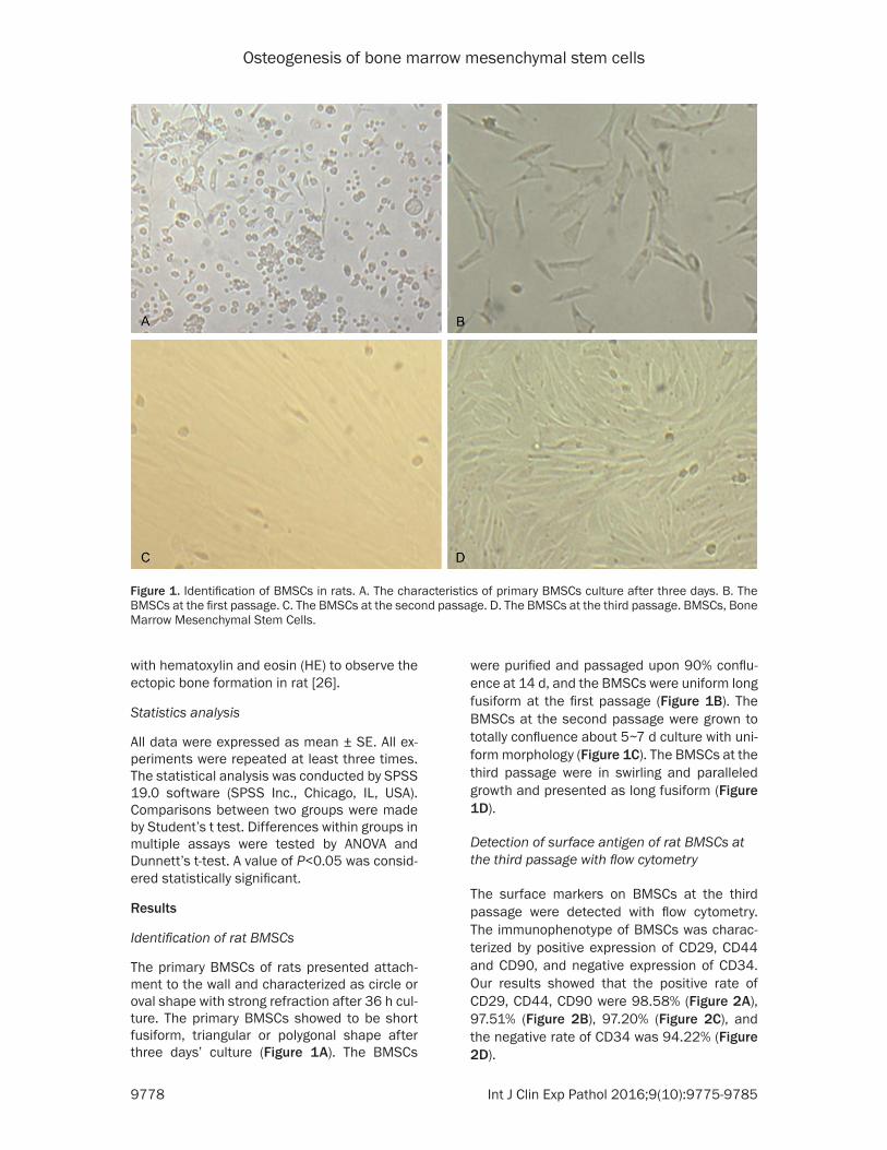
The surface markers on BMSCs at the third passage were detected with flow cytometry. The immunophenotype of BMSCs was charac-terized by positive expression of CD29, CD44 and CD90, and negative expression of CD34. Our results showed that the positive rate of CD29, CD44, CD90 were 98.58% (Figure 2A), 97.51% (Figure 2B), 97.20% (Figure 2C), and the negative rate of CD34 was 94.22% (Figure 2D).
Figure 1. Identification of BMSCs in rats. A. The characteristics of primary BMSCs culture after three days. B. The BMSCs at the first passage. C. The BMSCs at the second passage. D. The BMSCs at the third passage. BMSCs, Bone Marrow Mesenchymal Stem Cells.
Osteogenesis of bone marrow mesenchymal stem cells
9779 Int J Clin Exp Pathol 2016;9(10):9775-9785
Morphological characteristics of rat BMSCs on nHA/BC coposite scaffolds
Inverted phase contrast microscope was used to observe the characteristics of rat BMSCs on nHA/BC coposite scaffolds. BMSCs were ad- hered to nHA/BC coposite scaffolds after 24 h culture and presented as circle and short fusi-form (Figure 3A). At days 3, most of BMSCs were long fusiform and the gap of cells became smaller (Figure 3B). BMSCs were fused and fully covered nHA/BC scaffolds, and connected
ference in all groups. The p38 phosphorylation levels in group A and group B were higher than those in both group C and group D. Moreover, the level of phosphorylated p38 was higher in group A than group B, while the p38 phosphory-lation level group C was higher than group D (Figure 5).
Osteogenic differentiation of BMSCs in differ-ent groups
The osteogenic differentiation of BMSCs was evaluated by Alizarin Red staining. Positive
Figure 2. Detection of surface antigens of BMSCs in rats at the third pas-sage with flow cytometry. A. CD29 in BMSCs. B. CD44 in BMSCs. C. CD90 in BMSCs. D. CD34 in BMSCs. BMSCs, Bone Marrow Mesenchymal Stem Cells.
with the cells around nHA/BC scaffolds at 7 days (Figure 3C). BMSCs were fused and grown to one layer on nHA/BC scaffolds at 9 days (Figure 3D).
Proliferation of rat BMSCs on nHA/BC coposite scaffolds
The proliferation of rat BMSCs on nHA/BC coposite scaffolds was assessed at time points of 1, 3, 5, 7, and 9 d. BMSCs in two groups exhibited a loga-rithmic multiplication period from 3 to 7 days and displayed a growth inhibition at day 9. The BMSCs on nHA/BC co- posite scaffolds proliferated faster than the control group after the 3th day (Figure 4). These results indicated the nHA/BC scaffolds had a good cell biocompatibility, and may be able to promote prolifera-tion of BMSCs.
Phosphorylated p38 level in BMSCs of different groups
BMSCs were planted on nHA/BC scaffolds and cultured by osteogenic induction medium (Group A); BMSCs were cul-tured by osteogenic induction medium (Group B); BMSCs were planted on nHA/BC scaf-folds and cultured by DMEM- F12 medium (Group C); BMSCs were cultured by DMEM-F12 medium (Group D). Our results showed that the expression of total p38 protein had no significant dif-

Osteogenesis of bone marrow mesenchymal stem cells
9780 Int J Clin Exp Pathol 2016;9(10):9775-9785
Alizarin Red staining through the matrix at the surface layer of group A was greater than that
of the group B. The Alizarin Red staining was negative in group C and group D (Figure 6).
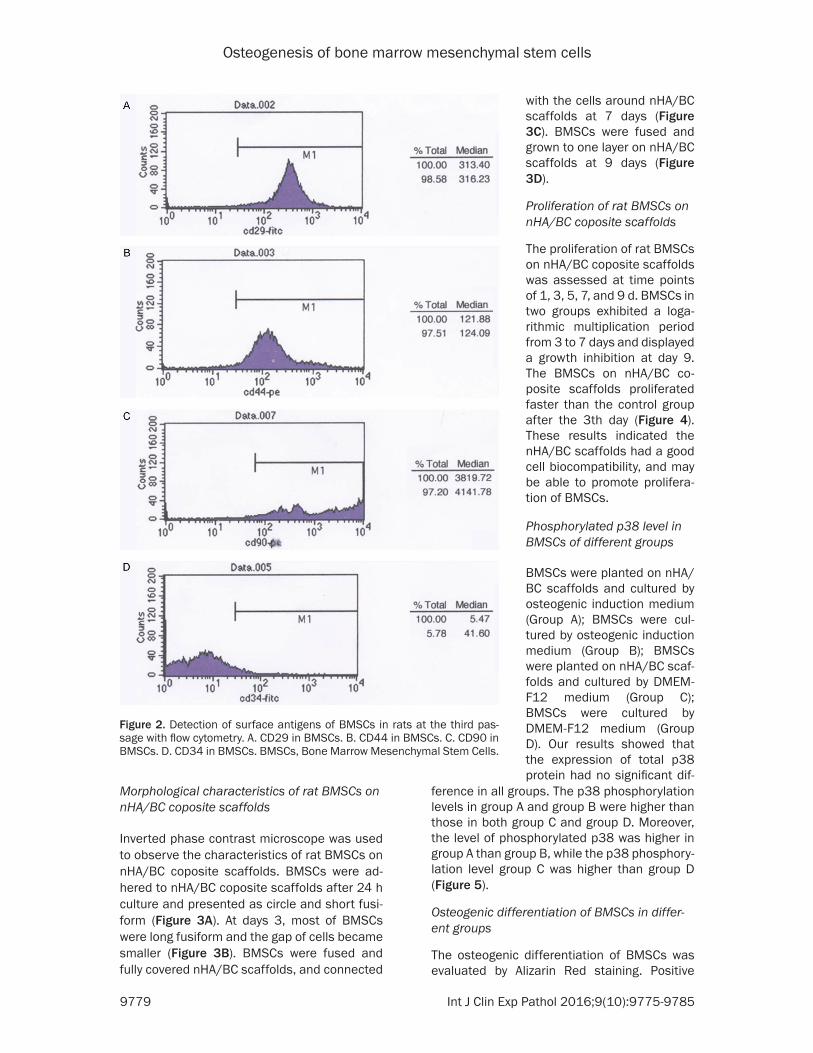
Osteogenesis in rat muscle pouch observed by CT scanning
The oval bone development of 1.0 cm×1.0 cm×0.3 cm appeared on the left muscle pouch (Figure 7A-C), while the non-induced group and control group had no bone development.
Bone formation of tissues in different groups
There was no new bone formation on the implanted materials in control group and non-induced group, the nHA/BC scaffolds in both control group and non-induced group was wrapped by thin fiber, the surface of tissues was darker than original materials (Figure 8A, 8B). The recombination nHA/BC scaffolds with BMSCs by osteogenic induction was entirely wrapped by white bone tissue with unclear boundary (Figure 8C).
Figure 3. Morphological characteristics of BMSCs in rats on nHA/BC coposite scaffold. A. BMSCs on nHA/BC co-posite scaffold after 24 h. B. BMSCs on nHA/BC coposite scaffold after 3 days. C. BMSCs on nHA/BC coposite scaf-fold after 7 days. D. BMSCs on nHA/BC coposite scaffold after 9 days. The arrow indicated the boundary of nHA/BC stent. BMSCs, Bone Marrow Mesenchymal Stem Cells.
Figure 4. The proliferation of rat BMSCs on nHA/BC coposite scaffold evaluated with CCK-8 kits at the indicated time. Values are mean ± SD. *P<0.05 vs. Control. n=6 for each group. The data in absorbance between groups at the same indicated time were compared with Student’s t test.

Osteogenesis of bone marrow mesenchymal stem cells
9781 Int J Clin Exp Pathol 2016;9(10):9775-9785
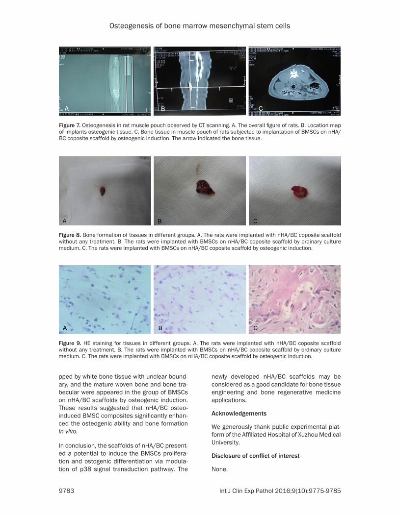
H&E staining for tissues in different groups
It is evidenced that more fibrous tissue, but no bone or cartilage tissue in the pore spaces of implanted nHA/BC scaffolds was present in the control group and non-induced group (Figure 9A, 9B). The mature woven bone and bone tra-becular were appeared in the group of BMSCs on nHA/BC scaffolds by osteogenic induction (Figure 9C).
Discussion
The reconstructive surgery requiring bone gra- fts is increasing in worldwide hospitals [27]. Autologous or allogenic bone graft is clinically used for bone repair in bone fractures and trau-ma-related injuries. However, it is recently revi- ewed the high risk of disease transmission and rejection in allografts and xenografts [28]. Bo- ne tissue engineering is recognized as an alter-native and developing option that has been in-troduced to improve the osteogenesis of the bone fractures and defects [29]. In the present study, we prepared nHA/BC scaffolds utilizing the biomimetic technique. Our results showed that (1) BMSCs could adhere and spread on nHA/BC scaffolds; (2) BMSCs proliferated fast-er on the nHA/BC scaffolds than pure BMSCs; (3) osteogenic differentiation of BMSCs on nHA/BC scaffolds was higher than those cul-tured upon the pure nHA/BC scaffolds in the presence or absence of osteogenic supplem- ents; (4) the new bone formation, mature woven bone and bone trabecular in the pores of the
and osteoconductive properties for bone regen-eration [14]. The appropriate bioactivity materi-als for bone tissue engineering include a vari-ety of factors: (1) the scaffolds can be able to regulate the adhesion, growth and differentia-tion of stem cells; (2) stem cells on scaffolds can undergo differentiation to osteoblasts; (3) the bioresorbable scaffolds can stimulate the cellular attachment, proliferation and differen-tiation [15]. Moreover, the combined use of scaffolds and BMSCs may open new insights of bone regenerative medicine [6]. The scaffolds of nHA have a good biocompatibility of BMSCs and display a good potential to support the pro-liferation and ostogenic differentiation of BMSCs [27]. BC has attracted extensive atten-tion all over the world due to its potential as nanofiber scaffolds for bone tissue engineering [20]. In the present study, our results showed that the primary BMSCs were expanded until passage 2, and BMSCs at the second passage were grown to totally confluence with uniform morphology under inverted phase contrast microscope, which are typical morphologic fea-tures of BMSCs. In addition, the BMSCs were CD29, CD44, CD90 positive, but CD34 nega-tive, which further confirmed the characters of BMSCs. We also found that BMSCs were adhered to nHA/BC coposite scaffolds and fully grown to one layer on nHA/BC scaffolds at 9 days. The BMSCs on nHA/BC coposite scaf-folds proliferated faster than the control group after the 3th day. These results indicated the nHA/BC scaffolds had a good cell biocompati-bility of BMSCs, and may be able to promote proliferation of BMSCs.
Figure 5. The phosphorylated p38 level in BMSCs of different groups. BM-SCs were planted on nHA/BC support and cultured by osteogenic induction medium (A); BMSCs were cultured by osteogenic induction medium (B); BM-SCs were planted on nHA/BC support and cultured by DMEM-F12 medium (C); BMSCs were cultured by DMEM-F12 medium (D). Values are mean ± SD. *P<0.05 vs. Group A; †P b 0.05 vs Group B; #P b 0.05 vs Group C. n=4 for each group. One-way ANOVA was used to compare the differences in phos-phorylated p38 level in different groups, and the comparisons between two groups were analyzed by One-way ANOVA post hoc Bonferroni test.
scaffolds were appeared in BMSCs on nHA/BC scaffolds by osteogenic induction; (5) the nHA/BC scaffolds gradu-ally degraded with bone for-mation. These results indicat-ed that the attachment, prolif-eration, and differentiation of BMSCs can be modulated by the nHA/BC scaffolds. We developed a combined nHA/BC scaffolds that exerted a biocompatibility of BMSCs, which may be clinically useful for bone tissue engineering.
Bone tissue engineering is used to develop tissue-engi-neered constructs that hol- ds osteogenic, osteoinductive

Osteogenesis of bone marrow mesenchymal stem cells
9782 Int J Clin Exp Pathol 2016;9(10):9775-9785
Previous studies disclosed that combined scaf-folds-BMSCs may have a better potential in in- ducing osteogenic differentiation in vitro and in vivo settings [30]. BMSCs with osteogenic dif-ferentiation have been described to be a poten-tial candidate for bone regeneration [31].
It is reported that nHA-chitosan nanocompos-ites can accelerate the adhesion, proliferation of BMSCs via activation of the integrin-BMP/Smad signaling pathway [32]. The p38-MAPK is critical for osteogenesis and differentiation of BMSCs [33]. Recent study indicates that aker-manite bioceramics promote osteogenesis for osteoporotic bone regeneration by activation of p38 signaling pathway [34]. In the present study, we found that BMSCs on nHA/BC scaf-folds cultured by osteogenic induction medium significantly promote deposition of calcified ma- trix on the surface of the culture dish evidenced by alizarin red staining, and stimulated the pho-
sphorylation of p38. These results hinted that nHA/BC scaffolds can promote osteogenesis and differen tiation of BMSCs associated with p38 activation in vitro.
It is revealed that more de novo bone and col-lagen formation in the pores of the scaffolds gradually increased from 2 weeks post-implan-tation in group of nHA-chitosan osteo-induced BMSC composites [35]. The scaffolds of calci-um phosphate, bone morphogenetic proteins and mesenchymal stem cells can more effec-tively repair bone defects than an autograft in vivo [36]. It is reported that the BMP-VEGF-PLGA-CPC scaffolds have osteogenic and an- giogenic activity by radiographic and histologi-cal analysis [37]. In present study, we showed that the oval bone development of 1.0 cm×1.0 cm×0.3 cm appeared on the left muscle pouch, the recombination nHA/BC scaffold with BM- SCs by osteogenic induction was entirely wra-
Figure 6. Osteogenic differentiation of BMSCs were observed with alizarin red staining in different groups. BMSCs were planted on nHA/BC support and cultured by osteogenic induction medium (A); BMSCs were cultured by os-teogenic induction medium (B); BMSCs were planted on nHA/BC support and cultured by DMEM-F12 medium (C); BMSCs were cultured by DMEM-F12 medium (D).
Osteogenesis of bone marrow mesenchymal stem cells
9783 Int J Clin Exp Pathol 2016;9(10):9775-9785
pped by white bone tissue with unclear bound-ary, and the mature woven bone and bone tra-becular were appeared in the group of BMSCs on nHA/BC scaffolds by osteogenic induction. These results suggested that nHA/BC osteo-induced BMSC composites significantly enhan- ced the osteogenic ability and bone formation in vivo.
In conclusion, the scaffolds of nHA/BC present-ed a potential to induce the BMSCs prolifera-tion and ostogenic differentiation via modula-tion of p38 signal transduction pathway. The
newly developed nHA/BC scaffolds may be considered as a good candidate for bone tissue engineering and bone regenerative medicine applications.
Acknowledgements
We generously thank public experimental plat-form of the Affiliated Hospital of Xuzhou Medical University.
Disclosure of conflict of interest
None.
Figure 9. HE staining for tissues in different groups. A. The rats were implanted with nHA/BC coposite scaffold without any treatment. B. The rats were implanted with BMSCs on nHA/BC coposite scaffold by ordinary culture medium. C. The rats were implanted with BMSCs on nHA/BC coposite scaffold by osteogenic induction.
Figure 7. Osteogenesis in rat muscle pouch observed by CT scanning. A. The overall figure of rats. B. Location map of Implants osteogenic tissue. C. Bone tissue in muscle pouch of rats subjected to implantation of BMSCs on nHA/BC coposite scaffold by osteogenic induction. The arrow indicated the bone tissue.
Figure 8. Bone formation of tissues in different groups. A. The rats were implanted with nHA/BC coposite scaffold without any treatment. B. The rats were implanted with BMSCs on nHA/BC coposite scaffold by ordinary culture medium. C. The rats were implanted with BMSCs on nHA/BC coposite scaffold by osteogenic induction.
Osteogenesis of bone marrow mesenchymal stem cells
9784 Int J Clin Exp Pathol 2016;9(10):9775-9785
Address correspondence to: Feng Yuan, Department of Spinal Surgery, The Affiliated Hospital of Xuzhou Medical University, 99 Huaihai Road West, Xuzhou 221000, Jiangsu, China. Tel: +86-0516-85609999; Fax: +86-0516-85609999; E-mail: yuanfengxuzhou @sina.com
References
[1] Meyer U, Neunzehn J and Wiesmann HP. Com- puter-aided approach for customized cell-bas- ed defect reconstruction. Methods Mol Biol 2012; 868: 27-43.
[2] Ryu TK, Oh MJ, Moon SK, Paik DH, Kim SE, Park JH and Choi SW. Uniform tricalcium phos-phate beads with an open porous structure for tissue engineering. Colloids Surf B Biointer- faces 2013; 112: 368-373.
[3] Oe K, Miwa M, Nagamune K, Sakai Y, Lee SY, Niikura T, Iwakura T, Hasegawa T, Shibanuma N, Hata Y, Kuroda R and Kurosaka M. Non-destructive evaluation of cell numbers in bone marrow stromal cell/beta-tricalcium phosph- ate composites using ultrasound. Tissue Eng Part C Methods 2010; 16: 347-353.
[4] Meyer U, Buchter A, Hohoff A, Stoffels E, Szuwart T, Runte C, Dirksen D and Wiesmann HP. Image-based extracorporeal tissue engi-neering of individualized bone constructs. Int J Oral Maxillofac Implants 2005; 20: 882-890.
[5] Li Q, Wang T, Zhang GF, Yu X, Zhang J, Zhou G and Tang ZH. A Comparative Evaluation of the Mechanical Properties of Two Calcium Phos- phate/Collagen Composite Materials and Their Osteogenic Effects on Adipose-Derived Stem Cells. Stem Cells Int 2016; 2016: 6409546.
[6] Elango J, Zhang J, Bao B, Palaniyandi K, Wang S, Wenhui W and Robinson JS. Rheological, biocompatibility and osteogenesis assessme- nt of fish collagen scaffold for bone tissue en-gineering. Int J Biol Macromol 2016; 91: 51-9.
[7] Amjadian S, Seyedjafari E, Zeynali B and Sha- bani I. The synergistic effect of nano-hydroxy-apatite and dexamethasone in the fibrous de-livery system of gelatin and poly(l-lactide) on the osteogenesis of mesenchymal stem cells. Int J Pharm 2016; 507: 1-11.
[8] Zheng K, Chen Y, Huang W, Lin Y, Kaplan DL and Fan Y. Chemically Functionalized Silk for Human Bone Marrow-Derived Mesenchymal Stem Cells Proliferation and Differentiation. ACS Appl Mater Interfaces 2016; 8: 14406-13.
[9] Yousefi AM, James PF, Akbarzadeh R, Subra- manian A, Flavin C and Oudadesse H. Prospect of Stem Cells in Bone Tissue Engineering: A Review. Stem Cells Int 2016; 2016: 6180487.
[10] Duarte Campos DF, Blaeser A, Buellesbach K, Sen KS, Xun W, Tillmann W and Fischer H. Bioprinting Organotypic Hydrogels with Impr-
oved Mesenchymal Stem Cell Remodeling and Mineralization Properties for Bone Tissue Engineering. Adv Healthc Mater 2016; 5: 1336-45.
[11] Wang H, Zhou Y, Chu TW, Li CQ, Wang J, Zhang ZF and Huang B. Distinguishing characteristics of stem cells derived from different anatomical regions of human degenerated intervertebral discs. Eur Spine J 2016; 25: 2691-704.
[12] Salerno A, Oliviero M, Di Maio E, Netti PA, Rofani C, Colosimo A, Guida V, Dallapiccola B, Palma P, Procaccini E, Berardi AC, Velardi F, Teti A and Iannace S. Design of novel three-phase PCL/TZ-HA biomaterials for use in bone regeneration applications. J Mater Sci Mater Med 2010; 21: 2569-2581.
[13] Udehiya RK, Amarpal, Aithal HP, Kinjavdekar P, Pawde AM, Singh R and Taru Sharma G. Com- parison of autogenic and allogenic bone mar-row derived mesenchymal stem cells for repair of segmental bone defects in rabbits. Res Vet Sci 2013; 94: 743-752.
[14] Zong C, Qian X, Tang Z, Hu Q, Chen J, Gao C, Tang R, Tong X and Wang J. Biocompatibility and bone-repairing effects: comparison betw- een porous poly-lactic-co-glycolic acid and na-no-hydroxyapatite/poly(lactic acid) scaffolds. J Biomed Nanotechnol 2014; 10: 1091-1104.
[15] Tamama K, Sen CK and Wells A. Differentiation of bone marrow mesenchymal stem cells into the smooth muscle lineage by blocking ERK/MAPK signaling pathway. Stem Cells Dev 2008; 17: 897-908.
[16] Lukasheva NV and Tolmachev DA. Cellulose Nanofibrils and Mechanism of their Minerali- zation in Biomimetic Synthesis of Hydroxyapa- tite/Native Bacterial Cellulose Nanocomposit- es: Molecular Dynamics Simulations. Langmuir 2016; 32: 125-134.
[17] Fang B, Wan YZ, Tang TT, Gao C and Dai KR. Proliferation and osteoblastic differentiation of human bone marrow stromal cells on hydroxy-apatite/bacterial cellulose nanocomposite sc- affolds. Tissue Eng Part A 2009; 15: 1091-1098.
[18] Grande CJ, Torres FG, Gomez CM and Bano MC. Nanocomposites of bacterial cellulose/hy- droxyapatite for biomedical applications. Acta Biomater 2009; 5: 1605-1615.
[19] Aminizadeh N, Tiraihi T, Mesbah-Namin SA and Taheri T. Stimulation of cell proliferation by glu-tathione monoethyl ester in aged bone marrow stromal cells is associated with the assistance of TERT gene expression and telomerase activ-ity. In Vitro Cell Dev Biol Anim 2016; 52: 772-81.
[20] Yue W, Yan F, Zhang YL, Liu SL, Hou SP, Mao GC, Liu N and Ji Y. Differentiation of Rat Bone
Osteogenesis of bone marrow mesenchymal stem cells
9785 Int J Clin Exp Pathol 2016;9(10):9775-9785
Marrow Mesenchymal Stem Cells Into Neuron-Like Cells In Vitro and Co-Cultured with Biological Scaffold as Transplantation Carrier. Med Sci Monit 2016; 22: 1766-1772.
[21] Wang C, Shi D, Song X, Chen Y, Wang L and Zhang X. Calpain inhibitor attenuates ER str- ess-induced apoptosis in injured spinal cord after bone mesenchymal stem cells transplan-tation. Neurochem Int 2016; 97: 15-25.
[22] Landim de Barros T, Brito VG, do Amaral CC, Chaves-Neto AH, Campanelli AP and Oliveira SH. Osteogenic markers are reduced in bone-marrow mesenchymal cells and femoral bone of young spontaneously hypertensive rats. Life Sci 2016; 146: 174-183.
[23] Sun HJ, Zhao MX, Ren XS, Liu TY, Chen Q, Li YH, Kang YM, Wang JJ and Zhu GQ. Salusin-beta Promotes Vascular Smooth Muscle Cell Migration and Intimal Hyperplasia After Vas- cular Injury via ROS/NFkappaB/MMP-9 Path- way. Antioxid Redox Signal 2016; 24: 1045-57.
[24] Sun HJ, Chen D, Han Y, Zhou YB, Wang JJ, Chen Q, Li YH, Gao XY, Kang YM and Zhu GQ. Relaxin in paraventricular nucleus contributes to sym-pathetic overdrive and hypertension via PI3K-Akt pathway. Neuropharmacology 2016; 103: 247-256.
[25] Gu Y, Zhou J, Wang Q, Fan W and Yin G. Ginsenoside Rg1 promotes osteogenic differ-entiation of rBMSCs and healing of rat tibial fractures through regulation of GR-dependent BMP-2/SMAD signaling. Sci Rep 2016; 6: 25282.
[26] Ailawadi S, Wang X, Gu H and Fan GC. Path- ologic function and therapeutic potential of exosomes in cardiovascular disease. Biochim Biophys Acta 2015; 1852: 1-11.
[27] Allsopp BJ, Hunter-Smith DJ and Rozen WM. Vascularized versus Nonvascularized Bone Gr- afts: What Is the Evidence? Clin Orthop Relat Res 2016; 474: 1319-1327.
[28] Oryan A, Alidadi S, Moshiri A and Maffulli N. Bone regenerative medicine: classic options, novel strategies, and future directions. J Orth- op Surg Res 2014; 9: 18.
[29] Nagasaki R, Mukudai Y, Yoshizawa Y, Nagasaki M, Shiogama S, Suzuki M, Kondo S, Shintani S and Shirota T. A Combination of Low-Intensity Pulsed Ultrasound and Nanohydroxyapatite Concordantly Enhances Osteogenesis of Adi- pose-Derived Stem Cells From Buccal Fat Pad. Cell Med 2015; 7: 123-131.
[30] Wu L, Zhao X, He B, Jiang J, Xie XJ and Liu L. The Possible Roles of Biological Bone Con- structed with Peripheral Blood Derived EPCs and BMSCs in Osteogenesis and Angiogenesis. Biomed Res Int 2016; 2016: 8168943.
[31] Zheng L, Tu Q, Meng S, Zhang L, Yu L, Song J, Hu Y, Sui L, Zhang J, Dard M, Cheng J, Murray D, Lian J, Stein G and Chen J. Runx2/DICER/miRNA Pathway in Regulating Osteogenesis. J Cell Physiol 2017; 232: 182-91.
[32] Liu H, Peng H, Wu Y, Zhang C, Cai Y, Xu G, Li Q, Chen X, Ji J, Zhang Y and OuYang HW. The pro-motion of bone regeneration by nanofibrous hydroxyapatite/chitosan scaffolds by effects on integrin-BMP/Smad signaling pathway in BMSCs. Biomaterials 2013; 34: 4404-4417.
[33] Zhou Y, Wu Y, Jiang X, Zhang X, Xia L, Lin K and Xu Y. The Effect of Quercetin on the Osteogen- esic Differentiation and Angiogenic Factor Ex-pression of Bone Marrow-Derived Mesenchy- mal Stem Cells. PLoS One 2015; 10: e0129- 605.
[34] Xia L, Yin Z, Mao L, Wang X, Liu J, Jiang X, Zhang Z, Lin K, Chang J and Fang B. Akermanite bioceramics promote osteogenesis, angiogen-esis and suppress osteoclastogenesis for os-teoporotic bone regeneration. Sci Rep 2016; 6: 22005.
[35] He Y, Dong Y, Cui F, Chen X and Lin R. Ectopic osteogenesis and scaffold biodegradation of nano-hydroxyapatite-chitosan in a rat model. PLoS One 2015; 10: e0135366.
[36] Sun H and Yang HL. Calcium phosphate scaf-folds combined with bone morphogenetic pro-teins or mesenchymal stem cells in bone tis-sue engineering. Chin Med J (Engl) 2015; 128: 1121-1127.
[37] Zhang HX, Zhang XP, Xiao GY, Hou Y, Cheng L, Si M, Wang SS, Li YH and Nie L. In vitro and in vivo evaluation of calcium phosphate compos-ite scaffolds containing BMP-VEGF loaded PLGA microspheres for the treatment of avas-cular necrosis of the femoral head. Mater Sci Eng C Mater Biol Appl 2016; 60: 298-307.