original article morphological changes in the sciatic ... · ic infusion pump zzb-ii fixed on the...
TRANSCRIPT

Int J Clin Exp Pathol 2015;8(11):13911-13920www.ijcep.com /ISSN:1936-2625/IJCEP0016263
Original Article Morphological changes in the sciatic nerve, skeletal muscle, heart and brain of rabbits receiving continuous sciatic nerve block with 0.2% ropivacaine
Yangning Zhou1,2, Miao He1, Tianxiao Zou1, Bin Yu1
1Department of Anesthesiology, Tongji Hospital, School of Medicine, Tongji University, Shanghai, China; 2Depart-ment of Anesthesiology, Rui Jin North Hospital, School of Medicine, Shanghai Jiaotong University, China
Received September 16, 2015; Accepted October 24, 2015; Epub November 1, 2015; Published November 15, 2015
Abstract: Objective: To investigate the morphological changes in various tissues of rabbits receiving sciatic nerve block with 0.2% ropivacaine for 48 h. Methods: Twenty healthy were randomly assigned to normal saline group (N group) and ropivacaine group (R group). The right sciatic nerve was exposed, and a nerve-blocking trocar cannula embedded. Animals received an injection of 0.5% ropivacaine hydrochloride at a dose of 0.75 ml/kg. Rabbit was then connected to an infusion pump containing 50 ml of normal saline in N group, or to a infusion pump containing 0.2% ropivacaine hydrochloride in R group at 0.25 ml/kg•h-1. Results: In both R group and N group, a small number of nerve cells exhibited pyknotic degeneration. More nerve cells with pyknotic degeneration were found in R group than in N group (P<0.001). At 48 h after surgery, there was a significant correlation between the abnormality of right hind limb and the degree of edema in sciatic nerve (P<0.01). Conclusion: Pyknotic degeneration of sciatic nerve increased after an infusion of 0.2% ropivacaine hydrochloride for 48 h, suggesting the neurotoxicity of ropivacaine. An infusion of 0.2% ropivacaine hydrochloride for 48 h may cause necrosis of skeletal muscle cells. The sciatic nerve edema would greatly affect the hindlimb motor while both pyknotic degeneration of sciatic nerve and skeletal muscle have little influence on the hindlimb movement. After an infusion of 0.2% ropivacaine hydrochloride for 48 h, the morphology of right atrium and brain tissues around the ventriculus tertius and medulla oblongata remained unchanged.
Keywords: Ropivacaine, morphology, continuous nerve block
Introduction
Nerve block anesthesia is a commonly em- ployed method for anesthesia. A proper nerve block may be used in patients of any age group [1]. Cardiovascular toxicity of local anesthetics is the consequence of interactions between several factors, such as a decrease in the depolarization velocity due to the impact on sodium channels [2], direct cardiac effects caused by negative inotropic effects on the myocardium [3], peripheral blood vessel dila-tion at normal anesthetic concentrations [4], and indirect effects on the cardiovascular sys-tem due to sympathetic or parasympathetic nerve blocking. Central nervous system toxicity of local anesthetics may also occur because the inhibitory pathway of the cerebral cortex is blocked, and both the excitatory pathway and
inhibitory pathway are inhibited with the in- crease in dosage [5]. Moreover, central nervous system toxicity of local anesthetics is also relat-ed to some excitatory and inhibitory transmit-ters [6]. Loca toxicity of local anesthetics can be divided into direct nerve injury and muscle injury. Direct nerve injury is related to the chem-ical property (lipophilicity) [7] and the concen-tration of local anesthetics [8], with apoptosis occurring at low concentrations and necrosis occurring at high concentrations, whereas mus-cle cell toxicity might be related to a mitochon-dria-mediated process [9]. Auroy et al conduct-ed a prospective study in 103,730 patients who received local anesthesia, and they found 32 patients suffered from asystole (7 died) and 34 patients displayed neurological complications (radiculopathy, cauda equina syndrome, and paraplegia) [10]. Arkoosh et al conducted a ran-

Morphological changes in after continuous sciatic nerve block with 0.2% ropivacaine
13912 Int J Clin Exp Pathol 2015;8(11):13911-13920
domized, double-blind, multicenter compara-tive study on the safety of continuous intrathe-cal labor analgesia and continuous epidural labor analgesia in 429 pregnant patients dur-ing which a 28-gauge catheter was used. They found that none displayed permanent impair-ment of neurological function, and the inci-dence of neurological complications due to intrathecal labor analgesia was less than 1% [11]. Although the incidence of nerve injury and serious cardiovascular events related to local anesthetic is low, it can result in irreparable consequences or even death. Ropivacaine has a lower efficacy as compared to bupivacaine in
the anesthesia, but its cardiac toxicity is con-siderably lower [12]. Ropivacaine is widely used due to its favorable safety.
In the present experiment, rabbits were contin-uously infused with ropivacaine at an analgesic concentration at the site close to the sciatic nerve for 48 h, aiming to simulate the peri-operative analgesia. Our findings will provide an experimental basis for the safe application of ropivacaine in postoperative anesthesia.
Materials and methods
Drugs and reagents
Ropivacaine hydrochloride (AstraZeneca AB, Lot number: H20020249), Pentobarbital sodi-um (Propbs Biotechnology Co., LTD. Beijing, China), 0.9% sodium chloride injection (Huayu [Wuxi] Pharmaceutical Co., Ltd, Lot number: H20003139), potassium chloride injection (China Otsuka Pharmaceutical Co., Ltd), and formalin solution (Kang Naixin Biological Me- dical Technology Co., Ltd. Zhongshan, China) were used in this study.
Apparatuses and instruments
Disposable nerve block needle (Tuoren Medical Instrument Co., Ltd; Xinxiang, Henan Province, China), KDL Disposable sterile syringe (5 ml and 50 ml; Shanghai Kindly Enterprise Development Group Co., Ltd), Apon automatic infusion pump ZZB-II (Nantong Apon Medical Instruments Co., Ltd; Nantong, Jiangsu Pro- vince, China), light microscope (Motic DMB5 with 1.35 megapixels; Motic China), and a com-puter with Motic Images Advance 2.0 program for digital microscope system were used in the present study.
Animals and grouping
Twenty male rabbits aged eight months and weighing 2.12-2.6 kg were purchased from the Experimental Animal Center of Tongji Hospital. Animals were allowed to acclimate to the envi-ronment in the laboratory for two days, and then received surgery. After surgery, rabbits were housed for another two days. Rabbits were then randomly assigned into ropivacaine group (R group) and normal saline group (N group) with a random number table (n=10 per group).
Figure 1. Location of nerve block needle cannula af-ter surgery.
Table 1. Body weight of rabbits in both groups (_x±s, n=20)
Group N Body weight (g) F PR group 10 2.43±0.16 11.244 0.777N group 10 2.41±0.06
Figure 2. Location of nerve block needle cannula and sciatic nerve
Morphological changes in after continuous sciatic nerve block with 0.2% ropivacaine
13913 Int J Clin Exp Pathol 2015;8(11):13911-13920
Surgical method
The rabbits fasted for 10-12 h before anesthe-sia. Sciatic nerve compression with modifica-tions was used in the present study [13]. In brief, rabbits were anesthetized with 3% pento-barbital sodium at 10 mg/kg via the rabbit ear and then fixed on the dissecting table. The right hind limbs were stretched backward to form a 135° angle with the trunk. The hair was removed and skin was sterilized at the pre-designed site. A perpendicular line (L1) was drawn from the femoral trochanter to the poste-rior midline, and then, the perpendicular bisec-tor (L2) of L1 was drawn. After that, subcutane-ous injection of approximately 0.1 ml of 0.5% ropivacaine was carried out at the midpoint of L1 to achieve local anesthesia. Then, a 1.5-cm incision was made from the midpoint of L1 to L2 along the caudal direction. The connective tissues were exposed, and the gluteus maxi-mus was pulled toward the caudal direction. The sciatic nerve was exposed. The diameter of sciatic nerve was 3-4 mm. A disposable nerve block needle was then employed to puncture the connective tissue membrane between the profundus layer of the gluteus medius muscle
and the sciatic nerve along the sciatic nerve. The needle cores were removed and replaced with cannulas, and the Y-shape connectors at the end of the cannulas were placed between the gluteus maximus and tensor fasciae latae. The soft pipe between the injection connectors and Y-shape connectors extended outside between the gluteus maximus and tensor fas-ciae latae. Then, the gluteus maximus, tensor fasciae latae, subcutaneous tissue and skin were sutured (Figure 1). The surgery was per-formed under an aseptic condition. This study was approved by the ethical committee of Tongji Hospital and all of the experiments were consistent with institution of Tongji Hospital for the care and use of laboratory animals.
The Meeh-Rubner formula was used to com-pare the estimated body surface area of rab-bits (0.181 m2) with the body surface area of an adult with a height of 1.70 m and body weight of 70 kg (1.800 m2). The dose of ropivacaine in rabbits was 2.93 times higher than in an adult. After surgery, rabbits received injection of 0.5% ropivacaine at 0.75 ml/kg via an Apon automat-ic infusion pump ZZB-II fixed on the lower back and not affected the limb movement or caused any damage. Then, 50 ml of 0.2% ropivacaine or normal saline was added to the pump. After the initial injection of ropivacaine through the nerve block needle, rabbits were observed for 1 h, and the right hind limb activity was record-ed after recovery from anesthesia. Then, ropi-vacaine or normal saline was pumped at a rate of 0.25 ml/kg·h-1 for 48 h. Thereafter, rabbits were observed, and the right hind limb activity was recorded.
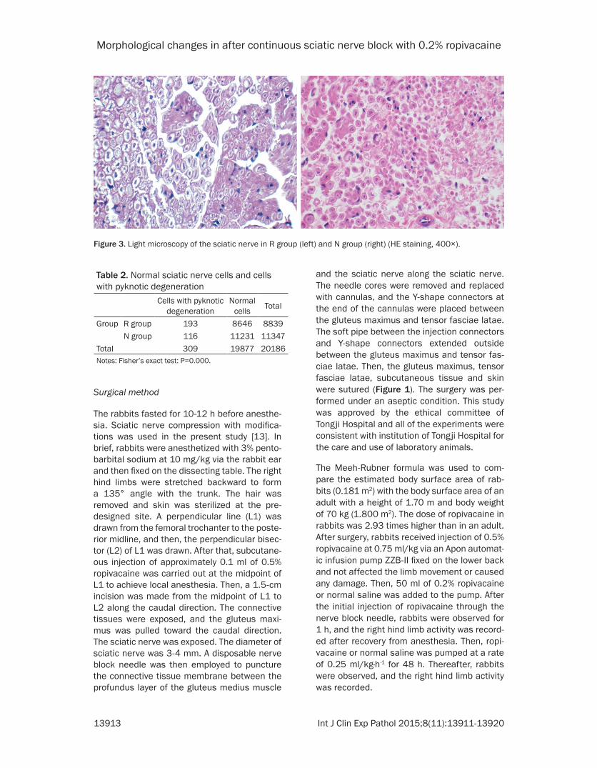
Figure 3. Light microscopy of the sciatic nerve in R group (left) and N group (right) (HE staining, 400×).
Table 2. Normal sciatic nerve cells and cells with pyknotic degeneration
Cells with pyknotic degeneration
Normal cells Total
Group R group 193 8646 8839N group 116 11231 11347
Total 309 19877 20186Notes: Fisher’s exact test: P=0.000.

Morphological changes in after continuous sciatic nerve block with 0.2% ropivacaine
13914 Int J Clin Exp Pathol 2015;8(11):13911-13920
Hind limb activity
After surgery, rabbits were housed in separate cages at 10-15°C and given ad libitum access to food and water with 12-h: 12-h light-dark cycle. At 2, 24 and 48 h after the initiation of drug infusion, inspections were carried out to guarantee normal operation of the infusion pump, smooth drug infusion, no liquid exuda-tion from the wound, and normal food and water intake. Motor Function (MF) assessment system was employed for the evaluation of hind limb activity [14]: 0, no obvious motor block and rapid contraction and withdrawal after a needle prick; 1, partial motor block and incomplete contraction after a needle prick; 2, complete motor block and no reaction to a needle prick.
Sample collection and processing
At the end of experiment, rabbits were sacri-ficed by injecting 10 ml of 10% KCl via the ear vein, and an indwelling needle was used to puncture the carotid artery on both sides, fol-lowed by bloodletting. Then, 300-400 ml of 10% formalin was injected through the punc-ture site via a tube. Then, the right atrium, brain tissues surrounding the third ventricle and
moved and flushed with 10% formalin. The right atrium was collected along the atrial septum and tricuspid valve and placed into 10% forma-lin immediately followed by fixation for 24 h.
Brain tissues: Craniotomy was performed and the brain was exposed. After removing the cra-nial nerves, the cerebrum, cerebellum and part of the spinal nerves were removed and collect-ed. The matter was carefully removed and remaining tissues were rinsed with 10% forma-lin. Then, these tissues were transected along the cleft between the cerebrum and the cere-bellum to the bulbopontine sulcus at the cen-ter. The cerebellum was separated and the medulla oblongata (approximately 1.5 cm×1 cm×0.8 cm) was harvested. The cerebral cortex containing the third ventricle (approximately 1.5 cm×1 cm×1 cm) was also prepared. The above tissues were fixed in 10% formalin for 24 h.
Sciatic nerve and skeletal muscles innervated by the sciatic nerve: An incision was made along the original incision and the subcutane-ous tissues were separated to expose the cath-eterized sciatic nerve. Then, the nerve block needle cannula was removed and sciatic nerve
Figure 4. Light microscopy of the skeletal muscle in R group (left) and N group (right) (HE staining, 400×).
Table 3. Correlation between the proportion of cells with pyknotic de-generation and grouping in each sample and the motor scores (Binary logistic)
B S.E, Wals df Sig. Exp (B)Cells with pyknotic degeneration -133.443 94.943 1.975 1 0.160 0.000Grouping 1.711 1.317 1.688 1 0.194 5.534Constant -0.241 1.099 0.048 1 0.826 0.785
medulla oblongata, sci-atic nerve and skeletal muscles innervated by the sciatic nerve were collected.
Right atrium: Thoracoto- my was performed and the heart was exposed. Then, the heart was re-
Morphological changes in after continuous sciatic nerve block with 0.2% ropivacaine
13915 Int J Clin Exp Pathol 2015;8(11):13911-13920
(2.5 cm in length) was obtained between the puncture site and the end of cannula. Me- chanical injury was avoided during the opera-
tion. The sciatic nerve was placed into 10% for-malin immediately and fixed for 24 h. Then, the semimembranosus muscle behind the sciatic
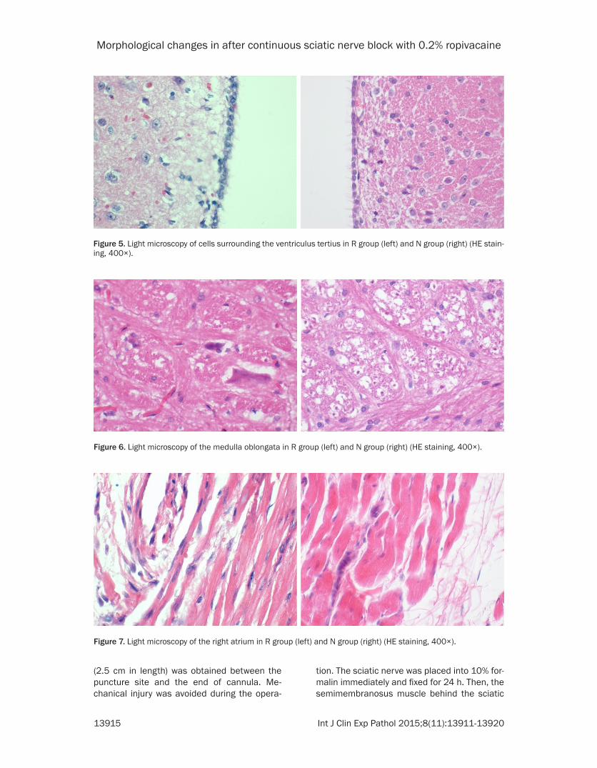
Figure 5. Light microscopy of cells surrounding the ventriculus tertius in R group (left) and N group (right) (HE stain-ing, 400×).
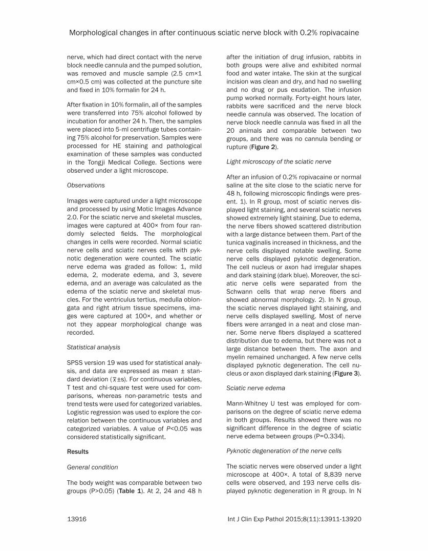
Figure 6. Light microscopy of the medulla oblongata in R group (left) and N group (right) (HE staining, 400×).
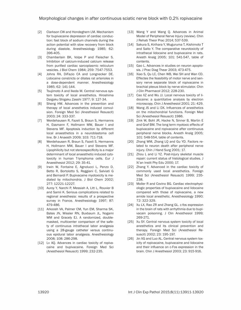
Figure 7. Light microscopy of the right atrium in R group (left) and N group (right) (HE staining, 400×).
Morphological changes in after continuous sciatic nerve block with 0.2% ropivacaine
13916 Int J Clin Exp Pathol 2015;8(11):13911-13920
nerve, which had direct contact with the nerve block needle cannula and the pumped solution, was removed and muscle sample (2.5 cm×1 cm×0.5 cm) was collected at the puncture site and fixed in 10% formalin for 24 h.
After fixation in 10% formalin, all of the samples were transferred into 75% alcohol followed by incubation for another 24 h. Then, the samples were placed into 5-ml centrifuge tubes contain-ing 75% alcohol for preservation. Samples were processed for HE staining and pathological examination of these samples was conducted in the Tongji Medical College. Sections were observed under a light microscope.
Observations
Images were captured under a light microscope and processed by using Motic Images Advance 2.0. For the sciatic nerve and skeletal muscles, images were captured at 400× from four ran-domly selected fields. The morphological changes in cells were recorded. Normal sciatic nerve cells and sciatic nerves cells with pyk-notic degeneration were counted. The sciatic nerve edema was graded as follow: 1, mild edema, 2, moderate edema, and 3, severe edema, and an average was calculated as the edema of the sciatic nerve and skeletal mus-cles. For the ventriculus tertius, medulla oblon-gata and right atrium tissue specimens, ima- ges were captured at 100×, and whether or not they appear morphological change was recorded.
Statistical analysis
SPSS version 19 was used for statistical analy-sis, and data are expressed as mean ± stan-dard deviation (
_x±s). For continuous variables,
T test and chi-square test were used for com-parisons, whereas non-parametric tests and trend tests were used for categorized variables. Logistic regression was used to explore the cor-relation between the continuous variables and categorized variables. A value of P<0.05 was considered statistically significant.
Results
General condition
The body weight was comparable between two groups (P>0.05) (Table 1). At 2, 24 and 48 h
after the initiation of drug infusion, rabbits in both groups were alive and exhibited normal food and water intake. The skin at the surgical incision was clean and dry, and had no swelling and no drug or pus exudation. The infusion pump worked normally. Forty-eight hours later, rabbits were sacrificed and the nerve block needle cannula was observed. The location of nerve block needle cannula was fixed in all the 20 animals and comparable between two groups, and there was no cannula bending or rupture (Figure 2).
Light microscopy of the sciatic nerve
After an infusion of 0.2% ropivacaine or normal saline at the site close to the sciatic nerve for 48 h, following microscopic findings were pres-ent. 1). In R group, most of sciatic nerves dis-played light staining, and several sciatic nerves showed extremely light staining. Due to edema, the nerve fibers showed scattered distribution with a large distance between them. Part of the tunica vaginalis increased in thickness, and the nerve cells displayed notable swelling. Some nerve cells displayed pyknotic degeneration. The cell nucleus or axon had irregular shapes and dark staining (dark blue). Moreover, the sci-atic nerve cells were separated from the Schwann cells that wrap nerve fibers and showed abnormal morphology. 2). In N group, the sciatic nerves displayed light staining, and nerve cells displayed swelling. Most of nerve fibers were arranged in a neat and close man-ner. Some nerve fibers displayed a scattered distribution due to edema, but there was not a large distance between them. The axon and myelin remained unchanged. A few nerve cells displayed pyknotic degeneration. The cell nu- cleus or axon displayed dark staining (Figure 3).
Sciatic nerve edema
Mann-Whitney U test was employed for com-parisons on the degree of sciatic nerve edema in both groups. Results showed there was no significant difference in the degree of sciatic nerve edema between groups (P=0.334).
Pyknotic degeneration of the nerve cells
The sciatic nerves were observed under a light microscope at 400×. A total of 8,839 nerve cells were observed, and 193 nerve cells dis-played pyknotic degeneration in R group. In N

Morphological changes in after continuous sciatic nerve block with 0.2% ropivacaine
13917 Int J Clin Exp Pathol 2015;8(11):13911-13920
group, 11,374 nerve cells were observed, and 116 nerve cells had pyknotic degeneration. The proportion of nerve cells with pyknotic degen-eration in R group was significantly higher than in N group (P<0.001) (Table 2).
Light microscopy of the skeletal muscles
HE staining showed the skeletal muscles of R group displayed light staining. The muscle fibers displayed swelling and were thickened. Moreover, the muscle fibers showed scattered distribution, had no smooth edges, and, in some areas, the cross-striations were unclear or even invisible. In one sample, many vacuoles were observed in cells, suggesting a dissolu-tion state, whereas, in other areas, the muscle fibers varied in size and exhibited distorted shapes, indicating necrosis. In N group, the skeletal muscles displayed light staining, some muscle fibers presented extremely light stain-ing, but they were arranged in a neat manner. The muscle fibers had normal shapes and clear cross-striations (Figure 4).
Skeletal muscle edema
The degree of skeletal muscle edema in both groups was compared. There was no significant difference in the degree of edema (P=0.193) between two groups.
Right hind limb activity
One hour later: All the 20 rabbits recovered from anesthesia, and both forelimbs and both left hind limbs were in a normal condition. The scores of right hind limb activity were com-pared. There was no significant difference in the score of right hind limb activity at 1 h after infusion between two groups (P1h=0.702).
48 hours later: The scores of right hind limb activity at 48 h after drug infusion were com-pared. Thus, there was no significant difference in the score of right hind limb activity at 48 h after infusion (P48h=0.374) between two groups.
Correlation between scores of right hind limb activity and morphological changes in the sci-atic nerve and skeletal muscle
Both the score of right hind limb activity and the overall score were linearly related to the de- gree of sciatic nerve edema (PR group<0.05, PN
group<0.05, Poverall<0.05). There was no signifi-cant correlation between the proportion of cells with pyknotic degeneration and the scores of right hind limb activity (P=0.16>0.05), as well as between the grouping and the scores of right hind limb activity (P=0.194>0.05) (Table 3).
Both the motor scores in each group and the overall motor score had no linear relationship with the degree of skeletal muscle edema (PR
group>0.05, PN group>0.05, Poverall>0.05).
Brain tissues surrounding the third ventricle
Light microscopy of the brain tissues surround-ing the third ventricle showed no notable abnor-malities in both groups (Figure 5). The cells below the ependyma showed regular spindle-like arrangements, and the cell nuclei were round or oval in shape. In some samples, the ependyma cells were in a single layer and neat-ly arranged. A few ependyma cells were in an irregular, tight multilayer arrangement.
Medulla oblongata
Light microscopy of the medulla oblongata of both groups failed to show abnormalities (Figure 6). The medulla oblongata of both groups was symmetric and the cell nuclei were normal in shape.
Right atrium
Light microscopy of the right atrium in both groups did not display abnormalities (Figure 7). The cardiocytes were normal in shape, and the cell nuclei were spindle-shaped. The muscle fibers displayed clear cross-striations and were irregularly arranged. Some muscle fibers were found to be filled with connective tissues, and sinoatrial nodes were observed in some sam-ples. P cell nuclei were large in size and round or oval in shape.
Discussion
Cell death can be classified as either apoptosis or necrosis. Apoptosis of nerve cells plays a critical role in the life cycle of both central and peripheral nerves. In fact, apoptosis can trans-form into necrosis, and when a large number of cells were dying, apoptosis dominates at the beginning, while cells display necrosis at the end [15].

Morphological changes in after continuous sciatic nerve block with 0.2% ropivacaine
13918 Int J Clin Exp Pathol 2015;8(11):13911-13920
Xiao et al [16] found that motor nerves and sen-sory nerves could be blocked with 0.2% ropiva-caine as a local anesthetic. Ropivacaine at 0.2% is also used clinically for postoperative analgesia. In the present experiment, some sci-atic nerves cells in two groups displayed pyk-notic degeneration, and had some features of apoptosis determined based on the morpholo-gy and distribution. Moreover, the proportion of nerve cells with pyknotic degeneration in R group was significantly higher than in N group (P<0.001). Thus, a prolonged exposure to ropi-vacaine, even a low concentration (0.2%), may cause morphological changes in the nerve cells. However, whether the pyknotic degenera-tion implies cell apoptosis and whether the cells that could not be clearly observed were necrotic ones remain unclear because a magni-fication of 400× failed to provide a clear field to observe the morphological changes after HE staining. Thus, light microscopy at a higher magnification or electron microscopy is needed in future studies to observe cell ultrastructure. Alternatively, phosphatidylserine detection or propidium iodide staining may be employed for quantitative analysis, which will confirm wheth-er apoptosis or necrosis occurs. In our experi-ment, a logistic regression was carried out to explore the correlation between the proportion of cells with pyknotic degeneration, grouping (based on drug infusion) and the scores of right hind limb activity. Results showed there were no significant correlations between the motor score and cell proportion and between the motor score and grouping (P>0.05). Additionally, the significance level for pyknotic cell degener-ation was lower than that for grouping. Thus, there was a higher correlation between the motor scores and rate of cell pyknotic degen-eration than between the motor scores and grouping. This result may be affected by the small sample size used in the present ex- periment.
Cell edema, also known as hydropic degenera-tion, is a type of reversible changes in the early stage of cell damage. Previous studies have shown that cell edema may also be caused by local anesthetics because local anesthetics may affect the function of mitochondria in cells [17, 18]. In the present experiment, many fac-tors could cause cell edema, including mechan-ical injury, nerve cell damage, and local inflam-mation. Mechanical injury may be caused by
the insertion of nerve block needle cannula near the sciatic nerve, the friction between the nerve block needle cannula and the sciatic nerve due to hind limb movement. Nerve cell damage is related to an increased local pres-sure due to continuous drug or normal saline infusion. In the present experiment, the sciatic nerves in both groups displayed edema of vari-ous degrees, and the morphological features of edema, such as cellular swelling, light staining, and cells being separated, were observed under a light microscope. However, there was no significant difference in the degree of edema between R group and N group, possibly because the edema was influenced by multiple factors.
When the sciatic nerve is partially damaged, the knee flexion and the biceps femoris muscle are weakened, but the semitendinosus and semimembranosus muscles are only slightly affected [1]. Thus, the semimembranosus mus-cle near the nerve block needle cannula was removed for observation. Compared with short-acting local anesthetics, long-acting local anes-thetics are more likely to cause injury to the skeletal muscles near the injection site [1]. Zink et al found that muscular toxicity caused by long-term (>6 h) injection of 0.75% ropivacaine was weaker than that of 0.5% bupivacaine, but both caused visible but irreversible calcified muscle lesions after 7-day and 28-day injection [19]. The semimembranosus muscles exam-ined in the present experiment also displayed different degrees of edema, and skeletal mus-cles of both groups displayed swelling and were pale in color. After cutting the skeletal muscle, the loose tissues were exposed. These tissues were rich in water and had fragile edges, and the section plane was in a jelly-like shape. There was no significant difference in the degree of edema between R group and N group (P>0.05). This may be explained as that the concentration of ropivacaine presenting our experiment (0.2%) was relatively low, and its muscle toxicity was lower than that of 0.75% ropivacaine. However, the continuous injection of drug or normal saline caused increased pres-sure at the injection site. When the normal envi-ronment of local tissues was disrupted, edema occurred. The muscle cell necrosis observed in our experiment may be related to the influence of local anesthetic on the mitochondria and the changes in calcium concentration within cells. However, muscle cell necrosis was observed in

Morphological changes in after continuous sciatic nerve block with 0.2% ropivacaine
13919 Int J Clin Exp Pathol 2015;8(11):13911-13920
only one sample, and thus future studies with a large sample size are needed to confirm our findings.
After long term injection of 0.2% ropivacaine or normal saline, no significant difference was observed in the scores of right hind limb activi-ty between two groups (P>0.05). Then, the cor-relation between motor scores and other fac-tors (proportion of cells with pyknotic degener-ation, degree of nerve edema, and degree of skeletal muscle edema) was further evaluated and results showed that there was significant correlation between the scores of right hind limb activity and the degree of nerve edema (P<0.05), but no significant correlation was observed between the scores of right hind limb activity and the proportion of cells with pyknot-ic degeneration, and between the scores of right hind limb activity and the degree of skele-tal muscle edema. Our findings were in accor-dance with the clinical observations, in which there is an extremely low incidence of nerve damage even though patients are exposed to multiple factors that may cause nerve damage. This phenomenon can be explained by the potent compensatory response in living organ-isms. In clinical practice, for patients displaying nerve damage after nerve blocking, the nerve and muscle damage is usually severely dam-aged, but for patients had no clinical manifes-tations or complaints, the nerves or muscles may be slightly damaged. However, more exper-iments are required to confirm these results. Moreover, according to previous studies, both nerve cells [20] and skeletal muscles [21] undergo self-repair when the damaging condi-tion changes. However, in our experiment, only the temporal condition of the rabbits was observed after infusion of 0.2% ropivacaine or normal saline. Thus, more observations are required to explore the long-term effect of a continuous nerve block.
Cardiac toxicity can manifest as the inhibition of myocardial contraction and arrhythmia [22]. In the present experiment, there was no obvi-ous morphological change in the right atrium of both groups, which may be ascribed to a rela-tively low dose of ropivacaine and/or a low car-diac toxicity of ropivacaine [23]. Additionally, local anesthetics primarily affect Purkinje fibers and ventricular muscle [2], and thus local anes-thetics will primarily cause conduction blocks rather than morphological changes.
Furthermore, local anesthetics can also medi-ate cardiac toxicity by affecting the nervous system, which is achieved not only through the inhibition of sympathetic nerves and fluctua-tion of blood pressure due to vascular dilation at the block site but also through influencing other regions of the brain, such as the solitary nucleus and trigeminal nerve subnucleus [24]. In our experiment, there was no obvious mor-phological change in the medulla oblongata of both groups, which was in accordance with the good condition of rabbits in both groups.
Local anesthetics may also cause central ner-vous system toxicity, which is often manifested as central nervous system excitation followed by convulsion. Central nervous system toxicity arises because the local anesthetic blocks the inhibitory pathway [25]. According to previous studies, the duration of convulsion caused by a local anesthetic is related to the number of neurons expressing c-Fos protein around the third ventricle [26]. In the present experiment, none in both groups exhibited convulsion, and light microscopy of brain sections after HE staining failed to display clear abnormalities at the third ventricle, which may be related to a low dose of ropivacaine. Moreover, local anes-thetic initially causes abnormality in electric activity in nerve cells, and then result in mor-phological changes.
Acknowledgements
This study was supported by the promotion project of the advanced appropriate technology of Shanghai Health System (No. 2013SY032). The authors thank to Prof. Peilin Zhao, Prof. Yan Zhao and Prof. Yanna Li in Department of Pathology of Tongji University School of Me- dicine and veterinarian Deju Chen for technical support.
Disclosure of conflict of interest
None.
Address correspondence to: Bin Yu, Department of Anesthesiology, Tongji Hospital, School of Me- dicine, Tongji University, Shanghai, China. E-mail: [email protected]; [email protected]
References
[1] Miller RD. Miller’s Anesthesia. Beijing: Peking University Medical Press; 2011.
Morphological changes in after continuous sciatic nerve block with 0.2% ropivacaine
13920 Int J Clin Exp Pathol 2015;8(11):13911-13920
[2] Clarkson CW and Hondeghem LM. Mechanism for bupivacaine depression of cardiac conduc-tion: fast block of sodium channels during the action potential with slow recovery from block during diastole. Anesthesiology 1985; 62: 396-405.
[3] Chamberlain BK, Volpe P and Fleischer S. Inhibition of calcium-induced calcium release from purified cardiac sarcoplasmic reticulum vesicles. J Biol Chem 1984; 259: 7547-7553.
[4] Johns RA, DiFazio CA and Longnecker DE. Lidocaine constricts or dilates rat arterioles in a dose-dependent manner. Anesthesiology 1985; 62: 141-144.
[5] Tsujimoto A and Ikeda M. Central nervous sys-tem toxicity of local anesthetics. Hiroshima Daigaku Shigaku Zasshi 1977; 9: 127-34.
[6] Sheng HW. Advances in the prevention and therapy of local anesthetics induced convul-sion. Foreign Med Sci (Anesthesiol Resuscit) 2003; 24: 333-337.
[7] Werdehausen R, Fazeli S, Braun S, Hermanns H, Essmann F, Hollmann MW, Bauer I and Stevens MF. Apoptosis induction by different local anaesthetics in a neuroblastoma cell line. Br J Anaesth 2009; 103: 711-718.
[8] Werdehausen R, Braun S, Fazeli S, Hermanns H, Hollmann MW, Bauer I and Stevens MF. Lipophilicity but not stereospecificity is a major determinant of local anaesthetic-induced cyto-toxicity in human T-lymphoma cells. Eur J Anaesthesiol 2012; 29: 35-41.
[9] Irwin W, Fontaine E, Agnolucci L, Penzo D, Betto R, Bortolotto S, Reggiani C, Salviati G and Bernardi P. Bupivacaine myotoxicity is me-diated by mitochondria. J Biol Chem 2002; 277: 12221-12227.
[10] Auroy Y, Narchi P, Messiah A, Litt L, Rouvier B and Samii K. Serious complications related to regional anesthesia: results of a prospective survey in France. Anesthesiology 1997; 87: 479-486.
[11] Arkoosh VA, Palmer CM, Yun EM, Sharma SK, Bates JN, Wissler RN, Buxbaum JL, Nogami WM and Gracely EJ. A randomized, double-masked, multicenter comparison of the safe- ty of continuous intrathecal labor analgesia using a 28-gauge catheter versus continu- ous epidural labor analgesia. Anesthesiology 2008; 108: 286-298.
[12] Lv AQ. Advances in cardiac toxicity of ropiva-caine and bupivacaine. Foreign Med Sci (Anesthesiol Resuscit) 1999; 232-235.
[13] Wang Y and Wang Q. Advances in Animal Model of Peripheral Nerve Injury (review). Chin J Rehab Theor Prac 2014; 537-539.
[14] Sakura S, Kirihara Y, Muguruma T, Kishimoto T and Saito Y. The comparative neurotoxicity of intrathecal lidocaine and bupivacaine in rats. Anesth Analg 2005; 101: 541-547, table of contents.
[15] Gao L. Advances in studies on neuron apopto-sis. J Prac Diag Thear 2003; 473-475.
[16] Xiao S, Qu LC, Chen WB, Wei SH and Wan CD. Effectes the feasibility of motor nerve and sen-sory nerve separate block of ropivacaine in brachial plexus block by nerve stimulator. Chin J Clin Pharmacol 2012; 228-230.
[17] Cao XZ and Wu JJ. Local nervous toxicity of li-docaine: a quantitative analysis by electron microscopy. Chin J Anesthesiol 2001; 21: 429.
[18] Wang JS and Li DX. Influences of anesthetics on the mitochondrial functions. Foreign Med Sci (Anesthesiol Resuscit) 1986.
[19] Zink W, Bohl JR, Hacke N, Sinner B, Martin E and Graf BM. The long term myotoxic effects of bupivacaine and ropivacaine after continuous peripheral nerve blocks. Anesth Analg 2005; 101: 548-554, table of contents.
[20] Zhang WM, Zhang LQ and Gu YD. Factors re-lated to neuron death after peripheral nerve injury. Chin J Hand Surg 2001; 17.
[21] Zhou L and Li YZ. Post-injury skeletal muscle repair: current status of histological studies. J Xi’an Instit Phy Edu 2000; 17.
[22] Zhang Y. Advanced in the cardiac toxicity of commonly used local anesthetics. Foreign Med Sci (Anesthesiol Resuscit) 1999; 235-238.
[23] Moller R and Covino BG. Cardiac electrophysi-ologic properties of bupivacaine and lidocaine compared with those of ropivacaine, a new amide local anesthetic. Anesthesiology 1990; 72: 322-329.
[24] Xu LX, Rao ZR and Zhang GL. c-fos expression in the brain of rats with arrhythmia due to bupi-vacain poisoning. J Clin Anesthesiol 1999; 269-271.
[25] Xu SY. Central nervous system toxicity of local anesthetics and its clinical prevention and therapy. Foreign Med Sci (Anesthesiol Re- suscit) 2002; 23: 195-197.
[26] Jin XG and Luo AL. Central nervous system tox-icity of ropivacaine, bupivacaine and lidocaine and their influence on c-Fos expression in the brain. Chin J Anesthesiol 2003; 23: 915-916.