original article laryngeal response patterns influence the
TRANSCRIPT
ORIGINAL ARTICLE
Laryngeal response patterns influence the efficacyof mechanical assisted cough in amyotrophic lateralsclerosisTiina Andersen,1,2,3 Astrid Sandnes,3 Anne Kristine Brekka,4 Magnus Hilland,5
Hege Clemm,3,6 Ove Fondenes,1 Ole-Bjørn Tysnes,7,8 John-Helge Heimdal,5,8
Thomas Halvorsen,3,6 Maria Vollsæter,1,3,6 Ola Drange Røksund4,6
▸ Additional material ispublished online only. To viewplease visit the journal online(http://dx.doi.org/10.1136/thoraxjnl-2015-207555).
For numbered affiliations seeend of article.
Correspondence toTiina Andersen, ThoracicDepartment, Norwegian Centreof Excellence for HomeMechanical Ventilation,Haukeland University Hospital,Bergen 5021, Norway; [email protected]
Received 17 July 2015Revised 22 March 2016Accepted 7 April 2016Published Online First12 May 2016
▸ http://dx.doi.org/10.1136/thoraxjnl-2016-208919
To cite: Andersen T,Sandnes A, Brekka AK,et al. Thorax 2017;72:221–229.
ABSTRACTBackground Most patients with amyotrophic lateralsclerosis (ALS) are treated with mechanical insufflation–exsufflation (MI-E) in order to improve cough. Thismethod often fails in ALS with bulbar involvement,allegedly due to upper-airway malfunction. We havestudied this phenomenon in detail with laryngoscopy tounravel information that could lead to better treatment.Methods We conducted a cross-sectional study of 20patients with ALS and 20 healthy age-matched and sex-matched volunteers. We used video-recorded flexibletransnasal fibre-optic laryngoscopy during MI-Eundertaken according to a standardised protocol,applying pressures of ±20 to ±50 cm H2O. Laryngealmovements were assessed from video files. ALS type andcharacteristics of upper and lower motor neuronsymptoms were determined.Results At the supraglottic level, all patients with ALSand bulbar symptoms (n=14) adducted their laryngealstructures during insufflation. At the glottic level, initialabduction followed by subsequent adduction was observedin all patients with ALS during insufflation and exsufflation.Hypopharyngeal constriction during exsufflation wasobserved in all subjects, most prominently in patients withALS and bulbar symptoms. Healthy subjects and patientswith ALS and no bulbar symptoms (n=6) coordinated theircough well during MI-E.Conclusions Laryngoscopy during ongoing MI-E inpatients with ALS and bulbar symptoms revealed laryngealadduction especially during insufflation but also duringexsufflation, thereby severely compromising the size of thelaryngeal inlet in some patients. Individually customisedsettings can prevent this and thereby improve and extendthe use of non-invasive MI-E.
INTRODUCTIONAmyotrophic lateral sclerosis (ALS) is an incurableand highly disabling neurodegenerative disease ofupper and lower motor neurons. Treatment islargely symptomatic, and average life expectancy atthe time of the diagnosis is 2–3 years unless ventila-tory assistance is provided.1
ALS is classified as ‘spinal’ if symptom onsetaffects the limbs predominantly, and as ‘bulbar’ ifthe disease presents with difficulty in speaking,swallowing or coughing. Paresis to predominantlyupper motor neurons leads primarily to spasticity,
whereas paresis of lower motor neurons leads toflaccidity.2 Regardless of the subtype, ALS pro-gresses and eventually encompasses all skeletalmuscles.3 Involvement of respiratory muscles limitsrespiratory function and cough, thereby leading tosecretion accumulation, lung infections and, even-tually, respiratory failure.3–6 Effective augmentationof cough is vital for clearance of airway secretionsin these patients and fundamental for the preven-tion and treatment of pneumonias.6 7
In a voluntary cough, inspiratory muscles increasethe lung volume, laryngeal muscles coordinateopening and closure of the glottis and expiratorymuscles increase the thoracoabdominal pressure.8
These interactions are disturbed in neuromusculardisorders.7 9 Mechanical insufflation-exsufflation(MI-E) is used widely to assist cough mechanicallyby applying positive and negative pressure changesto the airways, either non-invasively via a mask orinvasively via a tracheostomy.10 11 It has beenhypothesised that coordinated glottic movements arerequired for MI-E to be effective.12 Non-invasiveMI-E can be difficult to apply in patients with the
Key messages
What is the key question?▸ Mechanical insufflation–exsufflation (MI-E) is
an efficient tool used to improve cough in mostpatients with neuromuscular disorders, but themethod often fails when bulbar involvement ispresent.
What is the bottom line?▸ We used laryngoscopy during ongoing MI-E
and saw that patients with bulbar amyotrophiclateral sclerosis (ALS) were prone to adductlaryngeal structures throughout the variouspressure cycles, thereby severely obstructing theairflow and the effect of the treatment.
Why read on?▸ In patients with bulbar ALS, cough assistance
with MI-E should be delivered carefully andaccording to the criteria suggested in thepresent study.
Andersen T, et al. Thorax 2017;72:221–229. doi:10.1136/thoraxjnl-2015-207555 221
Non-invasive ventilation on F
ebruary 24, 2022 by guest. Protected by copyright.
http://thorax.bmj.com
/T
horax: first published as 10.1136/thoraxjnl-2015-207555 on 12 May 2016. D
ownloaded from
bulbar subtype of ALS. This problem may be due to dysfunctionof bulbar-innervated muscles, but the basic mechanisms are notunderstood.
The laryngeal response to MI-E in patients with ALS hasnever been studied. Here, we investigated the laryngeal responsepatterns to MI-E in ALS to improve the treatment that we canoffer to these severely ill patients.
METHODSNeurological assessment and definitionsALS was diagnosed by a senior neurologist (O-BT) in accord-ance with the revised criteria set by the El Escorial WorldFederation of Neurology.13 14 The disease was classified as‘spinal ALS’, ‘ALS with progressive bulbar palsy’ (hypotonicbulbar onset with dysarthria, tongue atrophy and absence of jawreflex) or ‘ALS with pseudobulbar palsy’ (spastic bulbar onsetwith dysarthria, exaggerated jaw reflex and no tongue atrophy).Patients were assessed using the ALS Functional Rating Scale-revised (ALSFRS-r).15 Bulbar impairment score (BIS) was evalu-ated from the ALSFRS-r, from where the items of speech andswallowing were calculated.16 Dysphagia was determined usingthe 100 mL water swallow test.17 18
SubjectsThis was a cross-sectional observational population-based studyof 20 patients with ALS who had not undergone tracheostomyand 20 neurologically healthy age-matched and sex-matchedcontrols. Exclusion criteria were age <18 years, history of laryn-gospasm, sensitisation to Xylocain (anaesthetic used duringlaryngoscopy), pneumothorax, additional lung disease, cancer,acute infection of the chest 1 month before study commence-ment and mental instability.
Approximately 20 patients with ALS who have not undergonetracheostomy are usually enrolled at all times at the ALS clinicat Haukeland University Hospital (Bergen, Norway), whichserves a population of ≈500 000 inhabitants. At the start of thisstudy, 17 patients were enrolled at the clinic and 20 newpatients were diagnosed and enrolled during the 1.5-yearrecruitment period from December 2011 to June 2013. All 37patients were informed about the study and invited to partici-pate. Thirteen patients declined and four died soon after beinginvited, leaving 20 participants. Reasons for non-participationwere severe disease/fatigue (n=7), or limb-onset ALS withoutbulbar symptoms and, therefore, no interest in participation(n=6). The study protocol was approved by the RegionalCommittee for Medical Research Ethics. Written informedconsent was obtained from all participants.
Pulmonary function and respiratory strengthSpirometry was undertaken with a Vmax 22 Encore system(SensorMedics, Yorba Linda, California, USA). FVC, FEV1 andpeak expiratory flow were measured seated, with a nose clip.Slow vital capacity was measured with a Respirometer (nSpireHealth, Hertford, UK). Peak cough flow was measured using ahand-held Peak Flow Meter (Vitalograph, Ennis, Ireland).Plateau values (average of 1 s) of the maximal inspiratory (Pimax)and expiratory (Pemax) muscle strength and sniff nasal inspira-tory pressure (SNIP) were measured seated using a RespiratoryPressure Meter (Micro RPM; Micro Medical, Rochester, UK).SNIP was measured at functional residual capacity, Pimax atresidual volume and Pemax at total lung capacity. The highestvalue from three or more attempts was selected for analyses andstandardised to predicted percentages.19–22
Video-recorded transnasal fibre-optic laryngoscopy duringMI-EVideo-recorded transnasal fibre-optic laryngoscopy (ENF-P3;Olympus, Tokyo, Japan) was used to visualise laryngeal anatomyat baseline and response patterns during MI-E (Cough Assist;Respironics, Murrysville, Pennsylvania, USA). We used a set-updescribed in detail previously, except that the laryngoscope wassupported manually (see online supplementary figure S1)instead of using a customised headgear.23 A standardised MI-Eprotocol was used.23 The protocol comprised 12 interventionarms with various combinations of pressures, instructions andmanual thoracic thrust (see online supplementary table S2).
Pressures of ±20, ±30, ±40 and ±50 cm H2O were usedwith specific instructions. For MI-E in automated mode with 2 sinsufflation, 2 s exsufflation and 1 s pause, the instructions wereto ‘inhale’ actively when insufflation was started and to (A)‘exhale’ or (B) ‘cough’ actively when the device switched toexsufflation. For MI in manual mode with 2 s insufflation fol-lowed by manually assisted thoracic thrust, the instructions wereto ‘inhale’ actively when insufflation was started and to ‘cough’actively during the thoracic thrust.
In case of patient discomfort, the procedure was stopped andhigher examination pressures were not applied.
Analyses of observationsAltogether, 480 recordings were scheduled for assessment, thatis, one recording from 12 intervention arms in 20 patients and20 control subjects. With respect to assessment of observations,MI-E cycles were edited into three phases of interest: (i) insuf-flation, (ii) pressure drop (from positive to negative) and (iii)active exsufflation or the voluntary cough with no negative pres-sure applied. The onset and offset of each phase were observedand defined from the parallel video recording of the MI-E man-ometer.23 Video recordings were assessed systematically, asdescribed previously,23 by two trained raters (TA and AKB).Main features were described at glottic, supraglottic and hypo-pharyngeal levels (see online supplementary figure S3).Laryngeal anatomy and motion at rest were evaluated by asenior laryngologist ( J-HH).
Statistical analysesThe χ2 test, or Fisher’s exact test if expected cell counts wereless than five, were applied to assess differences between groupswith regard to categorical data. Background data were given asgroup means with SDs. The number of subjects with thedescribed patterns of laryngeal movements during MI-E wasgiven as group counts and percentages. Statistical analyses wereconducted using SPSS V.21.0 (IBM, Armonk, USA). The two-sided significance level was set at 0.05.
RESULTSPatient characteristicsOf 20 participating patients with ALS, six had limb onset withno bulbar symptoms and 14 had bulbar symptoms (table 1); ofthese, seven had pseudobulbar (spastic) ALS and seven had pro-gressive bulbar (hypotonic) ALS. Lung-function characteristics inALS were lower than predicted (table 1). In patients with pro-gressive bulbar ALS, 4/7 subjects had an abnormal epiglottis:three had a juvenile and high-standing epiglottis, and in onepatient the epiglottis was considered ‘floppy’. Retention ofsecretions/sputum was observed in 4/7 patients with progressivebulbar ALS, in 2/7 cases with pseudobulbar ALS, in 2/6 subjectswith non-bulbar ALS and in 1/20 healthy controls.
222 Andersen T, et al. Thorax 2017;72:221–229. doi:10.1136/thoraxjnl-2015-207555
Non-invasive ventilation on F
ebruary 24, 2022 by guest. Protected by copyright.
http://thorax.bmj.com
/T
horax: first published as 10.1136/thoraxjnl-2015-207555 on 12 May 2016. D
ownloaded from
Laryngeal response to MI-EIn total, 453 (94%) of 480 scheduled recordings were analysed.Four patients with bulbar symptoms completed only parts ofthe MI-E protocol due to discomfort from the applied pres-sures, that is, one patient (progressive bulbar ALS) interruptedthe protocol after pressures of ±20 cm H2O (missing 9/12 inter-vention arms), one patient (pseudobulbar ALS) after±30 cm H2O (missing 6/12 intervention arms) and two patients(one pseudobulbar and one progressive bulbar ALS) after±40 cm H2O (both patients missing 3/12 intervention arms).Technical failures led to loss of video recordings in one healthycontrol at examining pressures of ±40 and ±50 cm H2O(missing 6/12 intervention arms).
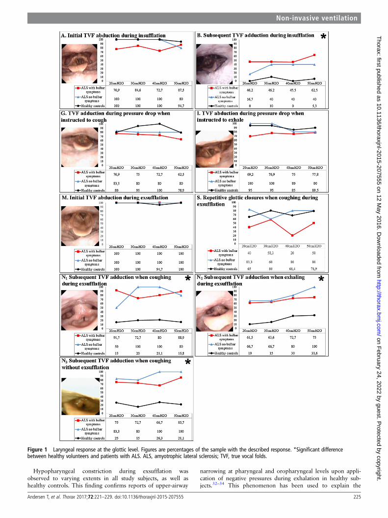
In general, the larynx moved downwards during applied insuf-flation and upwards (cranially) during exsufflation. (See table 2for overall descriptions and online supplementary video 1 for thelaryngeal response in a patient with non-bulbar ALS; onlinesupplementary video 2 in a healthy control; online supplemen-tary video 3 in a patient with progressive bulbar ALS; onlinesupplementary video 4 in a patient with pseudobulbar ALS.)Adequate laryngeal control was defined as described for normalcough in the literature,8 and presented as initial abduction of thetrue vocal folds (TVF) and aryepiglottic folds (AEF), and there-after glottic closure with subsequent rapid opening when cough-ing, abduction of the TVF and AEF followed by sequentialclosures and/or narrowing in the exhalation phase of the cough.
Response at the glottic levelObservations at the glottic level were not possible in some patientswith ALS and bulbar symptoms, because adduction of AEF and/orthe hypopharyngeal area obscured the view of TVF, particularly inthe high-pressure ranges of 40–50 cm H2O. Observations at theglottic level were based on successful visualisation of MI-E cycles(TVF responses A, B, G, I, M, S, N1, N2 and N3 in figure 1 andonline supplementary tables S4, S5 and S6).
There were significant differences between patients with ALSand healthy controls with respect to TVF adduction subsequent
to the initial abduction during insufflation (response B in figure 1and in online supplementary table S4) and exsufflation. Varyingthe instructions (to cough or exhale during negative pressures orto cough without applied negative pressure) did not influence thegroups differently (response N1, N2 and N3 in figure 1 andonline supplementary table S6).
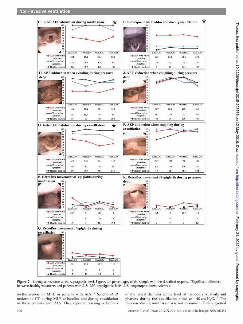
Response at the supraglottic levelAEF responses are presented as C, D, H, J, O and P (figure 2and online supplementary tables S4, S5 and S6). Medial rotationof the cuneiform tubercles accompanied by considerable adduc-tion of the AEF was observed during insufflation (initially orsubsequent to abduction) in all patients with bulbar ALS (onlinesupplementary table S4 and response C and D in figure 2).
A retroflex movement of epiglottis (a passive dorsal rotation)was observed to partly occlude the laryngeal inlet in some cases,either as a rapid movement or lasting throughout the insuffla-tion (responses E, K and Q (figure 2 and online supplementarytables S4, S5 and S6)).
Oesophageal opening was observed during insufflation in twopatients with progressive bulbar ALS. Both subjects wereobserved to burp afterwards, suggesting that (part of) the insuf-flation volume ended up in the oesophagus and stomach insteadof the lungs.
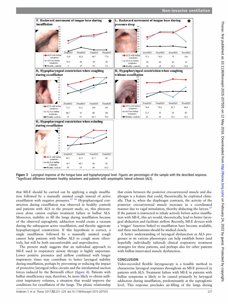
Response at the tongue base and at the hypopharyngeallevelThere were significant differences between healthy controls andpatients with ALS with regard to backward movement of thetongue base during insufflation and during the pressure drop(responses F and L in figure 3 and online supplementary tablesS4 and S5).
Constriction of the hypopharynx during exsufflation wasobserved in healthy controls and in patients with ALS, regard-less of the presence of bulbar symptoms. In patients with ALSand bulbar symptoms, hypopharyngeal constriction was moreprominent in those with progressive bulbar paresis. The
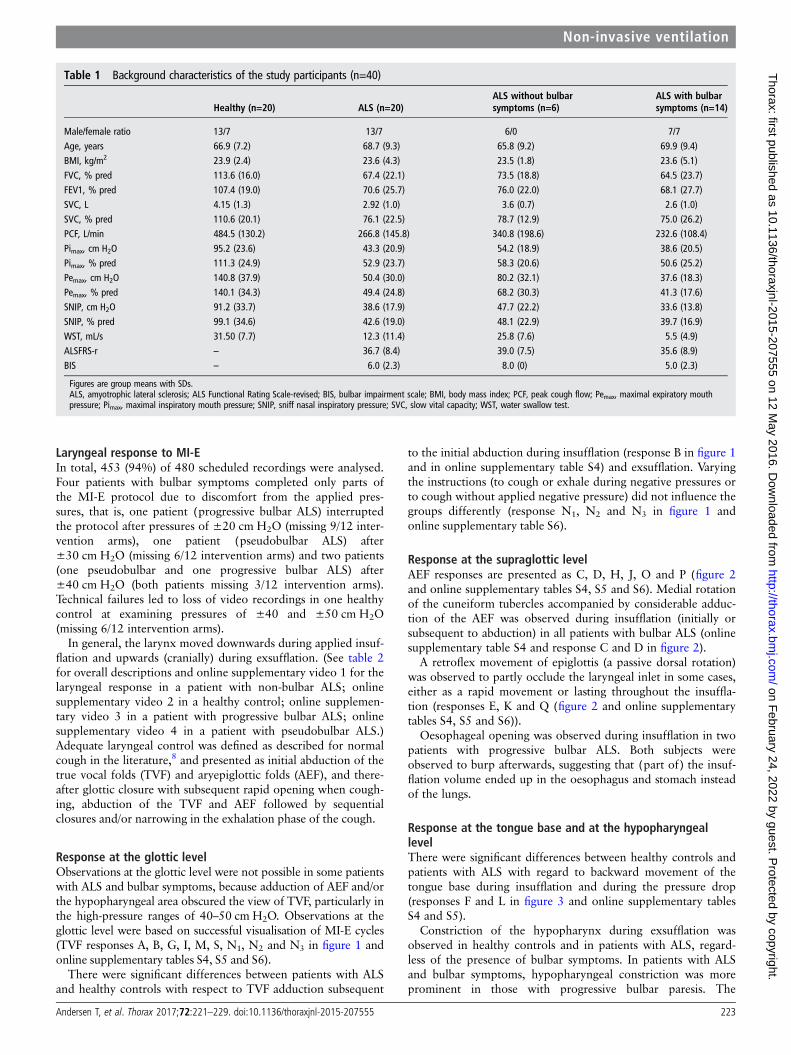
Table 1 Background characteristics of the study participants (n=40)
Healthy (n=20) ALS (n=20)ALS without bulbarsymptoms (n=6)
ALS with bulbarsymptoms (n=14)
Male/female ratio 13/7 13/7 6/0 7/7Age, years 66.9 (7.2) 68.7 (9.3) 65.8 (9.2) 69.9 (9.4)BMI, kg/m2 23.9 (2.4) 23.6 (4.3) 23.5 (1.8) 23.6 (5.1)FVC, % pred 113.6 (16.0) 67.4 (22.1) 73.5 (18.8) 64.5 (23.7)FEV1, % pred 107.4 (19.0) 70.6 (25.7) 76.0 (22.0) 68.1 (27.7)SVC, L 4.15 (1.3) 2.92 (1.0) 3.6 (0.7) 2.6 (1.0)SVC, % pred 110.6 (20.1) 76.1 (22.5) 78.7 (12.9) 75.0 (26.2)PCF, L/min 484.5 (130.2) 266.8 (145.8) 340.8 (198.6) 232.6 (108.4)Pimax, cm H2O 95.2 (23.6) 43.3 (20.9) 54.2 (18.9) 38.6 (20.5)Pimax, % pred 111.3 (24.9) 52.9 (23.7) 58.3 (20.6) 50.6 (25.2)Pemax, cm H2O 140.8 (37.9) 50.4 (30.0) 80.2 (32.1) 37.6 (18.3)Pemax, % pred 140.1 (34.3) 49.4 (24.8) 68.2 (30.3) 41.3 (17.6)SNIP, cm H2O 91.2 (33.7) 38.6 (17.9) 47.7 (22.2) 33.6 (13.8)SNIP, % pred 99.1 (34.6) 42.6 (19.0) 48.1 (22.9) 39.7 (16.9)WST, mL/s 31.50 (7.7) 12.3 (11.4) 25.8 (7.6) 5.5 (4.9)ALSFRS-r – 36.7 (8.4) 39.0 (7.5) 35.6 (8.9)BIS – 6.0 (2.3) 8.0 (0) 5.0 (2.3)
Figures are group means with SDs.ALS, amyotrophic lateral sclerosis; ALS Functional Rating Scale-revised; BIS, bulbar impairment scale; BMI, body mass index; PCF, peak cough flow; Pemax, maximal expiratory mouthpressure; Pimax, maximal inspiratory mouth pressure; SNIP, sniff nasal inspiratory pressure; SVC, slow vital capacity; WST, water swallow test.
Andersen T, et al. Thorax 2017;72:221–229. doi:10.1136/thoraxjnl-2015-207555 223
Non-invasive ventilation on F
ebruary 24, 2022 by guest. Protected by copyright.
http://thorax.bmj.com
/T
horax: first published as 10.1136/thoraxjnl-2015-207555 on 12 May 2016. D
ownloaded from
hypopharynx was totally constricted in 4/7 patients with pro-gressive bulbar paresis and in 1/7 patients with pseudobulbarparesis (responses R1, R2 and R3 in figure 3 and onlinesupplementary table S6).
Differences in laryngeal movements between patients withpseudobulbar and progressive bulbar ALS were not significant.A few significant values were observed between observations ofhealthy controls and patients with ALS and bulbar symptoms,and between patients with ALS with and without bulbarsymptoms. Due to a multiple-testing problem, these resultsshould be interpreted with caution. However, we saw a patternin comparison between controls and patients with ALS andbulbar symptoms with regard to backward movement of thetongue base during the pressure drop and in subsequent adduc-tion of TVF during exsufflation (see online supplementarytables S4–S6).
DISCUSSIONThis is the first study to show that video-recorded flexible laryn-goscopy is a feasible method to characterise laryngeal responsesthroughout MI-E in patients with ALS. Results clearly indicatedthat MI-E in patients with bulbar symptoms was associated withadduction of supraglottic laryngeal structures during insuffla-tion, and that this seemed to compromise airflow. Backwardmovement of the tongue base during insufflation, potentiallyobstructing airflow at the hypopharynx, was more prominent inpatients with ALS than in healthy controls. Moreover, patientswith ALS, irrespective of subtype, were more likely to adductthe vocal folds during insufflation and exsufflation. Patients withALS, without bulbar symptoms, could cough in a coordinatedway, similar to that seen in healthy controls.
The main strength of this study was provision of importantknowledge on a challenging clinical problem achieved usingobjective and verifiable methods in a population-based sampleof patients whose data were compared with those of healthymatched volunteers. The small study cohort was a limitation,complicating statistical handling and rendering the study at riskof particularly type-II errors (ie, failure to detect significant dif-ferences that may have been present). A priori power calculationcould not be undertaken, because the data distribution was notknown when planning the study.24
Transnasal fibre-optic laryngoscopy during ongoing MI-E inpatients with ALS has not been described previously, but hasbeen used to describe the larynx during simple tasks (eg, vocalis-ing, spontaneous cough and forced exhalation).25 26 We
encountered some technical challenges. First, as the larynxmoved downwards and upwards during insufflation and exsuf-flation, dynamic adjustments of the laryngoscope position wererequired. Sometimes, airway secretions led to poor-quality videorecordings, and pretreatment aiming to clear secretions couldhave been considered. Adduction of supraglottic structures pre-cluded visual access to the vocal folds in some patients.
The present study suggests that adduction of primarily supra-glottic laryngeal structures during insufflation may be a criticalissue when carrying out MI-E in patients with ALS and bulbarsymptoms. Conceivably, the observed adduction prevents lunginsufflation before exsufflation, thereby compromising the effectof MI-E. We cannot explain these response patterns, but can onlyspeculate. There is only one abductor muscle in the larynx, theposterior cricoarytenoid muscle, but several small intrinsicadductors.27 Intrinsic laryngeal muscles interact in a complexway during cough, speech and swallowing, but always act inconcert. Stimulation of extremely sensitive receptors in the supra-glottic larynx usually induces complex adductor reflexes that, forexample, prevent foreign bodies from entering the airways.27
This reflex circuit may be hyper-responsive or dysregulated inpatients with ALS and, therefore, lead to inappropriate laryngealclosure, comparable with the observations made in patients withParkinson’s disease or brainstem compression.28 29 Tomik et al25
observed early dysfunction of the vagal nerve before any clinicalsigns of bulbar dysfunction in patients with spinal ALS. Theobserved vocal fold adduction in our study supports this finding.
Differences in the two subtypes of bulbar ALS may influencelaryngeal response patterns to MI-E, that is, progressive (hypo-tonic) versus pseudobulbar (spastic) ALS. In pseudobulbar ALS,laryngeal adduction occurred mainly at the glottic level at rela-tively high insufflation pressures. It seems reasonable to suggestthat positive pressures more easily trigger laryngeal adductorreflexes in a disease that is predominantly spastic. AEF are rela-tively soft structures provided with only scattered muscle fibres.Therefore, adduction at the supraglottic level could, theoretically,be explained by the Bernoulli principle: increasing airflow initiatesnegative intraluminal pressures that eventually cause medial col-lapse.30 This mechanism may conceivably be particularly import-ant in progressive bulbar ALS characterised predominantly byhypotonic paresis. An abnormal high-standing epiglottis may havea practical implication by compromising the laryngeal inlet duringinsufflation due to retroflex movements caused by the positivepressures, as demonstrated also during treatment with CPAP inpatients with obstructive sleep apnoea.31
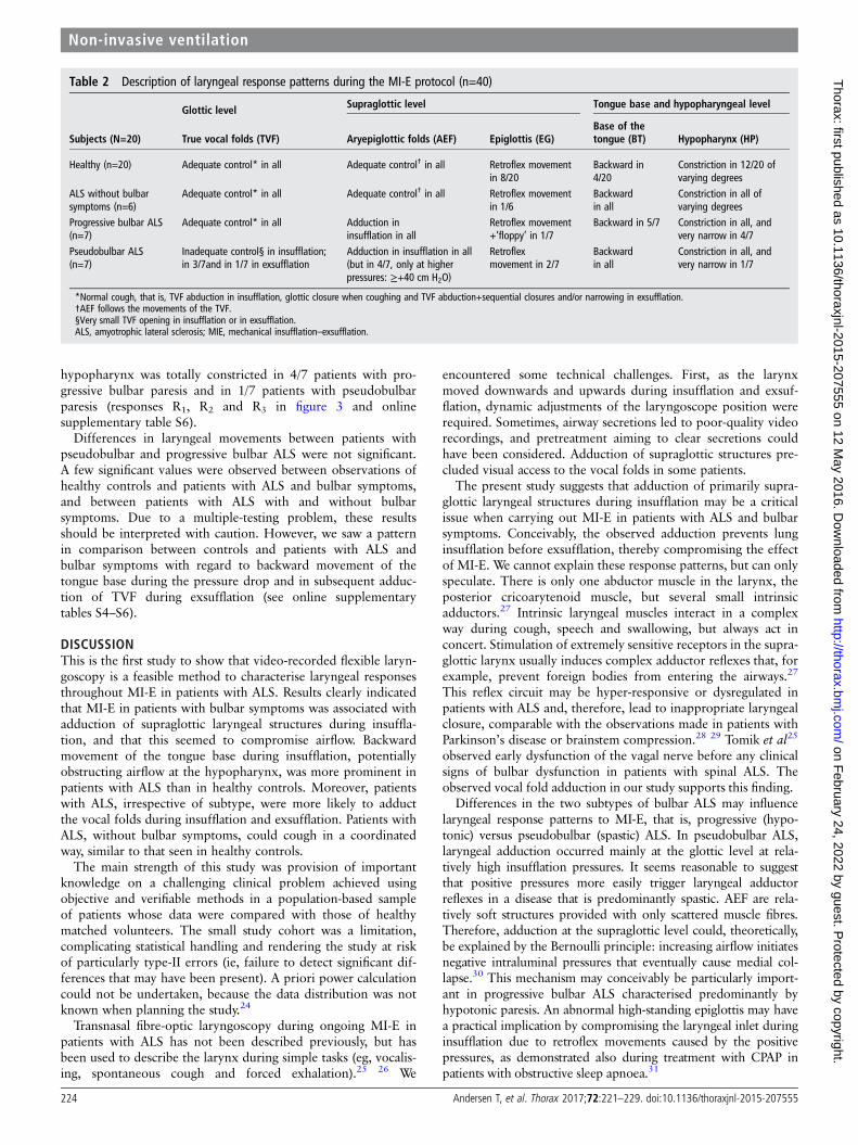
Table 2 Description of laryngeal response patterns during the MI-E protocol (n=40)
Glottic level Supraglottic level Tongue base and hypopharyngeal level
Subjects (N=20) True vocal folds (TVF) Aryepiglottic folds (AEF) Epiglottis (EG)Base of thetongue (BT) Hypopharynx (HP)
Healthy (n=20) Adequate control* in all Adequate control† in all Retroflex movementin 8/20
Backward in4/20
Constriction in 12/20 ofvarying degrees
ALS without bulbarsymptoms (n=6)
Adequate control* in all Adequate control† in all Retroflex movementin 1/6
Backwardin all
Constriction in all ofvarying degrees
Progressive bulbar ALS(n=7)
Adequate control* in all Adduction ininsufflation in all
Retroflex movement+‘floppy’ in 1/7
Backward in 5/7 Constriction in all, andvery narrow in 4/7
Pseudobulbar ALS(n=7)
Inadequate control§ in insufflation;in 3/7and in 1/7 in exsufflation
Adduction in insufflation in all(but in 4/7, only at higherpressures: ≥+40 cm H2O)
Retroflexmovement in 2/7
Backwardin all
Constriction in all, andvery narrow in 1/7
*Normal cough, that is, TVF abduction in insufflation, glottic closure when coughing and TVF abduction+sequential closures and/or narrowing in exsufflation.†AEF follows the movements of the TVF.§Very small TVF opening in insufflation or in exsufflation.ALS, amyotrophic lateral sclerosis; MIE, mechanical insufflation–exsufflation.
224 Andersen T, et al. Thorax 2017;72:221–229. doi:10.1136/thoraxjnl-2015-207555
Non-invasive ventilation on F
ebruary 24, 2022 by guest. Protected by copyright.
http://thorax.bmj.com
/T
horax: first published as 10.1136/thoraxjnl-2015-207555 on 12 May 2016. D
ownloaded from
Hypopharyngeal constriction during exsufflation wasobserved to varying extents in all study subjects, as well ashealthy controls. This finding confirms reports of upper-airway
narrowing at pharyngeal and oropharyngeal levels upon appli-cation of negative pressures during exhalation in healthy sub-jects.32–34 This phenomenon has been used to explain the
Figure 1 Laryngeal response at the glottic level. Figures are percentages of the sample with the described response. *Significant differencebetween healthy volunteers and patients with ALS. ALS, amyotrophic lateral sclerosis; TVF, true vocal folds.
Andersen T, et al. Thorax 2017;72:221–229. doi:10.1136/thoraxjnl-2015-207555 225
Non-invasive ventilation on F
ebruary 24, 2022 by guest. Protected by copyright.
http://thorax.bmj.com
/T
horax: first published as 10.1136/thoraxjnl-2015-207555 on 12 May 2016. D
ownloaded from
ineffectiveness of MI-E in patients with ALS.35 Sancho et alundertook CT during MI-E at baseline and during exsufflationin three patients with ALS. They reported varying reductions
of the lateral diameter at the level of nasopharynx, uvula andpharynx during the exsufflation phase at −40 cm H2O.12 Theresponse during insufflation was not examined. They suggested
Figure 2 Laryngeal response at the supraglottic level. Figures are percentages of the sample with the described response.*Significant differencebetween healthy volunteers and patients with ALS. AEF, aryepiglottic folds; ALS, amyotrophic lateral sclerosis.
226 Andersen T, et al. Thorax 2017;72:221–229. doi:10.1136/thoraxjnl-2015-207555
Non-invasive ventilation on F
ebruary 24, 2022 by guest. Protected by copyright.
http://thorax.bmj.com
/T
horax: first published as 10.1136/thoraxjnl-2015-207555 on 12 May 2016. D
ownloaded from
that MI-E should be carried out by applying a single insuffla-tion followed by a manually assisted cough instead of activeexsufflation with negative pressures.35 36 Hypopharyngeal con-striction during exsufflation was observed in healthy controlsand patients with ALS in the present study; so, this phenom-enon alone cannot explain treatment failure in bulbar ALS.Moreover, inability to fill the lungs during insufflation becauseof the observed supraglottic adduction would create a vacuumduring the subsequent active exsufflation, and thereby aggravatehypopharyngeal constriction. If this hypothesis is correct, asingle insufflation followed by a manually assisted coughcannot help patients with bulbar ALS to cough more effect-ively, but will be both uncomfortable and unproductive.
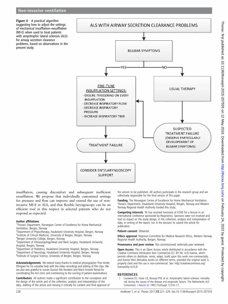
The present study suggests that an individual approach toMI-E used in respiratory airway therapy is highly important.Lower positive pressures and airflow combined with longerinspiratory times may contribute to better laryngeal stabilityduring insufflation, perhaps by preventing or reducing the impactof protective laryngeal reflex circuits and the intraluminal suctionforces induced by the Bernoulli effect (figure 4). Patients withbulbar insufficiency may, therefore, be more likely to obtain suffi-cient inspiratory volumes, a situation that would improve theconditions for exsufflation of the lungs. The phasic relationship
that exists between the posterior cricoarytenoid muscle and dia-phragm is a feature that could, theoretically, be exploited clinic-ally. That is, when the diaphragm contracts, the activity of theposterior cricoarytenoid muscle increases in a coordinatedmanner due to vagal stimulation, thereby abducting the larynx.27
If the patient is instructed to inhale actively before active insuffla-tion with MI-E, this act would, theoretically, lead to better laryn-geal abduction and facilitate airflow. Recently, MI-E devices witha ‘trigger’ function linked to insufflation have become available,and these mechanisms should be studied closely.
A better understanding of laryngeal dysfunction as ALS pro-gresses in its various phenotypes can help establish better (andhopefully individually tailored) clinical respiratory treatmentstrategies for these patients, and perhaps also for other patientswith bulbar-innervated muscle dysfunction.
CONCLUSIONVideo-recorded flexible laryngoscopy is a feasible method tocharacterise laryngeal responses throughout an MI-E protocol inpatients with ALS. Treatment failure with MI-E in patients withbulbar symptoms is likely to be caused primarily by laryngealadduction during insufflation, predominantly at the supraglotticlevel. This response precludes air-filling of the lungs during
Figure 3 Laryngeal response at the tongue base and hypopharyngeal level. Figures are percentages of the sample with the described response.*Significant difference between healthy volunteers and patients with amyotrophic lateral sclerosis (ALS).
Andersen T, et al. Thorax 2017;72:221–229. doi:10.1136/thoraxjnl-2015-207555 227
Non-invasive ventilation on F
ebruary 24, 2022 by guest. Protected by copyright.
http://thorax.bmj.com
/T
horax: first published as 10.1136/thoraxjnl-2015-207555 on 12 May 2016. D
ownloaded from
insufflation, causing discomfort and subsequent inefficientexsufflation. We propose that individually customised settingsfor pressure and flow can improve and extend the use of non-invasive MI-E in ALS, and that flexible laryngoscopy can be anefficient tool in this respect in selected patients who do notrespond as expected.
Author affiliations1Thoracic Department, Norwegian Centre of Excellence for Home MechanicalVentilation, Bergen, Norway2Department of Physiotherapy, Haukeland University Hospital, Bergen, Norway3Institute of Clinical Medicine, University of Bergen, Bergen, Norway4Bergen University College, Bergen, Norway5Department of Otolaryngology/Head and Neck Surgery, Haukeland UniversityHospital, Bergen, Norway6Department of Pediatrics, Haukeland University Hospital, Bergen, Norway7Department of Neurology, Haukeland University Hospital, Bergen, Norway8Institute of Surgical Science, University of Bergen, Bergen, Norway
Acknowledgements We extend many thanks to medical photographer Thor-AndreEllingsen for his valuable help with the video recordings and editing of film clips. Weare also very grateful to nurses Gunvor Mo Norstein and Marit Arnevik Renså forcoordinating the ALS clinic and contributing to the running of patient examinations.
Contributors All authors made a significant contribution to the conception andthe design of the article and of the collection, analysis and interpretation of thedata, drafting of the article and revising it critically for content and final approval of
the version to be published. All authors participate in the research group and arecollectively responsible for the final version of this paper.
Funding The Norwegian Centre of Excellence for Home Mechanical Ventilation,Thoracic Department, Haukeland University Hospital, Bergen, Norway and WesternNorway Regional Health Authority funded this study.
Competing interests TA has received honoraria of €500 for a lecture in aninternational conference sponsored by Respironics. Sponsors were not involved andhad no impact on the study design, in the collection, analysis and interpretation ofdata, in writing of the report, nor in the decision to submit the article forpublication.
Patient consent Obtained.
Ethics approval Regional Committee for Medical Research Ethics, Western NorwayRegional Health Authority, Bergen, Norway.
Provenance and peer review Not commissioned; externally peer reviewed.
Open Access This is an Open Access article distributed in accordance with theCreative Commons Attribution Non Commercial (CC BY-NC 4.0) license, whichpermits others to distribute, remix, adapt, build upon this work non-commercially,and license their derivative works on different terms, provided the original work isproperly cited and the use is non-commercial. See: http://creativecommons.org/licenses/by-nc/4.0/
REFERENCES1 Louwerse ES, Visser CE, Bossuyt PM, et al. Amyotrophic lateral sclerosis: mortality
risk during the course of the disease and prognostic factors. The Netherlands ALSConsortium. J Neurol Sci 1997;152(Suppl 1):S10–17.
Figure 4 A practical algorithmsuggesting how to adjust the settingsof mechanical insufflation–exsufflation(MI-E) when used to treat patientswith amyotrophic lateral sclerosis (ALS)for airway secretion clearanceproblems, based on observations in thepresent study.
228 Andersen T, et al. Thorax 2017;72:221–229. doi:10.1136/thoraxjnl-2015-207555
Non-invasive ventilation on F
ebruary 24, 2022 by guest. Protected by copyright.
http://thorax.bmj.com
/T
horax: first published as 10.1136/thoraxjnl-2015-207555 on 12 May 2016. D
ownloaded from
2 Howard RS, Orrell RW. Management of motor neurone disease. Postgrad Med J2002;78:736–41.
3 de Carvalho M, Matias T, Coelho F, et al. Motor neuron disease presenting withrespiratory failure. J Neurol Sci 1996;139Suppl:117–22.
4 Lechtzin N, Wiener CM, Clawson L, et al. Hospitalization in amyotrophic lateralsclerosis: causes, costs, and outcomes. Neurology 2001;56:753–7.
5 Bourke SC, Shaw PJ, Gibson GJ. Respiratory function vs sleep-disordered breathingas predictors of QOL in ALS. Neurology 2001;57:2040–4.
6 Lechtzin N. Respiratory effects of amyotrophic lateral sclerosis: problems andsolutions. Respir Care 2006;51:871–81.
7 Benditt JO, Boitano LJ. Pulmonary issues in patients with chronic neuromusculardisease. Am J Respir Crit Care Med 2013;187:1046–55.
8 Leith DE. The development of cough. Am Rev Respir Dis 1985;131:39–42.9 Woodson G. Management of neurologic disorders of the larynx. Ann Otol Rhinol
Laryngol 2008;117:317–26.10 Homnick DN. Mechanical insufflation-exsufflation for airway mucus clearance. Respir
Care 2007;52:1296–305.11 Anderson JL, Hasney KM, Beaumont NE. Systematic review of techniques to
enhance peak cough flow and maintain vital capacity in neuromuscular disease: thecase for mechanical insufflation-exsufflation. Phys Ther Rev 2013;10:25–33.
12 Sancho J, Servera E, Diaz J, et al. Efficacy of mechanical insufflation-exsufflation inmedically stable patients with amyotrophic lateral sclerosis. Chest 2004;125:1400–5.
13 Brooks BR. El Escorial World Federation of Neurology criteria for the diagnosis ofamyotrophic lateral sclerosis. Subcommittee on Motor Neuron Diseases/AmyotrophicLateral Sclerosis of the World Federation of Neurology Research Group onNeuromuscular Diseases and the El Escorial “Clinical limits of amyotrophic lateralsclerosis” workshop contributors. J Neurol Sci 1994;124(Suppl):96–107.
14 Brooks BR, Miller RG, Swash M, et al. El Escorial revisited: revised criteria for thediagnosis of amyotrophic lateral sclerosis. Amyotroph Lateral Scler Other MotorNeuron Disord 2009;1:293–9.
15 Cedarbaum JM, Stambler N, Malta E, et al. The ALSFRS-R: a revised ALS functionalrating scale that incorporates assessments of respiratory function. BDNF ALS StudyGroup (Phase III). J Neurol Sci 1999;169:13–21.
16 Volanti P, Cibella F, Sarvà M, et al. Predictors of non-invasive ventilationtolerance in amyotrophic lateral sclerosis. J Neurol Sci 2011;303:114–18.
17 Hughes TA, Wiles CM. Clinical measurement of swallowing in health and inneurogenic dysphagia. QJM 1996;89:109–16.
18 Wu MC, Chang YC, Wang TG, et al. Evaluating swallowing dysfunction using a100-ml water swallowing test. Dysphagia 2004;19:43–7.
19 [No authors listed]. Standardized lung function testing. Official statement of theEuropean Respiratory Society. Eur Respir J Suppl 1993;16:1–100.
20 Knudson RJ, Lebowitz MD, Holberg CJ, et al. Changes in the normal maximalexpiratory flow-volume curve with growth and aging. Am Rev Respir Dis1983;127:725–34.
21 Evans JA, Whitelaw WA. The assessment of maximal respiratory mouth pressures inadults. Respir Care 2009;54:1348–59.
22 Uldry C, Fitting JW. Maximal values of sniff nasal inspiratory pressure in healthysubjects. Thorax 1995;50:371–5.
23 Andersen T, Sandnes A, Hilland M, et al. Laryngeal response patterns to mechanicalinsufflation-exsufflation in healthy subjects. Am J Phys Med Rehabil2013;92:920–9.
24 Bacchetti P, Leung JM. Sample size calculations in clinical research. Anesthesiology2002;97:1028–9.
25 Tomik J, Tomik B, Partyka D, et al. Profile of laryngological abnormalities in patientswith amyotrophic lateral sclerosis. J Laryngol Otol 2007;121:1064–9.
26 Polkey MI, Lyall RA, Green M, et al. Expiratory muscle function in amyotrophiclateral sclerosis. Am J Respir Crit Care Med 1998;158:734–41.
27 Brancatisano TP, Dodd DS, Engel LA. Respiratory activity of posterior cricoarytenoidmuscle and vocal cords in humans. J Appl Physiol 1984;57:1143–9.
28 Koufman JA, Block C. Differential diagnosis of paradoxical vocal fold movement.Am J Speech Lang Pathol 2008;17:327–34.
29 Maschka DA, Bauman NM, McCray PB Jr, et al. A classification scheme forparadoxical vocal cord motion. Laryngoscope 1997;107:1429–35.
30 Fajdiga I. Snoring imaging: could Bernoulli explain it all? Chest 2005;128:896–901.
31 Dedhia RC, Rosen CA, Soose RJ. What is the role of the larynx in adult obstructivesleep apnea? Laryngoscope 2014;124:1029–34.
32 Suratt PM, Wilhoit SC, Cooper K. Induction of airway collapse with subatmosphericpressure in awake patients with sleep apnea. J Appl Physiol 1984;57:140–6.
33 Sanna A, Veriter C, Kurtansky A, et al. Contraction and relaxation of upper airwaymuscles during expiratory application of negative pressure at the mouth. Sleep1994;17:220–5.
34 Younes M, Sanii R, Patrick W, et al. An approach to the study of upper airwayfunction in humans. J Appl Physiol 1994;77:1383–94.
35 Bach JR. Mechanical insufflation/exsufflation: has it come of age? A commentary.Eur Respir J 2003;21:385–6.
36 Kang SW, Bach JR. Maximum insufflation capacity: vital capacity and cough flows inneuromuscular disease. Am J Phys Med Rehabil 2000;79:222–7.
Andersen T, et al. Thorax 2017;72:221–229. doi:10.1136/thoraxjnl-2015-207555 229
Non-invasive ventilation on F
ebruary 24, 2022 by guest. Protected by copyright.
http://thorax.bmj.com
/T
horax: first published as 10.1136/thoraxjnl-2015-207555 on 12 May 2016. D
ownloaded from