original article impaired cleavage of preproinsulin signal
TRANSCRIPT
Impaired Cleavage of Preproinsulin Signal PeptideLinked to Autosomal-Dominant DiabetesMing Liu,
1,2Roberto Lara-Lemus,
1Shu-ou Shan,
3Jordan Wright,
1Leena Haataja,
1Fabrizio Barbetti,
4
Huan Guo,1Dennis Larkin,
1and Peter Arvan
1
Recently, missense mutations upstream of preproinsulin’s signalpeptide (SP) cleavage site were reported to cause mutant INSgene-induced diabetes of youth (MIDY). Our objective was tounderstand the molecular pathogenesis using metabolic labelingand assays of proinsulin export and insulin and C-peptide pro-duction to examine the earliest events of insulin biosynthesis,highlighting molecular mechanisms underlying b-cell failure plusa novel strategy that might ameliorate the MIDY syndrome. We findthat whereas preproinsulin-A(SP23)S is efficiently cleaved, pro-ducing authentic proinsulin and insulin, preproinsulin-A(SP24)Dis inefficiently cleaved at an improper site, producing two subpo-pulations of molecules. Both show impaired oxidative folding andare retained in the endoplasmic reticulum (ER). Preproinsulin-A(SP24)D also blocks ER exit of coexpressed wild-type proinsulin,accounting for its dominant-negative behavior. Upon increasedexpression of ER–oxidoreductin-1, preproinsulin-A(SP24)D remainsblocked but oxidative folding of wild-type proinsulin improves,accelerating its ER export and increasing wild-type insulin pro-duction. We conclude that the efficiency of SP cleavage is linkedto the oxidation of (pre)proinsulin. In turn, impaired (pre)pro-insulin oxidation affects ER export of the mutant as well as thatof coexpressed wild-type proinsulin. Improving oxidative fold-ing of wild-type proinsulin may provide a feasible way to rescueinsulin production in patients with MIDY. Diabetes 61:828–837, 2012
Among the many monogenic causes of diabetes(1–3), mutant insulin gene syndrome was origi-nally used to describe three autosomal-dominantmutations associated with an adult-onset, type 2
diabetes-like phenotype (4). More recently, in addition torecessive INS gene mutations (5), new INS gene mutationshave been found to underlie a syndrome we call mutantINS gene-induced diabetes of youth (MIDY), in whichheterozygotes develop autosomal-dominant diabetes, withonset from neonatal life to adulthood (6–15). The MIDYmutations cause misfolding of the insulin precursor pro-tein (16,17), resulting in retention within the endoplasmicreticulum (ER) of b-cells (18,19). Importantly, such mis-folded proinsulin forms protein complexes that can in-corporate innocent bystander proinsulin (20), impairing
the ER exit of wild-type (WT) proinsulin and decreasinginsulin production (21). Moreover, recent studies indicatethat insulin deficiency precedes a net loss of b-cell mass(22,23), suggesting that this dominant-negative blockadecaused by mutants is sufficient to account for initial onsetof diabetes in MIDY (24,25).
In b-cells, insulin synthesis begins with the precursorpreproinsulin, which must undergo cotranslational trans-location into the ER, signal peptide (SP) cleavage anddownstream proinsulin folding. These earliest events arecritical to insulin biosynthesis, but they are relativelyunderstudied since the discovery of preproinsulin (26).Two preproinsulin SP mutations located at or next to theSP cleavage site have been recently reported to causehuman diabetes (2,3,6,8), which makes investigation of theseearliest events especially timely. One of them, preproinsulin-A(SP24)D, was reported to impair preproinsulin processing(26) and induce ER stress; yet, A(SP24)D reportedly pro-duced no significant adverse effect on insulin productionfrom a coexpressed WT allele (27). Thus, it is uncertainwhy A(SP24)D should not be a recessive rather than adominant mutation causing severe, early onset, insulin-deficient diabetes.
In this report, we have more closely examined A(SP23)Sand A(SP24)D, both reportedly associated with humanMIDY (6,7,10,12). We find that A(SP23)S has no detectablebiological defect and is likely to be a polymorphic variant.By contrast, upon translocation into the ER lumen, A(SP24)Dexhibits defective SP cleavage, resulting in abnormal oxi-dative folding of downstream proinsulin, forming disulfide-linked protein complexes that are retained in the ER. Wedemonstrate that A(SP24)D expression results in dominant-negative blockade of coexpressed proinsulin-WT, a com-mon mechanism in MIDY (20,24,25). Finally, we haveexplored a potential therapeutic mechanism that, in thepresence of A(SP24)D, can improve the native oxidativematuration of coexpressed proinsulin-WT, enhancing itsexport and increasing insulin production.
RESEARCH DESIGN AND METHODS
Materials. Guinea pig anti-porcine insulin, rat insulin (#RI-13 K), and humaninsulin-specific (#HI-14 K) and proinsulin-specific radioimmunoassay (#HPI-15K) were from Millipore; rabbit anti-Myc and anti-GFP were from ImmunologyConsultants Laboratories; Zysorbin was from Zymed; 35S-amino acid mixture(Met+Cys) and pure 35S-Met were from ICN; dithiothreitol (DTT), proteinA-agarose, digitonin, N-ethylmaleimide (NEM), and radioimmunoassay (RIA)-grade bovine serum albumin were from Sigma-Aldrich; Met/Cys-deficientDulbecco’s modified Eagle’s medium (DMEM) and all other tissue culturereagents were from Invitrogen. Plasmids encoding human Ero1a and Ero1bwere from Dr. P. Scherer (University of Texas Southwestern), and mouseprotein disulfide isomerase (PDI) was from Dr. M. Green (St. Louis University).Human and mouse preproinsulin mutagenesis. The human INS cDNA andmouse Ins2 cDNA with or without Myc tag as described (21) were subclonedinto pTarget and pCMS-GFP vectors. Mutations were introduced using theQuikChange site-directed mutagenesis kit (Stratagene) and confirmed by di-rect DNA sequencing.
From the 1Division of Metabolism, Endocrinology & Diabetes, University ofMichigan Medical Center, Ann Arbor, Michigan; 2Tianjin Medical UniversityGeneral Hospital, Tianjin, China; the 3Division of Chemistry and ChemicalEngineering, California Institute of Technology, Pasadena, California; andthe 4Laboratory of Molecular Endocrinology and Metabolism, BambinoGesù Children’s Hospital, Scientific Institute (Istituto Di Ricovero e Curaa Carattere Scientifico), Rome, Italy.
Corresponding authors: Ming Liu, [email protected], and Peter Arvan, [email protected].
Received 24 June 2011 and accepted 19 December 2011.DOI: 10.2337/db11-0878� 2012 by the American Diabetes Association. Readers may use this article as
long as the work is properly cited, the use is educational and not for profit,and the work is not altered. See http://creativecommons.org/licenses/by-nc-nd/3.0/ for details.
828 DIABETES, VOL. 61, APRIL 2012 diabetes.diabetesjournals.org
ORIGINAL ARTICLE
Cell culture, transfection, metabolic labeling, immunoprecipitation,
SDS-PAGE, Western blotting, and ER stress response. INS1 (rat) cells,Min6 (mouse) cells, or 293T (human) cells were plated onto 12-well plates 1 daybefore transfection with Lipofectamine (Invitrogen) using 1 to 2 mg plasmidDNA. At 48 h after transfection, cells were pulse-labeled with 35S-Met/Cys orpure 35S-Met and chased as indicated. Cells used for analysis of (pre)pro-insulin oxidative folding were preincubated with 20 mmol/L NEM in PBS onice for 10 min before lysis. Lysis buffer (1% Triton X-100, 0.1% SDS, Tris, pH7.4) contained 2 mmol/L NEM and a proteinase inhibitor cocktail. For im-munoprecipitation, samples were precleared with Zysorbin and immunopre-cipitated as described previously (21). Nonreducing or reducing Tris-Tricineurea–SDS-PAGE was performed as described (18). For Western blotting, 20 mgtotal lysate protein was boiled in SDS sample buffer with or without 100 mmol/LDTT, resolved by 4–12% NuPage, electrotransferred to nitrocellulose, andblotted with rabbit anti-Myc followed by anti-rabbit–HRP conjugate, with de-velopment by enhanced chemiluminescence. The BiP promoter-driven fireflyluciferase assay for Min6 cells 48 h posttransfection to express humanpreproinsulin-WT or mutants (normalized to Renilla luciferase activity) wasperformed as described (21) using the dual-luciferase reporter kit (Promega).Partial permeabilization of plasma membranes with digitonin. After la-beling with 35S-Cys/Met, 293T cells transfected with human preproinsulin-WT
or mutant were washed, resuspended, and incubated in 50 ml of 150 mmol/LNaCl, 2 mmol/L CaCl2, 50 mmol/L HEPES, pH 7.5, 6 0.01% digitonin on ice for10 min. The cells were spun at 14,000 rpm for 10 min at 4°C; the supernate wastransferred to a new tube containing 450 mL lysis buffer and the pellet lysed in500 mL lysis buffer. Both supernate and pellet were immunoprecipitated withanti-insulin or anti-GFP and analyzed using 4–12% NuPage.Radioimmunoassay of WT human proinsulin and insulin. 293T cells werecotransfected with human preproinsulin-WT and either mouse preproinsulin-WTor mutants, plus empty vector or a vector expressing PDI or ER–oxidoreductin-1(ERO1), at a DNA molar ratio of 1:2:3. At 40 h posttransfection, medium-bathingtransfected cells were collected for an additional 16 h and used to measuresecreted human proinsulin using the human proinsulin-specific RIA.
Min6 cells were transfected with vectors expressing human preproinsulin-WT or mutants. At 40 h posttransfection, cells were incubated with DMEMcontaining 25.5 mmol/L glucose and 10% FBS for 16 h. The media were thencollected and cells lysed with acid/ethanol (20) and human insulin measured byhuman insulin-specific RIA. In parallel, duplicate wells of each transfectionwere used for isolating total RNA. Human insulin mRNA levels were determinedby RT-PCR using forward primer: 59-GAACCAACACCTGTGCGGCTCAC-39 andreverse primer: 59-CGAGGCTTCTTCTACACACCCA-39. In this way, human in-sulin protein level was normalized to human insulin mRNA level.
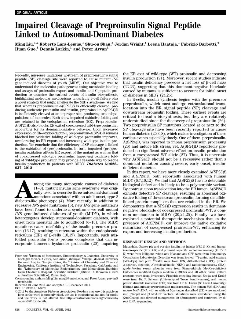
FIG. 1. A(SP23)S exhibits no abnormal phenotype, whereas A(SP24)D exhibits a defect in SP cleavage and insulin production and induces ERstress in b-cells. A: Signal sequence alignment of preproinsulins of various species. The arrow indicates the predicted signal peptide cleavage site;the 21 residue is boxed and a Ser-SP23 (22) residue in chimpanzee preproinsulin is circled. B: MIN6 cells were transiently transfected withplasmids encoding hProCpepMyc-WT, A(SP23)S, or A(SP24)D and lysed after 48 h posttransfection, and 20 mg total protein was used for anti-MycWestern blot as described in RESEARCH DESIGN AND METHODS. C: MIN6 cells were transiently transfected with human preproinsulin-WT, A(SP23)S, orA(SP24)D plasmids. At 40 h posttransfection, the cells were incubated with fresh DMEM medium containing 25.5 mmol/L glucose and 10% FBS foradditional 16 h; the media were collected and cells lysed as in RESEARCH DESIGN AND METHODS. Human insulin in the lysates (black bars) and media(white bars) were quantified using human-specific insulin RIA normalized to human-specific insulin mRNA (a measure of the efficiency of insulinproduction from the transfected translation product). Results shown are mean values 6 SD from three independent experiments. *P < 0.01compared with preproinsulin-WT or A(SP23)S. D: Min6 cells cotransfected to express BiP promoter-firefly luciferase, CMV promoter-driven R.luciferase, and preproinsulin-WT or mutants were lysed at 48 h posttransfection, and the ratio of firefly/R. luciferase was measured. Cellsexpressing WT proinsulin or empty vector (EV) served as a negative control. Results are expressed as mean 6 SD from three independentexperiments. *P < 0.05 compared with preproinsulin-WT.
M. LIU AND ASSOCIATES
diabetes.diabetesjournals.org DIABETES, VOL. 61, APRIL 2012 829
Statistical analysis. Statistical analyses were carried out by ANOVA fol-lowed by Bonferroni multiple comparison test using GraphPad Prism 5. Dataare presented as means 6 SD. A P value of ,0.05 was taken as statisticallysignificant.
RESULTS
SP cleavage of human preproinsulin-A(SP23)S andA(SP24)D. Evolutionarily, the preproinsulin SP divergesmore in sequence than does the insulin B-chain or A-chain(28). In tetrapods, residue A(SP24) predominates at thecleavage site (residue “21”), whereas there is considerablenatural variation at the penultimate “22” position (Fig. 1A).In chimpanzee, S(SP23) predominates (Fig. 1A), and thereis a polymorphic variation in this position even within thehuman genome (29). Thus, it was of interest to further in-vestigate the physiological significance of both A(SP23)Sand A(SP24)D substitutions.
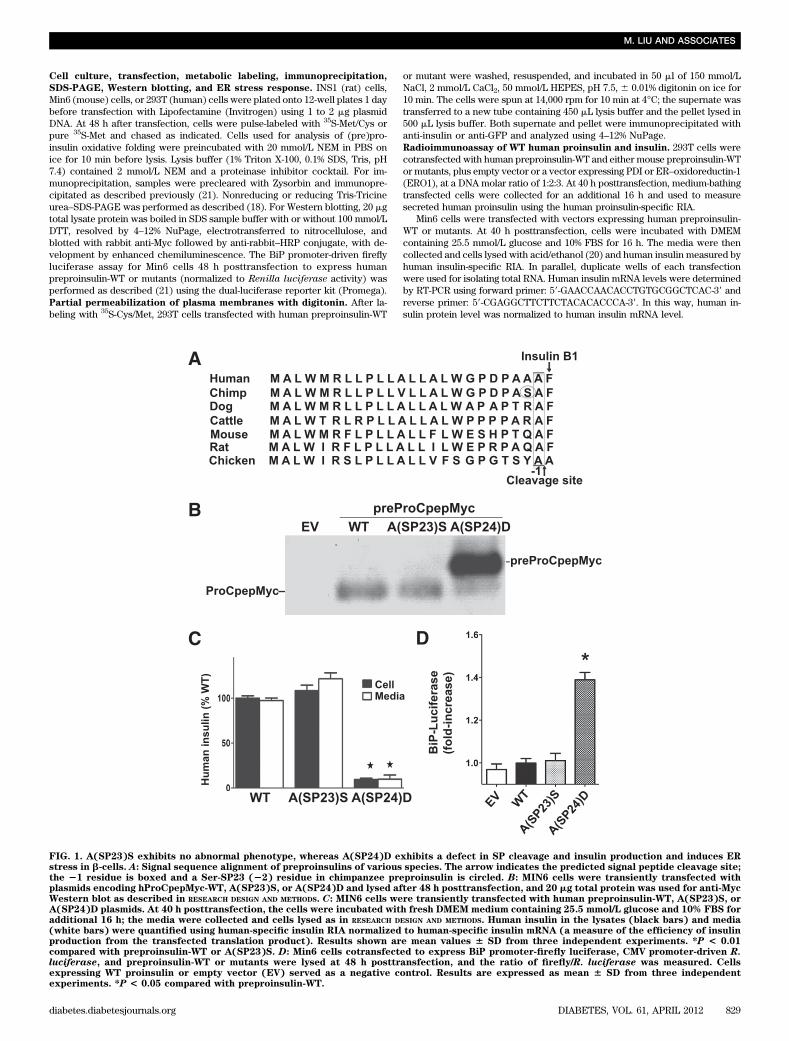
In MIN6 pancreatic b-cells transfected to express Myc-tagged human preproinsulin [preProCpepMyc (21)], A(SP24)Dwas predominantly uncleaved, with a minor cleaved bandmigrating near ProCpepMyc-WT (Fig. 1B). Impaired ef-ficiency of SP cleavage (Fig. 1B) can directly account fordecreased human insulin production (Fig. 1C). By contrast,A(SP23)S showed no defect of SP cleavage or humaninsulin production (Fig. 1B and C). Moreover, A(SP24)Dinduced ER stress in Min6 cells, whereas the ER stress re-sponse for A(SP23)S was similar to that for preproinsulin-WT (Fig. 1D). In 293T cells, newly synthesized A(SP23)Swas rapidly processed to proinsulin and secreted, exactlylike WT, whereas A(SP24)D showed little preproinsulinprocessing or secretion (Fig. 2A). From multiple suchexperiments, #10% of A(SP24)D underwent SP cleavage(Fig. 2B). We examined the subsequent fate of newly syn-thesized mutant preproinsulin for up to 4 h of chase.Although secretion of either uncleaved or SP-cleavedA(SP24)D was negligible, there was intracellular loss ofA(SP24)D, some of which was clearly blocked by theproteasome inhibitor MG132 (Fig. 2C). Although the data
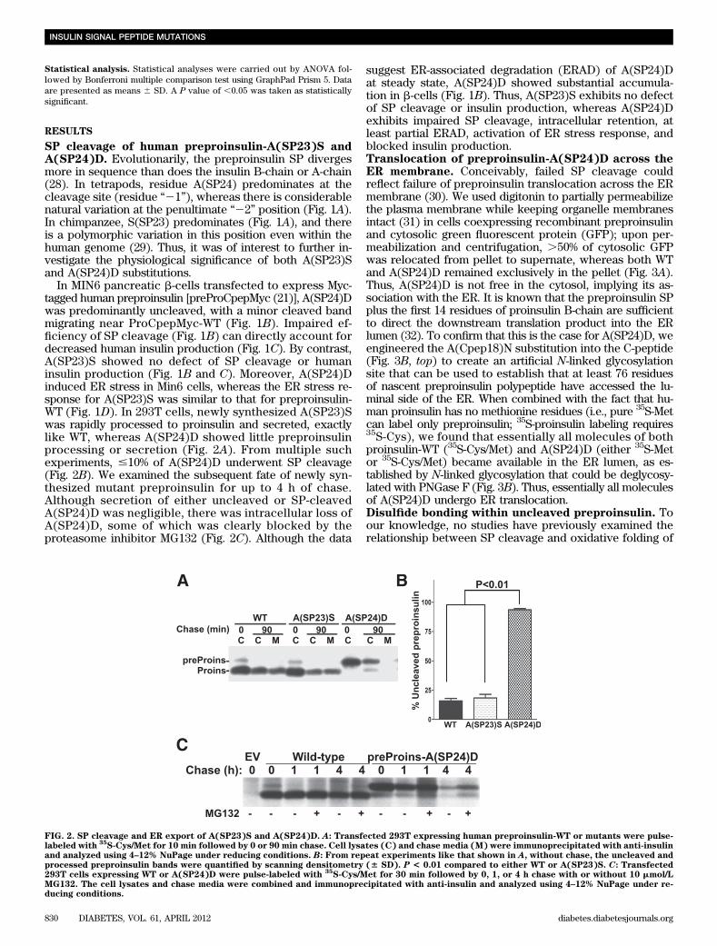
suggest ER-associated degradation (ERAD) of A(SP24)Dat steady state, A(SP24)D showed substantial accumula-tion in b-cells (Fig. 1B). Thus, A(SP23)S exhibits no defectof SP cleavage or insulin production, whereas A(SP24)Dexhibits impaired SP cleavage, intracellular retention, atleast partial ERAD, activation of ER stress response, andblocked insulin production.Translocation of preproinsulin-A(SP24)D across theER membrane. Conceivably, failed SP cleavage couldreflect failure of preproinsulin translocation across the ERmembrane (30). We used digitonin to partially permeabilizethe plasma membrane while keeping organelle membranesintact (31) in cells coexpressing recombinant preproinsulinand cytosolic green fluorescent protein (GFP); upon per-meabilization and centrifugation, .50% of cytosolic GFPwas relocated from pellet to supernate, whereas both WTand A(SP24)D remained exclusively in the pellet (Fig. 3A).Thus, A(SP24)D is not free in the cytosol, implying its as-sociation with the ER. It is known that the preproinsulin SPplus the first 14 residues of proinsulin B-chain are sufficientto direct the downstream translation product into the ERlumen (32). To confirm that this is the case for A(SP24)D, weengineered the A(Cpep18)N substitution into the C-peptide(Fig. 3B, top) to create an artificial N-linked glycosylationsite that can be used to establish that at least 76 residuesof nascent preproinsulin polypeptide have accessed the lu-minal side of the ER. When combined with the fact that hu-man proinsulin has no methionine residues (i.e., pure 35S-Metcan label only preproinsulin; 35S-proinsulin labeling requires35S-Cys), we found that essentially all molecules of bothproinsulin-WT (35S-Cys/Met) and A(SP24)D (either 35S-Metor 35S-Cys/Met) became available in the ER lumen, as es-tablished by N-linked glycosylation that could be deglycosy-lated with PNGase F (Fig. 3B). Thus, essentially all moleculesof A(SP24)D undergo ER translocation.Disulfide bonding within uncleaved preproinsulin. Toour knowledge, no studies have previously examined therelationship between SP cleavage and oxidative folding of
FIG. 2. SP cleavage and ER export of A(SP23)S and A(SP24)D. A: Transfected 293T expressing human preproinsulin-WT or mutants were pulse-labeled with
35S-Cys/Met for 10 min followed by 0 or 90 min chase. Cell lysates (C) and chase media (M) were immunoprecipitated with anti-insulin
and analyzed using 4–12% NuPage under reducing conditions. B: From repeat experiments like that shown in A, without chase, the uncleaved andprocessed preproinsulin bands were quantified by scanning densitometry (6 SD). P < 0.01 compared to either WT or A(SP23)S. C: Transfected293T cells expressing WT or A(SP24)D were pulse-labeled with
35S-Cys/Met for 30 min followed by 0, 1, or 4 h chase with or without 10 mmol/L
MG132. The cell lysates and chase media were combined and immunoprecipitated with anti-insulin and analyzed using 4–12% NuPage under re-ducing conditions.
INSULIN SIGNAL PEPTIDE MUTATIONS
830 DIABETES, VOL. 61, APRIL 2012 diabetes.diabetesjournals.org
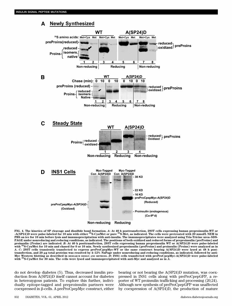
preproinsulin within the ER. We used nonreducing Tris-Tricine urea–SDS-PAGE (16) to examine disulfide bondformation in recombinant newly synthesized preproinsulin.Uncleaved preproinsulin-WT (labeled with 35S-Met) appearsfully reduced (Fig. 4A, lane 2). Upon SP removal, proinsulin-WT (labeled with 35S-Cys/Met) may begin as a fully re-duced species (Fig. 4A, lane 1, arrow) but proceeds tonative and isomeric oxidized proinsulin (lower bands).Within 10 min, reduced (pre)proinsulin nearly vanished,and oxidized native proinsulin became enriched (Fig. 4B,lane 2), indicating both rapid SP cleavage and downstreamproinsulin folding.
For A(SP24)D, the sequence of early events appeared al-tered: newly synthesized preproinsulin-A(SP24)D (35S-Met–labeled) started to become oxidized (Fig. 4A, lanes 7 and 8),and this increased after 10 min (Fig. 4B, lane 8). Bycomparing the recovery of fully reduced preproinsulin-A(SP24)D by nonreducing gel versus total preproinsulinrecovered under reducing conditions, we calculated (frommultiple experiments) that 42 6 2.7% of preproinsulin-A(SP24)D had oxidized (either properly or improperly, seebelow) at the zero chase time. The oxidized fraction ofpreproinsulin-A(SP24)D was 61% by 10 min of chase; 86%by 30 min of chase; and 90% under steady-state conditionsby Western blotting (Fig. 4C, right). By contrast, proinsulin-WT
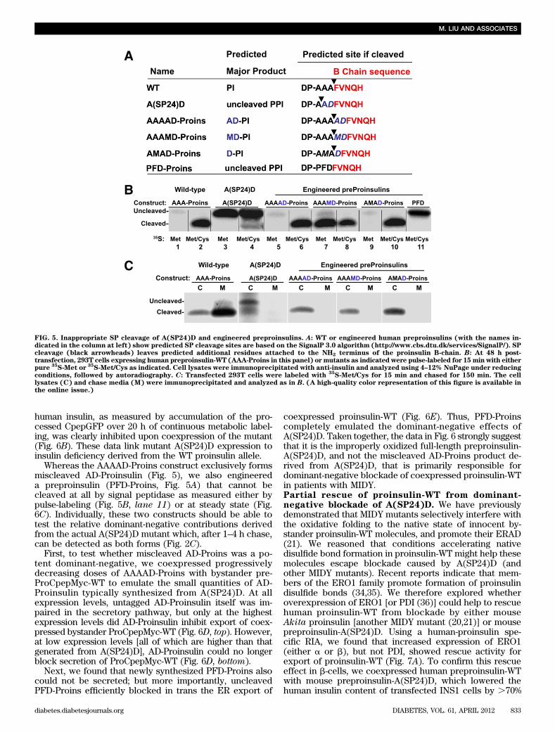
was .90% oxidized at the zero chase time, and there wasessentially no fully reduced proinsulin-WT remaining after10 min (Fig. 4B). Under steady-state conditions, #10% oxi-dized A(SP24)D remains as monomer recovered undernonreducing conditions (Fig. 4C, compare lanes 3 and 4),indicating that the majority forms disulfide-linked proteincomplexes. Most importantly, in b-cells, A(SP24)D coim-munoprecipitated coexpressed proinsulin-WT, which wasdetected only under reducing conditions; thus, this pro-insulin-WT must also have been recruited into disulfide-linked complexes (Fig. 4D). Altogether, the data in Figs. 3and 4 establish that impaired SP cleavage triggers defectiveoxidative folding within the ER, leaving exposed thiols onpreproinsulin that could create cellular problems by par-ticipating in inappropriate intermolecular thiol attack.Inappropriate SP cleavage of preproinsulin-A(SP24)Dand dominant-negative blockade of proinsulin-WT.Using the SignalP-3.0 computer-based algorithm, we notedthat whereas human preproinsulin-WT uses the AAA-proinsulin sequence to generate proinsulin beginning withthe appropriate F(B1) residue, the A(SP24)D sequence pre-dicts SP cleavage at low efficiency, generating a minorportion of AD-Proins that includes two extra residuespreceding the proinsulin B-chain (Fig. 5A). We thereforedesigned three additional preproinsulin mutants (AAAAD-Proins, AAAMD-Proins, and AMAD-Proins) intended tointerrogate the location of the preproinsulin cleavage sitewhile finding a high-efficiency means to generate the aber-rant AD-Proins cleavage product (Fig. 5A). By SDS-PAGE/autoradiography after metabolic labeling with 35S-Cys/Met,unlike the inefficient SP cleavage of A(SP24)D (Fig. 5B,lanes 3 and 4), other bioengineered preproinsulins un-derwent efficient (but aberrant) SP cleavage. In Fig. 5B,a comparison of lanes 2, 4, 6, 8, and 10 (each including35S-Cys label) showed the presence of a cleaved band inevery instance. However, when comparing lanes 3, 5, and7 (Fig. 5B) labeled with pure 35S-Met, the cleaved bandbecame undetectable except in lane 7 (AAAMD-Proins),when methionine was still found to be contained withinthe product that must have been cleaved upstream of theMet residue. By contrast, in lane 9 (AMAD-Proins; Fig. 5B)the cleaved product had lost the Met residue (i.e., a cleav-age site less than three residues upstream from the normalsite but containing at least two residues from the normalsite). These observations precisely match the cleavagesites predicted. Regardless of whether the cleavage reac-tion occurred at low efficiency or high efficiency, the ab-errant proinsulins bearing two extra NH2-terminal aminoacids in every instance failed to be secreted normally (Fig.5C). Thus, fidelity of SP cleavage, exposing the criticalproinsulin NH2-terminal arm, including phenylalanine-B1(33), is crucial to normal proinsulin folding and exportfrom the ER.
Because A(SP24)D forms aberrant disulfide-linked pro-tein complexes (Fig. 4C) that include coexpressed proinsulin-WT (Fig. 4D), we made A(SP24)D-DelCys (in which all sixcysteines were mutated) to test for dominant-negativebehavior. Interestingly, most of the blockade of WTproinsulin secretion was lost when thiols were elimi-nated from the A(SP24)D mutant (Fig. 6A). These datasupport the concept of thiol attack as a mechanism ofdominant-negative behavior of A(SP24)D (i.e., preciselythe danger mechanism that has been proposed for MIDY)(21,25).
Heterozygous patients bearing INS gene mutations thatcause decreased insulin biosynthesis from the mutant allele
FIG. 3. Translocation of preproinsulin-A(SP24)D across the ER mem-brane. A: At 40 h posttransfection, 293T cells coexpressing cytosolicGFP and preproinsulin-WT or -A(SP24)D were pulse-labeled with35S-Cys/Met for 15min and then treated with 0.01% digitonin on ice for 10
min to permeabilize the plasma membrane, which liberates a majorfraction of cytosolic GFP. Cells were then sedimented at 14,000 rpm at4°C for 10 min and each pellet (P) and supernate (S) analyzed se-quentially by anti-insulin and anti-GFP immunoprecipitation, 4–12%NuPage, under reducing conditions and autoradiography. B: The 18thresidue of human preproinsulin C-peptide was mutated from Ala to Asnto create an N-linked glycosylation site. At 40 h posttransfection, 293Tcells expressing human preproinsulin-A(Cpep18)N bearing or lackingthe A(SP24)D mutation were pulse-labeled with either pure
35S-Met or
mixed35S-Cys/Met for 15 min before lysis and immunoprecipitation
with anti-insulin. Immunoprecipitates were split in half and eitherdigested (+) or mock digested (2) with PNGase F at 37°C for 1 h beforeanalysis by 4–12% NuPage under reducing conditions. At this exposure,preproinsulin-WT is not detected with pure
35S-Met, indicating that the
vast majority of molecules have already undergone SP cleavage;thus, the same sample labeled with
35S-Cys/Met indicates glycosy-
lated (Glyco-Proins) or deglycosylated proinsulin (Proins). However,A(SP24)D remains labeled with pure
35S-Met, indicating the positions
of glycosylated (Glyco-preProins) or deglycosylated preproinsulins(preProins).
M. LIU AND ASSOCIATES
diabetes.diabetesjournals.org DIABETES, VOL. 61, APRIL 2012 831
do not develop diabetes (5). Thus, decreased insulin pro-duction from A(SP24)D itself cannot account for diabetesin heterozygous patients. To explore this further, indivi-dually epitope-tagged and preproinsulin partners werecoexpressed in b-cells. A preProCpepMyc construct, either
bearing or not bearing the A(SP24)D mutation, was coex-pressed in INS1 cells along with preProCpepGFP, a re-porter of WT proinsulin trafficking and processing (20,24).Although new synthesis of preProCpepGFP was unaffectedby coexpression of A(SP24)D, the production of mature
FIG. 4. The kinetics of SP cleavage and disulfide bond formation. A: At 40 h posttransfection, 293T cells expressing human preproinsulin-WT or-A(SP24)D were pulse-labeled for 10 min with either
35S-Cys/Met or pure
35S-Met, as indicated. The cells were pretreated with 20 mmol/L NEM in
PBS on ice for 10 min before lysis and immunoprecipitation with anti-insulin. The immunoprecipitates were analyzed using Tris-Tricine urea–SDS-PAGE under nonreducing and reducing conditions, as indicated. The positions of both oxidized and reduced forms of preproinsulin (preProins) andproinsulin (Proins) are indicated. B: At 40 h posttransfection, 293T cells expressing human preproinsulin WT or A(SP24)D were pulse-labeledwith
35S-Cys/Met for 10 min and chased for 0 or 10 min. Newly synthesized preproinsulin (preProins) and proinsulin (Proins) were analyzed as in
A. C: 293T cells transiently transfected to express preProCpepMyc-WT or the same construct bearing A(SP24)D were lysed at 48 h post-transfection, and 20 mg total proteins was resolved by 4–12% NuPage under nonreducing and reducing conditions, as indicated, followed by anti-Myc Western blotting as described in RESEARCH DESIGN AND METHODS. D: INS1 cells transfected with preProCpepMyc-A(SP24)D were pulse-labeledwith
35S-Cys/Met for 30 min. The cells were lysed and immunoprecipitated with anti-Myc and analyzed as in A.
INSULIN SIGNAL PEPTIDE MUTATIONS
832 DIABETES, VOL. 61, APRIL 2012 diabetes.diabetesjournals.org
human insulin, as measured by accumulation of the pro-cessed CpepGFP over 20 h of continuous metabolic label-ing, was clearly inhibited upon coexpression of the mutant(Fig. 6B). These data link mutant A(SP24)D expression toinsulin deficiency derived from the WT proinsulin allele.
Whereas the AAAAD-Proins construct exclusively formsmiscleaved AD-Proinsulin (Fig. 5), we also engineereda preproinsulin (PFD-Proins, Fig. 5A) that cannot becleaved at all by signal peptidase as measured either bypulse-labeling (Fig. 5B, lane 11) or at steady state (Fig.6C). Individually, these two constructs should be able totest the relative dominant-negative contributions derivedfrom the actual A(SP24)D mutant which, after 1–4 h chase,can be detected as both forms (Fig. 2C).
First, to test whether miscleaved AD-Proins was a po-tent dominant-negative, we coexpressed progressivelydecreasing doses of AAAAD-Proins with bystander pre-ProCpepMyc-WT to emulate the small quantities of AD-Proinsulin typically synthesized from A(SP24)D. At allexpression levels, untagged AD-Proinsulin itself was im-paired in the secretory pathway, but only at the highestexpression levels did AD-Proinsulin inhibit export of coex-pressed bystander ProCpepMyc-WT (Fig. 6D, top). However,at low expression levels [all of which are higher than thatgenerated from A(SP24)D], AD-Proinsulin could no longerblock secretion of ProCpepMyc-WT (Fig. 6D, bottom).
Next, we found that newly synthesized PFD-Proins alsocould not be secreted; but more importantly, uncleavedPFD-Proins efficiently blocked in trans the ER export of
coexpressed proinsulin-WT (Fig. 6E). Thus, PFD-Proinscompletely emulated the dominant-negative effects ofA(SP24)D. Taken together, the data in Fig. 6 strongly suggestthat it is the improperly oxidized full-length preproinsulin-A(SP24)D, and not the miscleaved AD-Proins product de-rived from A(SP24)D, that is primarily responsible fordominant-negative blockade of coexpressed proinsulin-WTin patients with MIDY.Partial rescue of proinsulin-WT from dominant-negative blockade of A(SP24)D. We have previouslydemonstrated that MIDY mutants selectively interfere withthe oxidative folding to the native state of innocent by-stander proinsulin-WT molecules, and promote their ERAD(21). We reasoned that conditions accelerating nativedisulfide bond formation in proinsulin-WT might help thesemolecules escape blockade caused by A(SP24)D (andother MIDY mutants). Recent reports indicate that mem-bers of the ERO1 family promote formation of proinsulindisulfide bonds (34,35). We therefore explored whetheroverexpression of ERO1 [or PDI (36)] could help to rescuehuman proinsulin-WT from blockade by either mouseAkita proinsulin [another MIDY mutant (20,21)] or mousepreproinsulin-A(SP24)D. Using a human-proinsulin spe-cific RIA, we found that increased expression of ERO1(either a or b), but not PDI, showed rescue activity forexport of proinsulin-WT (Fig. 7A). To confirm this rescueeffect in b-cells, we coexpressed human preproinsulin-WTwith mouse preproinsulin-A(SP24)D, which lowered thehuman insulin content of transfected INS1 cells by .70%
FIG. 5. Inappropriate SP cleavage of A(SP24)D and engineered preproinsulins. A: WT or engineered human preproinsulins (with the names in-dicated in the column at left) show predicted SP cleavage sites are based on the SignalP 3.0 algorithm (http://www.cbs.dtu.dk/services/SignalP/). SPcleavage (black arrowheads) leaves predicted additional residues attached to the NH2 terminus of the proinsulin B-chain. B: At 48 h post-transfection, 293T cells expressing human preproinsulin-WT (AAA-Proins in this panel) or mutants as indicated were pulse-labeled for 15 min with eitherpure
35S-Met or
35S-Met/Cys as indicated. Cell lysates were immunoprecipitated with anti-insulin and analyzed using 4–12% NuPage under reducing
conditions, followed by autoradiography. C: Transfected 293T cells were labeled with35S-Met/Cys for 15 min and chased for 150 min. The cell
lysates (C) and chase media (M) were immunoprecipitated and analyzed as in B. (A high-quality color representation of this figure is available inthe online issue.)
M. LIU AND ASSOCIATES
diabetes.diabetesjournals.org DIABETES, VOL. 61, APRIL 2012 833
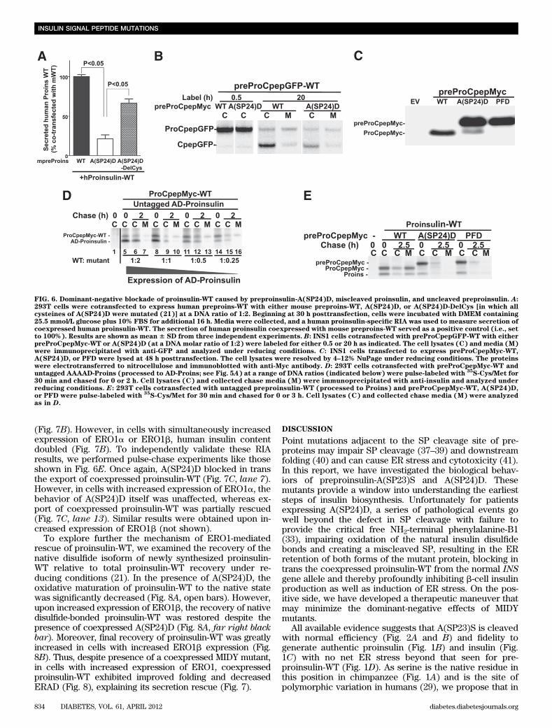
(Fig. 7B). However, in cells with simultaneously increasedexpression of ERO1a or ERO1b, human insulin contentdoubled (Fig. 7B). To independently validate these RIAresults, we performed pulse-chase experiments like thoseshown in Fig. 6E. Once again, A(SP24)D blocked in transthe export of coexpressed proinsulin-WT (Fig. 7C, lane 7).However, in cells with increased expression of ERO1a, thebehavior of A(SP24)D itself was unaffected, whereas ex-port of coexpressed proinsulin-WT was partially rescued(Fig. 7C, lane 13). Similar results were obtained upon in-creased expression of ERO1b (not shown).
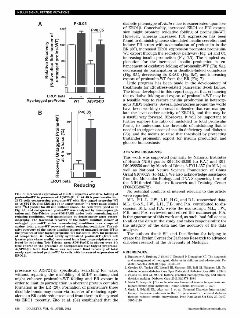
To explore further the mechanism of ERO1-mediatedrescue of proinsulin-WT, we examined the recovery of thenative disulfide isoform of newly synthesized proinsulin-WT relative to total proinsulin-WT recovery under re-ducing conditions (21). In the presence of A(SP24)D, theoxidative maturation of proinsulin-WT to the native statewas significantly decreased (Fig. 8A, open bars). However,upon increased expression of ERO1b, the recovery of nativedisulfide-bonded proinsulin-WT was restored despite thepresence of coexpressed A(SP24)D (Fig. 8A, far right blackbar). Moreover, final recovery of proinsulin-WT was greatlyincreased in cells with increased ERO1b expression (Fig.8B). Thus, despite presence of a coexpressed MIDY mutant,in cells with increased expression of ERO1, coexpressedproinsulin-WT exhibited improved folding and decreasedERAD (Fig. 8), explaining its secretion rescue (Fig. 7).
DISCUSSION
Point mutations adjacent to the SP cleavage site of pre-proteins may impair SP cleavage (37–39) and downstreamfolding (40) and can cause ER stress and cytotoxicity (41).In this report, we have investigated the biological behav-iors of preproinsulin-A(SP23)S and A(SP24)D. Thesemutants provide a window into understanding the earlieststeps of insulin biosynthesis. Unfortunately for patientsexpressing A(SP24)D, a series of pathological events gowell beyond the defect in SP cleavage with failure toprovide the critical free NH2-terminal phenylalanine-B1(33), impairing oxidation of the natural insulin disulfidebonds and creating a miscleaved SP, resulting in the ERretention of both forms of the mutant protein, blocking intrans the coexpressed proinsulin-WT from the normal INSgene allele and thereby profoundly inhibiting b-cell insulinproduction as well as induction of ER stress. On the pos-itive side, we have developed a therapeutic maneuver thatmay minimize the dominant-negative effects of MIDYmutants.
All available evidence suggests that A(SP23)S is cleavedwith normal efficiency (Fig. 2A and B) and fidelity togenerate authentic proinsulin (Fig. 1B) and insulin (Fig.1C) with no net ER stress beyond that seen for pre-proinsulin-WT (Fig. 1D). As serine is the native residue inthis position in chimpanzee (Fig. 1A) and is the site ofpolymorphic variation in humans (29), we propose that in
FIG. 6. Dominant-negative blockade of proinsulin-WT caused by preproinsulin-A(SP24)D, miscleaved proinsulin, and uncleaved preproinsulin. A:293T cells were cotransfected to express human preproins-WT with either mouse preproins-WT, A(SP24)D, or A(SP24)D-DelCys [in which allcysteines of A(SP24)D were mutated (21)] at a DNA ratio of 1:2. Beginning at 30 h posttransfection, cells were incubated with DMEM containing25.5 mmol/L glucose plus 10% FBS for additional 16 h. Media were collected, and a human proinsulin-specific RIA was used to measure secretion ofcoexpressed human proinsulin-WT. The secretion of human proinsulin coexpressed with mouse preproins-WT served as a positive control (i.e., setto 100%). Results are shown as mean 6 SD from three independent experiments. B: INS1 cells cotransfected with preProCpepGFP-WT with eitherpreProCpepMyc-WT or A(SP24)D (at a DNA molar ratio of 1:2) were labeled for either 0.5 or 20 h as indicated. The cell lysates (C) and media (M)were immunoprecipitated with anti-GFP and analyzed under reducing conditions. C: INS1 cells transfected to express preProCpepMyc-WT,A(SP24)D, or PFD were lysed at 48 h posttransfection. The cell lysates were resolved by 4–12% NuPage under reducing conditions. The proteinswere electrotransferred to nitrocellulose and immunoblotted with anti-Myc antibody. D: 293T cells cotransfected with preProCpepMyc-WT anduntagged AAAAD-Proins (processed to AD-Proins; see Fig. 5A) at a range of DNA ratios (indicated below) were pulse-labeled with
35S-Cys/Met for
30 min and chased for 0 or 2 h. Cell lysates (C) and collected chase media (M) were immunoprecipitated with anti-insulin and analyzed underreducing conditions. E: 293T cells cotransfected with untagged preproinsulin-WT (processed to Proins) and preProCpepMyc-WT, A(SP24)D,or PFD were pulse-labeled with
35S-Cys/Met for 30 min and chased for 0 or 3 h. Cell lysates (C) and collected chase media (M) were analyzed
as in D.
INSULIN SIGNAL PEPTIDE MUTATIONS
834 DIABETES, VOL. 61, APRIL 2012 diabetes.diabetesjournals.org

spite of our earlier report (12), impaired preproinsulin SPcleavage and proinsulin misfolding are not operative forthis particular variant. We cannot exclude that A(SP23)Smight cause diabetes through a different mechanism. Bycontrast, although A(SP24)D is successfully translocatedacross the ER membrane (Fig. 3), it is inefficiently pro-cessed at an inappropriate SP cleavage site, generatinga small subpopulation of miscleaved AD-Proins mole-cules (Figs. 1, 2, and 5–7). Both uncleaved and miscleavedA(SP24)D are retained intracellularly (Fig. 2A) and undergoERAD (Fig. 2C), suggesting that they are misfolded. Never-theless, at steady state, a major subpopulation of uncleavedA(SP24)D (along with the smaller subpopulation of SPcleaved) molecules accumulates in the ER (Fig. 1B), acti-vating an ER stress response (Fig. 1D).
The half-life of preproinsulin-WT is exceedingly short,making it technically difficult to follow uncleaved pre-proinsulin in live cells (Fig. 4B). Cleavage by signal pep-tidase generally requires that 60–80 amino acids of thenascent polypeptide are cotranslationally translocated(42), which represents most of the preproinsulin molecule.What the A(SP24)D demonstrates is that preproinsulin Cysresidues can be exposed to the oxidative environment ofthe ER even before SP cleavage (Fig. 4). To our knowl-edge, this is the first study to directly examine oxidativefolding of preproinsulin in the ER. Normally, uncleavedpreproinsulin-WT is fully reduced, and, upon SP removal,
proinsulin begins to quickly form both native and isomericdisulfide bonds (Fig. 4A and B). However, under con-ditions in which normal SP cleavage is interrupted [e.g., inA(SP24)D], uncleaved preproinsulin exhibits delayed andabnormal oxidative folding (Fig. 4A and B), causing theformation of disulfide-linked complexes (Fig. 4C), whichalso entraps coexpressed proinsulin-WT (Fig. 4D).
We have previously shown that acute ER stress is notitself sufficient to block export of coexpressed WT INSgene product (21). Moreover, recent studies indicate thatin MIDY, insulin deficiency precedes a net loss of b-cells(22,23). In this report, we demonstrate that A(SP24)Dblocks the export of coexpressed proinsulin-WT (Fig. 6)and thereby decreases insulin production (Figs. 6A and 7B,open bar), which may be sufficient to account for initiationof insulin deficiency in patients with MIDY. The evidencesuggests that it is primarily the uncleaved preproinsulinsubfraction rather than the minor misfolded AD-Proinssubfraction (Fig. 6D and E) that drives the dominant-negativeinsulin secretory behavior in patients bearing A(SP24)D.
Studies in humans suggest that one fully functional INSgene allele may be sufficient to maintain normoglycemia(5). We therefore reasoned that rescuing proinsulin-WTfrom dominant-negative blockade might restore insulinproduction to rescue from diabetes in MIDY. With this inmind, we have made a first attempt at a molecular thera-peutic approach to rescue proinsulin-WT export in the
FIG. 7. Dominant-negative blockade of human proinsulin by A(SP24)D is partially rescued by increased expression of ERO1. A: At 48 h post-transfection, media bathing 293T cells coexpressing human preproinsulin-WT, one of the mouse preproins-[WT, C(A7)Y, or A(SP24)D], and emptyvector (EV), PDI, or ERO1 (at a DNA molar ratio of 1:2:3) were collected for 16 h. Secreted human proinsulin was measured using a human-specificproinsulin RIA. Secretion of human proinsulin in the presence of mouse proinsulin-WT and empty vector was set to 100%. B: INS1 cells coex-pressing human preproinsulin and mouse preproinsulin-A(SP24)D plus ERO1a or ERO1b (at a DNA molar ratio of 1:3:4) were lysed with acid/ethanol to measure human insulin content using human-specific insulin RIA. The human insulin content of cells cotransfected with WT human andmouse preproinsulins was set to 100%. C: At 48 h posttransfection, 293T cells coexpressing preproinsulin-WT and either preProCpepMyc-WT(processed to Myc-tagged Proins) or preProCpepMyc-A(SP24)D (shown as Myc-tagged preProins) plus empty vector (Control) or ERO1a (ata DNA molar ratio of 1:2:3) were pulse-labeled with
35S-Cys/Met for 30 min and chased for 0 or 3 h. Newly synthesized (pre)proinsulins from cell
lysates (C) and chase media (M) were immunoprecipitated with anti-insulin, resolved by Tris-Tricine urea–SDS-PAGE under reducing conditions,and analyzed by autoradiography.
M. LIU AND ASSOCIATES
diabetes.diabetesjournals.org DIABETES, VOL. 61, APRIL 2012 835
presence of A(SP24)D; specifically searching for ways,without repairing the misfolding of MIDY mutants, thatmight enhance proinsulin-WT folding and ER export inorder to limit its participation in aberrant protein complexformation in the ER (20). Formation of proinsulin’s threedisulfide bonds may occur via transfer of reducing equiv-alents to ER oxidoreductases and from there to the cytosolvia ERO1; recently, Zito et al. (34) established that the
diabetic phenotype of Akita mice is exacerbated upon lossof ERO1b. Conceivably, increased ERO1 or PDI expres-sion might promote oxidative folding of proinsulin-WT.However, whereas increased PDI expression has beenfound to diminish glucose-stimulated insulin secretion andinduce ER stress with accumulation of proinsulin in theER (36), increased ERO1 expression promotes proinsulin-WT export through the secretory pathway (Fig. 7A and C),increasing insulin production (Fig. 7B). The simplest ex-planation for the increased insulin production is en-hancement of oxidative folding of proinsulin-WT (Fig. 8A),decreasing its participation in disulfide-linked complexes(Fig. 8A), decreasing its ERAD (Fig. 8B), and increasingexport of proinsulin-WT from the ER (Fig. 7).
Little progress has been made in the development oftreatments for ER stress-related pancreatic b-cell failure.The ideas developed in this report suggest that enhancingthe oxidative folding and export of proinsulin-WT may bea feasible way to restore insulin production in heterozy-gous MIDY patients. Several laboratories around the worldhave been working on small molecules that can manipu-late the level and/or activity of ERO1b, and this may bea useful way forward. Moreover, it will be important tofurther explore the ratio of misfolded to total proinsulinforms, to understand the threshold of misfolding that isneeded to trigger onset of insulin-deficiency and diabetes(23), and the means to raise that threshold by protectingbystander proinsulin export for insulin production andglucose homeostasis.
ACKNOWLEDGMENTS
This work was supported primarily by National Institutesof Health (NIH) grants R01-DK-48280 (to P.A.) and R01-DK-088856 and by March of Dimes 6-FY11-357 (to M.L.) aswell as National Nature Science Foundation of ChinaGrant 81070629 (to M.L.). We also acknowledge assistancefrom the Molecular Biology and DNA Sequencing Core ofthe NIH-funded Diabetes Research and Training Center(P60-DK-20572).
No potential conflicts of interest relevant to this articlewere reported.
M.L., R.L.-L., J.W., L.H., H.G., and D.L. researched data.M.L., S.-o.S., J.W., L.H., F.B., and P.A. contributed to dis-cussion. M.L. and P.A. wrote the manuscript. M.L., J.W.,F.B., and P.A. reviewed and edited the manuscript. P.A.is the guarantor of this work and, as such, had full accessto all of the data in the study and takes responsibility forthe integrity of the data and the accuracy of the dataanalysis.
The authors thank Bill and Dee Brehm for helping tocreate the Brehm Center for Diabetes Research to advancediabetes research at the University of Michigan.
REFERENCES
1. Hattersley A, Bruining J, Shield J, Njolstad P, Donaghue KC. The diagnosisand management of monogenic diabetes in children and adolescents. Pe-diatr Diabetes 2009;10(Suppl 12):33–42
2. Greeley SA, Tucker SE, Worrell HI, Skowron KB, Bell GI, Philipson LH. Up-date in neonatal diabetes. Curr Opin Endocrinol Diabetes Obes 2010;17:13–19
3. Fajans SS, Bell GI. MODY: history, genetics, pathophysiology, and clinicaldecision making. Diabetes Care 2011;34:1878–1884
4. Nishi M, Nanjo K. [The molecular mechanism of insulin biosynthesis andmutant insulin gene syndrome]. Nihon Rinsho 1994;52:2519–2527
5. Garin I, Edghill EL, Akerman I, et al; Neonatal Diabetes InternationalGroup. Recessive mutations in the INS gene result in neonatal diabetesthrough reduced insulin biosynthesis. Proc Natl Acad Sci USA 2010;107:3105–3110
FIG. 8. Increased expression of ERO1b improves oxidative folding ofproinsulin-WT in presence of A(SP24)D. A: At 48 h posttransfection,293T cells coexpressing preproins-WT with Myc-tagged preproins-WTor -A(SP24)D, plus ERO1b (+) or empty vector (2) were pulse-labeledwith
35S-Cys/Met for 30 min without chase. The cells were lysed as in
Fig. 4A, and coexpressed proins-WT was analyzed by immunoprecipi-tation and Tris-Tricine urea–SDS-PAGE under both nonreducing andreducing conditions, with quantitation by densitometry after autora-diography. The fractional recovery of the native disulfide isomer ofuntagged proins-WT under nonreducing conditions was comparedagainst total proins-WT recovered under reducing conditions. The rel-ative recovery of the native disulfide isomer of untagged proins-WT inthe presence of Myc-tagged preproins-WT was set to 100% for purposesof comparison. B: Total newly synthesized proins-WT (from celllysates plus chase media) recovered from immunoprecipitates ana-lyzed by reducing Tris-Tricine urea–SDS-PAGE is shown over 4-htime course in the presence of coexpressed Myc-tagged preproins-A(SP24)D. Note that there was increased final recovery of totalnewly synthesized proins-WT in cells with increased expression ofERO1b.
INSULIN SIGNAL PEPTIDE MUTATIONS
836 DIABETES, VOL. 61, APRIL 2012 diabetes.diabetesjournals.org
6. Støy J, Edghill EL, Flanagan SE, et al; Neonatal Diabetes InternationalCollaborative Group. Insulin gene mutations as a cause of permanentneonatal diabetes. Proc Natl Acad Sci USA 2007;104:15040–15044
7. Polak M, Dechaume A, Cavé H, et al; French ND (Neonatal Diabetes)Study Group. Heterozygous missense mutations in the insulin gene arelinked to permanent diabetes appearing in the neonatal period or in earlyinfancy: a report from the French ND (Neonatal Diabetes) Study Group.Diabetes 2008;57:1115–1119
8. Colombo C, Porzio O, Liu M, et al; Early Onset Diabetes Study Group of theItalian Society of Pediatric Endocrinology and Diabetes (SIEDP). Sevenmutations in the human insulin gene linked to permanent neonatal/infancy-onset diabetes mellitus. J Clin Invest 2008;118:2148–2156
9. Ahamed A, Unnikrishnan AG, Pendsey SS, et al. Permanent neonatal di-abetes mellitus due to a C96Y heterozygous mutation in the insulin gene.A case report. JOP 2008;9:715–718
10. Edghill EL, Flanagan SE, Patch AM, et al; Neonatal Diabetes InternationalCollaborative Group. Insulin mutation screening in 1,044 patients withdiabetes: mutations in the INS gene are a common cause of neonatal di-abetes but a rare cause of diabetes diagnosed in childhood or adulthood.Diabetes 2008;57:1034–1042
11. Molven A, Ringdal M, Nordbø AM, et al; Norwegian Childhood DiabetesStudy Group. Mutations in the insulin gene can cause MODY and autoantibody-negative type 1 diabetes. Diabetes 2008;57:1131–1135
12. Bonfanti R, Colombo C, Nocerino V, et al. Insulin gene mutations as causeof diabetes in children negative for five type 1 diabetes autoantibodies.Diabetes Care 2009;32:123–125
13. Rubio-Cabezas O, Edghill EL, Argente J, Hattersley AT. Testing formonogenic diabetes among children and adolescents with antibody-negativeclinically defined Type 1 diabetes. Diabet Med 2009;26:1070–1074
14. Boesgaard TW, Pruhova S, Andersson EA, et al. Further evidence thatmutations in INS can be a rare cause of Maturity-Onset Diabetes of theYoung (MODY). BMC Med Genet 2010;11:42
15. Meur G, Simon A, Harun N, et al. Insulin gene mutations resulting in early-onset diabetes: marked differences in clinical presentation, metabolicstatus, and pathogenic effect through endoplasmic reticulum retention.Diabetes 2010;59:653–661
16. Liu M, Ramos-Castañeda J, Arvan P. Role of the connecting peptide ininsulin biosynthesis. J Biol Chem 2003;278:14798–14805
17. Zhang BY, Liu M, Arvan P. Behavior in the eukaryotic secretory pathway ofinsulin-containing fusion proteins and single-chain insulins bearing variousB-chain mutations. J Biol Chem 2003;278:3687–3693
18. Liu M, Li Y, Cavener D, Arvan P. Proinsulin disulfide maturation andmisfolding in the endoplasmic reticulum. J Biol Chem 2005;280:13209–13212
19. Park SY, Ye H, Steiner DF, Bell GI. Mutant proinsulin proteins associ-ated with neonatal diabetes are retained in the endoplasmic reticulumand not efficiently secreted. Biochem Biophys Res Commun 2010;391:1449–1454
20. Liu M, Hodish I, Rhodes CJ, Arvan P. Proinsulin maturation, misfolding,and proteotoxicity. Proc Natl Acad Sci USA 2007;104:15841–15846
21. Liu M, Haataja L, Wright J, et al. Mutant INS-gene induced diabetes ofyouth: proinsulin cysteine residues impose dominant-negative inhibitionon wild-type proinsulin transport. PLoS ONE 2010;5:e13333
22. Gupta S, McGrath B, Cavener DR. PERK (EIF2AK3) regulates proinsulintrafficking and quality control in the secretory pathway. Diabetes 2010;59:1937–1947
23. Hodish I, Absood A, Liu L, et al. In vivo misfolding of proinsulin below thethreshold of frank diabetes. Diabetes 2011;60:2092–2101
24. Hodish I, Liu M, Rajpal G, et al. Misfolded proinsulin affects bystanderproinsulin in neonatal diabetes. J Biol Chem 2010;285:685–694
25. Liu M, Hodish I, Haataja L, et al. Proinsulin misfolding and diabetes: mu-tant INS gene-induced diabetes of youth. Trends Endocrinol Metab 2010;21:652–659
26. Sohma Y, Hua QX, Liu M, et al. Contribution of residue B5 to the foldingand function of insulin and IGF-I: constraints and fine-tuning in the evo-lution of a protein family. J Biol Chem 2010;285:5040–5055
27. Rajan S, Eames SC, Park SY, et al. In vitro processing and secretion ofmutant insulin proteins that cause permanent neonatal diabetes. AmJ Physiol Endocrinol Metab 2010;298:E403–E410
28. Steiner DF, Chan SJ, Welsh JM, Kwok SC. Structure and evolution of theinsulin gene. Annu Rev Genet 1985;19:463–484
29. Oda N, Nakai A, Fujiwara K, et al. Polymorphisms of the insulin geneamong Japanese subjects. Metabolism 2001;50:631–634
30. Prehn S, Tsamaloukas A, Rapoport TA. Demonstration of specific re-ceptors of the rough endoplasmic membrane for the signal sequence ofcarp preproinsulin. Eur J Biochem 1980;107:185–195
31. Miyamoto K, Yamashita T, Tsukiyama T, et al. Reversible membrane per-meabilization of mammalian cells treated with digitonin and its use forinducing nuclear reprogramming by Xenopus egg extracts. Cloning StemCells 2008;10:535–542
32. Eskridge EM, Shields D. The NH2 terminus of preproinsulin directs thetranslocation and glycosylation of a bacterial cytoplasmic protein bymammalian microsomal membranes. J Cell Biol 1986;103:2263–2272
33. Liu M, Hua QX, Hu SQ, et al. Deciphering the hidden informational contentof protein sequences: foldability of proinsulin hinges on a flexible arm thatis dispensable in the mature hormone. J Biol Chem 2010;285:30989–31001
34. Zito E, Chin KT, Blais J, Harding HP, Ron D. ERO1-beta, a pancreas-specificdisulfide oxidase, promotes insulin biogenesis and glucose homeostasis.J Cell Biol 2010;188:821–832
35. Khoo C, Yang J, Rajpal G, Wang Y, Liu J, Arvan P, Stoffers DA. Endo-plasmic reticulum oxidoreductase-1-like {beta} (ERO1l{beta}) regulatessusceptibility to endoplasmic reticulum stress and is induced by insulinflux in {beta}-cells. Endocrinology 2011;152:2599–2608
36. Zhang L, Lai E, Teodoro T, Volchuk A. GRP78, but not protein-disulfideisomerase, partially reverses hyperglycemia-induced inhibition of insulinsynthesis and secretion in pancreatic {beta}-cells. J Biol Chem 2009;284:5289–5298
37. Nothwehr SF, Gordon JI. Structural features in the NH2-terminal region ofa model eukaryotic signal peptide influence the site of its cleavage bysignal peptidase. J Biol Chem 1990;265:17202–17208
38. Racchi M, Watzke HH, High KA, Lively MO. Human coagulation factor Xdeficiency caused by a mutant signal peptide that blocks cleavage bysignal peptidase but not targeting and translocation to the endoplasmicreticulum. J Biol Chem 1993;268:5735–5740
39. Lyko F, Martoglio B, Jungnickel B, Rapoport TA, Dobberstein B. Signalsequence processing in rough microsomes. J Biol Chem 1995;270:19873–19878
40. Beuret N, Rutishauser J, Bider MD, Spiess M. Mechanism of endoplasmicreticulum retention of mutant vasopressin precursor caused by a signalpeptide truncation associated with diabetes insipidus. J Biol Chem 1999;274:18965–18972
41. Kang SW, Rane NS, Kim SJ, Garrison JL, Taunton J, Hegde RS. Substrate-specific translocational attenuation during ER stress defines a pre-emptivequality control pathway. Cell 2006;127:999–1013
42. Eskridge EM, Shields D. Cell-free processing and segregation of insulinprecursors. J Biol Chem 1983;258:11487–11491
M. LIU AND ASSOCIATES
diabetes.diabetesjournals.org DIABETES, VOL. 61, APRIL 2012 837