original article global incidence of oesophageal cancer … · global incidence of oesophageal...
TRANSCRIPT
ORIGINAL ARTICLE
Global incidence of oesophageal cancer byhistological subtype in 2012Melina Arnold, Isabelle Soerjomataram, Jacques Ferlay, David Forman
▸ Additional material ispublished online only. To viewplease visit the journal online(http://dx.doi.org/10.1136/gutjnl-2014-308124).
Section of Cancer Surveillance,International Agency forResearch on Cancer, Lyon,France
Correspondence toDr Melina Arnold, Section ofCancer Surveillance,International Agency forResearch on Cancer,150 Cours Albert Thomas,Lyon 69008, France;[email protected]
Received 24 July 2014Revised 26 August 2014Accepted 28 August 2014Published Online First15 October 2014
To cite: Arnold M,Soerjomataram I, Ferlay J,et al. Gut 2015;64:381–387.
ABSTRACTObjective The two major histological types ofoesophageal cancer—adenocarcinoma (AC) andsquamous cell carcinoma (SCC)—are known to differgreatly in terms of risk factors and epidemiology. Todate, global incidence estimates for individual subtypesare still lacking. This study for the first time quantifiedthe global burden of oesophageal cancer by histologicalsubtype.Design Where available, data from Cancer Incidence inFive Continents Vol. X (CI5X) were used to compute,age-specific, sex-specific and country-specific proportionsof AC and SCC. Nine regional averages were computedfor countries without CI5X data. The proportions werethen applied to all oesophageal cancer cases fromGLOBOCAN 2012 and age-standardised incidence ratescalculated for both histological types.Results Worldwide, an estimated 398 000 SCCs and52 000 ACs of the oesophagus occurred in 2012,translating to incidence rates of 5.2 and 0.7 per100 000, respectively. Although SCCs were mostcommon in South-Eastern and Central Asia (79% of thetotal global SCC cases), the highest burden of AC wasfound in Northern and Western Europe, NorthernAmerica and Oceania (46% of the total global ACcases). Men had substantially higher incidence thanwomen, especially in the case of AC (male to femaleratio AC: 4.4; SCC: 2.7).Conclusions These first global estimates ofoesophageal cancer incidence by histology suggested ahigh concentration of AC in high-income countries withmen being at much greater risk. This quantification ofincidence will aid health policy makers to planappropriate cancer control measures in the future.
INTRODUCTIONOesophageal cancer is the eighth most commoncancer worldwide, with an estimated 456 000 newcases and 400 000 deaths in 2012.1 Around 80%of all cases occurred in less-developed regions. Themajority of oesophageal cancers can be subdividedinto two main histological subtypes: adenocarcin-omas (AC) and squamous cell carcinomas (SCC).Whereas ACs typically develop in the lower thirdof the oesophagus and originate predominantlyfrom Barrett mucosa, SCC occurs mostly in flatcells lining the upper two-thirds of the oesopha-gus.2 High-grade dysplasia is the precursor for bothtypes, while the development of AC can addition-ally be characterised by a progression fromBarrett’s metaplasia to dysplasia and ultimatelyinvasive carcinoma.
The two subtypes differ greatly in terms of theirmale to female ratio, risk factors and incidence pat-terns across countries.2–4 In recent decades, enig-matic increases in the incidence of oesophageal AChave been observed among white populations in
Significance of this study
What is already known on this subject?▸ The two main types of oesophageal cancer,
squamous cell carcinoma (SCC) andadenocarcinoma (AC), differ greatly in theiraetiology and epidemiology. To date, however,no global incidence estimates exist.
▸ While the incidence of AC has been increasingin several high-income countries during thepast years, trends in SCC have remained stableor have decreased.
What are the new findings?▸ In this study, we quantified for the first time
the global burden oesophageal cancer byhistological type. In 2012, an estimated398 000 cases of SCC and 52 000 cases ofoccurred globally.
▸ The incidence of SCC was highest inSouth-Eastern and Central Asia, where 79% ofall SCC cases occurred. In contrast, the burdenof AC was highest in Northern and WesternEurope, Northern America and Oceania, whichaccounted for 46% of the global AC cases.
▸ While SCC is the more common type ofoesophageal cancer on the global scale,incidence rates of AC notably exceeded thoseof SCC in the Netherlands and in the UK. Thiswas also true for New Zealand, the USA,Canada, Ireland, Iceland, Australia, Norway,Malta, Sweden, Bahrain and Cyprus.
▸ For both sites, men had substantially higherincidence than women. This was morepronounced for AC, where incidence in menwas on average four times greater than inwomen and over eight times greater inNorthern America.
How might it impact on clinical practice inthe foreseeable future?▸ This quantification of incidence accords with
aetiological research and will aid health policymakers to plan appropriate cancer controlmeasures in the future.
Oesophagus
Arnold M, et al. Gut 2015;64:381–387. doi:10.1136/gutjnl-2014-308124 381
on 10 Septem
ber 2018 by guest. Protected by copyright.
http://gut.bmj.com
/G
ut: first published as 10.1136/gutjnl-2014-308124 on 15 October 2014. D
ownloaded from
high-income countries.5 This increase has largely been attributedto the rising prevalence of obesity, although some controversyexists around this hypothesis. In contrast, the declining inci-dence of SCC in more developed regions roughly mirrors thedecline in tobacco smoking, one of the most important riskfactors for SCC.3 SCC remains most common in less-developedregions of the world, including Africa6–8 and Eastern Asia.9 Thereasons for this are not yet entirely understood.
Although overall global and country-specific incidence esti-mates of oesophageal cancer have been available through theGLOBOCAN project since 1990,10–13 breakdown by histo-logical subtype has not been possible. Here, for the first time,we estimate the global, regional-specific and country-specificincidence of oesophageal cancer by histological type, based ondata from Cancer Incidence in Five Continents Vol. X (CI5X)14
and GLOBOCAN 2012.1
MATERIAL AND METHODSThe estimated total numbers of oesophageal cancers in 2012 byage, sex and country were obtained from GLOBOCAN 2012.1
For each country, we estimated the age-specific and sex-specificnumber of SCC and AC cases by multiplying the estimatednumber of oesophageal cancer cases in 2012 by the respectiveproportions of each histological subtype from relevant cancerregistry data extracted from CI5X14 using one of two methods.
Sex-specific and age-specific (<65 and ≥65 years) proportionsof AC and SCC were computed for all countries included inCI5X except for those with no cases of either AC or SCC inone of our four substrata (men and women <65 and≥65 years). Where data were available from multiple regionalregistries within a country, cases and populations were pooledto obtain national proportions. The sum of all carcinomas (AC+SCC+other carcinomas) was used as the denominator. Thehistological types were defined according to the third edition ofthe International Classification of Diseases for Oncology(ICD-O-3) which were presented in Cancer Incidence in FiveContinents Vol. IX (CI5IX)15 16; SCCs: 8050–8078 and 8083–8084; ACs: 8140–8141, 8143–8145, 8190–8231, 8260–8263,8310, 8401, 8480–8490, 8550–8551 and 8570–8574, 8576.Other histological subtypes, such as sarcomas, were found toconstitute <1% of all oesophageal cancer cases and were con-sidered negligible. Using this approach, country-specific propor-tions could be computed for 57 countries.
For the remaining 127 countries without data in CI5X orwith zero cases in a substratum, proportions were calculated fornine broad regions. The data for each region were derived fromCI5X and were complemented with data from CI5IX16 andfrom the African Cancer Registry Network (AFCRN) for thesub-Saharan African region. Regions are based on the followingUN geographical regions17: sub-Saharan Africa (includingEastern, Middle, Southern and Western Africa), Northern Africaand Western Asia, Central Asia (including India), Eastern andSouth-Eastern Asia (including China), Latin America and theCaribbean, Eastern Europe, Northern and Western Europe,Southern Europe and Oceania.
Age-standardised incidence rates (per 100 000 person-years)were calculated for each country and region and according tothe four categories of human development index (HDI) in201218 using the direct method and the World StandardPopulation.19
RESULTSIn 2012, an estimated 456 000 men and women developedoesophageal cancer, of which 398 000 cases were SCCs
(278 000 in men, 120 000 in women), 52 000 were ACs(41 000 in men, 11 000 in women) and 6000 other carcinomas.In more than 90% of all countries presented in GLOBOCAN2012, the rate of oesophageal SCC for both sexes combinedclearly exceeded that of AC. In contrast, a few countries hadhigher rates of AC than of SCC, which was most pronounced inthe Netherlands and in the UK but was also true for NewZealand, the USA, Canada, Ireland, Iceland, Australia, Norway,Malta, Sweden, Bahrain and Cyprus.
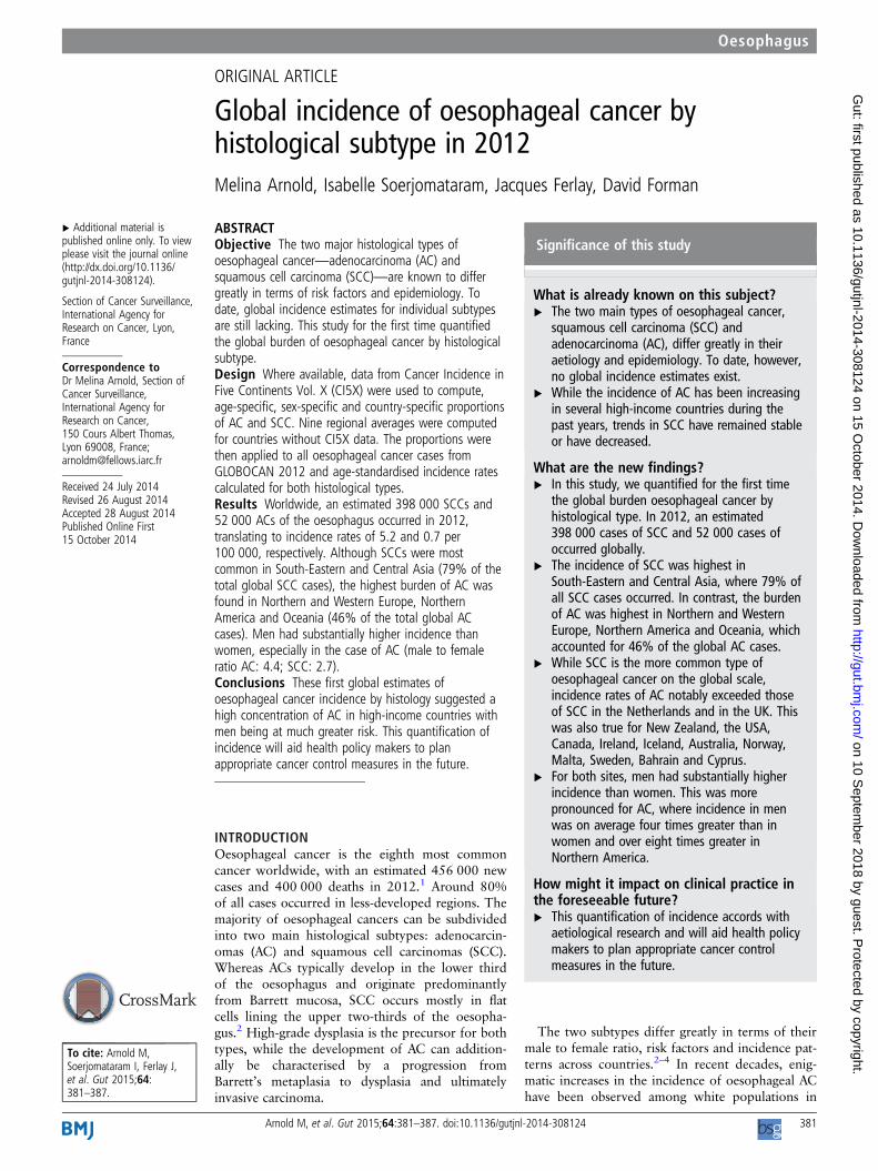
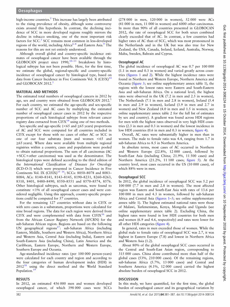
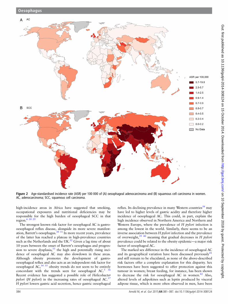
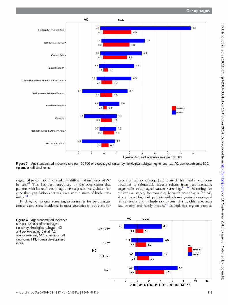
Oesophageal ACThe global incidence of oesophageal AC was 0.7 per 100 000(1.1 in men and 0.3 in women) and varied greatly across coun-tries (figures 1 and 2). While the highest incidence rates werefound in Northern and Western Europe, Northern America andOceania (figure 3; see online supplementary annex table 1), theregions with the lowest rates were Eastern and South-EasternAsia and sub-Saharan Africa. On a national level, the highestrates were observed in the UK (7.2 in men and 2.5 in women),The Netherlands (7.1 in men and 2.8 in women), Ireland (5.4in men and 2.9 in women), Iceland (3.9 in men and 2.7 inwomen) and New Zealand (4.0 in men and 1.5 in women; seeonline supplementary annex table 2 for the full list of estimatesby sex and country). A gradient was found across HDI regionsfor men with the highest rates observed in very high HDI coun-tries (2.3 in men and 0.3 in women) and the lowest incidence inlow HDI countries (0.6 in men and 0.3 in women; figure 4).
Overall, AC rates were substantially higher in men than inwomen. The male to female ratio was 4.4, ranging from 1.7 insub-Saharan Africa to 8.5 in Northern America.
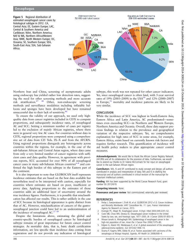
In absolute terms, most cases of AC occurred in Northernand Western Europe (22.8%, 12 000 cases), followed bySouth-East Asia (including China; 21.9%, 11 500 cases) andNorthern America (21.2%, 11 100 cases; figure 5). At thecountry level, most cases occurred in the US (10 000 cases), ofwhich 88% were in men.
Oesophageal SCCIn 2012, the global incidence of oesophageal SCC was 5.2 per100 000 (7.7 in men and 2.8 in women). The most affectedregion was Eastern and South-East Asia with rates of 13.6 per100 000 in men and 4.3 in women, followed by sub-SaharanAfrica and Central Asia (figures 1–3; see online supplementaryannex table 1). The highest estimated national rates were thoseof Malawi, Turkmenistan, Kenya, Mongolia and Uganda (seeonline supplementary annex table 2). Excluding China, thehighest rates were found in low HDI countries for both menand women (6.9 and 4.6, respectively) and rates were lower forall other HDI categories (figure 4).
In general, rates in men exceeded those of women. While theglobal male to female ratio of oesophageal SCC was 2.7, it washighest in Eastern Europe (7.8) and lowest in Northern Africaand Western Asia (1.2).
About 80% of the global oesophageal SCC cases occurred inthe Central and South-East Asian region, corresponding to315 000 cases. China alone contributed more than half of theglobal cases (53%, 210 000 cases). Of the remaining regions,sub-Saharan Africa (5.7%, 13 000 cases) and Central andSouthern America (4.1%, 12 000 cases) carried the highestabsolute burden of oesophageal SCC in 2012.
DISCUSSIONIn this study, we have quantified, for the first time, the globalburden of oesophageal cancer and its geographical variation by
Oesophagus
382 Arnold M, et al. Gut 2015;64:381–387. doi:10.1136/gutjnl-2014-308124
on 10 Septem
ber 2018 by guest. Protected by copyright.
http://gut.bmj.com
/G
ut: first published as 10.1136/gutjnl-2014-308124 on 15 October 2014. D
ownloaded from

histological subtype. While the incidence of SCC—the morecommon subtype globally—was highest in South-Eastern Asia,AC predominated in Northern and Western Europe and also inNorthern America and Oceania. For both subtypes, but particu-larly AC, men had substantially higher incidence of oesophagealcancer than women.
These results largely support the findings of previous studies.High incidence areas of oesophageal SCC have been identifiedin Northern Iran,4 Central Asia and China9 (together formingthe so-called ‘oesophageal cancer belt’) as well as parts ofEastern Africa.6 8 20 This pattern was also reflected in ourresults, where high rates of oesophageal SCC were mainly con-fined to those regions. In contrast, the incidence of oesophagealAC was greatest in high-income countries such as the US andthe UK, where incidence has increased substantially in recentdecades.3 5 Changing patterns in the epidemiology of the twotypes were also confirmed in countries where historically SCCwas the predominant type of oesophageal cancer and AC is nowon the rise. This was, for example, the case in Iran21 where
rates of AC, especially in women, were among the highest in theCentral Asian region.
In general, chronic irritation and inflammation of theoesophageal mucosa have been postulated to increase the risk ofoesophageal SCC. The two strongest risk factors are smokingand alcohol consumption, which have been found to accountfor more than 75% of all SCC cases in high-income coun-tries.22 23 Frequent consumption of extremely hot beverages iscommon risk factor for oesophageal SCC in less-developedregions. While for instance in Latin America, hot mate drinkinghas been shown to double the risk of oesophageal SCC,24 theconsumption of very hot beverages was associated with a nine-fold increase in risk in Southern China.25 In the latter study,high-temperature cooking methods, including barbecuing andfrying, were also found to increase the risk for oesophagealcancer. In Iran, opium use has been found to increase mortalityfrom oesophageal cancer by 50%.26 Also, low socioeconomicstatus, poverty and poor oral hygiene have been linked toan increased risk of oesophageal SCC.23 27 28 Studies from
Figure 1 Age-standardised incidence rate (ASR) per 100 000 of (A) oesophageal adenocarcinoma and (B) squamous cell carcinoma in men.AC, adenocarcinoma; SCC, squamous cell carcinoma.
Oesophagus
Arnold M, et al. Gut 2015;64:381–387. doi:10.1136/gutjnl-2014-308124 383
on 10 Septem
ber 2018 by guest. Protected by copyright.
http://gut.bmj.com
/G
ut: first published as 10.1136/gutjnl-2014-308124 on 15 October 2014. D
ownloaded from
high-incidence areas in Africa have suggested that smoking,occupational exposures and nutritional deficiencies may beresponsible for the high burden of oesophageal SCC in thatregion.6 20 29
The strongest known risk factor for oesophageal AC is gastro-oesophageal reflux disease, alongside its more severe manifest-ation, Barrett’s oesophagus.30 31 In more recent years, prevalenceof the latter has reached a plateau in high-prevalence countriessuch as the Netherlands and the UK.32 Given a lag time of about10 years between the onset of Barrett’s oesophagus and progres-sion to severe dysplasia,33 the high and potentially rising inci-dence of oesophageal AC may also slowdown in these areas.Although obesity promotes the development of gastro-oesophageal reflux and also acts as an independent risk factor foroesophageal AC,34 35 obesity trends do not seem to be entirelyconcordant with the trends seen for oesophageal AC.5 36
Recent evidence has suggested a possible role of Helicobacterpylori (H pylori) in the increasing rates of oesophageal AC.37
H pylori lowers gastric acid secretion, hence gastric oesophageal
reflux. Its declining prevalence in many Western countries38 mayhave led to higher levels of gastric acidity and therefore higherincidence of oesophageal AC. This could, in part, explain thehigh incidence observed in Northern America and Northern andWestern Europe, where the prevalence of H pylori infection isamong the lowest in the world. Similarly, there seems to be aninverse association between H pylori infection and the prevalenceof overweight,39 40 meaning that gradual decreases in H pyloriprevalence could be related to the obesity epidemic—a major riskfactor of oesophageal AC.
The marked sex difference in the incidence of oesophageal ACand its geographical variation have been discussed previously41
and still remain to be elucidated, as none of the above-describedrisk factors offer a complete explanation for this disparity. Sexhormones have been suggested to offer protection against thistumour in women; breast feeding, for instance, has been shownto decrease the risk for oesophageal AC in women.42 Also,altered levels of adipokines such as leptin produced by visceraladipose tissue, which is more often observed in men, have been
Figure 2 Age-standardised incidence rate (ASR) per 100 000 of (A) oesophageal adenocarcinoma and (B) squamous cell carcinoma in women.AC, adenocarcinoma; SCC, squamous cell carcinoma.
Oesophagus
384 Arnold M, et al. Gut 2015;64:381–387. doi:10.1136/gutjnl-2014-308124
on 10 Septem
ber 2018 by guest. Protected by copyright.
http://gut.bmj.com
/G
ut: first published as 10.1136/gutjnl-2014-308124 on 15 October 2014. D
ownloaded from
suggested to contribute to markedly differential incidence of ACby sex.43 This has been supported by the observation thatpatients with Barrett’s oesophagus have a greater waist circumfer-ence than population controls, even within strata of body massindex.44
To date, no national screening programmes for oesophagealcancer exist. Since incidence in most countries is low, costs for
screening (using endoscopy) are relatively high and risk of com-plications is substantial, experts refrain from recommendinglarger-scale oesophageal cancer screening.45 46 Screening forpreinvasive stages, for example, Barrett’s oesophagus for AC,should target high-risk patients with chronic gastro-oesophagealreflux disease and multiple risk factors, that is, older age, malesex, obesity and family history.45 In high-risk regions such as
Figure 3 Age-standardised incidence rate per 100 000 of oesophageal cancer by histological subtype, region and sex. AC, adenocarcinoma; SCC,squamous cell carcinoma.
Figure 4 Age-standardised incidencerate per 100 000 of oesophagealcancer by histological subtype, HDIand sex (excluding China). AC,adenocarcinoma; SCC, squamous cellcarcinoma; HDI, human developmentindex.
Oesophagus
Arnold M, et al. Gut 2015;64:381–387. doi:10.1136/gutjnl-2014-308124 385
on 10 Septem
ber 2018 by guest. Protected by copyright.
http://gut.bmj.com
/G
ut: first published as 10.1136/gutjnl-2014-308124 on 15 October 2014. D
ownloaded from
Northern Iran and China, screening of asymptomatic adultsusing endoscopy has yielded rather low detection rates, suggest-ing the need for other screening methods and more accuraterisk stratification.47 48 Other, non-endoscopic screeningmethods and surveillance modalities including inflatable bal-loons and sponges have been developed but have remainedunsuccessful due to their lack of sensitivity.46
To ensure the validity of our approach, we used only high-quality data from cancer registries included in CI5X to computeproportions, and subsequently incidence rates, of oesophagealAC and SCC. Setting a minimum number of cases per registryled to the exclusion of mainly African registries, where therewere in general very few AC cases. For countries without data inCI5X, regional proportions were computed using a comprehen-sive set of data from CI5 Vols. IX–X and from the AFCRN.Using regional proportions disregards any heterogeneity acrosscountries within the regions, for example, in the case of thesub-Saharan African and Central Asian region, where data camefrom only a very limited number of cancer registries with suffi-cient cases and data quality. However, in agreement with previ-ous reports, SCC accounted for over 90% of all oesophagealcancer cases in many sub-Saharan African countries6–8 and con-firmed the high burden of this subtype in the Eastern part ofthe continent.
It is also important to note that GLOBOCAN itself representsincidence estimates that are based on the best data available butnevertheless need to be interpreted with caution, especially forcountries where estimates are based on poor, insufficient orproxy data. Applying proportions to the estimates of thosecountries adds an additional layer of uncertainty around thetrue incidence figures. Furthermore, it is possible that misclassifi-cation has affected our results. This is rather unlikely in the caseof SCC because its histological appearance is quite distinct fromthat of AC. However, misclassification of cancers of the gastro-oesophageal junction could have led to an underestimation ofthe incidence of oesophageal AC.37 49
Despite the limitations above, estimating the global andcountry-specific burden of oesophageal cancer by histologicalsubtype remains of great importance for future cancer controlstrategies. As mortality data, that is, basic death certificationinformation, are less specific than incidence data coming fromregistration and do not provide any indication of histological
subtype, this work was not repeated for other cancer indicators.Yet, since oesophageal cancer is often fatal, with 5-year survivalrates of 19% (2003–2009) in the USA50 and 12% (2000–2007)in Europe,51 mortality and incidence patterns are likely to bevery similar.
CONCLUSIONWhile the incidence of SCC was highest in South-Eastern Asia,Eastern Africa and Latin America, AC predominated—some-times even exceeding SCC—in Northern and Western Europe,Northern America and Oceania. Overall, these data support pre-vious findings in relation to the prevalence and geographicalvariation of the respective subtypes. Yet, no comprehensiveexplanation for high rates of SCC in some areas, for example,Eastern Africa, exists based on currently known risk factors andrequires further research. This quantification of incidence willaid health policy makers to plan appropriate cancer controlmeasures.
Acknowledgements We would like to thank the African Cancer Registry Network(AFCRN) and all its collaborators for the provision of data. Furthermore, we wouldlike to extend our thanks to Dr Valerie McCormack for her input on oesophagealcancer in the sub-Saharan African region.
Contributors MA, IS and DF contributed to study concept and design; all authorscontributed in analysis and interpretation of data; MA and IS in drafting themanuscript and all authors contributed in critical revision of the manuscript forimportant intellectual content.
Funding MA has been supported by the World Cancer Research Fund, grantnumber SG 2012/619.
Competing interests None.
Provenance and peer review Not commissioned; externally peer reviewed.
REFERENCES1 Ferlay J, Soerjomataram I, Ervik M, et al. GLOBOCAN 2012 v1.0, Cancer Incidence
and Mortality Worldwide: IARC CancerBase No. 11. Lyon, France: InternationalAgency for Research on Cancer, 2013.
2 Enzinger PC, Mayer RJ. Esophageal cancer. N Engl J Med 2003;349:2241–52.3 Cook MB, Chow WH, Devesa SS. Oesophageal cancer incidence in the United
States by race, sex, and histologic type, 1977–2005. Br J Cancer 2009;101:855–9.4 Mosavi-Jarrahi A, Mohagheghi MA. Epidemiology of esophageal cancer in the
high-risk population of Iran. Asian Pac J Cancer Prev 2006;7:375–80.5 Edgren G, Adami HO, Weiderpass E, et al. A global assessment of the oesophageal
adenocarcinoma epidemic. Gut 2013;62:1406–14.6 Ocama P, Kagimu MM, Odida M, et al. Factors associated with carcinoma of the
oesophagus at Mulago Hospital, Uganda. Afr Health Sci 2008;8:80–4.
Figure 5 Regional distribution ofestimated oesophageal cancer cases byhistological subtype in 2012. CA,Central Asia; EE, Eastern Europe; LAC,Central & Southern America andCaribbean; NAm, Northern America;NAf & WA, Northern Africa/WesternAsia; NWE, North Western Europe; OC,Oceania; SE, Southern Europe; SEA,South-East Asia; SSA, Sub-SaharanAfrica.
Oesophagus
386 Arnold M, et al. Gut 2015;64:381–387. doi:10.1136/gutjnl-2014-308124
on 10 Septem
ber 2018 by guest. Protected by copyright.
http://gut.bmj.com
/G
ut: first published as 10.1136/gutjnl-2014-308124 on 15 October 2014. D
ownloaded from
7 White RE, Parker RK, Fitzwater JW, et al. Stents as sole therapy for oesophageal cancer:a prospective analysis of outcomes after placement. Lancet Oncol 2009;10:240–6.
8 Somdyala NI, Bradshaw D, Gelderblom WC, et al. Cancer incidence in a ruralpopulation of South Africa, 1998–2002. Int J Cancer 2010;127:2420–9.
9 Lin Y, Totsuka Y, He Y, et al. Epidemiology of esophageal cancer in Japan andChina. J Epidemiol 2013;23:233–42.
10 Ferlay J, Parkin DM, Pisani P. GLOBOCAN. Cancer Incidence and MortalityWorldwide: IARC CancerBase No. 3. Lyon, France: International Agency forResearch on Cancer, 1998.
11 Ferlay J, Bray F, Pisani P, et al. GLOBOCAN 2000. Cancer Incidence, Mortality andPrevalence Worldwide: IARC CancerBase No. 5. Lyon, France: International Agencyfor Research on Cancer, 2001.
12 Ferlay J, Bray F, Pisani P, et al. GLOBOCAN 2002. Cancer Incidence, Mortality andPrevalence Worldwide: IARC CancerBase No. 5 version 2.0. Lyon, France:International Agency for Research on Cancer, 2004.
13 Ferlay J, Shin HR, Bray F, et al. Estimates of worldwide burden of cancer in 2008:GLOBOCAN 2008. Int J Cancer 2010;127:2893–917.
14 Forman D, Brewster DH, Gombe Mbalawa C, et al. Cancer incidence in fivecontinents, Vol. X (electronic version). Lyon: IARC, 2013.
15 World Health Organization. International Classification of Diseases for Oncology.3rd edn, First Revision. Geneva, Switzerland: World Health Organization, 2013.
16 Curado MP, Edwards B, Shin HR, et al. Cancer incidence in five continents, Vol. IX.Lyon, France: International Agency for Research on Cancer, 2007.
17 World Population Prospects. The 2012 Revision. New York: United Nations,Department of Economic and Social Affairs, Population Division, 2013.
18 Human Development Report 2013. The Rise of the South: Human Progress in aDiverse World. New York: United Nations Development Programme (UNDP), 2013.
19 Segi M. Cancer mortality for selected sites in 24 countries (1950–57). Sendai,Japan: Department of Public Health, Tohoku University of Medicine, 1960.
20 Vizcaino AP, Parkin DM, Skinner ME. Risk factors associated with oesophagealcancer in Bulawayo, Zimbabwe. Br J Cancer 1995;72:769–73.
21 Ghasemi-Kebria F, Roshandel G, Semnani S, et al. Marked increase in the incidencerate of esophageal adenocarcinoma in a high-risk area for esophageal cancer. ArchIran Med 2013;16:320–3.
22 Pandeya N, Olsen CM, Whiteman DC. Sex differences in the proportion ofesophageal squamous cell carcinoma cases attributable to tobacco smoking andalcohol consumption. Cancer Epidemiol 2013;37:579–84.
23 Brown LM, Hoover R, Silverman D, et al. Excess incidence of squamous cellesophageal cancer among US Black men: role of social class and other risk factors.Am J Epidemiol 2001;153:114–22.
24 De Stefani E, Deneo-Pellegrini H, Ronco AL, et al. Diet patterns and risk ofsquamous cell oesophageal carcinoma: a case-control study in Uruguay. Asian Pac JCancer Prev 2014;15:2765–9.
25 Lin J, Zeng R, Cao W, et al. Hot beverage and food intake and esophageal cancerin southern China. Asian Pac J Cancer Prev 2011;12:2189–92.
26 Malekzadeh MM, Khademi H, Pourshams A, et al. Opium use and risk of mortalityfrom digestive diseases: a prospective cohort study. Am J Gastroenterol2013;108:1757–65.
27 Dar NA, Islami F, Bhat GA, et al. Poor oral hygiene and risk of esophagealsquamous cell carcinoma in Kashmir. Br J Cancer 2013;109:1367–72.
28 Tran GD, Sun XD, Abnet CC, et al. Prospective study of risk factors for esophagealand gastric cancers in the Linxian general population trial cohort in China. Int JCancer 2005;113:456–63.
29 van Rensburg SJ, Benade AS, Rose EF, et al. Nutritional status of Africanpopulations predisposed to esophageal cancer. Nutr Cancer 1983;4:206–16.
30 Lagergren J, Bergstrom R, Lindgren A, et al. Symptomatic gastroesophageal refluxas a risk factor for esophageal adenocarcinoma. N Engl J Med 1999;340:825–31.
31 Hvid-Jensen F, Pedersen L, Drewes AM, et al. Incidence of adenocarcinoma amongpatients with Barrett’s esophagus. N Engl J Med 2011;365:1375–83.
32 Masclee GM, Coloma PM, de Wilde M, et al. The incidence of Barrett’s oesophagusand oesophageal adenocarcinoma in the United Kingdom and the Netherlands islevelling off. Aliment Pharmacol Ther 2014;39:1321–30.
33 Umar A, Dunn BK, Greenwald P. Future directions in cancer prevention. Nat RevCancer 2012;12:835–48.
34 Lagergren J. Influence of obesity on the risk of esophageal disorders. Nat RevGastroenterol Hepatol 2011;8:340–7.
35 Hoyo C, Cook MB, Kamangar F, et al. Body mass index in relation to oesophagealand oesophagogastric junction adenocarcinomas: a pooled analysis from theInternational BEACON Consortium. Int J Epidemiol 2012;41:1706–18.
36 Kroep S, Lansdorp-Vogelaar I, Rubenstein JH, et al. Comparing trends in esophagealadenocarcinoma incidence and lifestyle factors between the United States, Spain,and the Netherlands. Am J Gastroenterol 2014;109:336–43; quiz 5, 44.
37 McColl KE, Going JJ. Aetiology and classification of adenocarcinoma of thegastro-oesophageal junction/cardia. Gut 2010;59:282–4.
38 Khalifa MM, Sharaf RR, Aziz RK. Helicobacter pylori: a poor man’s gut pathogen?Gut Pathog 2010;2:2.
39 Lender N, Talley NJ, Enck P, et al. Review article: associations between Helicobacterpylori and obesity—an ecological study. Aliment Pharmacol Ther 2014;40:24–31.
40 Lane JA, Murray LJ, Harvey IM, et al. Randomised clinical trial: Helicobacter pylorieradication is associated with a significantly increased body mass index in aplacebo-controlled study. Aliment Pharmacol Ther 2011;33:922–9.
41 Cook MB, Wild CP, Forman D. A systematic review and meta-analysis of the sexratio for Barrett’s esophagus, erosive reflux disease, and nonerosive reflux disease.Am J Epidemiol 2005;162:1050–61.
42 Cronin-Fenton DP, Murray LJ, Whiteman DC, et al. Reproductive and sex hormonalfactors and oesophageal and gastric junction adenocarcinoma: a pooled analysis.Eur J Cancer 2010;46:2067–76.
43 Chen Q, Zhuang H, Liu Y. The association between obesity factor and esophagealcaner. J Gastrointest Oncol 2012;3:226–31.
44 Kubo A, Cook MB, Shaheen NJ, et al. Sex-specific associations between body massindex, waist circumference and the risk of Barrett’s oesophagus: a pooled analysisfrom the international BEACON consortium. Gut 2013;62:1684–91.
45 Fitzgerald RC, di Pietro M, Ragunath K, et al. British Society of Gastroenterologyguidelines on the diagnosis and management of Barrett’s oesophagus. Gut2014;63:7–42.
46 Lao-Sirieix P, Fitzgerald RC. Screening for oesophageal cancer. Nat Rev Clin Oncol2012;9:278–87.
47 Lu XJ, Chen ZF, Guo CL, et al. Endoscopic survey of esophageal cancer in ahigh-risk area of China. World J Gastroenterol 2004;10:2931–5.
48 Roshandel G, Khoshnia M, Sotoudeh M, et al. Endoscopic screening forprecancerous lesions of the esophagus in a high risk area in Northern Iran. ArchIran Med 2014;17:246–52.
49 Lindblad M, Ye W, Lindgren A, et al. Disparities in the classification of esophagealand cardia adenocarcinomas and their influence on reported incidence rates. AnnSurg 2006;243:479–85.
50 Siegel R, Ma J, Zou Z, et al. Cancer statistics, 2014. CA Cancer J Clin2014;64:9–29.
51 De Angelis R, Sant M, Coleman MP, et al. Cancer survival in Europe 1999–2007 bycountry and age: results of EUROCARE—5-a population-based study. Lancet Oncol2014;15:23–34.
Oesophagus
Arnold M, et al. Gut 2015;64:381–387. doi:10.1136/gutjnl-2014-308124 387
on 10 Septem
ber 2018 by guest. Protected by copyright.
http://gut.bmj.com
/G
ut: first published as 10.1136/gutjnl-2014-308124 on 15 October 2014. D
ownloaded from