original article differential hrd1 expression and b-cell ... · disclosure. there are no financial...
TRANSCRIPT

1/18https://e-aair.org
ABSTRACTPurpose: Hrd1 has recently emerged as a critical regulator of B-cells in autoimmune diseases. However, its role in the pathogenesis of chronic rhinosinusitis with nasal polyps (CRSwNP) remains largely unexplored. This study aimed to examine Hrd1 expression and B-cell accumulation and their possible roles in CRSwNP.Methods: Quantitative real-time polymerase chain reaction, immunohistochemistry, enzyme-linked immunosorbent assay and Western blotting were used to assess gene and protein expression in nasal tissue extracts. Cells isolated from nasal tissues and peripheral blood mononuclear cells were characterized by flow cytometry. Local antibody production was measured in tissue extracts with a Bio-Plex assay. Additionally, changes in Hrd1 expression in response to specific inflammatory stimuli were measured in cultured dispersed polyp cells.Results: Nasal polyps (NPs) from patients with eosinophilic CRSwNP (ECRS) had increased levels of Hrd1, B-cells and plasma cells compared with NPs from patients with non-eosinophilic CRSwNP (non-ECRS) or other control subjects (P < 0.05). The average Hrd1 levels in B-cells in NPs from ECRS patients were significantly higher than those from non-ECRS patients and control subjects (P < 0.05). NPs also contained significantly increased levels of several antibody isotypes compared with normal controls (P < 0.05). Interestingly, Hrd1 expression in cultured polyp cells from ECRS patients, but not non-ECRS patients, was significantly increased by interleukin-1β, lipopolysaccharide and Poly(I:C) stimulation, and inhibited by dexamethasone treatment (P < 0.05).Conclusions: Differential Hrd1 expression and B-cell accumulation between the ECRS and non-ECRS subsets suggests that they can exhibit distinct pathogenic mechanisms and play important roles in NP.
Keywords: Hrd1; chronic rhinosinusitis; B-cells; nasal polyps; innate immunity
Allergy Asthma Immunol Res. 2018 Nov;10(6):e26https://doi.org/10.4168/aair.2018.10.e26pISSN 2092-7355·eISSN 2092-7363
Original Article
Received: May 9, 2018Revised: Jul 5, 2018Accepted: Jul 8, 2018
Correspondence toJun Yang, MD, PhDDepartment of Otolaryngology-Head and Neck Surgery, Xinhua Hospital, Shanghai Jiao Tong University School of Medicine, No. 1665, Kongjiang Road, Shanghai 200092, China. Tel: +86-21-25078532 Fax: +86-21-65156498E-mail: [email protected]
Huabin Li, MD, PhDDepartment of Otolaryngology-Head and Neck Surgery, Affiliated Eye, Ear, Nose and Throat Hospital, Fudan University, No. 83, Fenyang Road, Shanghai 200031, China. Tel: +86-21-64377134 Fax: +86-21-64377134E-mail: [email protected]
†Kun Chen, Miaomiao Han, and Mengyao Tang contributed equally to this study.
Copyright © 2018 The Korean Academy of Asthma, Allergy and Clinical Immunology • The Korean Academy of Pediatric Allergy and Respiratory DiseaseThis is an Open Access article distributed under the terms of the Creative Commons Attribution Non-Commercial License (https://creativecommons.org/licenses/by-nc/4.0/) which permits unrestricted non-commercial use, distribution, and reproduction in any medium, provided the original work is properly cited.
ORCID iDsHuabin Li https://orcid.org/0000-0002-2477-2879
Kun Chen,1,† Miaomiao Han,1,† Mengyao Tang,2,† Yadong Xie,3 Yuting Lai,4 Xianting Hu,4 Jia Zhang,4 Jun Yang,1 Huabin Li 1,4*
1 Department of Otolaryngology-Head and Neck Surgery, Xinhua Hospital, Shanghai Jiao Tong University School of Medicine, Shanghai, China
2 Department of Plastic and Reconstructive Surgery, Shanghai Tenth People's Hospital, Tongji University School of Medicine, Shanghai, China
3 Key Laboratory of Molecular Virology and Immunology, Vaccine Center, Institut Pasteur of Shanghai, Chinese Academy of Sciences, Shanghai, China
4 Department of Otolaryngology-Head and Neck Surgery, Affiliated Eye, Ear, Nose and Throat Hospital, Fudan University, Shanghai, China
Differential Hrd1 Expression and B-Cell Accumulation in Eosinophilicand Non-eosinophilic ChronicRhinosinusitis With Nasal Polyps
Provis
ional
Provis
ional

DisclosureThere are no financial or other issues that might lead to conflict of interest.
INTRODUCTION
Chronic rhinosinusitis (CRS) is a common disease that affects 4%–10% of the global population and is generally characterized by chronic inflammation of the mucosa of the nose and paranasal sinuses that persists for a minimum of 3 months. On the basis of whether visible nasal polyps (NPs) are present in the middle meatus, CRS can generally be classified into 2 distinct subtypes: chronic rhinosinusitis with nasal polyps (CRSwNP) and chronic rhinosinusitis without nasal polyps (CRSsNP).1,2 The tissue inflammatory response in CRSwNP patients is characterized by prominent eosinophilia with a Th2-skewed response, which reflects a complex interplay between environmental factors (bacteria, virus, fungi, etc.) and the host response.2 Based on the extent of tissue eosinophilia, it has been proposed that CRSwNP can be subclassified into eosinophilic (ECRS) and non-eosinophilic (non-ECRS) subtypes, each of which is characterized by distinct degrees, therapeutic strategies and prognoses.3,4 Pathogenic mechanisms of CRS have recently become the focus of intense investigations, and significant progress has been made. Defects in the innate immune function of nasal epithelial cells have been reported to play a role in the initial inflammatory response by producing and releasing cytokines like interleukin (IL)-25, IL-33 and thymic stromal lymphopoietin (TSLP). These cytokines subsequently recruit and activate eosinophils, neutrophils, mast cells, basophils and innate lymphoid cells (ILCs), which could further contribute to a chronic inflammatory response and directly activate adaptive immune cells including T and B-cells.5,6
In recent years, B-cells are emerging as a critical component of the adaptive immune response and are known to play several important roles in many inflammatory disorders and at mucosal sites.7,8 In addition to producing immunoglobulins that contribute to disease pathogenesis, B-cells can function as antigen-presenting or regulatory cells and produce a variety of cytokines and chemokines that can influence inflammation.7,9 Increased accumulation and activation of B-cells, driven by up-regulated expression of B-cell chemokines and activating factors, have been demonstrated in NPs.7,10 Accumulating data have demonstrated that B-cell class-switch recombination (CSR) to immunoglobulin (Ig) E and IgA can occur in NPs and subsequently mediate activation of local mast cells and eosinophils, respectively, in response to antigen exposure.7,9,11 Importantly, increased local IgE and IgG levels have been demonstrated to be associated with poorly controlled disease in patients with CRSwNP, even after surgical intervention.12,13 Collectively, these studies suggest that polyp tissue might provide a supportive environment for B-cell survival and immunoglobulin production, which can play important roles in the pathogenesis of CRSwNP.
Hrd1, also known as synoviolin, is a membrane-spanning protein on the endoplasmic reticulum (ER). As a ubiquitin ligase, Hrd1 is involved in recruiting misfolded cytoplasmic proteins and disease-related mutant proteins for ubiquitination and degradation by the endoplasmic reticulum-associated degradation (ERAD) machinery.14 It has been reported that Hrd1 is often highly expressed in synovial fibroblasts in patients with rheumatoid arthritis (RA), and proinflammatory cytokines including tumor necrosis factor (TNF)α and IL-1β are responsible for inducing its overexpression in synovial fibroblasts.15,16 Intriguingly, sustained high-level expression of Hrd1 in whole peripheral blood was related to a decreased clinical response in RA patients treated with TNF blockade.17 In addition, overexpression of Hrd1 in transgenic mice leads to advanced arthropathy caused by reduced synoviocyte apoptosis.15 We postulated that Hrd1 exerts anti-apoptotic effects via the ERAD pathway
2/18https://e-aair.org https://doi.org/10.4168/aair.2018.10.e26
Hrd1 and B-Cells in Nasal Polyps
Provis
ional
Pro
visio
nal

in autoimmune disease. Consistent with our hypothesis, we recently identified that Hrd1 is a positive regulator of T-cell activation and differentiation and that knockdown of Hrd1 in human CD4+ T-cells inhibits activation and differentiation to Th1 and Th17 cells.18 Additionally, we found that Hrd1 plays a critical role in protecting mature B-cell populations from activation-induced apoptosis via degradation of the death receptor Fas/CD95.19 These studies highlight the important role of Hrd1 in T-cell activation and B-cell immunity in peripheral immune responses. However, little is known about the role of Hrd1 during CRSwNP pathogenesis and whether its expression in the periphery might contribute to the generation of pathogenic antibody responses. This study aims to more fully assess whether Hrd1 might play a role in the nasal mucosa of patients with CRSwNP.
MATERIALS AND METHODS
This study was approved by the Ethics Committee of Xin Hua Hospital Affiliated to Shanghai Jiao Tong University School of Medicine (approval No. XHEC-D-2018-018), and written informed consent was obtained from each subject. Ninety-four patients with CRSwNP, 23 patients with CRSsNP and 22 normal control subjects were recruited from the Department of Otolaryngology, Head and Neck Surgery of Xinhua Hospital, Shanghai Jiaotong University School of Medicine. Patients with CRSwNP and CRSsNP were diagnosed on the basis of clinical history, nasal endoscopy and computed tomography (CT) results in accordance with the European Position Paper on Rhinosinusitis and Nasal Polyps (EPOS) guidelines.1 To evaluate the correlation between Hrd1 expression and the severity of CRSwNP, Lund-Mackay CT scores were assessed in patients with CRSwNP. The Lund-Mackay scoring system was used to grade the severity of CRSwNP. Each sinus group was assigned a numeric grade: 0 = no abnormality, 1 = partial opacification and 2 = total opacification. During endoscopic surgery, the following nasal tissues were collected: polyp tissue from patients with CRSwNP and uncinate process tissue from patients with CRSwNP and those with CRSsNP. The control group comprised excessive hypertrophic uncinate process from patients undergoing septoplasty for anatomic variations and middle turbinate tissue from patients with traumatic neuropathy undergoing endoscopic optical decompression. The atopic status of the patients and normal control subjects was evaluated by allergen skin prick tests. Asthma was diagnosed by a pneumologist based on clinical history and evaluation of airway responsiveness. None of the subjects used oral/nasal steroids or other medications (e.g., antibiotics or antileukotrienes) within 1 month before sample collection. Patients with an established immunodeficiency, acute infection or allergic fungal sinusitis were excluded from this study. According to eosinophil levels, the CRSwNP patient population was divided into ECRS and non-ECRS subgroups.20 Briefly, the cutoff value separating ECRS from non-ECRS was set at 8 eosinophils/high-power field (HPF); a polyp with ≥ 8 eosinophils/HPF was defined as ECRS. The demographic data of all subjects enrolled in this study are included in Table.
The tissues were divided into 3 portions. The first was stored immediately in RNA-stabilizing solution (RNAlater; Tiangen, Beijing, China) for subsequent RNA extraction; the second was fixed with 4% paraformaldehyde overnight and then embedded in paraffin for immunohistochemical (IHC) staining. The third was stored immediately at −80°C for Western blot analysis and protein isolation. In addition, dispersed NP cells and matched peripheral blood mononuclear cells (PBMCs) were collected for flow cytometry and/or in vitro assays.
3/18https://e-aair.org https://doi.org/10.4168/aair.2018.10.e26
Hrd1 and B-Cells in Nasal Polyps
Provis
ional
Pro
visio
nal

Quantitative real-time polymerase chain reaction (qRT-PCR)Hrd1, CD19, CD20, CD138 and B-cell activating factor (BAFF) mRNA expression levels were evaluated by using qRT-PCR analysis as previously described.20,21 Briefly, total RNA was extracted with TRIzol reagent (Invitrogen, Carlsbad, CA, USA), according to the manufacturer's instructions. Reverse transcription was performed, in which cDNA for quantitative PCR was synthesized from 2 μg of total RNA using an oligo (dT) 18 primer and M-MLV reverse transcriptase (Takara, Dalian, China). RNA integrity and the success of the reverse transcription reaction were monitored by PCR amplification of β-actin transcripts. Messenger RNA expression was determined by using an ABI PRISM 7500 Detection System (Applied Biosystems, Foster City, CA, USA) with SYBR Premix Taq (Takara). The primer sequences for each gene are listed in Supplementary Table S2. The qRT-PCR amplification protocol consisted of 40 cycles of a denaturation step at 95°C for 15 seconds and an annealing/extension cycle at 60°C for 45 seconds. Melting curve analysis was used to control for amplification specificity. The mean cycle threshold (Ct) values were normalized to those of β-actin, and the relative mRNA levels of the target genes were analyzed using the 2−△△Ct method. Experiments were performed in triplicate for each data point.
IHC stainingIHC staining for Hrd1 (Santa Cruz Biotechnology, Santa Cruz, CA, USA), CD19, CD20 and CD138 (all from Abcam, Cambridge, MA, USA) was performed as described elsewhere.20 Briefly, the IHC staining was performed using the Envision method. Paraffin-embedded human nasal tissues were cut into 4-μm sections and placed onto glass slides. The sections were rehydrated, and antigen retrieval was performed using protease digestion for 5 minutes. After blocking endogenous peroxidase with 3% hydrogen peroxide and 1% BSA, the sections were incubated overnight at 4°C in the presence of an anti-Hrd1 (1:200; Santa Cruz Biotechnology), anti-CD19 (1:100; Abcam), anti-CD20 (1:200; Abcam) or anti-CD138 (1:8,000; Abcam) antibody according to the manufacturer's instructions. This was followed by incubation with a secondary antibody and then with a horseradish peroxidase-labeled streptavidin complex (Zhongshanjinqiao, Beijing, China). Immunostaining was considered positive when brown cells were observed after treatment with 3% 3,3′-diaminobenzidine reagent. As a control, the sections were incubated with the isotype-matched IgG antibody at appropriate concentrations. The sections were examined by using an Olympus CX40
4/18https://e-aair.org https://doi.org/10.4168/aair.2018.10.e26
Hrd1 and B-Cells in Nasal Polyps
Table. Subjects' characteristicsCharacteristics ECRS non-ECRS CRSsNP patients Control subjectsNo. of subjects 49 45 23 22Gender (M/F) 31/18 24/21 14/9 13/9Age (yr) 41 (19–69) 37 (24–63) 32 (25–57) 28 (22–37)Duration (mon) 103 (32–309) 68 (21–116) 43 (20–95) -SPT (positive rate; %) 22.5 11.1 4.3 -Asthma history (positive rate; %) 7.5 2.8 - -Methodologies used
qRT-PCR 20 16 18 10IHC 17 13 11 10WB 23 20 22 15ELISA 22 14 23 15Immunoglobulin measurement 22 17 21 10Flow cytometry for tissue 8 7 - 5Flow cytometry for PBMC 7 6 - 7
In vitro NP cells culture assay 18 18 - -ECRS, eosinophilic chronic rhinosinusitis with nasal polyp; non-ECRS, non-eosinophilic chronic rhinosinusitis with nasal polyp; CRSsNP, chronic rhinosinusitis without nasal polyps; M, male; F, female; SPT, skin prick test; qRT-PCR, quantitative real-time polymerase chain reaction; IHC, immunohistochemical; WB, western blot; ELISA, enzyme-linked immunosorbent assay; PBMC, peripheral blood mononuclear cell; NP, nasal polyp.
Provis
ional
Pro
visio
nal

Microscope (Olympus Optical, Hamburg, Germany). The number of positive cells was counted in 5 HPFs (×400 magnification) by 2 independent observers in a blinded manner, and the relative positive cell rate was calculated as positive cell per total inflammatory cell.
Western blottingThe Hrd1 protein level was determined by using Western blot analysis as previously described.21 Briefly, the tissues or cells were dissociated on ice and homogenized in a radioimmunoprecipitation assay (RIPA) lysis buffer (Beyotime, Shanghai, China) containing 50 μL/mL protease inhibitor cocktail (Sigma-Aldrich, St. Louis, MO, USA) and 1 mM phenylmethylsulfonyl fluoride (PMSF) (Beyotime). The protein concentration in the supernatants was determined by the BCA method. Samples containing 20 µg of protein were boiled and subjected to sodium dodecyl sulfate-polyacrylamide gel electrophoresis (SDS-PAGE) on 10% Tris-glycine gels and electrophoretically transferred onto a polyvinylidene fluoride membrane. The membrane was blocked with 5% fat-free milk in Tris-buffered solution containing 0.05% Tween-20 for 1 hour at room temperature and then incubated with rabbit anti-human Hrd1 (1:5,000; Santa Cruz Biotechnology) overnight at 4°C. The membrane was washed and incubated with a horseradish peroxidase-linked secondary antibody (1:2,000, Beyotime) and finally processed by using an ECL chemiluminescence reaction kit (Millipore, Billerica, MA, USA), followed by exposure to medical film. The band density of the target protein relative to that of β-actin was quantified with Bio-Rad Quantity One 1-D Analysis Software (Bio-Rad Laboratories, Hercules, CA, USA).
Enzyme-linked immunosorbent assay (ELISA)BAFF protein levels in nasal extracts were measured by ELISA, as previously described.22 In brief, freshly obtained samples were weighed, placed in phosphate buffered saline (PBS; 1 mL of PBS/100 mg of tissue) containing 50 μL/mL protease inhibitor cocktail (Sigma-Aldrich), and homogenized on ice for 1 minute. The suspension was then centrifuged at 4,000 rpm for 20 minutes at 4°C. The supernatants were stored at −80°C for ELISA. The cytokine concentrations were measured with commercially available ELISA kits (R&D Systems, Minneapolis, MN, USA), according to the manufacturer's protocols. The detection limit for BAFF was 6.44 pg/mL. For convenient analysis, all values lower than the detectable limit were considered zero. To reduce errors, the measured levels were normalized to the total protein levels.
Measurement of immunoglobulinProtein levels of immunoglobulins in tissue homogenates were detected by using Bio-Plex suspension chip technology (Bio-Rad Laboratories) according to the manufacturer's instructions. Briefly, tissues samples were weighed, placed in PBS (1 mL of PBS/100 mg of tissue) containing 50 μL/mL protease inhibitor cocktail (Sigma-Aldrich), and homogenized on ice for 1 minute. The suspension was then centrifuged at 4,000 rpm for 20 minutes at 4°C, and the supernatants were harvested for later analysis. The samples were analyzed for total immunoglobulin isotype levels on a Bio-Plex MAGPIX System (Bio-Rad Laboratories). Total IgG was calculated as the sum of the 4 subclasses, as previously described.23 Values were normalized to total protein levels. The detection limit for the Bio-Plex assay is shown in Supplementary Table S3.
Flow cytometric analysisThe Hrd1 expression level in B-cells was determined by using flow cytometry. Briefly, fresh polyp tissues and healthy middle turbinate tissues were cut into pieces of approximately 1 mm and digested in incomplete RPMI-1640 medium (HyClone, Logan, UT, USA) with
5/18https://e-aair.org https://doi.org/10.4168/aair.2018.10.e26
Hrd1 and B-Cells in Nasal Polyps
Provis
ional
Pro
visio
nal

2 mg/mL collagenase I (Sigma-Aldrich) and 5 U/mL DNase I (Takara) for 1 hour at 37°C. The digested fragments were filtered through a cell strainer with a pore size of 70 µm to remove any undigested tissue. Mononuclear cells were then obtained from the dispersed cells by using Ficoll-Paque (GE Healthcare, Arlington Heights, IL, USA) density gradient centrifugation at 2,200 rpm for 20 minutes, and the cells were then washed twice with Hank's solution. To obtain a reliable number of cells for analysis, 1–5 × 10⁵ dispersed nasal tissue cells or PBMCs were suspended in complete RPMI-1640 medium containing 10% fetal bovine serum (FBS), 100 IU/mL penicillin, 100 IU/mL streptomycin and 1% L-glutamine. PBMCs of matched patients with CRSwNP and control subjects were isolated by using Ficoll-Paque density gradient centrifugation.
For surface staining, cells were incubated with anti-CD19-AF488 (1:20; BioLegend, San Diego, CA, USA) and anti-CD45-PE-Cy5 (1:5; BD Biosciences, San Jose, CA, USA) at 4°C in the dark for half an hour. After fixation and permeabilization with permeabilization/fixation buffer (BD Biosciences, San Jose, CA, USA), these cells were stained with a monoclonal antibody against Hrd1 (1:500; Abcam). The stained cells were washed twice before analysis with a FACSAria II cytometer (BD Biosciences). The gating strategy for lymphocytes in nasal tissues and PBMCs is indicated in each figure.
Dispersed polyp cell (DPC) culture and stimulationPrimary DPCs were randomly collected from patients with CRSwNP by using enzymatic digestion to establish in vitro cultures. Briefly, fresh polyp tissues were washed with PBS 3 times, cut into pieces of approximately 1 mm, digested in incomplete RPMI-1640 medium (HyClone) with 2 mg/mL collagenase I (Sigma-Aldrich) and 5 U/mL DNase I (Takara) and placed in a 37°C/5% CO2 incubator for 1 hour. After repeated vortexing, the cell suspensions were filtered through 70-μm cell strainers to remove undigested tissue. The cells were then transferred into RPMI 1640 medium with L-glutamine (HyClone) supplemented with 10% FBS and 1% penicillin/streptomycin and cultured on a 12-well plate in a humidified incubator infused with 5% CO2 at 37°C overnight. The cells were cultured in the presence of IL-1β (5 ng/mL; R&D Systems), lipopolysaccharide (LPS) (100 ng/mL; Invitrogen) and Poly (I:C) (10 μg/mL; Sigma-Aldrich), with or without dexamethasone (Dex) (1 μg/mL; Sigma-Aldrich). After 20 hours, the cells were harvested for qRT-PCR and Western blot analyses.
Statistical analysisData are expressed as medians and interquartile ranges, and statistical comparisons between the groups were made by the nonparametric Mann-Whitney U test; Spearman's rank correlation test was used when appropriate. Comparisons between multiple groups were made using one-way analysis of variance (ANOVA), with Tukey's correction for multiple comparisons. For in vitro assays, the data are expressed as the mean ± standard error of the mean. Calculations were done with GraphPad Prism version 5.0b software (GraphPad Software, San Diego, CA, USA). Significance was declared at P < 0.05.
RESULTS
Hrd1 is differentially expressed in polyp tissues from patients with ECRS and those with non-ECRSTo evaluate whether Hrd1 would be altered in polyp tissues from patients with CRSwNP, we first determined its mRNA levels in the nasal mucosa. As shown in Fig. 1A, Hrd1 mRNA
6/18https://e-aair.org https://doi.org/10.4168/aair.2018.10.e26
Hrd1 and B-Cells in Nasal Polyps
Provis
ional
Pro
visio
nal
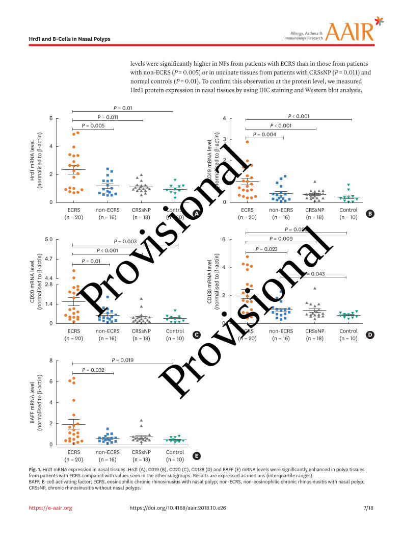
levels were significantly higher in NPs from patients with ECRS than in those from patients with non-ECRS (P = 0.005) or in uncinate tissues from patients with CRSsNP (P = 0.011) and normal controls (P = 0.01). To confirm this observation at the protein level, we measured Hrd1 protein expression in nasal tissues by using IHC staining and Western blot analysis.
7/18https://e-aair.org https://doi.org/10.4168/aair.2018.10.e26
Hrd1 and B-Cells in Nasal Polyps
B
0
2
6
4
Hrd
1 mRN
A le
vel
(nor
mal
ised
to β
-act
in)
ECRS(n = 20)
non-ECRS(n = 16)
P = 0.005
CRSsNP(n = 18)
Control(n = 10) A
P = 0.011
P = 0.01
0
2
4
3
CD19
mRN
A le
vel
(nor
mal
ised
to β
-act
in)
ECRS(n = 20)
non-ECRS(n = 16)
CRSsNP(n = 18)
Control(n = 10)
1
P = 0.004P < 0.001
P < 0.001
D
0
2
6
4
CD13
8 m
RNA
leve
l(n
orm
alis
ed to
β-a
ctin
)
ECRS(n = 20)
non-ECRS(n = 16)
CRSsNP(n = 18)
Control(n = 10)
P = 0.023
P = 0.043
P = 0.009
P = 0.002
0
2.8
5.0
4.4
CD20
mRN
A le
vel
(nor
mal
ised
to β
-act
in)
ECRS(n = 20)
non-ECRS(n = 16)
CRSsNP(n = 18)
Control(n = 10)
4.7
1.4
C
P = 0.01
P < 0.001
P = 0.003
0
8
4
BAFF
mRN
A le
vel
(nor
mal
ised
to β
-act
in)
ECRS(n = 20)
non-ECRS(n = 16)
CRSsNP(n = 18)
Control(n = 10)
6
2
E
P = 0.032
P = 0.019
Fig. 1. Hrd1 mRNA expression in nasal tissues. Hrd1 (A), CD19 (B), CD20 (C), CD138 (D) and BAFF (E) mRNA levels were significantly enhanced in polyp tissues from patients with ECRS compared with values seen in the other subgroups. Results are expressed as medians (interquartile ranges). BAFF, B-cell activating factor; ECRS, eosinophilic chronic rhinosinusitis with nasal polyp; non-ECRS, non-eosinophilic chronic rhinosinusitis with nasal polyp; CRSsNP, chronic rhinosinusitis without nasal polyps.
Provis
ional
Pro
visio
nal
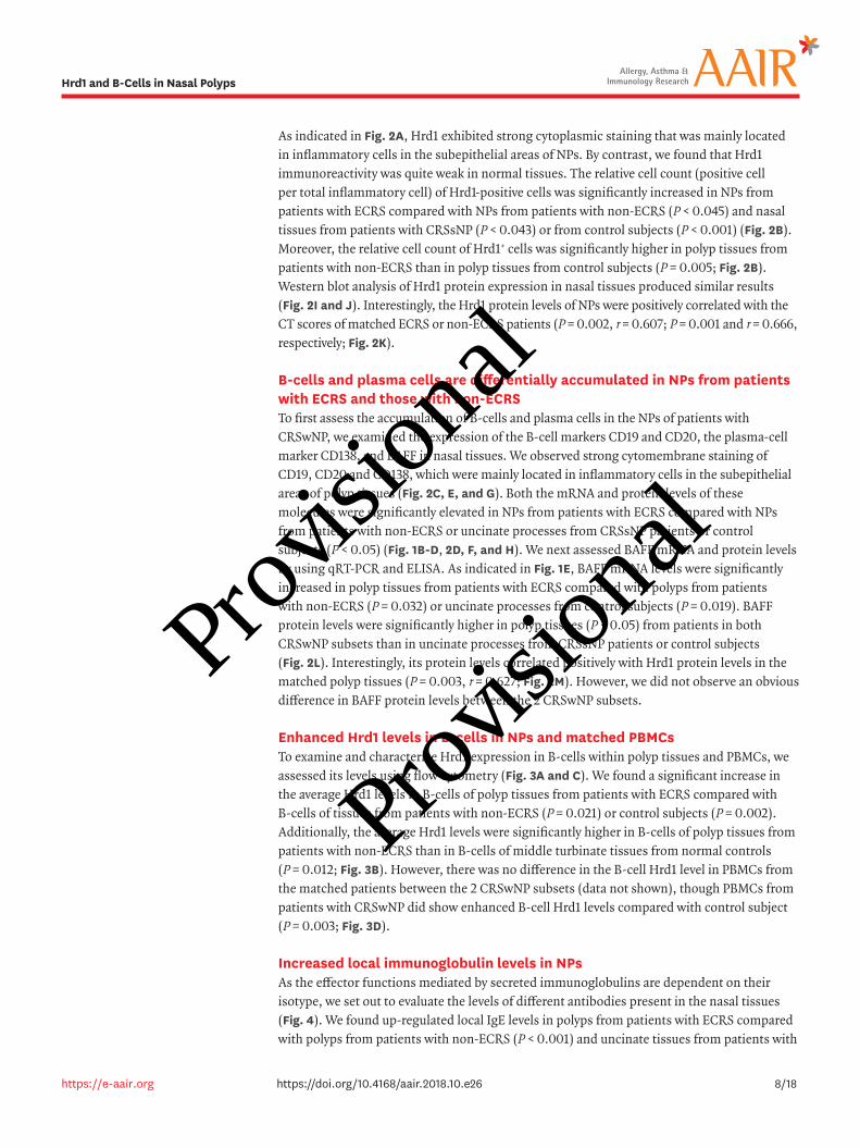
As indicated in Fig. 2A, Hrd1 exhibited strong cytoplasmic staining that was mainly located in inflammatory cells in the subepithelial areas of NPs. By contrast, we found that Hrd1 immunoreactivity was quite weak in normal tissues. The relative cell count (positive cell per total inflammatory cell) of Hrd1-positive cells was significantly increased in NPs from patients with ECRS compared with NPs from patients with non-ECRS (P < 0.045) and nasal tissues from patients with CRSsNP (P < 0.043) or from control subjects (P < 0.001) (Fig. 2B). Moreover, the relative cell count of Hrd1+ cells was significantly higher in polyp tissues from patients with non-ECRS than in polyp tissues from control subjects (P = 0.005; Fig. 2B). Western blot analysis of Hrd1 protein expression in nasal tissues produced similar results (Fig. 2I and J). Interestingly, the Hrd1 protein levels of NPs were positively correlated with the CT scores of matched ECRS or non-ECRS patients (P = 0.002, r = 0.607; P = 0.001 and r = 0.666, respectively; Fig. 2K).
B-cells and plasma cells are differentially accumulated in NPs from patients with ECRS and those with non-ECRSTo first assess the accumulation of B-cells and plasma cells in the NPs of patients with CRSwNP, we examined the expression of the B-cell markers CD19 and CD20, the plasma-cell marker CD138, and BAFF in nasal tissues. We observed strong cytomembrane staining of CD19, CD20 and CD138, which were mainly located in inflammatory cells in the subepithelial areas of polyp tissues (Fig. 2C, E, and G). Both the mRNA and protein levels of these molecules were significantly elevated in NPs from patients with ECRS compared with NPs from patients with non-ECRS or uncinate processes from CRSsNP patients or control subjects (P < 0.05) (Fig. 1B-D, 2D, F, and H). We next assessed BAFF mRNA and protein levels by using qRT-PCR and ELISA. As indicated in Fig. 1E, BAFF mRNA levels were significantly increased in polyp tissues from patients with ECRS compared with polyps from patients with non-ECRS (P = 0.032) or uncinate processes from control subjects (P = 0.019). BAFF protein levels were significantly higher in polyp tissues (P < 0.05) from patients in both CRSwNP subsets than in uncinate processes from CRSsNP patients or control subjects (Fig. 2L). Interestingly, its protein levels correlated positively with Hrd1 protein levels in the matched polyp tissues (P = 0.003, r = 0.627; Fig. 2M). However, we did not observe an obvious difference in BAFF protein levels between the 2 CRSwNP subsets.
Enhanced Hrd1 levels in B-cells in NPs and matched PBMCsTo examine and characterize Hrd1 expression in B-cells within polyp tissues and PBMCs, we assessed its levels using flow cytometry (Fig. 3A and C). We found a significant increase in the average Hrd1 levels in B-cells of polyp tissues from patients with ECRS compared with B-cells of tissues from patients with non-ECRS (P = 0.021) or control subjects (P = 0.002). Additionally, the average Hrd1 levels were significantly higher in B-cells of polyp tissues from patients with non-ECRS than in B-cells of middle turbinate tissues from normal controls (P = 0.012; Fig. 3B). However, there was no difference in the B-cell Hrd1 level in PBMCs from the matched patients between the 2 CRSwNP subsets (data not shown), though PBMCs from patients with CRSwNP did show enhanced B-cell Hrd1 levels compared with control subject (P = 0.003; Fig. 3D).
Increased local immunoglobulin levels in NPsAs the effector functions mediated by secreted immunoglobulins are dependent on their isotype, we set out to evaluate the levels of different antibodies present in the nasal tissues (Fig. 4). We found up-regulated local IgE levels in polyps from patients with ECRS compared with polyps from patients with non-ECRS (P < 0.001) and uncinate tissues from patients with
8/18https://e-aair.org https://doi.org/10.4168/aair.2018.10.e26
Hrd1 and B-Cells in Nasal Polyps
Provis
ional
Pro
visio
nal

9/18https://e-aair.org https://doi.org/10.4168/aair.2018.10.e26
Hrd1 and B-Cells in Nasal Polyps
A C E G
ECRS
Hrd
1
CD19
CD20
CD13
8
non-
ECRS
Cont
rol
CRSs
NP
I
Hrd
1
ECRS
β-ac
tin
non-
ECRS
Cont
rol
CRSs
NP
Bno
n-EC
RS(n
=13)
ECRS
(n=1
7)C
ontro
l(n
=10)
2CR
SsN
P(n
=11)P =
0.01
1
P =
0.00
5P
= 0.
045
P =
0.04
3P
< 0.
001
Relative Hrd1+ cells (%) 12 6 4810
D
P =
0.02
2
P =
0.00
1P
= 0.
036
P =
0.00
1P
< 0.
001
Relative CD19+ cells (%)
non-
ECRS
(n=1
3)EC
RS(n
=17)
Con
trol
(n=1
0)
50 20 103040 0CR
SsN
P(n
=11)
F
P =
0.01
2
P =
0.02
4P
= 0.
006
P <
0.00
1
Relative CD20+ cells (%)
non-
ECRS
(n=1
3)EC
RS(n
=17)
Con
trol
(n=1
0)
8 46 2CR
SsN
P(n
=11)
H
P =
0.02
8
P =
0.01
5P
< 0.
001
P <
0.00
1
Relative CD138+ cells (%)
non-
ECRS
(n=1
3)EC
RS(n
=17)
Con
trol
(n=1
0)
20 51015 0CR
SsN
P(n
=11)
Concentrations of BAFF in totalprotein (pg/mg)
Rela
tive
prot
ein
leve
l of H
rd1
21
50
34
M
1,00
0
400
200
600
800
P =
0.00
3r =
0.6
27n
= 20
Rela
tive
prot
ein
leve
l of H
rd1
K
Lund-Mackay CT scores
42
8
30 1020 06no
n-EC
RSEC
RS
P =
0.00
2r =
0.6
07
P =
0.00
1r =
0.6
66n
= 20
n =
23
Relative protein level of Hrd1
P <
0.00
1P
< 0.
001 P
< 0.
001
P <
0.00
1 P =
0.01
5
P =
0.00
3
J
8 246 0CR
SsN
P(n
=21)
ECRS
(n=2
2)Co
ntro
l(n
=10)
Non
-ECR
S(n
=17)
CRSs
NP
(n=2
1)EC
RS(n
=22)
Cont
rol
(n=1
0)N
on-E
CRS
(n=1
7)
Concentrations of BAFF in totalprotein (pg/mg)
P =
0.00
5P
= 0.
021 P
= 0.
008
P =
0.02
7
L
2,00
0
500 0
1,00
0
1,50
0
Fig.
2. I
ncre
ased
Hrd
1 pro
tein
leve
ls a
nd a
ccum
ulat
ion
of B
cel
ls a
nd p
lasm
a ce
lls in
NPs
. Rep
rese
ntat
ive
IHC
stai
ning
of H
rd1 (
A), C
D19
(C),
CD2
0 (E
), a
nd C
D138
(G) (
mag
nific
atio
n, ×
400
) in
nasa
l tis
sues
. Rel
ativ
e po
sitiv
e ce
lls o
f Hrd
1 (B)
, CD1
9 (D
), C
D20
(F),
and
CD1
38 (H
) wer
e si
gnifi
cant
ly in
crea
sed
in p
olyp
tiss
ues
from
pat
ient
s w
ith E
CRS
rela
tive
to th
ose
in o
ther
sub
grou
ps. R
epre
sent
ativ
e W
este
rn b
lott
ing
resu
lts (I
), d
ensi
tom
etric
ana
lysi
s (J
). P
ositi
ve c
orre
latio
n be
twee
n re
lativ
e H
rd1 p
rote
in le
vels
in th
e po
lyp
tissu
es a
nd L
und-
Mac
kay
CT s
core
s of
the
mat
ched
pat
ient
s (K
). E
LISA
of
BAFF
in n
asal
tiss
ues
(L).
Pos
itive
cor
rela
tion
betw
een
BAFF
and
Hrd
1 pro
tein
leve
ls in
the
mat
ched
NPs
(M).
Bar
s =
20 μ
m.
NP,
nas
al p
olyp
; ECR
S, e
osin
ophi
lic c
hron
ic rh
inos
inus
itis
with
nas
al p
olyp
; non
-ECR
S, n
on-e
osin
ophi
lic c
hron
ic rh
inos
inus
itis
with
nas
al p
olyp
; CRS
sNP,
chr
onic
rhin
osin
usiti
s w
ithou
t nas
al p
olyp
s;
HPF
, hig
h-po
wer
fiel
d; C
T, c
ompu
ted
tom
ogra
phy;
ELI
SA, e
nzym
e-lin
ked
imm
unos
orbe
nt a
ssay
; BAF
F, B
-cel
l act
ivat
ing
fact
or.
(con
tinue
d to
the
next
pag
e)
Provis
ional
Pro
visio
nal

10/18https://e-aair.org https://doi.org/10.4168/aair.2018.10.e26
Hrd1 and B-Cells in Nasal Polyps
A C E G
ECRS
Hrd
1
CD19
CD20
CD13
8
non-
ECRS
Cont
rol
CRSs
NP
I
Hrd
1
ECRS
β-ac
tin
non-
ECRS
Cont
rol
CRSs
NP
Bno
n-EC
RS(n
=13)
ECRS
(n=1
7)C
ontro
l(n
=10)
2CR
SsN
P(n
=11)P =
0.01
1
P =
0.00
5P
= 0.
045
P =
0.04
3P
< 0.
001
Relative Hrd1+ cells (%) 12 6 4810
D
P =
0.02
2
P =
0.00
1P
= 0.
036
P =
0.00
1P
< 0.
001
Relative CD19+ cells (%)
non-
ECRS
(n=1
3)EC
RS(n
=17)
Con
trol
(n=1
0)
50 20 103040 0CR
SsN
P(n
=11)
F
P =
0.01
2
P =
0.02
4P
= 0.
006
P <
0.00
1
Relative CD20+ cells (%)
non-
ECRS
(n=1
3)EC
RS(n
=17)
Con
trol
(n=1
0)
8 46 2CR
SsN
P(n
=11)
H
P =
0.02
8
P =
0.01
5P
< 0.
001
P <
0.00
1
Relative CD138+ cells (%)
non-
ECRS
(n=1
3)EC
RS(n
=17)
Con
trol
(n=1
0)
20 51015 0CR
SsN
P(n
=11)
Concentrations of BAFF in totalprotein (pg/mg)
Rela
tive
prot
ein
leve
l of H
rd1
21
50
34
M
1,00
0
400
200
600
800
P =
0.00
3r =
0.6
27n
= 20
Rela
tive
prot
ein
leve
l of H
rd1
K
Lund-Mackay CT scores
42
8
30 1020 06no
n-EC
RSEC
RS
P =
0.00
2r =
0.6
07
P =
0.00
1r =
0.6
66n
= 20
n =
23
Relative protein level of Hrd1
P <
0.00
1P
< 0.
001 P
< 0.
001
P <
0.00
1 P =
0.01
5
P =
0.00
3
J
8 246 0CR
SsN
P(n
=21)
ECRS
(n=2
2)Co
ntro
l(n
=10)
Non
-ECR
S(n
=17)
CRSs
NP
(n=2
1)EC
RS(n
=22)
Cont
rol
(n=1
0)N
on-E
CRS
(n=1
7)
Concentrations of BAFF in totalprotein (pg/mg)
P =
0.00
5P
= 0.
021 P
= 0.
008
P =
0.02
7
L
2,00
0
500 0
1,00
0
1,50
0
Fig.
2. (
Cont
inue
d) In
crea
sed
Hrd
1 pro
tein
leve
ls a
nd a
ccum
ulat
ion
of B
cel
ls a
nd p
lasm
a ce
lls in
NPs
. Rep
rese
ntat
ive
IHC
stai
ning
of H
rd1 (
A), C
D19
(C),
CD2
0 (E
), a
nd C
D138
(G) (
mag
nific
atio
n,
× 40
0) in
nas
al ti
ssue
s. R
elat
ive
posi
tive
cells
of H
rd1 (
B), C
D19
(D),
CD2
0 (F
), a
nd C
D138
(H) w
ere
sign
ifica
ntly
incr
ease
d in
pol
yp ti
ssue
s fr
om p
atie
nts
with
ECR
S re
lativ
e to
thos
e in
oth
er s
ubgr
oups
. Re
pres
enta
tive
Wes
tern
blo
ttin
g re
sults
(I),
den
sito
met
ric a
naly
sis
(J).
Pos
itive
cor
rela
tion
betw
een
rela
tive
Hrd
1 pro
tein
leve
ls in
the
poly
p tis
sues
and
Lun
d-M
acka
y CT
sco
res
of th
e m
atch
ed
patie
nts
(K).
ELI
SA o
f BAF
F in
nas
al ti
ssue
s (L
). P
ositi
ve c
orre
latio
n be
twee
n BA
FF a
nd H
rd1 p
rote
in le
vels
in th
e m
atch
ed N
Ps (M
). B
ars
= 20
μm
. N
P, n
asal
pol
yp; E
CRS,
eos
inop
hilic
chr
onic
rhin
osin
usiti
s w
ith n
asal
pol
yp; n
on-E
CRS,
non
-eos
inop
hilic
chr
onic
rhin
osin
usiti
s w
ith n
asal
pol
yp; C
RSsN
P, c
hron
ic rh
inos
inus
itis
with
out n
asal
pol
yps;
H
PF, h
igh-
pow
er fi
eld;
CT,
com
pute
d to
mog
raph
y; E
LISA
, enz
yme-
linke
d im
mun
osor
bent
ass
ay; B
AFF,
B-c
ell a
ctiv
atin
g fa
ctor
.
Provis
ional
Pro
visio
nal

11/18https://e-aair.org https://doi.org/10.4168/aair.2018.10.e26
Hrd1 and B-Cells in Nasal Polyps
0
2,00
0
10,0
00
8,00
0
MFI level of Hrd1
DCR
SwN
P(n
=13)
P =
0.00
3
Cont
rol
(n=7
)
4,00
0
6,00
0
C
250K
200K
150K
100K 50
K 00
−103
103
104
105
250K
200K
150K
100K 50
K 00
−103
103
104
105
250K
250K
200K
150K
100K
50K
200K
150K
100K 50
K 00 FS
C-A
CD45
CD19
0−103
103
104
105
0
100 80 60 40 20
Hrd
1
SSC-A
FSC-H
FSC-H
Isot
ype
Cont
rol
CRSw
NP
0
5,00
0
15,0
00
10,0
00
MFI level of Hrd1
BN
on-E
CRS
(n=7
)EC
RS(n
=8)
Cont
rol
(n=5
)
P =
0.01
2
P =
0.02
1P =
0.00
2
SSC-A
FSC-
A
A
0−103
103
104
105
250K
200K
150K
100K 50
K 00
−103
103
104
105
250K
200K
150K
100K 50
K 0
250K
250K
200K
150K
100K
50K
200K
150K
100K 50
K 00
CD45
CD19
0−103
103
104
105
100 80 60 40 20 0
Hrd
1
FSC-H
FSC-H
Isot
ype
Cont
rol
Non
-ECR
SEC
RS
Fig.
3. E
nhan
ced
Hrd
1 exp
ress
ion
in B
-cel
ls fr
om p
olyp
tiss
ues
and
mat
ched
PBM
Cs. G
atin
g st
rate
gy a
nd re
pres
enta
tive
flow
plo
ts fo
r nas
al ti
ssue
s (A
). M
FI le
vels
of H
rd1 i
n CD
19+ B
-cel
ls w
ere
sign
ifica
ntly
incr
ease
d in
pol
yp ti
ssue
s fr
om p
atie
nts
with
ECR
S co
mpa
red
with
thos
e in
in p
olyp
s fr
om p
atie
nts
with
non
-ECR
S, m
iddl
e tu
rbin
ates
from
con
trol
sub
ject
s (B
). G
atin
g st
rate
gy a
nd
repr
esen
tativ
e flo
w p
lots
for P
BMCs
(C).
MFI
leve
ls o
f Hrd
1 in
CD19
+ B-c
ells
wer
e si
gnifi
cant
ly in
crea
sed
in P
BMCs
from
pat
ient
s w
ith C
RSw
NP
com
pare
d w
ith th
ose
in P
BMCs
from
hea
lthy
cont
rols
(D).
PB
MC,
per
iphe
ral b
lood
mon
onuc
lear
cel
l; M
FI, m
ean
fluor
esce
nce
inte
nsity
; ECR
S, e
osin
ophi
lic c
hron
ic rh
inos
inus
itis
with
nas
al p
olyp
; non
-ECR
S, n
on-e
osin
ophi
lic c
hron
ic rh
inos
inus
itis
with
nas
al
poly
p; C
RSw
NP,
chr
onic
rhin
osin
usiti
s w
ith n
asal
pol
yps.
Provis
ional
Pro
visio
nal

12/18https://e-aair.org https://doi.org/10.4168/aair.2018.10.e26
Hrd1 and B-Cells in Nasal Polyps
01020 15
Concentrations of IgM in totalprotein (µg/mg)
3
P =
0.01
4r =
0.5
42n
= 20
Rela
tive
prot
ein
leve
l of H
rd1
14
25
5
0
100
200
150
Concentrations of IgG in totalprotein (µg/mg)
3
P <
0.00
1r =
0.7
14n
= 20
Rela
tive
prot
ein
leve
l of H
rd1
14
25
50
060100 80
Concentrations of IgG1 in totalprotein (µg/mg)
3
P =
0.02
2r =
0.5
08n
= 20
Rela
tive
prot
ein
leve
l of H
rd1
14
25
40 20
04080 60
Concentrations of IgG2 in totalprotein (µg/mg)
3
P =
0.01
5r =
0.5
37n
= 20
Rela
tive
prot
ein
leve
l of H
rd1
14
25
20
050150
100
µg/mg total protein
CRSs
NP
(n=2
1)
IgA
ECRS
(n=2
2)Co
ntro
l(n
=10)
Non
-ECR
S(n
=17)
050200
150
µg/mg total protein
CRSs
NP
(n=2
1)
IgG
ECRS
(n=2
2)Co
ntro
l(n
=10)
Non
-ECR
S(n
=17)
100
02070 50
µg/mg total protein
CRSs
NP
(n=2
1)
IgE
ECRS
(n=2
2)Co
ntro
l(n
=10)
60
Non
-ECR
S(n
=17)
40 10
0624
µg/mg total protein
CRSs
NP
(n=2
1)
IgM
ECRS
(n=2
2)Co
ntro
l(n
=10)
18
Non
-ECR
S(n
=17)
12 3
P <
0.00
1P
= 0.
001
P =
0.00
1P
< 0.
001
P <
0.00
1
P =
0.00
2
P <
0.00
1P
< 0.
001
P <
0.00
1P
= 0.
001
P =
0.00
4P
< 0.
001
P =
0.00
3P
< 0.
001
P =
0.00
1
P =
0.00
2
060100 80
µg/mg total protein
CRSs
NP
(n=2
1)
IgG
1
ECRS
(n=2
2)Co
ntro
l(n
=10)
Non
-ECR
S(n
=17)
40 20
0515 10
µg/mg total protein
CRSs
NP
(n=2
1)
IgG
3
ECRS
(n=2
2)Co
ntro
l(n
=10)
Non
-ECR
S(n
=17)
020100 60
µg/mg total protein
CRSs
NP
(n=2
1)
IgG
2
ECRS
(n=2
2)Co
ntro
l(n
=10)
80
Non
-ECR
S(n
=17)
40P
= 0.
001
P =
0.02
9
P <
0.00
1
015
µg/mg total protein
CRSs
NP
(n=2
1)
IgG
4
ECRS
(n=2
2)Co
ntro
l(n
=10)
10
Non
-ECR
S(n
=17)
5
P =
0.04
3P
= 0.
001
P <
0.00
1
P <
0.00
1
P <
0.00
1P
< 0.
001
P <
0.00
1P
< 0.
001
P =
0.00
3P
< 0.
001
P =
0.00
1
P =
0.00
2
P <
0.00
1P
= 0.
016
Fig.
4. I
ncre
ased
loca
l im
mun
oglo
bulin
leve
ls in
pol
yp ti
ssue
s. P
rote
in le
vels
of I
gA, I
gE, I
gM, I
gG a
nd Ig
G s
ubcl
asse
s in
tiss
ue h
omog
enat
es fr
om E
CRS,
non
-ECR
S, C
RSsN
P, a
nd c
ontr
ol ti
ssue
are
de
tect
ed b
y us
ing
the
Bio-
Plex
ass
ay. P
ositi
ve c
orre
latio
ns a
re fo
und
betw
een
rela
tive
Hrd
1 pro
tein
leve
ls a
nd Ig
M, I
gG, I
gG1,
and
IgG
2.
Ig, i
mm
unog
lobu
lin; E
CRS,
eos
inop
hilic
chr
onic
rhin
osin
usiti
s w
ith n
asal
pol
yp; n
on-E
CRS,
non
-eos
inop
hilic
chr
onic
rhin
osin
usiti
s w
ith n
asal
pol
yp; C
RSsN
P, c
hron
ic rh
inos
inus
itis
with
out n
asal
pol
yps.
Provis
ional
Pro
visio
nal

CRSsNP (P = 0.001) or control individuals (P = 0.004). Moreover, the levels of total IgG, IgG1, IgG2, IgG3, IgG4, IgM, and IgA were increased similarly in both patients with ECRS and those with non-ECRS compared with control subjects (P < 0.05). Interestingly, we found the Hrd1 protein levels correlated positively with IgM, IgG, IgG1, and IgG2 levels in the matched polyp tissues (P < 0.05; r = 0.542, r = 0.714, r = 0.508 and r = 0.537, respectively).
IL-1β, LPS, and Poly(I:C) differentially regulate Hrd1 expression in DPCs from patients with ECRS and those with non-ECRS in vitroTo determine the critical roles of Hrd1 in innate immune response, we attempted to establish a direct relationship between Toll-like receptors (TLRs) and innate immunity as well as between proinflammatory cytokines and innate immunity in the nasal mucosa. Thus, we investigated the effects of TLR activation and IL-1β stimulation on Hrd1 expression in DPCs in vitro. As indicated in Fig. 5A-C, Hrd1 mRNA and protein levels were significantly increased in DPCs from patients with ECRS after stimulation with IL-1β, LPS and Poly(I:C) (P < 0.05). However, when DPCs from patients with non-ECRS were stimulated with IL-1β, LPS, or Poly(I:C), we did not observe obvious changes in Hrd1 mRNA and protein expression (Fig. 5D-F). We also examined the effect of Dex on Hrd1 mRNA and protein expression in DPCs in vitro. After incubating cells with Dex, Hrd1 mRNA and protein levels were significantly down-regulated in the absence or presence of TLR agonists and proinflammatory cytokines (IL-1β, LPS and Poly(I:C); P < 0.05; Fig. 5).
DISCUSSION
It is well known that CRSwNP is characterized by type 2 inflammation and eosinophilia.2-4 However, recent evidence has suggested that the local proliferation and activation of B-cells and plasma cells are of central pathogenic importance in airway diseases including CRSwNP. Studies have demonstrated an increase in B-cell chemoattractants, B and plasma cell numbers, and antigen-specific IgE in the nasal mucosa of CRSwNP patients,7,9,10,24 and more recently increases in BAFF have also been demonstrated by other study groups and ours.10,25 This chemokine is thought to not only promote B-cell survival, proliferation, and maturation but also facilitate immunoglobulin CSR, a process central to IgA production, eosinophil activation, and subsequent polyp formation.9,10,26 Although numerous studies have demonstrated the critical roles of B-cells in the pathophysiology of CRSwNP, precise molecular mechanisms underlying the inflammatory pattern in patients with CRSwNP have not yet been completely elucidated.
In the present study, we have expanded on our earlier work and found that B-cells exhibited eosinophilic accumulation in NPs from patients with ECRS.25 Polyp tissues from patients with ECRS not only showed increased expression of BAFF but also contained significantly more B-cells and plasma cells compared with NPs from patients with non-ECRS or uncinate tissues from control subjects (Figs. 1 and 2). Whether these B-cells enter the tissue as naive cells and become activated or if they enter as memory cells primed to respond within the tissue is not yet clear. Previous studies have demonstrated that eosinophils not only produce a variety of proinflammatory molecules, including BAFF, that can contribute type 2 inflammation but also play an important role in the long-term maintenance of plasma cells and have been shown to activate T-cells during inflammatory responses.10,27,28 Further studies are needed to determine whether B-cells can be activated within NPs themselves and what mechanisms might be involved.
13/18https://e-aair.org https://doi.org/10.4168/aair.2018.10.e26
Hrd1 and B-Cells in Nasal Polyps
Provis
ional
Pro
visio
nal

Hrd1 is known to exert antiapoptotic effects in a variety of immune responses.17-19 However, little is known about the expression of Hrd1 and its possible roles in the polyp tissues of CRSwNP patients. In the current study, we found a significant increase in both Hrd1 gene and protein expression in NPs from patients with ECRS compared with NPs from patients with non-ECRS and uncinate tissues from CRSsNP patients or normal controls (Fig. 1A, Fig. 2A, B, I and J). We also showed that Hrd1 relative protein levels correlated with disease severity indicated by Lund-Mackay CT scores (Fig. 2K). In addition, we observed increased Hrd1 expression in B-cells in polyp tissues and matched PBMCs from CRSwNP patients (Fig. 3). As we previously showed that Hrd1 has an anti-apoptotic function in B-cell development,19 it is reasonable to speculate that Hrd1 expression could play an anti-apoptotic role in NPs. Interestingly, we found that Hrd1 mRNA and protein levels were strongly increased in polyp
14/18https://e-aair.org https://doi.org/10.4168/aair.2018.10.e26
Hrd1 and B-Cells in Nasal Polyps
−
Hrd1
β-actin
IL1βDex − −
−++
++
Hrd
1 mRN
A le
vel
(nor
mal
ised
to β
-act
in)
0
10
15
5
‡
†
*
†
A−
Hrd1
β-actin
LPSDex − −
−++
++
Hrd
1 mRN
A le
vel
(nor
mal
ised
to β
-act
in)
0
3
4
2
1
‡
‡
‡
†
†
B−
Hrd1
β-actin
Poly (I:C)Dex − −
−++
++
Hrd
1 mRN
A le
vel
(nor
mal
ised
to β
-act
in)
0
6
8
4
2
†
†
**
*
†
C
−
Hrd1
β-actin
IL1βDex − −
−++
++
Hrd
1 mRN
A le
vel
(nor
mal
ised
to β
-act
in)
0
1.5
2.0
0.5
1.0
**
**
D−
Hrd1
β-actin
LPSDex − −
−++
++
Hrd
1 mRN
A le
vel
(nor
mal
ised
to β
-act
in)
0
1.0
1.5
0.5
* ***
†
†
E−
Hrd1
β-actin
Poly (I:C)Dex − −
−++
++
Hrd
1 mRN
A le
vel
(nor
mal
ised
to β
-act
in)
0
3
4
1
2
*
F
Fig. 5. Hrd1 mRNA and protein levels in cultured DPCs in response to IL-1β, LPS, Poly(I:C) and Dex. mRNA expression and representative western blot results of DPCs lysates from ECRS patients after 20-hour stimulation (A-C). mRNA expression and representative western blot results of DPCs lysates from non-ECRS patients after 20-hour stimulation (D-F). Results represent mean values from 3 independent experiments. Data are expressed as means (standard error of the means). DPC, dispersed polyp cell; IL, interleukin; LPS, lipopolysaccharide; Poly(I:C), polyinosinic-polycytidylic acid; Dex, dexamethasone; ECRS, eosinophilic chronic rhinosinusitis with nasal polyp;non-ECRS, non-eosinophilic chronic rhinosinusitis with nasal polyp. *P < 0.05; †P < 0.01; ‡P < 0.001.
Provis
ional
Pro
visio
nal

tissue homogenates (Fig. 1A, Fig. 2A, B, I and J), as well as in B-cells in NPs, but not PBMCs (data not shown) (Fig. 3B and D), from ECRS patients compared with those from non-ECRS patients. These data might suggest that Hrd1 expression is associated with eosinophil recruitment and localization in polyp tissues in patients with CRSwNP. Several groups have shown that immunoglobulins potentially play an important role in the pathogenesis of NPs.10,11,24 In this study, we found that IgA, IgG and IgM, but not IgE, were similarly enhanced in eosinophilic and non-eosinophilic polyps compared with uncinate tissues from control subjects (Fig. 4). These findings were in accordance with those reported by Zhang et al.29 Moreover, we found that local Hrd1 protein levels in NPs were positively associated with IgM, IgG, IgG1, and IgG2 levels in the matched patients with CRSwNP (Fig. 4). Therefore, increased Hrd1 expression might contribute to immunoglobulin production in polyp tissues. Collectively, the studies described here further suggest that Hrd1 plays a potential role in disease pathogenesis, although future studies are needed to confirm whether Hrd1 is involved in activating local plasma-cell responses in patients with CRSwNP.
The innate immune response is the first line of host defense and is responsible for immediate recognition and control of microbial invasion. The cytokines IL-25, IL-33, and TSLP released by damaged epithelial cells subsequently recruit and activate eosinophils, basophils, mast cells and ILCs, which promote type 2 inflammation. These innate immune cells and released cytokines further contribute to the chronic inflammatory response and directly activate adaptive immune cells including T and B-cells.5,6 One of the primary families of molecules responsible for innate immunity in humans are the TLRs, expressed by epithelial cells and immune cells in the nose and sinuses, could activate expression of inflammatory mediators and host defense molecules.30 Among them, abnormalities in the immune response or expression of TLR3 and TLR4 in CRSwNP have been reported by other study groups and ours.21,31 Activation of TLR3 or TLR4 can lead to the activation of nuclear factor-κB (NF-κB), which results in the expression of proinflammatory cytokines that are critical for both innate and adaptive immune responses.32 In this study, Hrd1 mRNA and protein levels were significantly up-regulated in eosinophilic DPCs after stimulation with IL-1β, Poly(I:C) (TLR3 ligand) and LPS (TLR4 ligand) in vitro. However, we did not observe obvious changes in the Hrd1 mRNA and protein levels in non-eosinophilic DPCs after treatment with the same stimuli. Our findings suggest that Hrd1 participates in the proinflammatory process and innate immunity and that the eosinophilic response might play a role in inducing Hrd1 expression in patients with CRSwNP. B-cells express MHC class II molecules, along with co-stimulatory molecules, and thus play a key role in the activation of T-cell responses.32,33 Although the immune regulatory function of Hrd1 has been reported in some studies,18,19,34 the observed positive effects of Hrd1 on B-cells in cultured eosinophilic DPCs after TLR agonist and proinflammatory cytokine stimulation in this study cannot be ignored. Notably, we found that Dex decreased Hrd1 mRNA and protein expression, independent of the presence or absence of IL-1β, Poly(I:C) and LPS in both eosinophilic and non-eosinophilic DPCs. Although the underlying molecular mechanisms require further characterization, this finding demonstrates that Dex treatment decreases proinflammatory cytokine-induced Hrd1 expression. Therefore, Hrd1 might represent a potential therapeutic target for patients with CRSwNP.
In summary, to the best of our knowledge, this is the first report of differential expression of Hrd1 in NPs from patients with ECRS and those with non-ECRS. We showed that NPs from patients with ECRS had abundant accumulation of B-cells and plasma cells. Not surprisingly, we also observed significantly increased levels of several immunoglobulins in NPs. Finally, we
15/18https://e-aair.org https://doi.org/10.4168/aair.2018.10.e26
Hrd1 and B-Cells in Nasal Polyps
Provis
ional
Pro
visio
nal

showed that Hrd1 was differentially expressed in B-cells in eosinophilic and non-eosinophilic polyp tissues and participated in the innate immune response. These findings indicate that the differential expression of Hrd1 in NPs might facilitate a better understanding of the pathogenesis involved in these 2 CRSwNP subsets and provide novel insights into the development of improved therapeutic interventions.
ACKNOWLEDGMENTS
This study was supported by National Natural Science Foundation of China grants 81470673,81470689 and 81725004 (H.L.), 81470689 (J.Y.) and 81725004 (M.H.), and a grant from Shanghai Tenth Hospital's improvement plan for NSFC: SYGZRPY2017032 (M.T.).
SUPPLEMENTARY MATERIALS
Supplementary Table S1Subjects' characteristics
Click here to view
Supplementary Table S2Primers used for qRT-PCR analysis
Click here to view
Supplementary Table S3Detection limit for Bio-Plex assay
Click here to view
Supplementary Fig. S1Enhanced Hrd1 expression in T cells from nasal polyps.
Click here to view
Supplementary Fig. S2Polyp tissue continuous section (3-μm) to detect whether Hrd1 sourced from mast cells (indicated by tryptase). As shown in the figure, Hrd1 was not sourced from mast cells.
Click here to view
Supplementary Fig. S3Increased local inflammatory cytokines in nasal polyps. Protein levels of IFN-γ, IL-4, IL-5, IL-13 and IL-17A in tissue homogenates from ECRS, non-ECRS, CRSsNP and control tissue are detected by using the ELISA assay.
Click here to view
16/18https://e-aair.org https://doi.org/10.4168/aair.2018.10.e26
Hrd1 and B-Cells in Nasal Polyps
Provis
ional
Pro
visio
nal

REFERENCES
1. Fokkens WJ, Lund VJ, Mullol J, Bachert C, Alobid I, Baroody F, et al. EPOS 2012: European position paper on rhinosinusitis and nasal polyps 2012. A summary for otorhinolaryngologists. Rhinology 2012;50:1-12.PUBMED
2. Hsu J, Peters AT. Pathophysiology of chronic rhinosinusitis with nasal polyp. Am J Rhinol Allergy 2011;25:285-90. PUBMED | CROSSREF
3. Akdis CA, Bachert C, Cingi C, Dykewicz MS, Hellings PW, Naclerio RM, et al. Endotypes and phenotypes of chronic rhinosinusitis: a PRACTALL document of the European Academy of Allergy and Clinical Immunology and the American Academy of Allergy, Asthma & Immunology. J Allergy Clin Immunol 2013;131:1479-90. PUBMED | CROSSREF
4. Payne SC, Borish L, Steinke JW. Genetics and phenotyping in chronic sinusitis. J Allergy Clin Immunol 2011;128:710-20. PUBMED | CROSSREF
5. Hulse KE. Immune mechanisms of chronic rhinosinusitis. Curr Allergy Asthma Rep 2016;16:1. PUBMED | CROSSREF
6. Khalmuratova R, Park JW, Shin HW. Immune cell responses and mucosal barrier disruptions in chronic rhinosinusitis. Immune Netw 2017;17:60-7. PUBMED | CROSSREF
7. Hulse KE, Stevens WW, Tan BK, Schleimer RP. Pathogenesis of nasal polyposis. Clin Exp Allergy 2015;45:328-46. PUBMED | CROSSREF
8. Dörner T, Radbruch A, Burmester GR. B-cell-directed therapies for autoimmune disease. Nat Rev Rheumatol 2009;5:433-41. PUBMED | CROSSREF
9. Kato A, Hulse KE, Tan BK, Schleimer RP. B-lymphocyte lineage cells and the respiratory system. J Allergy Clin Immunol 2013;131:933-57. PUBMED | CROSSREF
10. Kato A, Peters A, Suh L, Carter R, Harris KE, Chandra R, et al. Evidence of a role for B cell-activating factor of the TNF family in the pathogenesis of chronic rhinosinusitis with nasal polyps. J Allergy Clin Immunol 2008;121:1385-92, 1392.e1-2.PUBMED | CROSSREF
11. Song J, Wang H, Zhang YN, Cao PP, Liao B, Wang ZZ, et al. Ectopic lymphoid tissues support local immunoglobulin production in patients with chronic rhinosinusitis with nasal polyps. J Allergy Clin Immunol 2018;141:927-37. PUBMED | CROSSREF
12. Van Zele T, Gevaert P, Watelet JB, Claeys G, Holtappels G, Claeys C, et al. Staphylococcus aureus colonization and IgE antibody formation to enterotoxins is increased in nasal polyposis. J Allergy Clin Immunol 2004;114:981-3. PUBMED | CROSSREF
13. Tan BK, Li QZ, Suh L, Kato A, Conley DB, Chandra RK, et al. Evidence for intranasal antinuclear autoantibodies in patients with chronic rhinosinusitis with nasal polyps. J Allergy Clin Immunol 2011;128:1198-1206.e1. PUBMED | CROSSREF
14. Guerriero CJ, Brodsky JL. The delicate balance between secreted protein folding and endoplasmic reticulum-associated degradation in human physiology. Physiol Rev 2012;92:537-76. PUBMED | CROSSREF
15. Amano T, Yamasaki S, Yagishita N, Tsuchimochi K, Shin H, Kawahara K, et al. Synoviolin/Hrd1, an E3 ubiquitin ligase, as a novel pathogenic factor for arthropathy. Genes Dev 2003;17:2436-49. PUBMED | CROSSREF
16. Gao B, Lee SM, Chen A, Zhang J, Zhang DD, Kannan K, et al. Synoviolin promotes IRE1 ubiquitination and degradation in synovial fibroblasts from mice with collagen-induced arthritis. EMBO Rep 2008;9:480-5. PUBMED | CROSSREF
17. Toh ML, Marotte H, Blond JL, Jhumka U, Eljaafari A, Mougin B, et al. Overexpression of synoviolin in peripheral blood and synoviocytes from rheumatoid arthritis patients and continued elevation in nonresponders to infliximab treatment. Arthritis Rheum 2006;54:2109-18. PUBMED | CROSSREF
17/18https://e-aair.org https://doi.org/10.4168/aair.2018.10.e26
Hrd1 and B-Cells in Nasal Polyps
Provis
ional
Pro
visio
nal

18. Xu Y, Zhao F, Qiu Q, Chen K, Wei J, Kong Q, et al. The ER membrane-anchored ubiquitin ligase Hrd1 is a positive regulator of T-cell immunity. Nat Commun 2016;7:12073. PUBMED | CROSSREF
19. Kong S, Yang Y, Xu Y, Wang Y, Zhang Y, Melo-Cardenas J, et al. Endoplasmic reticulum-resident E3 ubiquitin ligase Hrd1 controls B-cell immunity through degradation of the death receptor CD95/Fas. Proc Natl Acad Sci U S A 2016;113:10394-9. PUBMED | CROSSREF
20. Wen W, Liu W, Zhang L, Bai J, Fan Y, Xia W, et al. Increased neutrophilia in nasal polyps reduces the response to oral corticosteroid therapy. J Allergy Clin Immunol 2012;129:1522-1528.e5. PUBMED | CROSSREF
21. Wei Y, Xia W, Ye X, Fan Y, Shi J, Wen W, et al. The antimicrobial protein short palate, lung, and nasal epithelium clone 1 (SPLUNC1) is differentially modulated in eosinophilic and noneosinophilic chronic rhinosinusitis with nasal polyps. J Allergy Clin Immunol 2014;133:420-428.e10. PUBMED | CROSSREF
22. Ba L, Du J, Liu F, Yang F, Han M, Liu S, et al. Distinct inflammatory profiles in atopic and nonatopic patients with chronic rhinosinustis accompanied by nasal polyps in Western china. Allergy Asthma Immunol Res 2015;7:346-58. PUBMED | CROSSREF
23. Lindqvist M, van Lunzen J, Soghoian DZ, Kuhl BD, Ranasinghe S, Kranias G, et al. Expansion of HIV-specific T follicular helper cells in chronic HIV infection. J Clin Invest 2012;122:3271-80. PUBMED | CROSSREF
24. Hulse KE, Norton JE, Suh L, Zhong Q, Mahdavinia M, Simon P, et al. Chronic rhinosinusitis with nasal polyps is characterized by B-cell inflammation and EBV-induced protein 2 expression. J Allergy Clin Immunol 2013;131:1075-83, 1083.e1-7.PUBMED | CROSSREF
25. Dilidaer, Zheng Y, Liu Z, Hu X, Zhang J, Hu L, et al. Increased BAFF expression in nasal polyps is associated with local IgE production, Th2 response and concomitant asthma. Eur Arch Otorhinolaryngol 2017;274:1883-90. PUBMED | CROSSREF
26. Feldman S, Kasjanski R, Poposki J, Hernandez D, Chen JN, Norton JE, et al. Chronic airway inflammation provides a unique environment for B cell activation and antibody production. Clin Exp Allergy 2017;47:457-66. PUBMED | CROSSREF
27. Chu VT, Fröhlich A, Steinhauser G, Scheel T, Roch T, Fillatreau S, et al. Eosinophils are required for the maintenance of plasma cells in the bone marrow. Nat Immunol 2011;12:151-9. PUBMED | CROSSREF
28. Jacobsen EA, Helmers RA, Lee JJ, Lee NA. The expanding role(s) of eosinophils in health and disease. Blood 2012;120:3882-90. PUBMED | CROSSREF
29. Zhang YN, Song J, Wang H, Wang H, Zeng M, Zhai GT, et al. Nasal IL-4(+)CXCR5(+)CD4(+) T follicular helper cell counts correlate with local IgE production in eosinophilic nasal polyps. J Allergy Clin Immunol 2016;137:462-73. PUBMED | CROSSREF
30. Schleimer RP. Immunopathogenesis of chronic rhinosinusitis and nasal polyposis. Annu Rev Pathol 2017;12:331-57. PUBMED | CROSSREF
31. Hamilos DL. Host-microbial interactions in patients with chronic rhinosinusitis. J Allergy Clin Immunol 2014;133:640-653.e4. PUBMED | CROSSREF
32. Ishii KJ, Koyama S, Nakagawa A, Coban C, Akira S. Host innate immune receptors and beyond: making sense of microbial infections. Cell Host Microbe 2008;3:352-63. PUBMED | CROSSREF
33. Lund FE, Randall TD. Effector and regulatory B cells: modulators of CD4+ T cell immunity. Nat Rev Immunol 2010;10:236-47. PUBMED | CROSSREF
34. Yang H, Qiu Q, Gao B, Kong S, Lin Z, Fang D. Hrd1-mediated BLIMP-1 ubiquitination promotes dendritic cell MHCII expression for CD4 T cell priming during inflammation. J Exp Med 2014;211:2467-79. PUBMED | CROSSREF
18/18https://e-aair.org https://doi.org/10.4168/aair.2018.10.e26
Hrd1 and B-Cells in Nasal Polyps
Provis
ional
Pro
visio
nal