original article difference in vitamin d levels between
TRANSCRIPT

81https://pghn.org
ABSTRACT
Purpose: A steady increase in Clostridioides difficile enteritis (CDE) has been reported recently. CDE is associated with intestinal dysbiosis, and vitamin D receptors are known to play an important role in this microbial imbalance as immunological regulators. We investigated the difference in vitamin D levels between children with CDE and those with other acute infectious enteritis.Methods: This retrospective study was conducted on children below 18 years of age who visited the Gil hospital, underwent investigation to assess vitamin D levels, and had confirmed gastrointestinal infection between January 2015 and December 2018. Patients were divided into two groups: the “CDE group” (n=18) and the “other infectious enteritis group” (n=88); their clinical characteristics, other laboratory results, and vitamin D levels were analyzed.Results: There was no difference in gender, age, and seasonal distributions between the CDE and other infectious enteritis groups. Other laboratory results were not significantly different between two groups, excluding serum albumin level (4.52±0.45 g/dL vs. 4.31±0.28 g/dL, p=0.011). The mean 25-hydroxy vitamin D level in the CDE group was higher than that in the control group (18.75±8.11 ng/mL vs. 14.50±6.79 ng/mL, p=0.021).Conclusion: Vitamin D levels in the CDE group were lower than normal but higher than the other infectious enteritis group. These results suggested that CDE has a different mechanism or susceptibility associated with vitamin D in children, and even marginal changes in vitamin D levels can act as a risk factor for infection.
Keywords: Clostridioides difficile; Children; Vitamin D; Receptors, calcitriol
INTRODUCTION
Clostridioides difficile is an anaerobic gram-positive spore-forming bacillus that is known to occur in both toxic and non-toxic forms. C. difficile enteritis (CDE) can cause symptoms that range from mild diarrhea and toxic megacolon to death [1].
C. difficile is the cause of both hospital- and community-acquired infections worldwide, with hospital-acquired infections being the most common. Toxinotype V is one of the hypervirulent strains of C. difficile, and is the common cause of death associated with hospital-
Pediatr Gastroenterol Hepatol Nutr. 2021 Jan;24(1):81-89https://doi.org/10.5223/pghn.2021.24.1.81pISSN 2234-8646·eISSN 2234-8840
Original Article
Received: May 17, 2020Revised: Sep 2, 2020Accepted: Sep 19, 2020
Correspondence toEell RyooDepartment of Pediatrics, Gachon Children's Hospital, Gachon University College of Medicine, 21 Namdong-daero 774beon-gil, Namdong-gu, Incheon 21565, Korea.E-mail: [email protected]
Copyright © 2021 by The Korean Society of Pediatric Gastroenterology, Hepatology and NutritionThis is an open-access article distributed under the terms of the Creative Commons Attribution Non-Commercial License (https://creativecommons.org/licenses/by-nc/4.0/) which permits unrestricted non-commercial use, distribution, and reproduction in any medium, provided the original work is properly cited.
ORCID iDsSang Woo Park https://orcid.org/0000-0002-0021-8339Young June Lee https://orcid.org/0000-0002-7873-5969Eell Ryoo https://orcid.org/0000-0002-0785-5314
Conflict of InterestThe authors have no financial conflicts of interest.
Sang Woo Park , Young June Lee , and Eell Ryoo
Department of Pediatrics, Gachon University Gil Medical Center, Incheon, Korea
Difference in Vitamin D Levels Between Children with Clostridioides difficile Enteritis and Those with Other Acute Infectious Enteritis
acquired C. difficile enteritis (HA-CDE) [2]. Almost all hospital-acquired infections are associated with the use of broad-spectrum antibiotics that disrupt the indigenous intestinal microbiota. CDE is the most common cause of diarrhea in hospitalized patients [3]. CDE in adults and children is accompanied by general digestive symptoms, the most common being mild to moderate diarrhea. Asymptomatic C. difficile carriage rates are significantly high in the first year of life [4]. The duration and severity of symptoms are shorter and weaker in children than in adults, and the recurrence rate is 10% lower in children [5].
CDE incidence has been reported to increase with age and the increasing use of antibiotics [3,6]. However, regardless of the use of antibiotics, incidence of community-acquired C. difficile enteritis (CA-CDE) is steadily increasing in the pediatric population [6-8]. Although not yet proven, CA-CDE appears to be acquired through food, such as meat, seafood and raw products. Moreover, zoonosis and environmental factors have been reported to have an impact on the condition [9].
Recent studies have suggested that vitamin D is associated with the innate immune response of the body [10]; 1,25-dihydroxy vitamin D, the metabolized form of vitamin D, plays a significant role in innate and adaptive immune responses and is known to induce cathelicidin and beta-defensin proteins [11]. Based on the relationship between cathelicidin and vitamin D, a study reported that a lower concentration of 25-hydroxy vitamin D (25-OH vitamin D) was associated with the risk of CDE in patients with inflammatory bowel disease, and the mean plasma 25-OH vitamin D level was remarkably lower in CDE patients [12]. Although the mechanism of CDE in children is not clearly defined, its prevalence in this population has increased in recent years [13]. Additionally, most CDE cases in children have atypical features, regardless of prior antibiotic treatment.
A recent study identified that vitamin D deficiency impairs the gut microbiota and intestinal mucus barrier in a mouse model [14]. Moreover, in patients with Crohn's disease, vitamin D can modulate the composition of intestinal microbiota. These results suggest a relationship between vitamin D levels and intestinal dysbiosis [15].
The aim of the present study is to investigate the relationship of vitamin D levels with CDE compared to that with other acute infectious enteritis in children.
MATERIALS AND METHODS
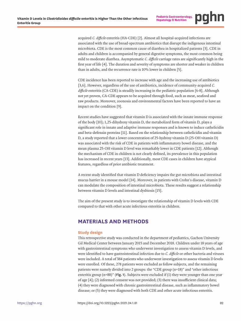
Study designThis retrospective study was conducted in the department of pediatrics, Gachon University Gil Medical Center between January 2015 and December 2018. Children under 18 years of age with gastrointestinal symptoms who underwent investigation to assess vitamin D levels, and were identified to have gastrointestinal infection due to C. difficile or other bacteria and viruses were included. A total of 384 patients who underwent investigation to assess vitamin D levels were enrolled. Of these, 278 patients were excluded as follow subjects, and the remaining patients were namely divided into 2 groups: the “CDE group (n=18)” and “other infectious enteritis group (n=88)” (Fig. 1). Subjects were excluded if (1) they were younger than one year of age [4]; (2) informed consent was not provided; (3) there was insufficient clinical data; (4) they were diagnosed with chronic gastrointestinal disease, such as inflammatory bowel disease; or (5) they were diagnosed with both CDE and other acute infectious enteritis.
82https://pghn.org https://doi.org/10.5223/pghn.2021.24.1.81
Vitamin D Levels in Clostridioides difficile enteritis is Higher Than the Other Infectious Enteritis Group

Clinical data, such as sex, age, predisposing factors, presence of underlying diseases, prior antibiotic use within three months, treatments, and laboratory findings (complete blood count, serum albumin [g/dL], serum creatinine [mg/dL], and vitamin D level), were obtained. CDE was defined as identified antigens of C. difficile toxin A or B infections and/or positive PCR and/or culture assays. Other bacterial infections were confirmed with multiplex real-time PCR assays (GeneXpert; Cepheid Xpert C. difficile assay, Cepheid, CA, USA) or stool cultures. Viral infection was diagnosed using multiplex real-time PCR (xTAG GPP; Luminex System LX200, Luminex, Toronto, ON, Canada).
To detect toxins A and B in C. difficile, VIDAS C. difficile toxin A and B (bioMerieux, Marcy-I'Etoile, France) was used, and for culture analysis, a culture medium containing a selective antibiotic was used (cycloserine cefoxitine fructose egg yolk agar). Anaerobic incubation was performed for 24 to 48 hours by inoculating the growth medium.
Patients were classified as those with CA-CDE if symptoms occurred within two days of hospitalization, or if they had never been hospitalized in the last three months. In contrast, patients were considered to have HA-CDE if symptoms occurred after two days of hospitalization in the last three months.
The Institutional Review Board (IRB) of Gachon University Gil Medical Center (IRB No. C2017042) waived the need for informed consent and approved this study.
Statistical analysesMeans with standard deviations or medians with interquartile ranges were used for continuous variables, while numbers with percentages were used for categorical variables. Among the epidemiological and clinical characteristics of patients, continuous variables were compared using Student's t-test, while categorical variables were compared using Chi-square or Fisher's exact tests. IBM SPSS Statistics for Windows, version 24.0 (IBM Co., Armonk, NY, USA) was used for analyses, and p-values <0.05 were considered to indicate statistical significance.
83https://pghn.org https://doi.org/10.5223/pghn.2021.24.1.81
Vitamin D Levels in Clostridioides difficile enteritis is Higher Than the Other Infectious Enteritis Group
384 patients under 18 years of agewho performed 25-OH vitamin D levelfrom January 2015 to December 2018
88 patients diagnosed withother acute infectious enteritis
18 patients diagnosedwith CDE
Exclusion criteria-Patients who didn t undergo stool exam.-Patients who were younger than one year of age.-Patients for whom informed consent was not obtained.-Patients who were diagnosed with chronic gastrointestinal
diseases, such as inflammatory bowel disease.-Patients who were diagnosed both with CDE and
other acute infectious enteritis.
Fig. 1. Selection algorithm for CDE enteritis group and other infectious enteritis group. CDE: C. difficile enteritis, 25-OH vitamin D: 25-hydroxy vitamin D.

RESULTS
Comparison of laboratory results and characteristics between C. difficile enteritis and other infectious enteritisNo differences were observed between the groups with respect to sex and age. Seasonal distribution was observed in all patients in the CDE group, with six patients acquiring the infection in spring (March–May) (33.3%), six in summer (June–August) (33.3%), two in autumn (September–November) (11.1%) and four in winter (December-February) (22.3%). Similarly, in the other infectious enteritis group, 13 patients (14.8%) acquired the infection in spring, 29 in summer (33.0%), 21 in autumn (23.9%) and 25 in winter (28.4%). There was no difference in seasonal distributions between the two groups. Laboratory findings, such as white blood cell count and creatinine levels, were not different between the two groups (p>0.05), but albumin levels were higher in the CDE group than in the other infectious enteritis group (4.52±0.45 g/dL vs. 4.31±0.28 g/dL, p=0.011) (Table 1).
In the other infectious enteritis group, 26 cases were viral enteritis, 61 cases were bacterial enteritis, and one case included duplicated infections. Causative agents of viral enteritis were rotavirus (n=20) and norovirus (n=6). Campylobacter (n=21) was the most common bacteria enteritis and others were as follows: Salmonella (n=19), Clostridium perfringens (n=11), Aeromonas (n=3), Escherichia coli (n=2), Shigella (n=2), Yersinia (n=2), and Vibrio species (n=1).
Medical characteristics of children with C. difficile enteritisThe number of outpatients (n=9, 50.0%) was the same as that of inpatients (n=9, 50.0%). The mean age of patients was 4.83±4.50 years and most patients (n=16, 88.9%) were under 9 years of age (Table 2).
Diarrhea was the most common symptom (n=13, 72.2%), and other symptoms were as follows: abdominal pain (n=11, 61.1%), hematochezia (n=9, 50.0%), vomiting (n=5, 27.8%), fever (n=4, 22.2%), and nausea (n=1, 5.6%).
CA-CDE (n=13, 72.2%) was more common than HA-CDE (n=5, 27.8%).
A predisposing factor was identified in 5 patients (27.8%), of which 3 patients (16.7%) had prior history of antibiotic use and 2 (11.1%) had underlying diseases, including intussusception and hydronephrosis.
84https://pghn.org https://doi.org/10.5223/pghn.2021.24.1.81
Vitamin D Levels in Clostridioides difficile enteritis is Higher Than the Other Infectious Enteritis Group
Table 1. Comparison of laboratory results and characteristics between CDE and other infectious enteritisVariable CDE group (n=18) Other infectious enteritis group (n=88) p-valueSex 0.609
Male 11 (61.1) 48 (54.5)Female 7 (38.9) 40 (45.5)
Age at diagnosis (yr) 4.83±4.50 7.37±5.09 0.052Seasonal distribution 0.274
March to May 6 (33.3) 12 (14.8)June to August 6 (33.3) 8 (33.0)September to November 2 (11.1) 6 (23.9)December to February 4 (22.3) 25 (28.4)
White blood cells (/mm3) 10.31±6.09 10.89±5.40 0.685Albumin (g/dL) 4.52±0.45 4.31±0.28 0.011Creatinine (mg/dL) 0.34±0.15 0.39±0.18 0.232Values are presented as number (%) or mean±standard deviation.CDE: C. difficile enteritis.

The majority of patients were managed with conservative care without the use of antibiotics (n=13, 72.2%). Three patients (16.7%) were prescribed metronidazole for initial therapy, and two (11.1%) were treated with other antibiotics.
Comparison of mean 25-OH vitamin D levels between the groupsVitamin D levels were different between the groups (Fig. 2). Mean 25-OH vitamin D levels were higher in the CDE group than in the other infectious enteritis group (18.75±8.11 ng/mL vs. 14.50±6.79 ng/mL, p=0.021) (Fig. 2).
Comparison of mean 25-OH vitamin D levels between groups according to the type of infectionMean 25-OH vitamin D levels were not different between the viral (n=26, 16.05±6.83 ng/mL) and bacterial enteritis groups, except the CDE group (n=61, 13.69±6.65 ng/mL). Mean 25-OH
85https://pghn.org https://doi.org/10.5223/pghn.2021.24.1.81
Vitamin D Levels in Clostridioides difficile enteritis is Higher Than the Other Infectious Enteritis Group
Table 2. Medical characteristics of children with CDECharacteristic Value (n=18)Associated symptoms
Diarrhea 13 (72.2)Nausea 1 (5.6)Vomiting 5 (27.8)Fever 4 (22.2)Abdominal pain 11 (61.1)Hematochezia 9 (50.0)
Community/Hospital-acquired CDECA-CDE 13 (72.2)HA-CDE 5 (27.8)
Predisposing factorPrior use of antibiotics 3 (16.7)Underlying disease* 2 (11.1)
TreatmentObservation or discontinuation of antibiotics 13 (72.2)Metronidazole for initial therapy 3 (16.7)Other antibiotics for initial therapy 2 (11.1)
Values are presented as number (%).CDE: C. difficile enteritis, CA-CDE: community-acquired C. difficile enteritis, HA-CDE: hospital-acquired C. difficile enteritis.*Underlying diseases; intussusception and hydronephrosis.
CDE group
40.00
30.00
20.00
10.00
0.00Other infectiousenteritis group
25-O
Hvitam
inD
level(n
g/m
L)
p=0.021
Fig. 2. Comparison of mean 25-OH vitamin D levels between the groups. Mean 25-OH vitamin D level in the CDE group was 18.75±8.11 ng/mL and mean 25-OH vitamin D level in the other infectious enteritis group was 14.50± 6.79 ng/mL. p=0.021. 25-OH vitamin D: 25-hydroxy vitamin D, CDE: C. difficile enteritis.

vitamin D levels also were not different between the viral (n=26, 16.05±6.83 ng/mL) and the bacterial enteritis groups, including the CDE group (n=79, 14.84±7.27 ng/mL). The mean 25-OH vitamin D levels of the CDE group were higher than those of the bacteria enteritis group, except for the CDE group (n=18, 18.75±8.11 ng/mL vs. n=61, 13.69±6.65 ng/mL, p=0.009) (Fig. 3).
DISCUSSION
The mean vitamin D levels in the CDE group were higher than other bacterial and viral enteritis groups. Moreover, when comparing CDE groups with other bacterial enteritis except CDE groups, the difference between vitamin D levels was greater. These results suggest that the susceptibility to vitamin D Levels may differ between the CDE and other infectious enteritis groups.
Previous studies have reported the effects of vitamin D on the severity, recurrence, and outcomes of CDE, as well as on intestinal microbiota [16-18]. The importance of gut microbiota in the pathogenesis of C. difficile infection is well recognized, and marked decreases in microbial diversity have been reported in CDE patients [19]. Jin et al. [20] reported that intestinal dysbiosis in CDE could be related to a decrease in the expression of vitamin D receptors (VDR). VDRs appear to be an important immunological regulator in the human body, and the impairments of which result in the dysbiosis of the gut microbiome [21]. In the normal intestine, VDRs regulate bacterial invasion and suppress inflammatory responses. Intercellular complexes of receptors physically interact with NF-κB in osteoblasts and suppress inflammatory responses to a common invasive enteric bacterial pathogen [22].
Contrary to prior studies in adults, the our study showed that vitamin D levels in the other infectious enteritis group were lower than that in the CDE group among children [17]. These results suggest that intestinal dysbiosis, which was related to a decrease in VDR expressions, is a more sensitive factor in children than adult patients [20]. Impaired VDRs in CDE patients could be associated with variations in vitamin D levels, and even marginal changes in such levels can act as a more risk factor for infection. However, in the other acute infectious enteritis
86https://pghn.org https://doi.org/10.5223/pghn.2021.24.1.81
Vitamin D Levels in Clostridioides difficile enteritis is Higher Than the Other Infectious Enteritis Group
40.00
30.00
20.00
10.00
0.00
25-O
Hvitam
inD
level(n
g/m
L)
CDE Bacterial enteritisexcept CDE
Viralenteritis
Bacterial enteritisinclude CDE
*
Fig. 3. Comparison of mean 25-OH vitamin D levels between groups according to the type of infection. *CDE (n=18, 18.75±8.11 ng/mL) vs. bacterial enteritis except CDE (n=61, 13.69±6.65 ng/mL) (p=0.009). †Viral enteritis group (n=26, 16.05±6.83 ng/mL) vs. bacterial enteritis group except CDE (n=61, 13.69±6.65 ng/mL) (p=0.137). ‡Viral enteritis group (n=26, 16.05±6.83 ng/ml) vs. bacterial enteritis group including CDE (n=79, 14.84±7.27 ng/ mL) (p=0.458). 25-OH vitamin D: 25-hydroxy vitamin D, CDE: C. difficile enteritis.

patients with normally expressed VDR, the risk of infection will increase only in cases of significant variation in vitamin D levels in children. This may support our findings regarding the difference in vitamin D levels between the CDE and other infectious enteritis groups.
There were no differences between the groups with respect to sex and age. Several studies have found a co-seasonality of CDE and respiratory tract infection. Spring is associated with increased CDE incidence due to increases in the use of antibiotics [23]. Although there was no statistical significance, in this study, more CDE cases were reported in spring and summer. However, this could be due to the small sample size in the CDE group (n=18).
Diarrhea (n=13, 72.2%) was the most common symptom in patients with CDE, which is consistent with the findings reported in previous studies [24].
Vitamin D levels in the CDE group were compared with a control group only in adult patients in previous studies [10,17]. However, vitamin D deficiency is more common in children, and low levels of vitamin D are associated with various infections in children. The present work is the first retrospective study to compare vitamin D levels between patients with CDE and other infectious enteritis in a pediatric population.
There are several limitations of the present study. First, the sample size of the CDE group was small. Second, the study was retrospective in design and hence could not compare vitamin D levels between healthy individuals and patients with CDE. Third, the patients in the other infectious enteritis group could not be analyzed by categorizing them based on the individual bacteria and viruses. Therefore, prospective studies are needed to compare the difference in vitamin D levels between CDE and healthy control groups in a large pediatric population.
In conclusion, vitamin D levels in patients with CDE were lower than normal but higher than those with other infectious enteritis. In particular, patients with CDE had higher levels of vitamin D than those with other bacterial enteritis. These results suggested that CDE has different mechanism or susceptibility associated with vitamin D in children, and even marginal changes in vitamin D levels can act as a risk factor for infection.
REFERENCES
1. Cózar-Llistó A, Ramos-Martinez A, Cobo J. Clostridium difficile infection in special high-risk populations. Infect Dis Ther 2016;5:253-69. PUBMED | CROSSREF
2. Lamont JT, Theodore E. Theodore E. Woodward Award. How bacterial enterotoxins work: insights from in vivo studies. Trans Am Clin Climatol Assoc 2002;113:167-80; discussion 180-1.PUBMED
3. Migriauli I, Meunargia V, Chkhaidze I, Sabakhtarishvili G, Gujabidze K, Khokrishvili G, et al. Epidemiology of Clostridium difficile infection in hospitalised pediatric patients in Georgia. Georgian Med News 2018;(Issue):172-6.PUBMED
4. Jangi S, Lamont JT. Asymptomatic colonization by Clostridium difficile in infants: implications for disease in later life. J Pediatr Gastroenterol Nutr 2010;51:2-7. PUBMED | CROSSREF
5. McFarland LV, Ozen M, Dinleyici EC, Goh S. Comparison of pediatric and adult antibiotic-associated diarrhea and Clostridium difficile infections. World J Gastroenterol 2016;22:3078-104. PUBMED | CROSSREF
87https://pghn.org https://doi.org/10.5223/pghn.2021.24.1.81
Vitamin D Levels in Clostridioides difficile enteritis is Higher Than the Other Infectious Enteritis Group

6. Kociolek LK, Gerding DN. Is pediatric Clostridium difficile infection associated with prior antibiotic exposure? Future Microbiol 2014;9:825-8. PUBMED | CROSSREF
7. Borali E, Ortisi G, Moretti C, Stacul EF, Lipreri R, Gesu GP, et al. Community-acquired Clostridium difficile infection in children: a retrospective study. Dig Liver Dis 2015;47:842-6. PUBMED | CROSSREF
8. Cho HJ, Ryoo E, Sun YH, Cho KH, Son DW, Tchah H. Epidemiology and clinical characteristics of Clostridium difficile-associated disease in children: comparison between community- and hospital-acquired infections. Korean J Pediatr Gastroenterol Nutr 2010;13:146-53. CROSSREF
9. Warriner K, Xu C, Habash M, Sultan S, Weese SJ. Dissemination of Clostridium difficile in food and the environment: significant sources of C. difficile community-acquired infection? J Appl Microbiol 2017;122:542-53. PUBMED | CROSSREF
10. Sahay T, Ananthakrishnan AN. Vitamin D deficiency is associated with community-acquired clostridium difficile infection: a case-control study. BMC Infect Dis 2014;14:661. PUBMED | CROSSREF
11. Lehmann J, Retz M, Harder J, Krams M, Kellner U, Hartmann J, et al. Expression of human beta-defensins 1 and 2 in kidneys with chronic bacterial infection. BMC Infect Dis 2002;2:20. PUBMED | CROSSREF
12. Ananthakrishnan AN, Cagan A, Gainer VS, Cheng SC, Cai T, Szolovits P, et al. Higher plasma vitamin D is associated with reduced risk of Clostridium difficile infection in patients with inflammatory bowel diseases. Aliment Pharmacol Ther 2014;39:1136-42. PUBMED | CROSSREF
13. Lees EA, Miyajima F, Pirmohamed M, Carrol ED. The role of Clostridium difficile in the paediatric and neonatal gut - a narrative review. Eur J Clin Microbiol Infect Dis 2016;35:1047-57. PUBMED | CROSSREF
14. Zhu W, Yan J, Zhi C, Zhou Q, Yuan X. 1,25(OH)2D3 deficiency-induced gut microbial dysbiosis degrades the colonic mucus barrier in Cyp27b1 knockout mouse model. Gut Pathog 2019;11:8. PUBMED | CROSSREF
15. Schäffler H, Schmidt M, Huth A, Reiner J, Glass Ä, Lamprecht G. Clinical factors are associated with vitamin D levels in IBD patients: a retrospective analysis. J Dig Dis 2018;19:24-32. PUBMED | CROSSREF
16. Furuya-Kanamori L, Wangdi K, Yakob L, McKenzie SJ, Doi SAR, Clark J, et al. 25-Hydroxyvitamin D concentrations and Clostridium difficile Infection: a meta-analysis. JPEN J Parenter Enteral Nutr 2017;41:890-5. PUBMED | CROSSREF
17. van der Wilden GM, Fagenholz PJ, Velmahos GC, Quraishi SA, Schipper IB, Camargo CA Jr. Vitamin D status and severity of Clostridium difficile infections: a prospective cohort study in hospitalized adults. JPEN J Parenter Enteral Nutr 2015;39:465-70. PUBMED | CROSSREF
18. Abdelfatah M, Nayfe R, Moftakhar B, Nijim A, El Zoghbi M, Donskey CJ, et al. Low vitamin D level and impact on severity and recurrence of Clostridium difficile infections. J Investig Med 2015;63:17-21. PUBMED | CROSSREF
19. Antharam VC, Li EC, Ishmael A, Sharma A, Mai V, Rand KH, et al. Intestinal dysbiosis and depletion of butyrogenic bacteria in Clostridium difficile infection and nosocomial diarrhea. J Clin Microbiol 2013;51:2884-92. PUBMED | CROSSREF
20. Jin D, Wu S, Zhang YG, Lu R, Xia Y, Dong H, et al. Lack of vitamin D receptor causes dysbiosis and changes the functions of the murine intestinal microbiome. Clin Ther 2015;37:996-1009.e7. PUBMED | CROSSREF
21. Wu S, Zhang YG, Lu R, Xia Y, Zhou D, Petrof EO, et al. Intestinal epithelial vitamin D receptor deletion leads to defective autophagy in colitis. Gut 2015;64:1082-94. PUBMED | CROSSREF
22. Wu S, Liao AP, Xia Y, Li YC, Li JD, Sartor RB, et al. Vitamin D receptor negatively regulates bacterial-stimulated NF-kappaB activity in intestine. Am J Pathol 2010;177:686-97. PUBMED | CROSSREF
23. Furuya-Kanamori L, McKenzie SJ, Yakob L, Clark J, Paterson DL, Riley TV, et al. Clostridium difficile infection seasonality: patterns across hemispheres and continents - a systematic review. PLoS One 2015;10:e0120730. PUBMED | CROSSREF
88https://pghn.org https://doi.org/10.5223/pghn.2021.24.1.81
Vitamin D Levels in Clostridioides difficile enteritis is Higher Than the Other Infectious Enteritis Group

24. Borali E, De Giacomo C. Clostridium difficile infection in children: a review. J Pediatr Gastroenterol Nutr 2016;63:e130-40. PUBMED | CROSSREF
89https://pghn.org https://doi.org/10.5223/pghn.2021.24.1.81
Vitamin D Levels in Clostridioides difficile enteritis is Higher Than the Other Infectious Enteritis Group