original article combined haemophilus influenzae ...methods see online supplement for additional...
TRANSCRIPT

ORIGINAL ARTICLE
Combined Haemophilus influenzae respiratoryinfection and allergic airways disease drives chronicinfection and features of neutrophilic asthma
Ama-Tawiah Essilfie,1 Jodie L Simpson,1,2 Margaret L Dunkley,3
Lucy C Morgan,4 Brian G Oliver,5 Peter G Gibson,1,2 Paul S Foster,1
Philip M Hansbro1
ABSTRACTBackground 20e30% of patients with asthma haveneutrophilic airway inflammation and reducedresponsiveness to steroid therapy. They often havechronic airway bacterial colonisation and Haemophilusinfluenzae is one of the most commonly isolatedbacteria. The relationship between chronic airwaycolonisation and the development of steroid-resistantneutrophilic asthma is unclear.Objectives To investigate the relationship between Hinfluenzae respiratory infection and neutrophilic asthmausing mouse models of infection and ovalbumin (OVA)-induced allergic airways disease.Methods BALB/c mice were intratracheally infectedwith H influenzae (day 10), intraperitoneally sensitised(day 0) and intranasally challenged (day 12e15) withOVA. Treatment groups were administereddexamethasone intranasally during OVA challenge.Infection, allergic airways disease, steroid sensitivity andimmune responses were assessed (days 11, 16 and 21).Results The combination of H influenzae infection andallergic airways disease resulted in chronic lung infectionthat was detected on days 11, 16 and 21 (21, 26 and31 days after infection). Neutrophilic allergic airwaysdisease and T helper 17 cell development were induced,which did not require active infection. Importantly, allfeatures of neutrophilic allergic airways disease weresteroid resistant. Toll-like receptor 4 expression andactivation of phagocytes was reduced, but mostsignificantly the influx and/or development ofphagocytosing neutrophils and macrophages into theairways was inhibited.Conclusions The combination of infection and allergicairways disease promotes bacterial persistence, leadingto the development of a phenotype similar to steroid-resistant neutrophilic asthma and which may result fromdysfunction in innate immune cells. This indicates thattargeting bacterial infection in steroid-resistant asthmamay have therapeutic benefit.
INTRODUCTIONAsthma is a chronic inflammatory condition of theairways, frequently characterised by abnormalimmune responses to environmental antigens,leading to recurrent episodes of cough, wheezingand breathlessness.1 Patients with neutrophilicasthma make up between 20% and 30% of patientswith asthma, and are characterised by substantial
increases in airway neutrophils. Their diseasesymptoms and neutrophilic inflammation arepoorly responsive to corticosteroids, the mainstayof asthma therapy, and it has been suggested thatairway neutrophilia may be a consequence ofsteroid treatment.2 3 Neutrophilic airway inflam-mation is also common to many obstructiveairway diseases, including neutrophilic asthma,chronic obstructive pulmonary disease (COPD)and bronchiectasis.2 4 5
The cellular innate immune system of therespiratory tract includes epithelial cells, neutro-phils, macrophages and dendritic cells. It isconsidered the first line of defence against invadingpathogens, mounts an immediate non-specificresponse, and promotes antigen-specific responsesby the adaptive component.6 Defects in innateimmunity may be important in the development ofsteroid-resistant neutrophilic asthma, and increasedsputum neutrophils, interleukin (IL)-8 levels andexpression of toll-like receptors (TLRs) have beendemonstrated.4 7
Neutrophils and macrophages have critical rolesin innate immunity and are important contributorsto airway inflammation. Therefore, dysfunction inthese cells may lead to the development or wors-ening of steroid-resistant neutrophilic asthma.Airway neutrophils from patients with asthma areless activated compared with healthy controls,which may lead to impaired local defence and
Key messages
What is the key question?< How is chronic bacterial infection associated
with steroid-resistant neutrophilic asthma.
What is the bottom line?< The combination of infection and allergic
airways disease induces the complete suppres-sion of phagocytic cells in the lung that leads tochronic infection, neutrophilic inflammation andsteroid resistance.
Why read on?< This indicates how bacterial infections may be
associated with steroid-resistant neutrophilicasthma.
< Additional materials arepublished online only. To viewthese files please visit thejournal online (http://thorax.bmj.com/content/67/7.toc).1Centre for Asthma andRespiratory Disease and HunterMedical Research Institute,University of Newcastle,Newcastle, Australia2Department of Respiratory andSleep Medicine, John HunterHospital, New Lambton, NewSouth Wales, Australia3Hunter Immunology Ltd,Newcastle, New South Wales,Australia4Department of ThoracicMedicine, Concord RepatriationGeneral Hospital, Concord, NewSouth Wales, Australia5Sydney Medical School andWoolcock Institute of MedicalResearch, University of Sydney,Sydney, Australia
Correspondence toProfessor Philip Hansbro,Infection and Immunity, TheDavid Maddison ClinicalSciences Building, University ofNewcastle, Cnr King and WattSts, Newcastle, NSW 2300,Australia; [email protected]
Received 10 March 2011Accepted 5 January 2012Published Online First3 March 2012
588 Thorax 2012;67:588e599. doi:10.1136/thoraxjnl-2011-200160
Asthma
on May 20, 2020 by guest. P
rotected by copyright.http://thorax.bm
j.com/
Thorax: first published as 10.1136/thoraxjnl-2011-200160 on 3 M
arch 2012. Dow
nloaded from

increased susceptibility to infections.8 Indeed, in COPD, whichis also underpinned by neutrophilic bronchitis, airway neutro-phils have impaired chemotaxis, and alveolar macrophages havereduced TLR2 expression.9 10
Infections by viruses and bacteria have pathogenic roles inasthma and induce asthma exacerbations.11e14 Chronic bacterialinfection occurs frequently in airway diseases such as COPD,and bronchiectasis, and may be important in steroid-resistantneutrophilic asthma.7 15 16 In neutrophilic asthma H influenzae isone of the most commonly isolated bacteria and we haverecently shown that infection in allergic airways disease drivesa neutrophilic phenotype that is dependent on IL-17.7 15 17
However, the relationship between chronic H influenzae infec-tion and the development of steroid-resistant neutrophilicasthma is not understood.
In this study we employed murine models of H influenzaeinfection and OVA-induced allergic airways disease to investi-gate if and how the combination of infection and allergicairways disease may interact to promote chronic infection andthe development of steroid-resistant neutrophilic asthma.
METHODSSee online supplement for additional details.
Experimental protocolsFemale BALB/c mice, 6e8 weeks old, were inoculated intra-tracheally with 53105 colony-forming units (CFU) non-type-able H influenzae (NTHi-289, in 30 ml phosphate-buffered saline)or 53105 CFU ethanol-killed NTHi-289 and allergic airwaysdisease was induced 10 days later (figures 1A and 2A).18 Micewere sensitised to OVA (50 mg, intraperitoneal injection, Sigma-Aldrich, Sydney, Australia), with Rehydrogel (1 mg, Reheis,Berkeley Heights, New Jersey, USA) in saline (200 ml), andsubsequently challenged intranasally with OVA (10 mg/50 mlsaline) on days 12e15 to induce allergic airways disease. Salinecontrol groups received saline sensitisation with Rehydrogel byintraperitoneal injection and subsequent OVA challenges ondays 12e15.19 Infection, immune responses and allergic airwaysdisease were assessed on days 11, 16 and 21.
Bacterial recoverySerial dilutions of lung homogenates and bronchoalveolar lavagefluid (BALF) were plated onto chocolate agar plates (Oxoid,Thebarton, Australia) and incubated overnight (378C, 5% CO2).Colonies were enumerated and bacterial numbers in lungs andBALF were determined and combined.17
Cellular inflammationBALF cytospins and blood slides were stained with May-Grunwald Giemsa and differential leucocyte counts obtainedusing light microscopy.19
T-cell cytokinesMediastinal lymph node (MLN) T-cell cultures were re-stimu-lated with OVA or ethanol-killed H influenzae and cytokineconcentrations in supernatants assessed by ELISA.20
Lung cytokine expressionIL-13 and interferon g (IFNg) expression was determined relativeto the reference gene hypoxanthine-guanine phosphoribosyl-transferase in TRIZOL-extracted RNA from whole lung tissueaccording to manufacturer ’s instructions (Invitrogen, MountWaverly, Australia).19 Primers used were hypoxanthine-guaninephosphoribosyltransferase Fwd 59-aggccagactttgttggatttgaa-39,
Rev 59-caacttgcgctcatcttaggcttt-39; IL-13, Fwd 59-aaacatgagtccagggagagcttt-39, Rev 59-actgagcttcccagatcacagagg-39; IFNg, Fwd59-gctcatcttaaggccagact-39, Rev 59-atttgaatccagggatccgaatt-39.
Lung functionAirway hyperresponsiveness (AHR) was measured in responseto increasing doses of methacholine by whole body invasiveplethysmography as previously described.18
Th2 and Th17 cellsSingle-cell lung and MLN preparations were stimulated withphorbol 12-myristate 13-acetate (PMA, 0.1 mg/ml) and iono-mycin (1 mg/ml) in the presence of Brefeldin A (8 mg/ml, Sigma-Aldrich). Cells were stained for surface markers CD3, CD4 (BDBioscience, San Diego, California, USA), fixed, permeabilised andstained for intracellular IL-13 and IL-17A (eBioscience, SanDiego, California, USA). Cells were analysed by flow cytometry(FACS Canto, BD Bioscience).19e21
Dexamethasone treatmentDexamethasone (Sigma-Aldrich) was prepared in sterile saline(1 mg/kg) and administered intranasally (50 ml, days 13e15,figure 3A).
Mucus secreting cellsLungs were imbedded in paraffin, sectioned (4e6 mm) andstained with periodic acid Schiff. Mucus secreting cell (MSC)
Figure 1 Allergic airways disease during Haemophilus influenzaeinfection promotes chronicity of infection. (A) Mice were inoculatedintratracheally with 53105 colony-forming units (CFU) of H influenzae,then 10 days later sensitised intraperitoneally (day 0) and challengedintranasally (days 12e15) with ovalbumin (OVA). Controls received salinesensitisation and OVA challenge. Allergic airways disease, immuneresponses and infection were assessed (days 11, 16 and 21) andcompared with controls. (B) The effects of allergic airways disease on Hinfluenzae recovery from lungs and bronchoalveolar lavage fluid combinedwere characterised. Data represent the mean 6 SEM from six to eightmice. Significant differences between infected non-allergic (H influenza(Hi)) and infected allergic (Hi/OVA) groups are shown as ***p<0.001.
Thorax 2012;67:588e599. doi:10.1136/thoraxjnl-2011-200160 589
Asthma
on May 20, 2020 by guest. P
rotected by copyright.http://thorax.bm
j.com/
Thorax: first published as 10.1136/thoraxjnl-2011-200160 on 3 M
arch 2012. Dow
nloaded from

numbers within the airway lumen (103100 mm fields) wereenumerated.20
Ciliary beat frequencyCiliary beat frequency was measured for 1 min per section intracheal transverse slices using transmitted light photometryand mean frequency (Hz) was calculated.22
Phenotyping of immune cellsMacrophages (CD11b+F4/80+, eBioscience) and neutrophils(CD11bhiGr-1hi, eBioscience) were stained for the activationmarker CD62L, and TLR2 and TLR4 (eBioscience) and wereanalysed using flow cytometry.
PhagocytosisCarboxyfluorescein succinimidyl ester (CFSE, 5 mM) labelledNTHi were incubated with BALF cells as previously described.23
BALF cells were stained for CD11b, F4/80 and Gr-1 andphagocytosis of CFSE-labelled NTHi was assessed using flowcytometry.
Oxidative burstLung cells were incubated with dihydroethidium (DHE, Sigma-Aldrich) for 15 min.23 Reactions were terminated by placing onice, and cells were washed and fixed. Oxidative burst wasassessed in fixed cells using flow cytometry.
Serum antibodiesHi-specific immunoglobulin G1 (IgG1, using H influenzae as thecapture antigen) was measured in serum by ELISA.21
Statistical analysesData are presented as mean 6 SEM. Statistical significance formultiple comparisons was determined by one-way analysis of
Figure 2 Killed Haemophilusinfluenzae (KHi) suppresses hallmarkfeatures of eosinophilic allergic airwaysdisease. (A) Mice were administeredwith ethanol-killed H influenzae thensensitised (day 0) and challenged (days12e15) with ovalbumin (OVA). Theeffects of KHi in allergic airwaysdisease on the numbers of (B) totalcells, (C) eosinophils and (D)neutrophils in bronchoalveolar lavagefluid (BALF) and OVA-induced (E)interleukin (IL)-5, (F) IL-13 and (G)interferon g (IFNg) release from MLNT-cell were assessed. Airwayhyperresponsiveness measured as (H)dynamic compliance and (I)transpulmonary resistance in responseto increasing doses of methacholinewas determined. Significant differencesbetween allergic (OVA) and non-allergic(saline) groups are shown as###p<0.001, ##p<0.01,#p<0.05. Significant differencesbetween infected allergic (H influenzae/OVA) and uninfected allergic (OVA)groups are shown as ***p<0.001,**p<0.01. Significant differences forresistance and compliance are for theentire doseeresponse curve.
590 Thorax 2012;67:588e599. doi:10.1136/thoraxjnl-2011-200160
Asthma
on May 20, 2020 by guest. P
rotected by copyright.http://thorax.bm
j.com/
Thorax: first published as 10.1136/thoraxjnl-2011-200160 on 3 M
arch 2012. Dow
nloaded from

Figure 3 Chronic Haemophilus influenzae (Hi) infection in allergic airways disease induces steroid resistance. The effects of chronic Hi infectionin allergic airways disease on responsiveness to steroid treatment were investigated. (A) Infected allergic groups were treated intranasally (i.n.)with dexamethasone (Dex) during ovalbumin (OVA) challenge (days 13e15) and allergic airways disease was assessed (days 16 and 21) andcompared with controls. (B) To determine the effect of treatment on infection, bacterial recovery was assessed. To determine the effectof treatment on neutrophilic allergic airways disease, numbers of (C) eosinophils and (D) neutrophils in bronchoalveolar lavage fluid (BALF), (E)Hi-induced interleukin (IL)-17, OVA-induced (F) IL-5, (G) IL-13 and (H) interferon g (IFNg) were assessed. Airway hyperresponsiveness, measuredas (I) dynamic compliance and (J) transpulmonary resistance in response to increasing doses of methacholine, was determined. Representative(K) compliance and (L) resistance data at 5 mg/ml are also shown. Data represent the mean 6 SEM from six to eight mice. Significant differencesbetween allergic (OVA) and non-allergic (saline) groups are shown as ###p<0.001, ##p<0.01, #p<0.05. Significant differences compared
Thorax 2012;67:588e599. doi:10.1136/thoraxjnl-2011-200160 591
Asthma
on May 20, 2020 by guest. P
rotected by copyright.http://thorax.bm
j.com/
Thorax: first published as 10.1136/thoraxjnl-2011-200160 on 3 M
arch 2012. Dow
nloaded from

variance (ANOVA) with the Bonferroni post-test. One-wayrepeated measures ANOVA with Bonferroni post-test wasused to analyse AHR data (GraphPad Prism Software, La Jolla,California, USA).
RESULTSAllergic airways disease during infection leads to chronic Hinfluenzae infectionTo investigate the relationship between infection and allergicairways disease, we first assessed the effect of allergic airwaysdisease on H influenzae infection. In a recent study we showedthat after inoculation with 53105 CFU, H influenzae infection inthe absence of allergic airways disease peaks after 5 days(1.563106 6 93103 CFU), declines after 10 days (43102 613102 CFU) and is undetectable by 16 days.17
Next, mice were inoculated with H influenzae and 10 dayslater, after the majority of H influenzae had been cleared, allergicairways disease was induced (figure 1A). The induction ofallergic airways disease promoted the development of chronicinfection with substantial levels of H influenzae recovered 21(day 11 of the model), 26 (day 16 of the model) and 31 (day 21 ofthe model) days after inoculation of infected allergic (H influ-enzae/OVA) groups compared with infected non-allergic(H influenzae) controls (figure 1B). Uninfected allergic controlswere also assessed and neither H influenzae nor commensalbacteria were detected (data not shown), ruling out the possi-bility that allergic airways disease induces substantial alterationsin commensal species.
Chronic H influenzae infection in allergic airways diseasesuppresses eosinophilic inflammationWe have recently shown that infection during or after sensiti-sation to OVA suppresses eosinophilic allergic airways disease.17
To investigate if similar effects were induced by chronicH influenzae infection, mice were infected, allergic airwaysdisease induced (figure 1A) and key features of allergic airwaysdisease were assessed.
The induction of allergic airways disease (OVA groups)resulted in eosinophilic inflammation, OVA-induced IL-5, IL-13and IFN-g release from MLN T-cell and AHR (decreaseddynamic compliance and increased transpulmonary resistance)compared with non-allergic (saline) controls (figure 4AeK).Infection alone had no effects on these features. However,infection during allergic airways disease significantly reduced thenumbers of total cells and eosinophils in BALF compared withuninfected allergic (OVA) controls (figure 4A,B). The suppres-sion of airway responses was accompanied by reductions inOVA-induced IL-5, IL-13 and IFN-g release from T-cell (days 16and 21), Th2 cells in the MLN (figure 4CeF) and expression ofIL-13 and IFN-g mRNA in the lungs at days 16 and 21 (figure4G). Infection during allergic airways disease also significantlyreduced AHR (increased airway dynamic compliance andreduced transpulmonary resistance), which however remainedelevated compared with non-allergic controls. Specifically, ininfected allergic (H influenzae/OVA) groups compared withuninfected non-allergic (saline) controls, dynamic compliancewas reduced across the whole methacholine dose response curveon day 16 of the model (figure 4H) and at the highest meth-
acholine dose (10 mg/ml) on day 21 (figure 4J). Transpulmonaryresistance was also increased but was only statistically signifi-cantly so at the highest methacholine dose (10 mg/ml) on day16 (figure 4I).
Chronic H influenzae infection in allergic airways diseaseinduces neutrophilic inflammation and Th17 cell and IL-17responsesH influenzae has frequently been isolated from patients withneutrophilic asthma who also have increased sputum neutro-philic inflammation. We have also recently shown that infec-tion during or after sensitisation to OVA induces neutrophilicallergic airways disease that is dependent on IL-17.17 Therefore,we investigated whether chronic H influenzae infection inallergic airways disease also induced enhanced neutrophilicinflammation.The induction of allergic airways disease was associated with
mild neutrophilic inflammation (figure 5A,B). The combinationof chronic infection and allergic airways disease signi-ficantly increased the influx of neutrophils into the airway andblood compared with uninfected allergic groups on day 16(figure 5A,B).Patients with asthma have increased sputum levels of IL-17
mRNA, and IL-17 stimulates the recruitment of neutrophils.Therefore, we assessed the numbers and levels of Th17 cells andIL-17 in infection and allergic airways disease. The induction ofallergic airways disease increased the numbers of Th17 cells inlung tissue and IL-17 release from MLN T-cell stimulated withH influenzae on day 16 (figure 5C,D). Infection in allergic airwaysdisease significantly increased the numbers of Th17 cells in thelung and IL-17 release from MLN T-cell (figure 5C,D). Therewere no differences in OVA-induced IL-17 production (data notshown). In other studies we used monoclonal antibody blockingstudies to show that IL-17 is required for the development of theinfection-induced neutrophilic inflammatory phenotype.18
Killed H influenzae suppresses hallmark features of eosinophilicallergic airways diseaseTo investigate whether an active infection was required for theinduction of a neutrophilic phenotype, the effect of killed H influ-enzae on allergic airways disease was assessed. Mice were admin-istered with 53105 CFU of ethanol-killed bacteria and the effect ofallergic airways disease was assessed on day 16 (figure 2A).The combination of killed H influenzae and allergic airways
disease (killed H influenzae/OVA) significantly decreased totalcells, eosinophils and neutrophils in the BALF (figure 2BeD) andOVA-induced IL-13 and IFNg release from T-cell compared withallergic controls (figure 2EeG). Killed H influenzae did not haveany effect on AHR (figure 2HeI).
Chronic H influenzae infection in allergic airways diseaseinduces steroid resistanceMany patients with neutrophilic asthma do not respondwell to steroid therapy. Therefore, we investigated the sensi-tivity of infection and neutrophilic allergic airways disease todexamethasone treatment.Allergic and infected allergic groups were administered dexa-
methasone (1 mg/kg) intranasally during OVA challenge (days
with uninfected allergic (OVA) groups are shown as ***p<0.001, **p<0.01, *p<0.05. Significant differences between untreated and treatedgroups are shown as +p<0.05. Significant differences for resistance and compliance are for the entire doseeresponse curve. NS,non-significant.
[Continued]
592 Thorax 2012;67:588e599. doi:10.1136/thoraxjnl-2011-200160
Asthma
on May 20, 2020 by guest. P
rotected by copyright.http://thorax.bm
j.com/
Thorax: first published as 10.1136/thoraxjnl-2011-200160 on 3 M
arch 2012. Dow
nloaded from

Figure 4 Chronic Haemophilus influenzae (Hi) infection suppresses hallmark features of eosinophilic allergic airways disease. The effects of chronicHi infection in allergic airways disease on the numbers of (A) total cells and (B) eosinophils in bronchoalveolar lavage fluid (BALF) were assessed. Theeffects of infection on ovalbumin (OVA)-induced (C) interleukin (IL)-5, (D) IL-13 and (E) interferon g (IFNg) release from mediastinal lymph node T-cell,(F) mediastinal lymph node (LN) T helper 2 cells, and (G) IL-13 and IFNg mRNA expression relative to hypoxanthine-guanine phosphoribosyl-transferase (HPRT) in the lungs were also assessed. (H, I) Airway hyperresponsiveness, measured as dynamic compliance and transpulmonaryresistance in response to increased doses of methacholine, was determined on day 16 and (J, K) day 21, respectively. Significant differences betweenallergic (OVA) and non-allergic (saline) groups are shown as ###p<0.001, ##p<0.01, #p<0.05. Significant differences between infected allergic
Thorax 2012;67:588e599. doi:10.1136/thoraxjnl-2011-200160 593
Asthma
on May 20, 2020 by guest. P
rotected by copyright.http://thorax.bm
j.com/
Thorax: first published as 10.1136/thoraxjnl-2011-200160 on 3 M
arch 2012. Dow
nloaded from

13e15, figure 3A), and infection and allergic airways diseasewere assessed. Steroid treatment caused a small reduction inbacterial numbers in infected allergic (H influenzae/OVA/dexa-methasone) groups at day 16 but not at day 21. Treatmentsignificantly increased bacterial numbers in treated infected(H influenzae/dexamethasone) groups compared with untreatedinfected (H influenzae) controls at days 16 and 21 (figure 3B).
Dexamethasone treatment of uninfected allergic (OVA/dexa-methasone) groups resulted in a significant reduction in all thekey features of allergic airways disease at days 16 and 21 (figure3CeJ). In stark contrast, treatment of infected allergic groupsdid not reduce eosinophil or neutrophil recruitment to theairways. There were also no effects on H influenzae-induced IL-17, OVA-induced IL-5 or IL-13 release from MLN T-cell or AHRcompared with untreated infected allergic groups at day 16 orday 21 (figure 3CeJ). The exception was IFNg, which wasslightly but significantly increased by dexamethasone treatmenton day 21 (figure 3H). To enhance clarity of the AHR data wehave included a magnified online supplementary figure 1. Tosimplify the AHR data, we have shown dynamic compliance(figure 3K) and resistance (figure 3L) for all groups at onemethacholine dose (5 mg/ml).
Collectively these data suggest that the induction of allergicairways disease promotes chronic infection, despite the presenceof increased numbers of neutrophils, and leads to the develop-ment of neutrophilic allergic airways disease that is steroidresistant.
Chronic H influenzae infection in allergic airways disease doesnot affect MSC numbers or ciliary beat frequencyWe then investigated the mechanisms involved in promotingchronic infection. Reduced mucus production and ciliary func-tion in the airways may compromise bacterial clearance.Therefore, we assessed the effect of infection in allergic airwaysdisease on MSC numbers and ciliary beat frequency in airwayepithelia.
The induction of allergic airways disease significantlyincreased MSC numbers, but had no effect on ciliary beatfrequency (figure 6A,B). Infection in allergic airways disease hadno effect on either MSC numbers or cilia beat frequencycompared with uninfected allergic controls (figure 6A,B).
Chronic H influenzae infection in allergic airways diseaseinhibits innate immune cell activation and functionInnate immune responses are impaired in neutrophilic asthma.Therefore, we examined if chronic infection and neutrophilicallergic airways disease were associated with alterations inphagocyte function by assessing TLR expression and activationand function of neutrophils and macrophages from the lungs.
TLR2 and TLR4 expression is indicative of increased activa-tion of neutrophils and macrophages. The induction of allergicairways disease increased the numbers of macrophages that werepositive for TLR4 on days 16 and 21 but had no effect on thenumber of neutrophils expressing TLR2 or TLR4 or macro-phages positive for TLR2 compared with uninfected allergiccontrols (figure 7AeD). Infection in allergic airways diseaseresulted in an increase in TLR2-positive macrophages but had noeffect on TLR2 or four positive neutrophils or TLR4-positive
macrophages on day 16. However, on day 21 the numbers ofTLR4-positive neutrophils and macrophages were significantlyreduced compared with allergic controls (figure 7AeD).Increased CD62L expression is a marker of reduced activation
of innate immune cells. The induction of allergic airways diseasehad no effect on the number of CD62L-positive neutrophils andmacrophages. However, infection in allergic airways diseaseresulted in a significant increase in CD62L-positive macrophagesand neutrophils on day 16, indicating reduced activationcompared with uninfected allergic controls (figure 7E,F).We then investigated the function of neutrophils and
macrophages in terms of phagocytosis and oxidative burst. Theinduction of allergic airways disease increased the numbers ofphagocytosing neutrophils and macrophages in the lungs, butnot oxidative burst in lung tissue. Critically, infection in allergicairways disease completely suppressed the numbers of phago-cytosing neutrophils and macrophages in the airways (figure 7G)but increased oxidative burst compared with uninfected allergicgroups on day 16 (figure 7H). Indeed the numbers of phagocy-tosing neutrophils and macrophages was reduced to the samelevels as in uninfected non-allergic (saline) controls.Allergic airways disease also reduced serum IgG1 responses to
infection compared with saline sensitised groups (day 21, notday 16, figure 7I).
Chronic H influenzae infection and OVA sensitisation suppressesthe numbers of phagocytosing macrophagesWe then assessed whether the mechanisms that lead to chronicinfection occur earlier in the timecourse, that is, with infectionand OVA sensitisation, but prior to OVA challenge (day 11) toinduce allergic airways disease (figure 8A).Alveolar macrophage and neutrophil numbers in BALF were
determined and there were no differences between groups (figure8B). Macrophages were the predominant immune cell at thistimepoint, however the number of phagocytosing macrophageswas reduced in infected compared with uninfected OVA sensi-tised and infected non-sensitised controls (figure 8C). OVAsensitisation increased serum IgG1 responses to infectioncompared with saline sensitised groups (figure 8D), which mayhave been a result of the increased infection at this timepoint.Collectively these results suggest that decreased activation and
function of innate immune cells may contribute to the estab-lishment of chronic infection in steroid-resistant neutrophilicallergic airways disease.
DISCUSSIONWe have recently showed that H influenzae drives the develop-ment of neutrophilic allergic airways disease that is dependenton IL-17.17 Here we extend these studies by showing that thecombination of H influenzae infection and allergic airwaysdisease results in chronic infection that drives the developmentof neutrophilic allergic airways disease that is steroid resistant.We also demonstrate that chronic infection in allergic airwaysdisease impairs the activation and function of airway neutro-phils and macrophages, which may promote bacterial persis-tence and airway neutrophilia. Notably the effects of infectionwere persistent and were evident before OVA challenge and forat least 5 days after the induction of allergic airways disease.
(Hi /OVA) and uninfected allergic (OVA) groups are shown as ***p<0.001, **p<0.01, *p<0.05. Significant differences in resistance and complianceare for the entire doseeresponse curve (HeK), although significant differences between infected allergic (Hi/OVA) and uninfected non-allergic (saline)groups for the whole curve or at 10 mg/ml methacholine (I, J) are shown as ^p<0.05.
[Continued]
594 Thorax 2012;67:588e599. doi:10.1136/thoraxjnl-2011-200160
Asthma
on May 20, 2020 by guest. P
rotected by copyright.http://thorax.bm
j.com/
Thorax: first published as 10.1136/thoraxjnl-2011-200160 on 3 M
arch 2012. Dow
nloaded from

Chronic infection is commonly associated with many chronicairways diseases that are characterised by neutrophilic inflam-mation, such as COPD and bronchiectasis, and potentiallyneutrophilic asthma.4 7 15 16 24 Few studies have investigated thechronicity of infection in neutrophilic asthma, however anunexplained relationship between H influenzae and patients withneutrophilic asthma has been shown.4 15 This study contributesto our understanding of this relationship. A better under-standing of the association may also identify the optimal targetsfor therapy (infection vs inflammation/immunity) that wouldbenefit patients with neutrophilic asthma and those with otherchronic airways diseases. A recent study showed that the airwaysecretions of 15% for all patients with asthma and 41% forpatients with neutrophilic asthma had a significant load ofpotentially pathogenic bacteria, and that H influenzae wasdetected in 60% of the patients with a neutrophilic phenotype.15
Our data provide an understanding of this observation bydemonstrating that the combination of an allergic environmentand H influenzae infection results in the suppression of innateimmune cell activation and function, which may lead to chronic
infection. Interestingly, we show that allergic airways diseasedecreased antibody responses to infection compared with sham-sensitised groups, which may contribute to the persistence ofinfection.Our results also show that a live H influenzae infection and the
administration of killed H influenzae in allergic airways diseasesuppressed features of eosinophilic asthma, including eosino-philic inflammation and allergen-induced Th2 cytokines. Infec-tion in allergic airways disease simultaneously induced featuresof neutrophilic asthma, including intense airway neutrophilicinflammation and AHR that was reduced compared witheosinophilic allergic airways disease but still elevated above non-allergic controls, as is the case in patients with neutrophilicasthma. Thus it is clear that exposure to bacteria suppresses theinduction of allergic airways disease, which does not require theinfectious process and may occur through altering sensitisationor challenge or both. Investigation of the mechanisms involvedwould require a detailed study of the effects of infection ondifferent antigen-presenting cell, B-cell and T-cell populations,which is beyond the scope of this study. We have previouslydemonstrated that respiratory infection with Chlamydia alsodrives the development of neutrophilic allergic airways disease,and that the suppression of neutrophils during infectionprevents the development of this phenotype.20
An important feature of patients with neutrophilic asthma isthat they do not respond well to corticosteroids, which are themainstay of asthma therapy.3 Our study demonstrates that allof the key features of infection-induced neutrophilic allergicairways disease, including airway eosinophils and neutrophils,Th1, Th2 and Th17 cytokine responses and AHR are resistant todexamethasone treatment. Another study showed that severecombined immunodeficiency (SCID) mice reconstituted withTh17 cells were resistant to dexamethasone treatment, whiletreatment in mice reconstituted with Th2 cells significantlyreduced inflammation and AHR. Importantly, in vitro, neutro-phil apoptosis is resistant to steroid action, demonstrating thatsteroids prolong neutrophil survival.25 Several molecular mech-anisms have been suggested to explain steroid resistance, such asincreases in glucocorticoid receptor (GR)-b which binds to DNAand not steroids.3 Interestingly, IL-17 is able to induce the
Figure 5 Chronic Haemophilusinfluenzae (Hi) infection in allergicairways disease induces neutrophilicinflammation and T helper 17responses. The effects of chronic Hiinfection in allergic airways disease onthe levels of neutrophils in (A) theairways and (B) blood, (C) Th17 cellnumbers in the lung and (D) Hi-inducedinterleukin (IL)-17 release frommediastinal lymph node T-cell wereassessed. Data represent the mean 6SEM from six to eight mice. Significantdifferences between allergic ovalbumin(OVA) and non-allergic (saline) groupsare shown as ###p<0.001,#p<0.05. Significant differencesbetween infected allergic (Hi/OVA) anduninfected allergic (OVA) groups areshown as ***p<0.001, **p<0.01,*p<0.05. BALF, bronchoalveolar lavagefluid.
Figure 6 Chronic Haemophilus influenzae (Hi) infection in allergicairways disease does not affect mucus-secreting cell (MSC) numbers orciliary beat frequency. The effect of chronic H influenzae infection inallergic airways disease on (A) tissue MSCs and (B) ciliary beatfrequency were assessed (day 16). Data represent the mean 6 SEMfrom six to eight mice. Significant differences between allergicovalbumin (OVA) and non-allergic (saline) groups are shown as###p<0.001.
Thorax 2012;67:588e599. doi:10.1136/thoraxjnl-2011-200160 595
Asthma
on May 20, 2020 by guest. P
rotected by copyright.http://thorax.bm
j.com/
Thorax: first published as 10.1136/thoraxjnl-2011-200160 on 3 M
arch 2012. Dow
nloaded from
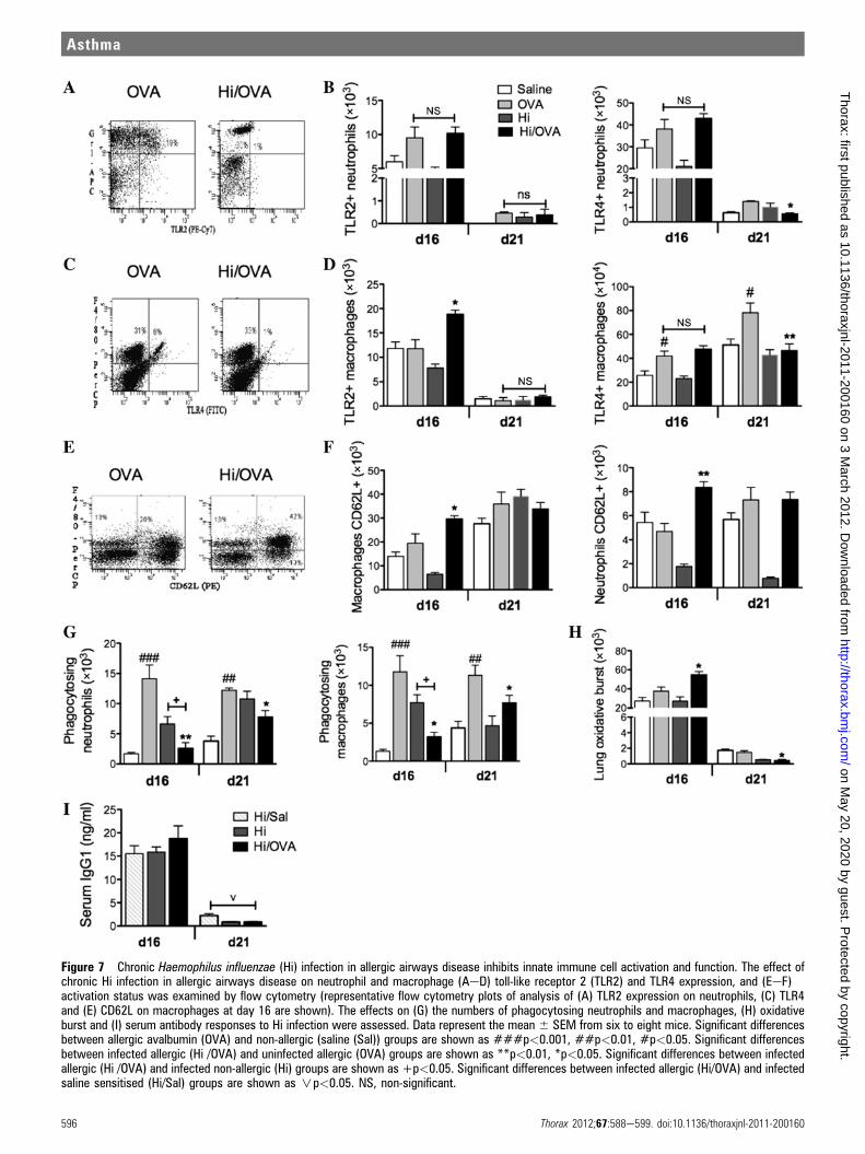
Figure 7 Chronic Haemophilus influenzae (Hi) infection in allergic airways disease inhibits innate immune cell activation and function. The effect ofchronic Hi infection in allergic airways disease on neutrophil and macrophage (AeD) toll-like receptor 2 (TLR2) and TLR4 expression, and (EeF)activation status was examined by flow cytometry (representative flow cytometry plots of analysis of (A) TLR2 expression on neutrophils, (C) TLR4and (E) CD62L on macrophages at day 16 are shown). The effects on (G) the numbers of phagocytosing neutrophils and macrophages, (H) oxidativeburst and (I) serum antibody responses to Hi infection were assessed. Data represent the mean 6 SEM from six to eight mice. Significant differencesbetween allergic avalbumin (OVA) and non-allergic (saline (Sal)) groups are shown as ###p<0.001, ##p<0.01, #p<0.05. Significant differencesbetween infected allergic (Hi /OVA) and uninfected allergic (OVA) groups are shown as **p<0.01, *p<0.05. Significant differences between infectedallergic (Hi /OVA) and infected non-allergic (Hi) groups are shown as +p<0.05. Significant differences between infected allergic (Hi/OVA) and infectedsaline sensitised (Hi/Sal) groups are shown as np<0.05. NS, non-significant.
596 Thorax 2012;67:588e599. doi:10.1136/thoraxjnl-2011-200160
Asthma
on May 20, 2020 by guest. P
rotected by copyright.http://thorax.bm
j.com/
Thorax: first published as 10.1136/thoraxjnl-2011-200160 on 3 M
arch 2012. Dow
nloaded from

expression of GR-b mRNA in asthmatic epithelial cells in vitro.26
Taken together, this study and others suggest that infection-induced increases in neutrophilic inflammation, Th17 cells andIL-17 production may be important in promoting steroid resis-tance in patients with neutrophilic asthma.
Steroid treatment caused a small reduction in bacterialnumbers in allergic airways disease at day 16. Treatmentsubstantially exacerbated infection in non-allergic groups afterthe infection had apparently cleared. This suggests that theinfection persisted at undetectable levels, perhaps intracellularly,and could be reactivated by steroid treatment, which is knownto suppress immune responses.3
We also investigated the mechanisms involved in the induc-tion of chronic infection. Chronic infection was maintained inallergic airways disease despite the increase in airway neutro-phils in infected allergic groups compared with uninfectedallergic controls. In the healthy airway, normal mucociliaryclearance of inhaled particles and bacteria requires the interac-tion of normally beating respiratory cilia and the overlyingmucus blanket. Reduction of ciliary function and mucusproduction in the airways may compromise bacterial clearance.Infection and inflammatory cells have been reported to impairciliary beat frequency and mucociliary clearance.22 However, wefound that there were no reductions in MSCs or ciliary beatfrequency in infected allergic compared with uninfected allergicgroups, suggesting that alterations in these innate immunedefences are not responsible for chronic infection in neutrophilicasthma.
The most important cellular components of innate immunityare neutrophils and macrophages, which when activated mountan immediate non-specific response and clear bacteria byphagocytosis. We therefore investigated the combined effects of
infection and allergic airways disease on the activation andfunction of airway neutrophils and macrophages. TLRs areexpressed on neutrophils and macrophages and increases in theiractivation and signalling are involved in the activation, chemo-kine receptor expression and function of these phagocytic cells.27
Although TLR2 is important, TLR4 is crucial for host defenceand clearance of pulmonary H influenzae infection.28 We showhere that in infected allergic groups, TLR2-positive cells wereunchanged compared with uninfected allergic controls (at days16 and 21), the only exception being an increase in TLR2-posi-tive macrophages on day 16. Interestingly, we show that, on day21, TLR4-positive neutrophils and macrophages were reducedcompared with allergic controls, demonstrating that H influenzaein allergic airways disease suppresses the influx of TLR4-positiveinnate immune cells, which may contribute to the persistence ofinfection.Neutrophil and macrophage activation was also investigated.
Activation of neutrophils and macrophages occurs in stages andincludes a range of phenotypic and functional changes. Lowlevels of activation lead to the modulation of surface receptorsand priming to subsequent stimuli, while higher levels enhancefunction.29 30 CD62L (L-selectin) is a surface receptor that ishighly expressed on resting neutrophils and circulating mono-cytes, and mediates their migration to the site of inflammation.During normal activation, migration and maturation, CD62Lexpression is downregulated by shedding and its expression isindicative of reduced activation and maturity.30e32 Our studydemonstrates that chronic H influenzae infection during allergicairways disease results in increased numbers of neutrophils andmacrophages expressing CD62L. This indicates that H influenzaemay suppress the activation of these cells, which maycontribute to chronic infection in neutrophilic asthma.
Figure 8 Chronic Haemophilusinfluenzae (Hi) infection and ovalbumin(OVA) sensitisation suppress thenumbers of phagocytosingmacrophages. (A) Mice were infected,sensitised 10 days later, and infectionand immune responses assessed onday 11. The effect of infection andsensitisation on (B) macrophage andneutrophil numbers in bronchoalveolarlavage fluid (BALF), (C) numbers ofphagocytosing macrophages andneutrophils, and (D) antibody responsesto Hi infection were assessed. Datarepresent the mean 6 SEM from six toeight mice. Significant differencesbetween infected allergic (Hi/OVA) anduninfected allergic (OVA) groups areshown as *p<0.05. Significantdifferences between infected allergic(Hi/OVA) and infected non-allergic (Hi)groups are shown as +p<0.05.Significant differences between infectedallergic (Hi/OVA) and infected salinesensitised (Hi/Sal) groups are shown asnp<0.05.
Thorax 2012;67:588e599. doi:10.1136/thoraxjnl-2011-200160 597
Asthma
on May 20, 2020 by guest. P
rotected by copyright.http://thorax.bm
j.com/
Thorax: first published as 10.1136/thoraxjnl-2011-200160 on 3 M
arch 2012. Dow
nloaded from

The function of neutrophils and macrophages was alsoexamined. Once activated, neutrophils and macrophages gainmicrobicidal functions, the most important of which is phago-cytosis and elimination of invading pathogens.33 34 Critically weshow that infection in allergic airways disease completelyinhibited the influx and/or development of phagocytosingneutrophils and macrophages in the airways compared withuninfected allergic controls. The mechanisms that underpin thisinhibition of phagocytosis in infected allergic groups arecurrently unknown and are under investigation. However,possible mechanisms include increased apoptosis of phagocy-tosing neutrophils and macrophages, increased necrosis of thesecells, or altered pathogen recognition receptor signalling. Therewere significantly less phagocytosing neutrophils and macro-phages in infected allergic compared with uninfected allergicgroups at days 11, 16 and 21. There were also fewer of thesephagocytosing cells in infected allergic compared with infectedalone groups but only at days 11 and 16 and not at day 21. Theseresults suggest that it may be the asthma phenotype, ratherthan the effects of infection, that plays the dominant role indriving reduced phagocytosis. However, infection may alsocontribute to the decreased function of phagocytes.
The lack of activation and influx of phagocytes may be thereasons for the development of chronic infection in neutrophilicallergic airways disease and in neutrophilic asthma. However,this does not rule out the possibility that other mechanisms ofinnate immune dysfunction may be responsible for chronicinfection and neutrophilic asthma. There is the potential thatthe increased neutrophilic inflammation observed in neutro-philic asthma may be the result of the increase in neutrophilinflux to compensate for their decreased function.
Our results also show that lung cells from infected allergicgroups generated more oxidative burst than uninfected allergiccontrols. Oxidative burst increases oxidative stress and hassignificant effects on the pathophysiology of asthma, such asincreasing inflammation and airway smooth muscle contrac-tility.35 Increased oxidative stress occurs in patients with asthmawho have increased bacterial load compared with healthycontrols, which is probably due to the bacterial load rather thanincreased neutrophilic inflammation.15
Early in the model, we show increased antibody responses toH influenzae infection in OVA-sensitised groups, which may bea result of increased infection. However, later, the combinationof infection and allergic airways disease reduced antibodyresponses and this could also contribute to chronic infection.The reduction in antibody responses presumably results froma suppression of B-cell activity that requires T-cell help. Weshow similar suppressive effects on T-cell activity.
Taken together these results may explain the association ofinfection with neutrophilic asthma. Allergic airways diseasepromotes persistent infection and induces the influx of defectiveinnate immune cells that fail to clear the bacteria but mayincrease oxidative stress.
In conclusion, we show that the combination of allergicairways disease and H influenzae infection leads to chronicinfection. Chronic infection together with infection-inducedincreases in Th17 cells and IL-17 production may lead to thedevelopment of neutrophilic allergic airways disease andasthma. We demonstrate that this phenotype is steroid resistant,which may be the result of a combination of increased neutro-phils and Th17 cells. Chronic infection probably results from thesuppression of innate cell activation and function, howeverfurther studies of the mechanisms of neutrophilic asthma arerequired to better understand disease progression. Such studies
would enable the development of more effective treatments forpatients with this disease phenotype, whereby targeting theinfection and asthmatic responses may be beneficial.
Funding Hunter Medical Research Institute, the National Health and MedicalResearch Council of Australia, The Australian Research Council, and University ofNewcastle.
Competing interests None.
Ethics approval All experiments were approved by the Animal Care and EthicsCommittee, University of Newcastle.
Provenance and peer review Not commissioned; externally peer reviewed.
REFERENCES1. Umetsu DT, McIntire JJ, Akbari O, et al. Asthma: an epidemic of dysregulated
immunity. Nat Immunol 2002;3:715e20.2. Simpson JL, Scott R, Boyle MJ, et al. Inflammatory subtypes in asthma:
assessment and identification using induced sputum. Respirology 2006;11:54e61.3. Wang W, Li JJ, Foster PS, et al. Potential therapeutic targets for steroid-resistant
asthma. Curr Drug Targets 2010;11:957e70.4. Simpson JL, Phipps S, Gibson PG. Inflammatory mechanisms and treatment of
obstructive airway diseases with neutrophilic bronchitis. Pharmacol Ther2009;124:86e95.
5. Stockley RA. Neutrophils and the pathogenesis of COPD. Chest2002;121:151Se5S.
6. Zaas AK, Schwartz DA. Innate immunity and the lung: defense at the interfacebetween host and environment. Trends Cardiovasc Med 2005;15:195e202.
7. Simpson JL, Grissell TV, Douwes J, et al. Innate immune activation in neutrophilicasthma and bronchiectasis. Thorax 2007;62:211e18.
8. Baines KJ, Simpson JL, Scott RJ, et al. Immune responses of airway neutrophils areimpaired in asthma. Exp Lung Res 2009;35:554e69.
9. Droemann D, Goldmann T, Tiedje T, et al. Toll-like receptor 2 expression isdecreased on alveolar macrophages in cigarette smokers and COPD patients. RespirRes 2005;6:68.
10. Yoshikawa T, Dent G, Ward J, et al. Impaired neutrophil chemotaxis in chronicobstructive pulmonary disease. Am J Respir Crit Care Med 2007;175:473e9.
11. Hansbro NG, Horvat JC, Wark PA, et al. Understanding the mechanisms of viralinduced asthma: new therapeutic directions. Pharmacol Ther 2008;117:313e53.
12. Hansbro PM, Beagley KW, Horvat JC, et al. Role of atypical bacterial infection of thelung in predisposition/protection of asthma. Pharmacol Ther 2004;101:193e210.
13. Jackson DJ, Johnston SL. The role of viruses in acute exacerbations of asthma. JAllergy Clin Immunol 2010;125:1178e87.
14. Bisgaard H, Hermansen MN, Buchvald F, et al. Childhood asthma after bacterialcolonization of the airway in neonates. N Engl J Med 2007;357:1487e95.
15. Wood LG, Simpson JL, Hansbro PM, et al. Potentially pathogenic bacteria culturedfrom the sputum of stable asthmatics are associated with increased 8-isoprostaneand airway neutrophilia. Free Radic Res 2010;44:146e54.
16. Murphy TF, Brauer AL, Schiffmacher AT, et al. Persistent colonization byHaemophilus influenzae in chronic obstructive pulmonary disease. Am J Respir CritCare Med 2004;170:266e72.
17. Essilfie AT, Simpson JL, Horvat JC, et al. Haemophilus influenzae infection drives IL-17-mediated neutrophilic allergic airways disease. PLoS Pathog 2011;7:e1002244.
18. Preston JA, Essilfie A-T, Horvat JC, et al. Inhibition of allergic airways disease byimmunomodulatory therapy with whole killed Streptococcus pneumoniae. Vaccine2007;25:8154e62.
19. Horvat JC, Starkey MR, Kim RY, et al. Early-life chlamydial lung infection enhancesallergic airways disease through age-dependent differences in immunopathology. JAllergy Clin Immunol 2010;125:617e25, 625.e1e625.e6.
20. Horvat JC, Starkey MR, Kim RY, et al. Chlamydial respiratory infection duringallergen sensitization drives neutrophilic allergic airways disease. J Immunol2010;184:4159e69.
21. Thorburn AN, O’Sullivan BJ, Ranjeny T, et al. Pneumococcal conjugate vaccine-induced regulatory T cells suppress the development of allergic airways disease.Thorax 2010;65:1053e60.
22. Rayner CF, Rutman A, Dewar A, et al. Ciliary disorientation in patients with chronicupper respiratory tract inflammation. Am J Respir Crit Care Med 1995;151:800e4.
23. Preston JA, Beagley KW, Gibson PG, et al. Genetic background affects susceptibilityin nonfatal pneumococcal bronchopneumonia Eur Respir J 2004;23:224e31.
24. Angrill J, Agusti C, de Celis R, et al. Bacterial colonisation in patients withbronchiectasis: microbiological pattern and risk factors. Thorax 2002;57:15e19.
25. Cox G. Glucocorticoid treatment inhibits apoptosis in human neutrophils. Separationof survival and activation outcomes. J Immunol 1995;154:4719e25.
26. Vazquez-Tello A, Semlali A, Chakir J, et al. Induction of glucocorticoid receptor-b expression in epithelial cells of asthmatic airways by T-helper type 17 cytokines.Clin Exp Allergy 2010;40:1312e22.
27. Parker LC, Whyte MK, Dower SK, et al. The expression and roles of toll-likereceptors in the biology of the human neutrophil. J Leukoc Biol 2005;77:886e92.
28. Wieland CW, Florquin S, Maris NA, et al. The MyD88-dependent, but not theMyD88-independent, pathway of TLR4 signaling is important in clearing nontypeableHaemophilus influenzae from the mouse lung. J Immunol 2005;175:6042e9.
598 Thorax 2012;67:588e599. doi:10.1136/thoraxjnl-2011-200160
Asthma
on May 20, 2020 by guest. P
rotected by copyright.http://thorax.bm
j.com/
Thorax: first published as 10.1136/thoraxjnl-2011-200160 on 3 M
arch 2012. Dow
nloaded from

29. van Eeden SF, Klut ME, Walker BA, et al. The use of flow cytometry to measureneutrophil function. J Immunol Methods 1999;232:23e43.
30. Springer TA. Traffic signals for lymphocyte recirculation and leukocyte emigration:the multistep paradigm. Cell 1994;76:301e14.
31. Spertini O, Luscinskas FW, Gimbrone MA, et al. Monocyte attachment to activatedhuman vascular endothelium in vitro is mediated by leukocyte adhesion molecule-1(L-selectin) under nonstatic conditions. J Exp Med 1992;175:1789e92.
32. von Andrian UH, Chambers JD, McEvoy LM, et al. Two-step model ofleukocyteeendothelial cell interaction in inflammation: distinct roles for LECAM-1 and
the leukocyte beta 2 integrins in vivo. Proc Natl Acad Sci United States America1991;88:7538e42.
33. Neufert C, Pai RK, Noss EH, et al. Mycobacterium tuberculosis 19-kDa lipoproteinpromotes neutrophil activation. J Immunol 2001;167:1542e9.
34. Luscinskas FW, Kansas GS, Ding H, et al. Monocyte rolling, arrest and spreading onIL-4-activated vascular endothelium under flow is mediated via sequential action of L-selectin, beta 1-integrins, and beta 2-integrins. J Cell Biol 1994;125:1417e27.
35. Wood LG, Gibson PG, Garg ML. Biomarkers of lipid peroxidation, airwayinflammation and asthma. Eur Respir J 2003;21:177e86.
Journal club
Mechanisms underlying the increase in mycobacterialinfections in cystic fibrosis patients on azithromycinThis study investigated the increased incidence of non-tuberculous mycobacterium (NTM)infections in cystic fibrosis (CF) patients on azithromycin. The authors were particularlyinterested in autophagia, a process whereby intracellular material is isolated into phagosomesand then destroyed by being fused with acid-rich lysosomes. It is disrupted by othermacrolides and is crucial for the clearance of intracellular pathogens.In vitro macrophages showed decreased degradation of phagosomes when exposed to
azithromycin in both healthy controls and CF patients. This was demonstrated to be mainlydue to lysosomal pH being raised to levels that inhibited destructive enzymes and also due toimpaired phagosomeelysosome fusion. Further disruption to immune function was caused byinhibition of cytokine release, as evidenced by decreased levels of interferon g (IFNg). In vivoeffects were verified by infecting mice with resistant strains of Mycobacterium abscessus.Control groups normally cleared the pathogen within 1 month whereas mice treated withazithromycin were much more likely to develop persistent disease and show consistentlyaltered levels of pulmonary cytokines. The authors speculated that such immunomodulatoryeffects may explain anti-inflammatory properties associated with azithromycin, but fear theymight also render concurrently administered IFNg therapy less effective.The findings offer a plausible explanation for the predisposition that CF patients on
azithromycin have to NTM infections; however, the processes described are not specific tothis group. The increasing use of prophylactic azithromycin in chronic obstructive pulmonarydisease means that physicians should be vigilant for the development of NTM infections inthis cohort as well.
< Renna M, Schaffner C, Brown K, et al. Azithromycin blocks autophagy and may predispose cystic fibrosis patients tomycobacterial infection. J Clin Invest 2011;121:3554e63.
Gareth Jones
Correspondence to Dr Gareth Jones, ST5, Amanda Unit, Liverpool Heart & Chest Hospital, Thomas Drive, Liverpool L14 3PE,UK; [email protected]
Provenance and peer review Not commissioned; internally peer reviewed.
Published Online First 21 November 2012
Thorax 2012;67:599. doi:10.1136/thoraxjnl-2011-201327
Thorax July 2012 Vol 67 No 7 599
Asthma
on May 20, 2020 by guest. P
rotected by copyright.http://thorax.bm
j.com/
Thorax: first published as 10.1136/thoraxjnl-2011-200160 on 3 M
arch 2012. Dow
nloaded from