original article burn trauma disrupts circadian rhythms … · original article burn trauma...
TRANSCRIPT

Int J Burn Trauma 2016;6(2):12-25www.IJBT.org /ISSN:2160-2026/IJBT0029198
Original ArticleBurn trauma disrupts circadian rhythms in rat liver
Rohit Rao1, Qian Yang1, Mehmet A Orman1, Francois Berthiaume2, Marianthi G Ierapetritou1, Ioannis P Androulakis1,2
1Chemical and Biochemical Engineering Department, Rutgers, The State University of New Jersey, Piscataway, NJ 08854, USA; 2Biomedical Engineering Department, Rutgers, The State University of New Jersey, Piscataway, NJ 08854, USA
Received March 26, 2016; Accepted April 29, 2016; Epub June 1, 2016; Published June 15, 2016
Abstract: Circadian rhythms play an important role in maintaining homeostasis and solid organ function. The purpose of this study is to assess the implications of burn injury in rats on the underlying circadian patterns of gene expres-sion in liver. Circadian-regulated genes and burn-induced genes were identified by applying consensus clustering methodology to temporally differentially expressed probe sets obtained from burn and sham-burn data sets. Of the liver specific genes which we hypothesize that exhibit circadian rhythmicity, 88% are not differentially expressed following the burn injury. Specifically, the vast majority of the circadian regulated-genes representing central carbon and nitrogen metabolism are “up-regulated” after the burn injury, indicating the onset of hypermetabolism. In addi-tion, cell-cell junction and membrane structure related genes showing rhythmic behavior in the control group were not differentially expressed across time in the burn group, which could be an indication of hepatic damage due to the burn. Finally, the suppression of the immune function related genes is observed in the postburn phase, implying the severe “immunosuppression”. Our results demonstrated that the short term response (24-h post injury) mani-fests a loss of circadian variability possibly compromising the host in terms of subsequent challenges.
Keywords: Circadian clock, liver, burn injury, microarray analysis, rats, differentially expressed gene clusters
Introduction
The circadian clock is one of the most critical biological regulators for all living entities con-trolling behavioral, physiological and biochemi-cal processes. Acting like a multifunctional timer, the circadian clock organizes the homeo-static system, including sleep and wakefulness, hormonal secretions and various bodily func-tions with a roughly 24-h cycle [1]. The circadi-an clock provides fitness advantages by endow-ing organisms with anticipatory mechanisms predicting periodic events, as opposed to responding in a reactive way to external signals [2]. In mammals the primary circadian “clock”, is located in the suprachiasmatic nucleus (SCN), a pair of distinct groups of cells located in the hypothalamus [3]. Independent circadian rhythms are also found in many organs and cells outside the SCN, such as the oesophagus, lungs, liver, pancreas, spleen, thymus, and the skin [4]. Given that circadian rhythmicity and host immunity are interrelated, numerous stud-
ies have demonstrated that external stressors can influence circadian timekeeping processes, while cytokines and immune mediators impact clock gene expression [5].
Liver is one of the important organs regulating the metabolic activity of body as well as produc-ing acute phase proteins. Using microarray technology to generate a rich time series within a 24 h cycle, it has been already elucidated that a wide variety of biological pathways show cir-cadian rhythms in the rat liver [6]. Since the genes exhibiting statistically validated-rhythmic behavior are differentially expressed across the time, Ovacik et al. [7] applied “consensus” clus-tering approach [8] to statistically significant patterns of gene expression levels obtained from the microarray data. They identified five distinct clusters exhibiting circadian rhythm in the rat liver, which are related to energy metab-olism, amino acid metabolism, lipid metabo-lism, and DNA replication and protein synthe-sis. Since an external stressor activates the

Circadian disruption after burn injury
13 Int J Burn Trauma 2016;6(2):12-25
host immune system through stimulation of a series of local and systemic responses, includ-ing the release of proinflammatory cytokines, up-regulation of glucose, fatty acid, and amino acid turnover in the liver [9], the question is raised as to whether, and how, this affects the circadian rhythms in the liver. The purpose of this study is to demonstrate the possible sup-pression of circadian variability in liver-specific gene expression within the first 24 hours follow-ing burn injury by using a standard rat model of cutaneous burn injury covering 20% of the total body surface area (TBSA), and its correspond-ing sham-burn (control of burn). Applying the “consensus clustering” approach in an unsu-pervised manner to the expression profiles of temporally differentially expressed genes in the control group resulted in four different patterns associated with the immune system, energy and amino acid metabolism, cell-cell junction and membrane structure, and DNA replication and repair [10]. Investigating the expression of these clusters in the burn group in the present work, we identified that 88% of genes in these clusters were not differentially expressed across time following the burn injury, demon-strating the disruption of circadian rhythms as a result of severe trauma.
Materials and methods
Animal model
Male Sprague-Dawley rats (Charles River Labs, Wilmington, MA) weighing between 150 and 200 g were utilized for this study. The animals were housed in a temperature-controlled envi-ronment (25°C) with a 12-hour light-dark cycle and provided water and standard chow ad libi-tum. All experimental procedures were carried out in accordance with National Research Council guidelines and approved by the Rutgers University Animal Care and Facilities Committee.
A systemic hypermetabolic response was in- duced by applying a full-thickness burn on an area of the dorsal skin corresponding to 20% of the total body surface area (TBSA) as described elsewhere [11]. This model was chosen be- cause it has nearly 100% long-term survival, no evidence of systemic hypoperfusion, and no significant alterations on feeding patterns [12]. Rats were first randomized into two groups: burn and sham burn (control group). Rats were anesthetized by intraperitoneal injection of 80 to 100 mg/kg ketamine + 12 to 10 mg/kg xyla-
zine, and all hair removed from the dorsal abdominal area using electric clippers. The ani-mal’s back was immersed in water at 100°C for 10 s to produce a full-thickness scald injury covering 20% TBSA. Immediately after burns, the animals were resuscitated with 50 mL/kg of saline injected intraperitoneally. Negative controls (sham burn) consisted of animals treated identically but immersed in warm water (37°C). Rats were single caged after burn or sham burn and given standard rat chow and water ad libitum until sacrifice. No post-burn analgesics were administered, consistent with other studies with this full thickness burn model since the nerve endings in the skin are destroyed and the skin becomes insensate [13]. Furthermore, after animals woke up, they ate, drank and moved freely around the cage, responded to external stimuli, and did not show clinical signs of pain or distress. Animal body weights were monitored daily and found to increase at the same rate in both groups.
Starting at 9am, animals are sacrificed at dif-ferent time points (0, 2, 4, 8, 16 and 24 hr post-treatment, i.e., sham burn and burn) and liver tissues were collected and frozen for microar-ray analysis (n=3 per time point per group). We limited ourselves to three samples at each time point for reasons of practicality due to the num-ber of time points being analyzed in our study. Ideally, in future studies the selected time points would be equally spaced in the latter parts of the day and the night in order to prop-erly characterize the entire time course of circa-dian expression. However, the choice of time points in the present study was dictated by our previous study to identify changes in global hepatic gene expression within the first 24 hours after burn injury. The tissues were lysed and homogenized using Trizol, and the RNAs were further purified and treated with DNase using RNeasy columns (Qiagen). Then cRNAs prepared from the RNAs of liver tissues using protocols provided by Affymetrix were utilized to hybridize Rat Genome 230 2.0 Array (GeneChip, Affymetrix) comprised of more than 31,000 probe sets. The microarray dataset obtained in our experiments has been submit-ted to an open-access data repository [14].
Data analysis
In this study, genome expression data analysis includes normalization, filtering, clustering and merging. DNA chip analyzer (dChip) software

Circadian disruption after burn injury
14 Int J Burn Trauma 2016;6(2):12-25
[15] was used with invariant-set normalization and perfect match (PM) model to generate expression values. The analysis of variance (ANOVA) test implemented in R [16] was applied to filter differentially expressed probe sets in shamburn dataset and burn dataset. Probesets with p values less than 0.001 were selected as demonstrating a statistically significant differ-ence in expression overtime in shamburn liver and burn-injured liver respectively. Moreover, applying the method proposed by Storey et al. [17], a q-value cutoff of 0.01 was used to con-trol the false discovery rate. Strict thresholds
were used due to data limitations, which result-ed in several well-known circadian genes not being present in the filtered data set (see Supplementary material; sheet ‘filtering’). Subsequently, consensus clustering [8] was applied in order to identify coherent clusters of co-expressed genes in an unsupervised man-ner. Finally, merging is used to combine the clusters which have a similar expression pat-tern but are separated into two or more clus-ters [18]. We characterize the biological rele-vance of the intrinsic responses by evaluating the enrichment of the corresponding subsets in
Figure 1. Circadian patterns in liver. Left Panel: Expression of 52, 64, 153 and 83 probe sets in 4 clusters in sham-burn rats at 0, 2, 4, 8, 16, 24 h are exhibited on heatmap. Green indicates the lowest level while red indicates the highest and black average level. Right Panel: Expression patterns are shown by plotting the average normalized (z-score) expression values of 52, 64, 153, and 83 probe sets in 4 clusters in sham-burn rats (means ± SEM). n=3 rats per time point.

Circadian disruption after burn injury
15 Int J Burn Trauma 2016;6(2):12-25
circadian rhythm specific pathways by using KEGG database through ARRAYTRACK [19] as well as analyzing the functions of individual genes extensively. We perform paired t-tests to determine significant changes at correspond-ing time points between the clusters in the burn and shamburn treatment groups.
In our previous study [10], we further explored the underlying regulatory mechanisms of circa-dian-relevant genes, by hypothesizing that the core circadian transcription factor (TF) CLOCK: BMAL1 as well as TFs that are downstream tar-gets of CLOCK: BMAL1 might regulate the expression of temporally expressed genes we identified in the above analysis. Briefly, we used MatInspector [20] to scan for TF binding sites on the promoter regions of each of the corre-sponding differentially expressed genes. Genes reported by MatBase, which also consist of CLOCK: BMAL1 binding sites were considered clock-controlled TFs. Additionally PARF and RORA were also included in the analysis as these are well-known core clock TFs [18]. Following this, we identified significant sets of clock TFs that were commonly present on the promoter regions of the differentially expressed genes in each cluster, since we hypothesized that these common sets of TFs were more likely to regulate the expression of the clusters in a combinatorial manner more representative of eukaryote transcriptional regulation. Finally, in order to determine the relations of the clock TFs to each of the clusters, we estimated the statistical significance of the common sets of clock TFs for each cluster compared to the GC-matched background set by the method proposed by Bozek et al [21].
Results
Identification of circadian rhythm regulated genes
Transcriptional analysis: In the sham-burn group, 545 probe sets are identified as differ-entially expressed. Consensus clustering iden-tifies four statistically dominant transcriptional clusters all exhibiting circadian oscillations, composed of 52, 64, 153, 83 probe sets respectively. In our previous study [10], we fur-ther used Fisher’s exact g-test to determine whether the genes in the identified cluster were expressed in a circadian manner. Briefly, a new vector is created for the test by randomly dis-
tributing the three replicates at each time point to three consecutive time points in a 24-hour period; t, t+24 and t+48. We found that all 352 genes identified in the four clusters passed the test (with a q-value<0.01), implying that these genes are indeed periodically expressed and are thus, considered circadian-relevant in this context. The characteristic patterns of the four clusters are depicted in Figure 1. The gene list and expression levels for each probe set are provided in the Supplementary file “shamBurn_Burn.suppData.xls”. It is important to note that all probe sets coding for the same gene exhib-ited similar expression patterns (i.e., were assigned to the same cluster) further enhanc-ing our confidence in our gene expression mea-surements. Thus, the probe sets could be mapped to their corresponding genes, and therefore the clusters are composed of 46, 41, 132, 70 genes respectively.
Functional characterization of circadian expres-sion patterns: Functional annotation enables a succinct description of the key roles each clus-ter plays. Specifically:
Cluster 1, displays a maximum activity during the light phase and was found to be primarily composed of genes associated with the immune system. Genes in this temporal class are involved in the Chemokine signaling path-way (Chuk, Gnai3, Stat3), RIG-I-like receptor signaling pathway (Chuk, Ddx3x), Toll-like recep-tor signaling pathway (Chuk, Myd88). Numerous genes regulated by the circadian rhythm and related to immune functions are also found in this cluster. BTG1, one of components of immune function responses was examined and determined to be rhythmic under a light dark cycle [22]. Moreover, it is reported that circadi-an transcriptional regulation could be found at every level of LPS induced immune response in macrophages including SFPQ which regulates cytokine mRNA stability and localization [23]. FKBP5 is observed to show strong circadian dynamics in rat liver whose isomerase activity is inhibited by immunosuppressants [6].
Cluster 2 reaches its maximum during the day-time and quickly returns to its baseline before the dark phase and is found to contain genes with diverse functions including energy produc-tion (Ppargc1a, Coq10b), cell-cell junction (Tanc1, Epb4.1l5), and membrane structure (Ubr4, Tmem106b). The genes associated with

Circadian disruption after burn injury
16 Int J Burn Trauma 2016;6(2):12-25
cell junction (Tanc1, Epb4.1l5) and component of membrane (Ubr4, Tmem106b) play a critical role in the integrity of the endothelial barrier function. In addition, some genes regulating cir-cadian rhythms are found in this cluster. One of the isoforms generated from the Crem gene, a transcription factor involved in output clock function, regulates melatonin synthesis in the mouse [24]. klf9, which is a Krüppel-like family transcription factor is identified as putative clock-regulated gene in gene expression profil-ing experiments performed in liver [25]. Moreover, other genes owing diverse functions are reported to fluctuate during the day. DAPK1, whose expression has strong circadian charac-teristics [6], is associated with regulation of apoptosis. Genome-wide expression analysis reveals that the expression of LPIN1 which reg-ulates the fatty acid catabolic process is circa-dian dependent [26].
Cluster 3 is composed of genes whose activity peaks at night and is enriched in functions associated with metabolism, including amino acid metabolism such as nitrogen metabolism (Car14, Car5a, Cth, Gls2), alanine, aspartate and glutamate metabolism (Abat, Agxt2, Gls2, LOC641316), glycine, serine and threonine metabolism (Agxt2, Cth, Gldc), arginine and proline metabolism (Agmat, Gls2), beta-alanine metabolism (Abat, Dpys), and carbohydrate metabolism such as glycosaminoglycan degra-dation (Gns, Gusb), propanoate metabolism (Abat, Acacb), starch and sucrose metabolism (Gusb, Pygl). In addition, an important clock
thesis of steroids, fluctuate during the day [26]. KYNU and AKR1C18 regulating the cofactor metabolism rhythmically [6] appear in this group.
Finally, Cluster 4 reaches its peak activity around midnight and is enriched in functions associated with DNA replication and repair, such as mismatch repair (Rfc2, Rfc5), DNA rep-lication (Rfc2, Rfc5), and non-homologous end joining (Rad50). Similarly, some genes regulat-ed by circadian rhythm reported by previous studies are found in this cluster. Timeless, an output circadian gene whose mRNA expression has also been detected in most peripheral tis-sues examined in both human and mouse [27]. Calm1 is a time dependent gene in human skel-etal muscle [22].
Comparison of circadian expression patterns in sham and burn conditions
In order to determine whether the expression of temporally expressed genes identified in the sham group is disrupted we explored the dif-ferentially expressed genes following induction of the burn injury. Following burn injury, 1034 probe sets are identified to be significantly. Careful analysis of the data reveals the follow-ing significant results indicating that the major-ity of the probesets differentially expressed in the sham group change expression patterns following thermal injury. Generally, of the 352 probe sets constituting these 4 dominant circa-dian patterns, 309 (88%) have lost their tempo-
Figure 2. Animal weight change (%). Time of burn or sham burn injury is day 0. Solid line: Burn group; Dashed line: Sham-burn group. n=18 per group on day 0, n=6 per group on day 1, and n=3 per group on day 2. Note that n is decreasing with respect to time, since a portion of the animals is sacrificed at each time point to collect liver tissue.
gene product BMAL1 which plays a role in regulation of glucose homeostasis and metabolism [6] is identified in this cluster. Many other genes relating to the metab-olism are transcribed in a cyclic fashion. GCJR and DHTKD1, which regulate glucose homeostasis [6], are under the control of the circa-dian rhythm. CTH controlling he- patic synthesis of glutathione, Abat catalyzing gamma-aminobu-tyric acid (GABA) and GLDC being associated with glycine metabo-lism are regulated by circadian rhythmcity [6]. Moreover, the expressions of genes such as Chka, Cth, and Agxt2, whose func-tions are associated with biosyn-

Circadian disruption after burn injury
17 Int J Burn Trauma 2016;6(2):12-25
ral circadian variability. Specifically, 51 out of 52 probes constituting sham cluster 1 are not differentially expressed following burn injury whereas of the 83 probe sets of sham cluster 4, none are circadian expressed after burn. Moreover, 142 out of 153 probesets in cluster 3 in the sham group are not differentially expressed in the burn condition, while cluster 2 genes show the least disruption relative to other clusters with about half of the probe sets in cluster 2 not being differentially expressed following burn injury.
Figure 3 shows the detailed transcriptional pat-terns of the four clusters under sham and burn
conditions. t-tests between corresponding time points reveal that following burn injury, the average expression profile of cluster 1 genes shows a marked decrease in peak expression value (at ~ZT8) and also a significant increase in nadir expression value (at ~ZT24). Cluster 3 shows a significant increase in the nadir expres-sion value (~ZT8), and a decrease in peak expression value (~ZT24). The average expres-sion profiles of both these cluster show a marked decrease in amplitude after the burn injury is induced. On the other hand, the aver-age expression profile of the genes in pattern 2, which show a peak in activity level (ZT2) soon after those of pattern 1, remains particularly
Figure 3. Comparison of dynamics of temporally expressed shamburn treated clusters after burn injury injury. Aver-age z-scored temporal gene expression patterns in burn (red) and sham (blue) groups. Paired t-tests were used to determine significant changes (p<0.005) between sham and burn clusters at corresponding time points.
Circadian disruption after burn injury
18 Int J Burn Trauma 2016;6(2):12-25
unaffected in the burn injury group with no sig-nificant changes in expression between sham and burn groups at any of the corresponding time points.
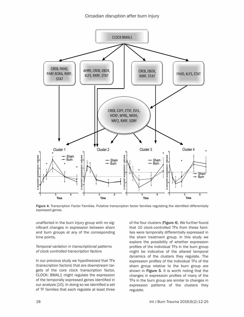
Temporal variation in transcriptional patterns of clock controlled transcription factors
In our previous study we hypothesized that TFs (transcription factors) that are downstream tar-gets of the core clock transcription factor, CLOCK: BMAL1 might regulate the expression of the temporally expressed genes identified in our analysis [10]. In doing so we identified a set of TF families that each regulate at least three
of the four clusters (Figure 4). We further found that 10 clock-controlled TFs from these fami-lies were temporally differentially expressed in the sham treatment group. In this study we explore the possibility of whether expression profiles of the individual TFs in the burn group might be indicative of the altered temporal dynamics of the clusters they regulate. The expression profiles of the individual TFs of the sham group relative to the burn group are shown in Figure 5. It is worth noting that the changes in expression profiles of many of the TFs in the burn group are similar to changes in expression patterns of the clusters they regulate.
Figure 4. Transcription Factor Families. Putative transcription factor families regulating the identified differentially expressed genes.

Circadian disruption after burn injury
19 Int J Burn Trauma 2016;6(2):12-25
Discussion
Robust circadian entrainment is associated with fitness, while abnormal rhythms are characteristic of stress and have been linked with illness [28, 29]. Thus, time-of-day va- riations in immune response are ubiquitous and the implica-tions are profound. As early as fifty years ago it was docu-mented that LPS-induced en- dotoxin shock in mice depends on when, during the circadian cycle, LPS is administered [30]. More recently Liu et al. documented that mice survival rate following LPS injection drops from 70-80% when LPS is administered at 2am or 9am to about 40% when adminis-tered at 2pm or 9pm [31]. These, and many other, results indicate the significance of cir-cadian rhythmicity in the im- mune response.
The liver expresses a diverse set of genes and previous research indicated that many of hepatic genes are under direct or indirect circadian con-trol [6]. Applying a very strin-gent filtering and clustering strategy, which reduces the possibility of selecting non cir-cadian genes, our results show that 352 probe sets oscillate in a circadian manner. Our func-tional enrichment analysis revealed circadian regulated functions associated with the immune system, energy pro-
Figure 5. Temporal dynamics of differentially expressed transcrip-tion factors. Temporal variation in transcription factor dynami- cs in burn (red) and sham (blue) groups. t-tests were used to deter-mine a significant change in aver-age z-scored expression values at corresponding time points for burn and shamburn treatment groups (p<0.05).

Circadian disruption after burn injury
20 Int J Burn Trauma 2016;6(2):12-25
duction, metabolism and DNA replication and repair. Immune system functions, including lymphocyte proliferation, natural killer cell activity, humoral immune response, absolute and relative numbers of circulating white blood cells and their subsets, cytokine levels, and serum cortisol, are known to be entrained by circadian cues [23]. It has also been previously reported that TNFα secretion was significantly increased in burn vs. sham isolated spleno-cytes cells only when injury took place in the morning [32] further verifying that the response to injury is time-of-day dependent as a result of the circadian variability of the immune func-tions. Energy production is also time-of-day dependent. It is reported that the production of ATP in rat liver is also regulated by the circadian rhythm and is lower in the dark phase [33]. The liver plays an important role in the orchestra-tion of metabolic functions such as the regula-tion of metabolic fuels, protein synthesis, iron and vitamin storage, as well as nutrient intake [34]. As might be expected, animals subjected to a light-dark schedule display pronounced rhythms in glycogen content, with a peak occur-ring late in the night, following the main period of food intake. This is also in agreement with our result that metabolic functions, especially glucose metabolism, reach their peak during the dark phase. The strategy of adaptive circa-dian clocks could be also timing of UV-sensitive cellular processes to occur at night to avoid UV-induced damage [35]. Our analysis showed that functions related to DNA replication/repair (cluster 4 in shamburn) achieve a maximum activity around midnight.
Following burn injury, hepatic responses are triggered and the functional characterization demonstrates that the expressed probe sets are related to the early pro-inflammatory, later pro-inflammatory and anti-inflammatory res- ponses. The activation of the pro-inflammatory response is known to involve a number of inter-related steps forming a complex signal trans-duction cascade. Once the pro-inflammatory response has mounted it serves as a subse-quent signal for stimulating the anti-inflamma-tory response, which helps to inhibit pro-inflam-matory signals and drive the system back to homeostasis.
In order to explore how the burn injury affects the circadian regulated genes’ expression, we take all the identified circadian regulated probe-
sets and check how they are expressed in the burn condition. It is informative to study the characteristics and functions of all these circa-dian regulated probesets including those both losing circadian variability and being temporally expressed in burn condition:
The genes constituting sham clusters one and four appear to have lost their temporal dynamics after burn. Specifically, of the 52 probes of sham cluster 1, only one is tempo-rally expressed after burn, whereas of the 83 probe sets of sham cluster 4 none are expressed after burn. Further analysis reveals the average expression profile of cluster 1 shows a marked decrease in peak expression value (at ~ZT8), while cluster 4 also shows a significant decrease in the amplitude of oscillation at ~ZT8 and ~ZT24, similar to that seen in patterns 1 and 3 (Figure 3). The obser-vation that the entire set (almost) of genes constituting sham cluster 1 is not differentially expressed in the burn condition indicates that sham cluster 1, representative of the immune system response, is suppressed post burn. Severe injury, such as burn, induces a state of immune suppression that predisposes burn and trauma patients to infection and sepsis. Genes which show a marked decrease in expression at ~ZT8 such as Myd88, Gnai3, Chuk, and Crp are associated with IL-1 signal-ing, T-cell activaiton and the acute phase response signaling. Extensive thermal trauma results in impaired immune function which has been attributed to a reduction in T lymphocyte numbers, increased suppressor cell activity, serum suppressive factors, altered receptor expression on T cells [36]. This is also supported by our prior gene expression stud-ies, which reveal that an initial upregulation of pro-inflammatory cytokines was resolved within 8 hours post administration of the burn injury [37]. The suppression of the expression of cluster 1 here may be an indication for postburn immune suppression.
Of the 64 probe sets constituting the circadian pattern of sham cluster 2, 31 are temporally expressed following burn in a coherent manner, whereas the remaining probes are not circadi-an expressed after burn. The average expres-sion profile of the genes in this cluster remains particularly unaffected in the burn injury group with no significant changes in expression between sham and burn groups at any of the

Circadian disruption after burn injury
21 Int J Burn Trauma 2016;6(2):12-25
corresponding time points. The clustered heat map for pattern 2 shows that nearly none of the genes in this cluster shows a significant change in expression. This may be an indica-tion that in fact these are genes whose ex- pression is not affected by the burn injury. Among those we identify genes involved mito-chondrial energy production (Ppargc1a), as well as diverse functions such as cell-cycle maintenance (Crem, Gch1). It can therefore be hypothesized that the criticality of these func-tions drives their robustness in the face of an external stressor. Our previous study [10] has shown that genes in cluster 2 might be under greater circadian control in comparison to those in other clusters (further discussed in the following section). Our data from the present study supports this hypothesis, since the expression profile of cluster 2 genes is relative-ly unaffected following burn injury. Interestingly this cluster also consists of genes such as Lpin1, whose activity is inversely associated with the secretion of very low-density lipopro-tein (VLDL) [38, 47]. Under homeostasis, increases in hepatic triglycerides are accompa-nied by increases in VLDL-triglycerides resulting in decreased accumulation of hepatic triglycer-ides [41]. However, following burn injury, the rate of VLDL-triglyceride synthesis has been previously observed to be unchanged contrib-uting to the incidence of hepatic steatosis [39]. Therefore, the relatively unchanged expression of genes associated with VLDL secretion (Lpin1) might indicate a maladaptive response to burn injury. Moreover, this cluster includes genes associated with cell-cell junc-tion and membrane integrity of endothelial monolayer. Though no direct report on the dam-age of the liver endothelial cells by burn injury is available, many studies reveal that intestinal permeability is increased in burn patient shortly after injury partly due to the junction integrity alterations [40]. Suppression of probe sets functioning as cell-cell junctions and membrane structural integrity may be indica-tion of the damage of the burn to liver.
Of the 153 probes constituting sham cluster three, 11 are also expressed under burn. It is important to note that the common probes under burn exhibit a fundamentally different expression dynamics (early up-regulation) com-pared to sham cluster 3 (early-down-regula-tion). Among them, Pygl, functioning as regula-
tor of glucose homeostasis and glycogen cata-bolic process is activated by burn. Following burn, catabolism is one of the most significant signatures for burn-induced symptoms. It is also interesting to notice that the remaining 142 probe sets in this cluster are not tempo-rally expressed after burn, with significant upregulation at ZT8 and downregulation at ZT24 compared to the sham group. Amongst the cluster 1 genes acacb inhibits the beta-oxidation of fatty acids to acetyl coA and their entry into the tricarboxylic cycle (TCA), while daglb promotes lipolysis by the break down of diacylglycerol. The increased concentration of free fatty acids due to increased lipolysis can either be oxidized or re-synthesized to triglycer-ides [41]. However, since it is possible that beta-oxidation, regulated by the expression of Acacb is impaired after burn injury step this could imply an accumulation of hepatic triglyc-erides, which is well-documented in burn patients. Furthermore, this cluster also con-sists of genes involved in glucose metabolism such as Cth, which increases gluconeogenesis and glycogenolysis. Thus, it may be hypothe-sized that shamburn cluster 3 functions as an indicator of metabolism, activated following burn. Significant evidence suggests that the metabolic rate in burns is extremely high; ener-gy requirements are immense and are met by the mobilization of proteins and amino acids. The hypermetabolism is demonstrated by accelerated metabolic rates, increased gluco-neogenesis, increased nitrogen loss and loss of lean body mass, stimulated acute-phase protein synthesis in the liver, and abnormalities in lipid metabolism [42]. Thus, our result may be an early indication for the burn-associated hypermetabolism.
It is well known that feeding/fasting cycles are dominant zeitgebers of circadian clocks and, feeding regimens may potentially be altered fol-lowing burn injury because of burn associated anorexia. In our study, the 20% TBSA thermal injury model was chosen because it has nearly 100% long-term survival, no evidence of sys-temic hypoperfusion, and no significant impact on feeding patterns. As seen in Figure 2, ani-mal body weights were monitored daily and found to increase at the same rate in both groups. Thus, the desynchronization of rat liver following burn was not likely due to associated anorexia.

Circadian disruption after burn injury
22 Int J Burn Trauma 2016;6(2):12-25
Hepatocytes are by far the main cellular com-ponent of the liver constituting approximately 78% of the parenchymal volume whereas endo-thelial cells, Kupffer cells, fat storing cells and blood cells account the rest [43]. It might be speculated that the composition of the liver could change following burn injury (for example due to inflammatory cell infiltration), which in turn could impact on the measured gene expression patterns. Cakir et al. did not report any inflammatory cell infiltration by histological analysis of liver whereas they observed a mas-sive inflammatory cell infiltration in the gastric and colonic tissues in 30% TBSA burn injury rat models [44]. Basakaran and co-workers observed minimal neutrophil recruitment in liver following a 20% TBSA burn in rats; howev-er, the cell infiltration in liver was found to be about two orders of magnitude lower when compared to that of lung [45]. It has also been reported that the circulating leukocyte concen-tration profile does not change significantly in the 30% TBSA burn model in rats [46]. Therefore, we could safely assume that in our 20% TBSA burn injury model used herein, it is very unlikely that there was a change in the composition of the rat liver that could signifi-cantly impact on the measured gene expres-sion profile.
Temporal variation in transcriptional patterns of clock controlled transcription factors
Changes in the expression patterns of many of the individual TFs in the burn group have trends similar to the changes in expression of the clus-ters they regulate. Transcription factors with an early peak such as Stat5a, Foxo3, and Rarb regulate cluster 3 and their expression profiles show an early upregulation (ZT4-ZT8) similar to the changes seen in cluster 1 (Figure 5). Similarly, Klf9, and Ahr, which regulate cluster 2, do not show an appreciable change in expression after the burn injury is adminis-tered, while Atf6 and Stat3, which show puta-tive binding sites on the promoter set of cluster 1 show a trend towards being downregulated at ~ZT8 and upregulated at ~ZT24 respectively.
Though transcription factor activity is not always indicative of transcriptional dynamics, the above noted similarities might imply a coop-erative transcriptional influence on dynamics of the circadian relevant genes. For instance, studies have shown that an increase in Stat5
signaling results in an upregulation of acetyl CoA carboxylase (in cluster 3) a gene whose expression is related to hepatic insulin resis-tance and inhibition of beta-oxidation of lipids [37, 49]. Furthermore, Stat5a expression is inversely related to Stat3 expression as seen in Figure 5. A loss of stat5a is associated with an increase in the incidence of liver fibrosis, apop-tosis as well as hepatic steatosis through increased TGF-β and Stat3 signaling [50]. Therefore, a relative decrease in Stat5a and increase in stat3 signaling at ~ZT24 could pro-mote increased liver damage.
Our analysis of the TF families shows that clus-ter 2 has the multiple overlapping levels of reg-ulation [10] (Figure 4). We hypothesized that this would result in genes of cluster 2 being more robust to disruptions in temporal expres-sion. Indeed, we see that the expression of cluster 2 genes is relatively unchanged after burn treatment (Figure 3). This suggests that these TFs may contribute to the robustness of circadian rhythm of pattern 2 genes through multiple levels of transcriptional regulation pro-viding redundancy. On the other hand, cluster 4 genes are only indirectly regulated by TFs influ-enced by clock-controlled and possibly have the weakest circadian regulation relative to the three other clusters identified. Thus, the lack of overlapping layers of regulation might be indic-ative of their sensitivity to circadian disruption. This hypothesis is supported by our results, where none of the genes in cluster 4 are differ-entially expressed after burn injury. Genes of cluster 1 and cluster 3 are predominantly asso-ciated with innate immunity and metabolism respectively, and are under similar circadian regulation despite being significantly separated in circadian time. Recent studies have shown that metabolic and immune functions are high-ly coordinated in regulating host response to injury. Our results show that cluster 1 and clus-ter 3 genes are particularly sensitive and might be indicative of the closely linked incidence of hypermetabolism and immune-suppression in response to the burn treatment [47].
Conclusions
The mechanisms of disruption of host rhyth-micity have been observed and studied exten-sively by applying various external stressors since the interrelationship between immune system and circadian rhythms have been dis-

Circadian disruption after burn injury
23 Int J Burn Trauma 2016;6(2):12-25
covered. It has been reported that in vivo endo-toxin synchronizes and suppresses clock gene expression in human peripheral blood leuko-cytes [5] while continuous administration of IFN-alpha significantly decreased CLOCK and BMAL1 protein levels in the SCN and liver in mice, thereby preventing oscillations in the expression of clock and clock-controlled output genes [1]. Furthermore, as reported by Cavadini [48] TNF-alpha suppresses the expression of clock genes in mice liver by interfering with E-box-mediated transcription. These results might point to the possibility of suppressing the rhythmcity of key clock genes through stimula-tion of the innate immune system by various immune mediators.
In this study we explored the hypothesis that stimulation and rapid release of diverse pro-inflammatory mediators triggered by burn injury could alter the circadian rhythm regulated gene expression and disrupt primary homeostatic dynamics in liver. Study limitations include the use of mixed cell populations; although primary hepatocytes are the major cell type of the liver, our results might also capture the expression due to the resident inflammatory cell popula-tions that are influenced by the induction of the burn injury. Furthermore, due to data limita-tions, we use strict cut-offs in identifying differ-ential expressed genes, which resulted in many well-known circadian genes being excluded from our filtered data set (see Supplementary material, sheet ‘filtering’). Our results revealed that the burn disrupts the expression of the genes, which are primarily regulated by clock and clock-controlled TFs. Specifically, first, we observed that majority of the probesets in sham cluster 3 representative of nutrition metabolism are not differentially expressed under burn, thus “up-regulated” relative to sham indicating thermal injury significantly up-regulated metabolism related gene. This result suggests a potential mechanism to explain the onset of hypermetabolism, a well-known char-acteristic of burn injury with potential adverse outcome. Secondly, we find that many of genes in cluster do not show a significant disruption in their expression after induction of the burn inju-ry. However, we find that cell-cell junction and membrane structure related genes in sham cluster 2 are not differentially expressed follow-ing burn injury. The suppression could be an indication of hepatic damage due to the burn
response. Third, we observed a suppression of the immune function related gene expression in shamburn cluster 1, which may be associat-ed with the severe immunosuppression seen in the postburn phase. An intriguing possibility is whether the disruption of the circadian rhythms could play a role in the dysregulation of the immune system following burn injury. The con-sequences of these changes and in particular on the response of the host to subsequent challenges-such as nosocomial infection-war-rant further investigation.
Acknowledgements
National Institute of Health (NIH) GM082974. The authors acknowledge support from NIH though grant R01GM082974.
Disclosure of conflict of interest
None.
Address correspondence to: Dr. Ioannis P Androu- lakis, Biomedical Engineering, Rutgers University, 599 Taylor Road, Piscataway, NJ 08854, USA. Tel: 732-445-4500 x6212; Fax: 732-445-3753; E-mail: [email protected]
References
[1] Koyanagi S and Ohdo S. Alteration of intrinsic biological rhythms during interferon treatment and its possible mechanism. Mol Pharmacol 2002; 62: 1393-1399.
[2] Vitalini MW, de Paula RM, Park WD and Bell-Pedersen D. The rhythms of life: circadian out-put pathways in Neurospora. J Biol Rhythms 2006; 21: 432-444.
[3] Bartness TJ, Song CK and Demas GE. SCN ef-ferents to peripheral tissues: implications for biological rhythms. J Biol Rhythms 2001; 16: 196-204.
[4] Zanello SB, Jackson DM and Holick MF. Expression of the circadian clock genes clock and period1 in human skin. J Invest Dermatol 2000; 115: 757-760.
[5] Haimovich B, Calvano J, Haimovich AD, Calvano SE, Coyle SM and Lowry SF. In vivo en-dotoxin synchronizes and suppresses clock gene expression in human peripheral blood leukocytes. Crit Care Med 2010; 38: 751-758.
[6] Almon RR, Yang E, Lai W, Androulakis IP, DuBois DC and Jusko WJ. Circadian variations in rat liver gene expression: relationships to drug actions. J Pharmacol Exp Ther 2008; 326: 700-716.

Circadian disruption after burn injury
24 Int J Burn Trauma 2016;6(2):12-25
[7] Ovacik MA, Sukumaran S, Almon RR, DuBois DC, Jusko WJ, Androulakis IP. Circadian signa-tures in rat liver: from gene expression to path-ways. BMC Bioinformatics 2010; 11: 540.
[8] Nguyen TT, Nowakowski RS and Androulakis IP. Unsupervised selection of highly coexpressed and noncoexpressed genes using a consensus clustering approach. OMIC 2009; 13: 219-237.
[9] Vemula M, Berthiaume F, Jayaraman A and Yarmush ML. Expression profiling analysis of the metabolic and inflammatory changes fol-lowing burn injury in rats. Physiological Genomics 2004; 18: 87-98.
[10] Nguyen TT, Mattick JSA, Yang Q, Orman MA, Ierapetritou MG, Berthiaume F and Androulakis IP. Bioinformatics analysis of transcriptional regulation of circadian genes in rat liver. BMC Bioinformatics 2014; 15: 83.
[11] Banta S, Vemula M, Yokoyama T, Jayaraman A, Berthiaume F and Yarmush ML. Contribution of gene expression to metabolic fluxes in hy-permetabolic livers induced through burn inju-ry and cecal ligation and puncture in rats. Biotechnol Bioeng 2007; 97: 118-137.
[12] Jayaraman A, Maguire T, Vemula M, Kwon DW, Vannucci M, Berthiaume F and Yarmush ML. Gene Expression Profiling of Long-Term Changes in Rat Liver Following Burn Injury. J Surg Res 2009; 152: 3-17.
[13] Valenti LM, Mathieu J, Chancerelle Y, De Sousa M, Levacher M, Tuan Dinh-Xuan A and Florentin I. High levels of endogenous nitric oxide pro-duced after burn injury in rats arrest activated T lymphocytes in the first G1 phase of the cell cycle and then induce their apoptosis. Exp Cell Res 2005; 306: 150-167.
[14] Rao R, Orman MA, Berthiaume F and Androulakis IP. Dynamics of hepatic gene ex-pression and serum cytokine profiles in single and double-hit burn and sepsis animal mod-els. Data Brief 2015; 3: 229-233.
[15] Li C and Wong WH. Model-based analysis of oligonucleotide arrays: expression index com-putation and outlier detection. Proc Natl Acad Sci U S A 2001; 98: 31-36.
[16] Gentleman RC, Carey VJ, Bates DM, Bolstad B, Dettling M, Dudoit S, Ellis B, Gautier L, Ge Y, Gentry J, Hornik K, Hothorn T, Huber W, Iacus S, Irizarry R, Leisch F, Li C, Maechler M, Rossini AJ, Sawitzki G, Smith C, Smyth G, Tierney L, Yang JY and Zhang J. Bioconductor: open soft-ware development for computational biology and bioinformatics. Genome Biol 2004; 5: R80.
[17] Storey JD, Xiao W, Leek JT, Tompkins RG and Davis RW. Significance analysis of time course microarray experiments. Proc Natl Acad Sci U S A 2005; 102: 12837-12842.
[18] Nguyen TT, Almon RR, Dubois DC, Jusko WJ and Androulakis IP. Comparative analysis of acute and chronic corticosteroid pharmacoge-nomic effects in rat liver: transcriptional dy-namics and regulatory structures. BMC Bioinformatics 2010; 11: 515.
[19] Tong W, Cao X, Harris S, Sun H, Fang H, Fuscoe J, Harris A, Hong H, Xie Q and Perkins R. ArrayTrack--supporting toxicogenomic research at the US Food and Drug Administration National Center for Toxicological Research. Environmental Health Perspectives 2003; 111: 1819-1826.
[20] Cartharius K, Frech K, Grote K, Klocke B, Haltmeier M, Klingenhoff A, Frisch M, Bayerlein M and Werner T. MatInspector and beyond: promoter analysis based on transcription fac-tor binding sites. Bioinformatics (Oxford, England) 2005; 21: 2933-2942.
[21] Bozek K, Relogio A, Kielbasa SM, Heine M, Dame C, Kramer A and Herzel H. Regulation of clock-controlled genes in mammals. PLoS One 2009; 4: e4882.
[22] Zambon AC, McDearmon EL, Salomonis N, Vranizan KM, Johansen KL, Adey D, Takahashi JS, Schambelan M and Conklin BR. Time- and exercise-dependent gene regulation in human skeletal muscle. Genome Biol 2003; 4: R61.
[23] Keller M, Mazuch J, Abraham U, Eom GD, Herzog ED, Volk HD, Kramer A and Maier B. A circadian clock in macrophages controls in-flammatory immune responses. Proc Natl Acad Sci U S A 2009; 106: 21407-21412.
[24] Cermakian N and Sassone-Corsi P. Multilevel regulation of the circadian clock. Nat Rev Mol Cell Biol 2000; 1: 59-67.
[25] Guillaumond F, Grechez-Cassiau A, Subra- maniam M, Brangolo S, Peteri-Brunback B, Staels B, Fievet C, Spelsberg TC, Delaunay F and Teboul M. Kruppel-like factor KLF10 is a link between the circadian clock and metabo-lism in liver. Mol Cell Biol 2010; 30: 3059-3070.
[26] Hirao J, Niino N, Arakawa S, Shibata S, Mori K, Ando Y, Furukawa T, Sanbuissho A, Manabe S, Mori Y and Nishihara M. Circadian modulation of hepatic transcriptome in transgenic rats ex-pressing human growth hormone. J Toxicol Sci 2010; 35: 673-685.
[27] King DP and Takahashi JS. Molecular genetics of circadian rhythms in mammals. Annu Rev Neurosci 2000; 23: 713-742.
[28] Lowry SF. The stressed host response to infec-tion: the disruptive signals and rhythms of sys-temic inflammation. Surg Clin North Am 2009; 89: 311-326.
[29] Seely AJ and Macklem PT. Complex systems and the technology of variability analysis. Crit Care 2004; 8: R367-384.

Circadian disruption after burn injury
25 Int J Burn Trauma 2016;6(2):12-25
[30] Halberg F, Johnson EA, Brown BW and Bittner JJ. Susceptibility rhythm to E. coli endotoxin and bioassay. Proc Soc Exp Biol Med 1960; 103: 142-144.
[31] Liu J, Malkani G, Shi X, Meyer M, Cunningham-Runddles S, Ma X and Sun ZS. The circadian clock Period 2 gene regulates gamma interfer-on production of NK cells in host response to lipopolysaccharide-induced endotoxic shock. Infect Immun 2006; 74: 4750-4756.
[32] Holzheimer RG, Curley P, Saporoschetz IB, Doherty JM, Mannick JA and Rodrick ML. Circadian Rhythm of Cytokine Secretion Following Thermal Injury in Mice: Implications for Burn and Trauma Research. Shock 2002; 17: 527-529.
[33] Robinson JL, Foustock S, Chanez M, Bois-Joyeux B and Peret J. Circadian variation of liv-er metabolites and amino acids in rats adapt-ed to a high protein, carbohydrate-free diet. J Nutr 1981; 111: 1711-1720.
[34] Davidson AJ, Castanon-Cervantes O and Stephan FK. Daily oscillations in liver function: diurnal vs circadian rhythmicity. Liver Int 2004; 24: 179-186.
[35] Nikaido SS and Johnson CH. Daily and circadi-an variation in survival from ultraviolet radia-tion in Chlamydomonas reinhardtii. Photochem Photobiol 2000; 71: 758-765.
[36] Barlow Y. T lymphocytes and immunosuppres-sion in the burned patient: a review. Burns 1994; 20: 487-490.
[37] Mao J, Molenaar AJ, Wheeler TT and Seyfert HM. STAT5 binding contributes to lactational stimulation of promoter III expressing the bo-vine acetyl-CoA carboxylase alpha-encoding gene in the mammary gland. J Mol Endocrinol 2002; 29: 73-88.
[38] Coleman RA and Mashek DG. Mammalian Triacylglycerol Metabolism: Synthesis, Lipolysis and Signaling. Chem Rev 2011; 111: 6359-6386.
[39] Aarsland A, Chinkes D, Wolfe RR, Barrow RE, Nelson SO, Pierre E and Herndon DN. Beta-blockade lowers peripheral lipolysis in burn patients receiving growth hormone. Rate of he-patic very low density lipoprotein triglyceride secretion remains unchanged. Ann Surg 1996; 223: 777-787; discussion 787-789.
[40] Bjarnason I, MacPherson A and Hollander D. Intestinal permeability: an overview. Gastroen- terology 1995; 108: 1566-1581.
[41] Jeschke MG. The Hepatic Response to Thermal Injury: Is the Liver Important for Postburn Outcomes? Mol Med 2009; 15: 337-351.
[42] Biolo G, Toigo G, Ciocchi B, Situlin R, Iscra F, Gullo A and Guarnieri G. Metabolic response to injury and sepsis: changes in protein metabo-lism. Nutrition 1997; 13 Suppl: 52S-57S.
[43] Blouin A, Bolender RP and Weibel ER. Distribution of organelles and membranes be-tween hepatocytes and nonhepatocytes in the rat liver parenchyma. A stereological study. J Cell Biol 1977; 72: 441-455.
[44] Cakir B, Cevik H, Contuk G, Ercan F, Ekşioğlu-Demiralp E, Yeğen BC. Leptin ameliorates burn-induced multiple organ damage and modulates postburn immune response in rats. Regul Pept 2005; 125: 135-144.
[45] Baskaran H, Yarmush ML and Berthiaume F. Dynamics of Tissue Neutrophil Sequestration after Cutaneous Burns in Rats. J Surg Res 2000; 93: 88-96.
[46] Peter FW, Schuschke DA, Barker JH, Fleishcher-Peter B, Pierangeli S, Vogt PM and Steinau HU. The effect of severe burn injury on proinflam-matory cytokines and leukocyte behavior: its modulation with granulocyte colony-stimulat-ing factor. Burns 1999; 25: 477-486.
[47] Williams FN, Herndon DN and Jeschke MG. The hypermetabolic response to burn injury and interventions to modify this response. Clin Plast Surg 2009; 36: 583-596.
[48] Cavadini G, Petrzilka S, Kohler P, Jud C, Tobler I, Birchler T and Fontana A. TNF-alpha sup-presses the expression of clock genes by inter-fering with E-box-mediated transcription. Proc Natl Acad Sci U S A 2007; 104: 12843-12848.
[49] Savage DB, Choi CS, Samuel VT, Liu ZX, Zhang D, Wang A, Zhang XM, Cline GW, Yu XX, Geisler JG, Bhanot S, Monia BP, Shulman GI. Reversal of diet-induced hepatic steatosis and hepatic insulin resistance by antisense oligonucleotide inhibitors of acetyl-CoA carboxylases 1 and 2. J Clin Invest 2006; 116: 817-824.
[50] Hosui A, Kimura A, Yamaji D, Zhu BM, Na R, Hennighausen L. Loss of STAT5 causes liver fi-brosis and cancer development through in-creased TGF-{beta} and STAT3 activation. J Exp Med 2009; 206: 819-831.