organization of the nervous system - weebly · into two types of primitive nervous system cells:...
TRANSCRIPT
Neuroanatomic Basis of Cognition
Organization of the Nervous System
Gregory P. Lee, Ph.D.

Overview of Neuroanatomy
• CNS cell types• Phylogenetic/embryologic development• Spinal cord• Brainstem structures• Cranial nerves• Cerebellum• Diencephalon =thalamus, hypothalamus

Overview of Neuroanatomy
• Cortical anatomy• Cytoarchitecture of cortex• Limbic structures (amygdala,
hippocampus)• Basal ganglia• Ventricular system• Blood supply to brain

Complexity of Brain
• Brain composed of > 180 billion cells• > 80 billion of engaged in information
processing• Brain has 10 - 13 billion neurons• Each cell receives up to 15,000
connections from other cells
Neurons & Glia
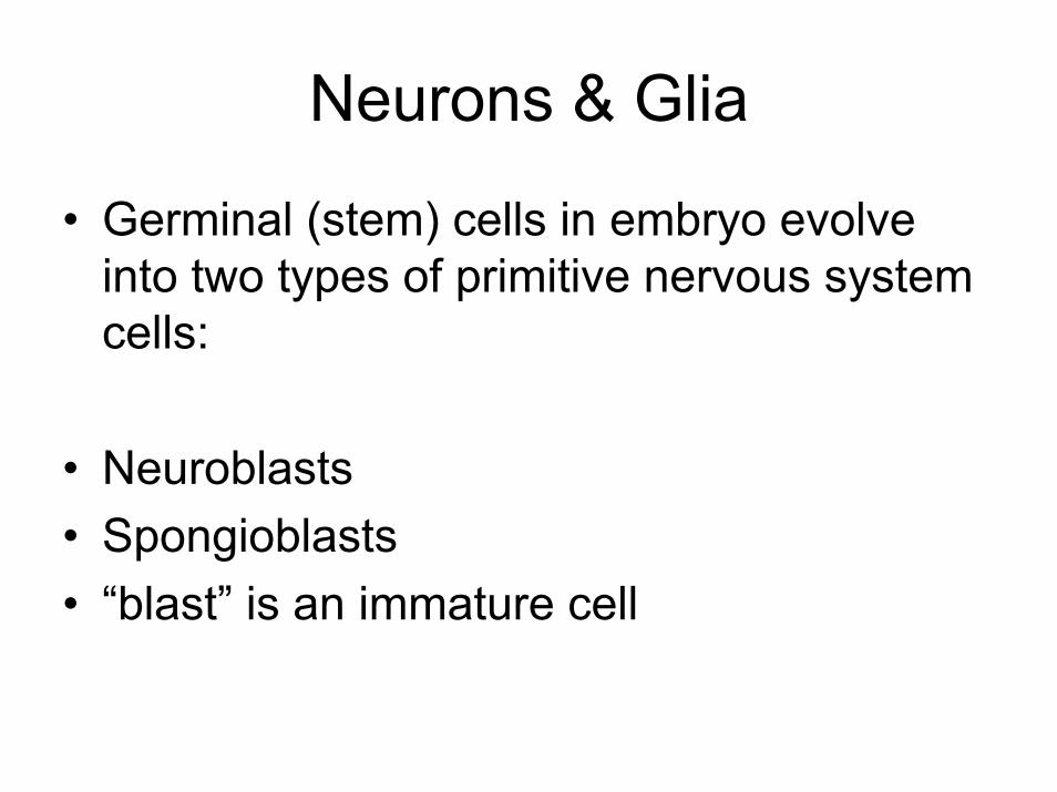
• Germinal (stem) cells in embryo evolve into two types of primitive nervous system cells:
• Neuroblasts• Spongioblasts• “blast” is an immature cell

Neurons & Glia
• Neuroblasts develop into neurons (nerve cells)
• Spongioblasts develop into glial cells (provide support functions to neurons)
• Neurons consist of a cell body with a dendrite on one side and an axon on the other

Neuron - Examples

Neuron - Examples


Glial Cells
• Astroglia – give structural support & repair neurons.
• Oligodendroglia – insulate & increase speed of transmission.
• Schwann cells – oligodendrocytes of peripheral nervous system

Glial Cells
• Microglia – perform phagocytosis (engulf foreign particles-lymphatic system’s defenses)
• Ependymal cells – line the brain’s ventricles & produce cerebrospinal fluid


Gray & White Matter
• Gray matter – named for the color of nerve cell bodies (cortex, deep nuclei)
• White matter – axons extend from neurons to form connections with other neurons. Insulating covering (oligodendrocytes) is composed of lipids & is white in color.

Gray/White Matter in Brain


Nuclei
• In CNS, a large number of cell bodies grouped together is called a nucleus.
• Nuclei have a particular function.
• In PNS, large cell body collections are referred to as ganglia.

Tracts
• Large collection of axons from a nucleus is called a tract
• Or fasciculi, fiber pathway• Carries information from one place to
another• E.g., optic tract - from eye to brain

Approaches to Study of Nervous System
• Comparative (relative to other creatures)• Developmental (changes in structure and
size during individual development)
• Cytoarchitectual (architecture of cells)• Biochemical (neurotransmitters, hormones)

Comparative Approach
• Traces development of cord and brain from simple to complex creatures.
• Phyla of animals developed through stages from floating to swimming, crawling, walking, climbing, and flying.
• Brain complexity correlated with each successive behavioral development.

Comparative Approach
• Much is still unknown• E.g., limbic system (a middle brain layer) first
developed in reptiles/amphibians• But why is still unknown• To control new modes of locomotion?• Mediate new forms of social behavior?• Support more advanced learning ability?

Comparative Approach
• This approach indicates mammals differ from other animals in large size of cortex.
• Cortex is particularly large in humans.• Cortex is thought to confer abilities unique
to mammals.• So human neuropsychology focuses on
the study of the cortex.

Developmental Approach
• Ontogenetic approach• Changes in brain structure & size followed
over course of development in an individual
• Individual brain development goes through the same stages as animal species do in their evolution.
• “Ontogeny recapitulates phylogeny”

Developmental Approach
• Cortex & its connections are immature in newborn infants.
• Correlating developing brain changes with specific behaviors is a powerful method of uncovering
• Relations between brain structures and behavioral functions

Cytoarchitectonic Approach
• Neuroantomists study the architecture of cells in cortex
• Differences in cell structure, size, shape, connections and distribution throughout cortex are studied
• Produce detailed cellular maps of cortex.


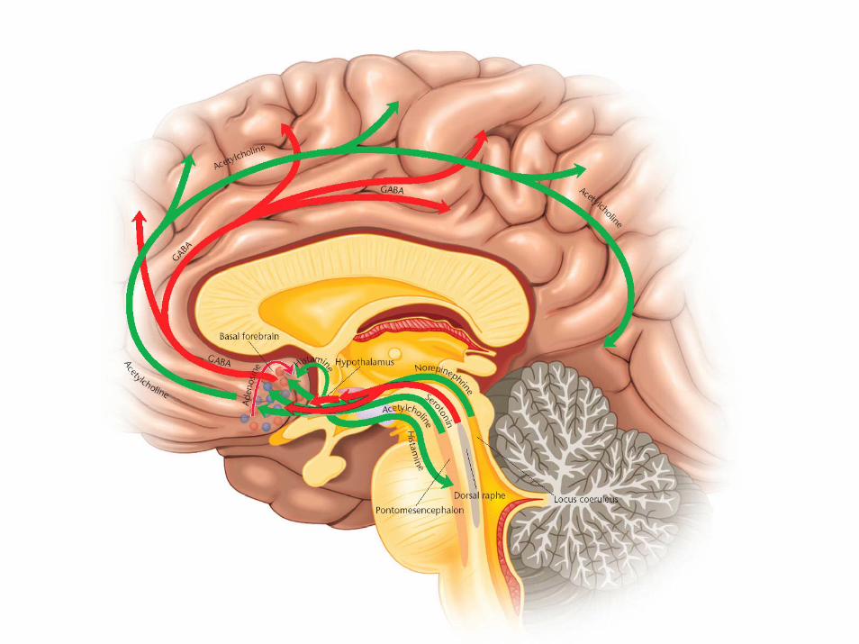
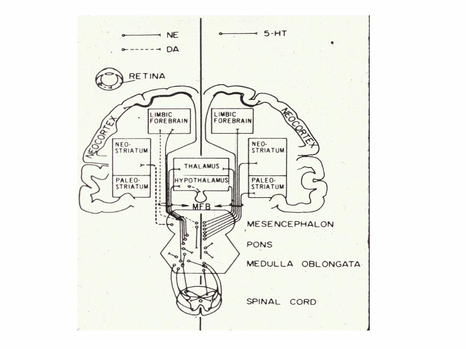
Biochemical Approach
• Cells contain specific neurotransmitters that allow them to play their special roles intracellular communication.
• Specific neurotransmitters have been related to specific behaviors and disorders
• E.g., reduction of acetylcholine (Ach) in Alzheimer’s
• Decreased dopamine (DA) in Parkinson’s

Biochemical Approach
• Increased dopamine (DA) - psychosis• Too little serotonin (5 HT) – depression
• Identification of the types of cells and distribution of transmitters across cortex helps lead to understanding biochemical basis of behavior.
Development of Brain
• Nervous system was first a spinal cord.
• Fibers from each cord segment connect to each segment of the body.
• E.g., earthworm has these organizational features.

Cord & Brainstem in Embryo

Development of Brain
• In more complex animals, one end of the animal “goes first.”
• Develops variety of receptors (nose, eyes, ears) to tell it where its going.
• A brain develops at the front end of the cord to receive this sensory information.

Development of Brain
• This primitive brain developed to receive information and tell the body what to do.
• E.g., a fish’s brain is representative of this stage.
• Mammalian equivalent = brainstem• This primitive brain consists of 3
enlargements (Table 3.4, p. 53).

Primitive (Fish) Brain
• Primitive brainstem divisions:
• Prosencephalon (“front brain”)
• Mesencephalon (“middle brain”)
• Rhombencephalon (hindbrain)

Brainstem in Primitive Brain

Mammalian Brain
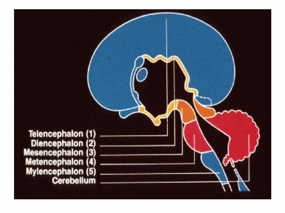
• In mammals, prosencephalon develops to form the
• Telencephalon (endbrain) and• Diencephalon (between-brain)
• Midbrain (mesencephalon) stays the same
Mammalian Brain
• Back part of primitive brain (rhombencephalon) becomes:
• Metencephalon (across-brain) and
• Myelencephalon (spinal brain)

Mammalian Brain


Mammalian Brain
• TELENCEPHALON is composed of:• Neocortex• Basal ganglia• Limbic system• Olfactory bulb• Lateral ventricles

Mammalian Brain
• DIENCEPHALON is composed of:
• Thalamus• Epithalamus (pineal body)• Hypothalamus• Third ventricle

Mammalian Brain
• MESENCEPHALON is composed of:• Tectum• Tegmentum• Cerebral aqueduct

Mammalian Brain
• METENCEPHALON is composed of:• Cerebellum• Pons• Fourth ventricle• MYELENCEPHALON is composed of:• Medulla oblongata• Fourth ventricle


Orientation of Structures
• Superior (top)• Lateral (side)• Medial or mesial (middle)• Ventral (towards the belly or s.t.’s bottom)• Dorsal (towards the back or s.t.’s top)• Anterior (front)• Posterior (back)

Planes of the Body
• Midsagittal – the plane vertically dividing the body through the midline into right and left halves.
• Sagittal – any plane parallel to the midsagittal line dividing the body into right and left portions.

Planes of the Body
• Transverse (horizontal) – any plane dividing the body into superior and inferior portions.
• Coronal (frontal) – any plane dividing body into anterior and posterior portions; at right angle to sagittal plane.


Mid-Saggital View of Brain

Transverse View of Brain

Coronal View of Brain


Orientation of Structures
• Ipsilateral – when two structures lie on the same side.
• Contralateral – if they lie on opposite sides.
• Bilateral – if one is on each side.

Orientation of Structures
• Proximal – structures that are close to one another; also closer to the midline of body.
• Distal – structures that are far away from one another; also ones that are farther away from midline
• Afferent – an approaching pathway• Efferent – pathway that is traveling away from

Spinal Cord
• Tube of nerve cells divided into segments.
• Each segment receives fibers from sensory receptors of the part of the body adjacent to it
• And sends back fibers to muscles in that part of the body.

Dermatomes
• Area of skin supplied with sensory (afferent) nerve fibers by a single spinal segment.
• Dermatomes encircle the body in a ring formation.
• Distorted in humans due to upright posture – imagine humans down on all fours.


CNS & PNS
• Central Nervous System (CNS) = brain and spinal cord
• Peripheral Nervous System = 12 cranial nerves and 31 pairs of spinal nerves.

CNS & PNS

Spinal Nerves
• 31 pairs of spinal nerves:• 8 Cervical (C)• 12 Thoracic (T)• 5 Lumbar (L)• 5 Sacral (S) and 1 Coccygeal• Cord segments connect with body
dermatomes of the same number.

Dorsal Root
• Fibers entering the dorsal portion of spinal cord carry information from sensory receptors.
• As fibers approach cord, they collect together
• This collection is called the dorsal root.• Dorsal root = sensory
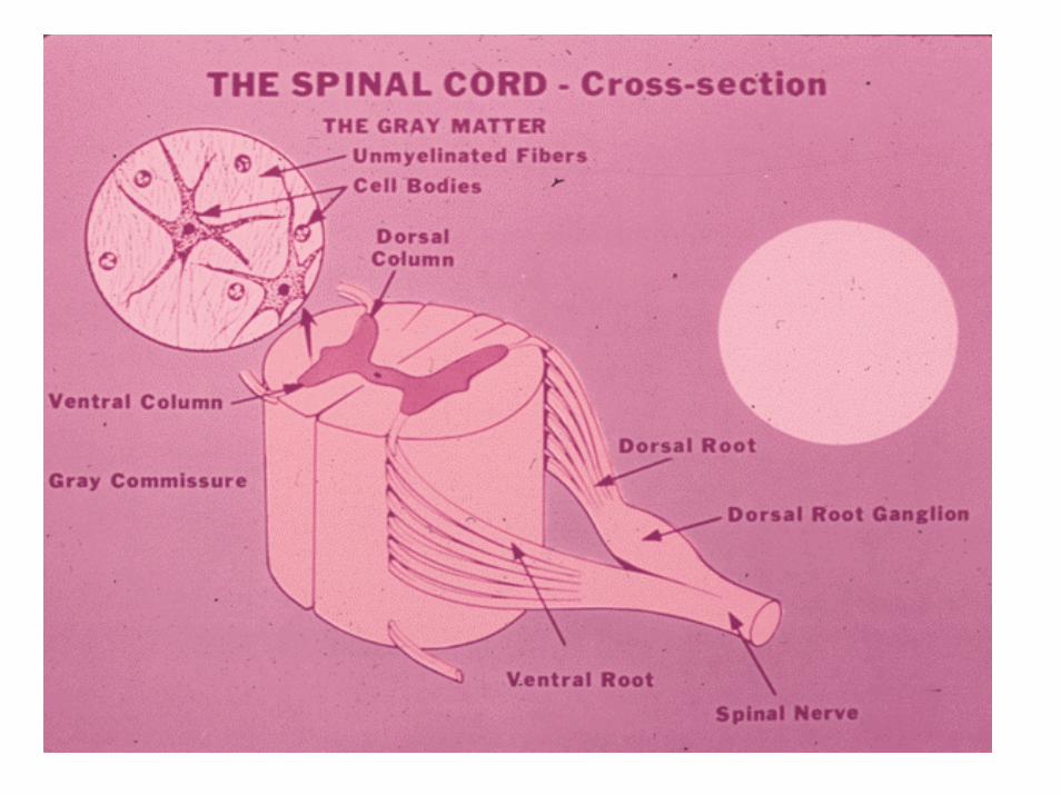

Ventral Root
• Fibers leaving the ventral (anterior) portion of cord carry information from spinal cord to muscles
• Form the ventral root.
• Ventral root = motor


Composition of Cord
• Outer portion of cord consists of white matter or tracts.
• Inner portion consists of gray matter, composed of nerve cell bodies.


Spinal Cord – Cross Section

Bell-Magendie Law
• Charles Bell – Scottish neurophysiologist• Francois Magendie – French physiologist• Cutting dorsal root caused loss of sensation.• Cutting ventral root caused loss of motor
function.• Entire nervous system organized in this fashion.
Dermatomes of Internal Organs
• Organs (liver, kidney, heart, lungs) in the body are also arranged segmentally
• Organs have no sensory representation in brain.
• Pain in organs is felt within the body portion of the dermatome it is represented in

Referred Pain
• Pain that is felt in a body part away from the site of disease or injury.
• E.g., pain originating from heart is felt in the left shoulder and left arm.
• E.g., kidney pain is felt in the back.• Brain interprets pain as coming from the
spinal segment the organ enters.

Spinal Cord Motor Defects
• If spinal cord is cut in its lower portions (lumbar, sacral), patient loses control over legs – called paraplegia.
• If cord is cut at higher levels (cervical) so patient loses control over both legs and arms – called quadriplegia

Spinal Reflex Arc
• Pinprick damages skin and stimulates pain receptors;
• Stimulated receptor activates a pain neuron which conducts information to cord;
• Pain fibers activate interneurons in cord;• Interneurons activate motor neurons to muscle;• Muscle contracts and flexes limb.

Flexor Withdrawal Reflex

Withdrawal Reflexes
• Reflex is proportional to intensity of stimulus.
• Small pain = movement of finger or hand toward body
• Large pain = movement of entire limb bringing it toward the body.
• Stimulation of pain & temperature receptors cause flexor withdrawal reflex.

Extensor Reflexes
• Tactile stimulation activates tactile & pressure receptors;
• These neurons stimulate interneurons in the cord and the motor neurons then cause the limb to extend.
• Stimulating fine (discriminative) touch and pressure receptors produce extensor movements – move limb away from body.

Brain Stem
• HINDBRAIN– Medulla Oblongata– Pons– Cerebellum
MIDBRAIN (Mesencephalon)
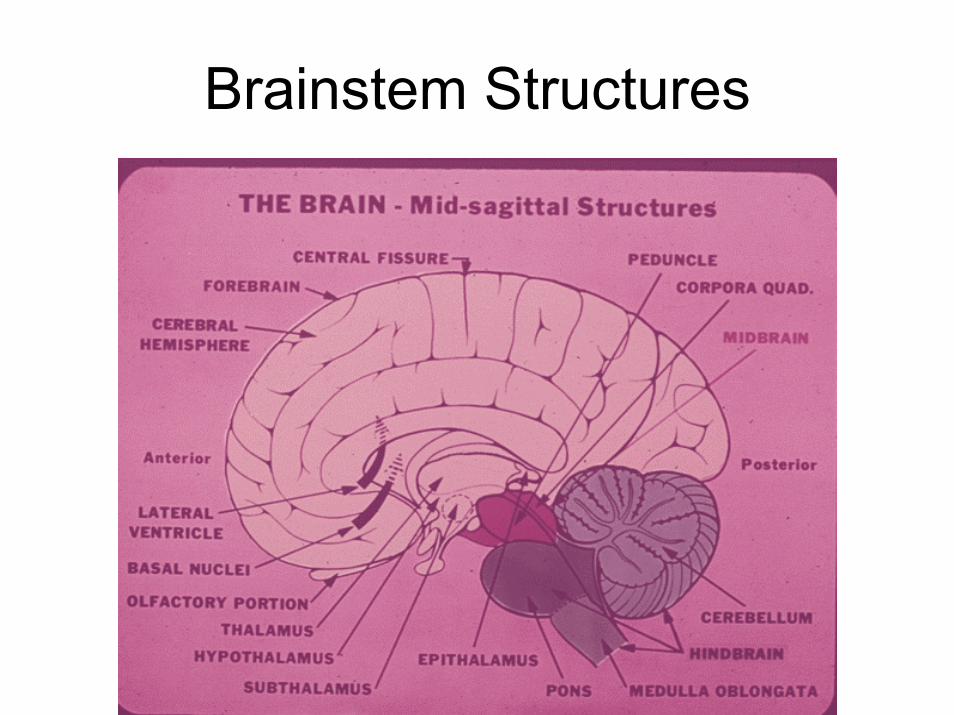
Brainstem Structures


Brainstem
• Sensory (afferent) fibers from spinal cord on way to thalamus pass through here.
• Motor (efferent) fibers from cortex pass through on way to anterior horn of cord.
• Brainstem packed with fibers and cranial nerve nuclei;
• So even small lesions have large effects.

Reticular Activating Center
• Located in central core of brainstem in midbrain, pons, & superior medulla.
• Maintenance of consciousness• Controls sleep and wakefulness • Controls arousal levels• Composed of many different brainstem
nuclei
Midbrain
• Mesencephalon – two subdivisions• Tectum (roof) – above cerebral aqueduct• Tegmentum (floor) – below aqueduct• Tectum contains 2 sets of bilaterally
symmetrical nuclei:– Superior colliculi (upper hills)– Inferior colliculi (lower hills)


Midbrain
• Superior colliculi – receives input from retina; mediates visual behaviors.
• Inferior colliculi – input from ear; mediates auditory-related behaviors.
• Tegmentum – contains some cranial nerve nuclei (primarily motor), substantia nigra, and the VTA.


Cranial Nerves
• 12 sets of nerves lying within brainstem• Sensory information from specialized
sensory systems of head to brain• Control of muscle movement of head• Important for neurological diagnosis and
localization of lesion


Cranial Nerves Function
• I = Olfactory – Smell• II = Optic – Vision• III= Occulomotor – Eye movement /
pupil constriction• IV= Trochlear – Eye movement• V = Trigeminal –Face sensation / jaw
movement• VI = Abducens – Eye movement
Cranial Nerves Function
• VII = Facial – Facial movement• VIII= Cochleo-Vestibular – Hearing/
Equilibrium• IX = Glossopharyngeal – Taste / Pharynx• X = Vagus – Heart, vessels, viscera /
movement of larynx & pharynx• XI = Spinal Accessory – Neck muscles• XII = Hypoglossal – Tongue muscles

Sensory Cranial Nerve Nuclei

Motor Cranial Nerve Nuclei
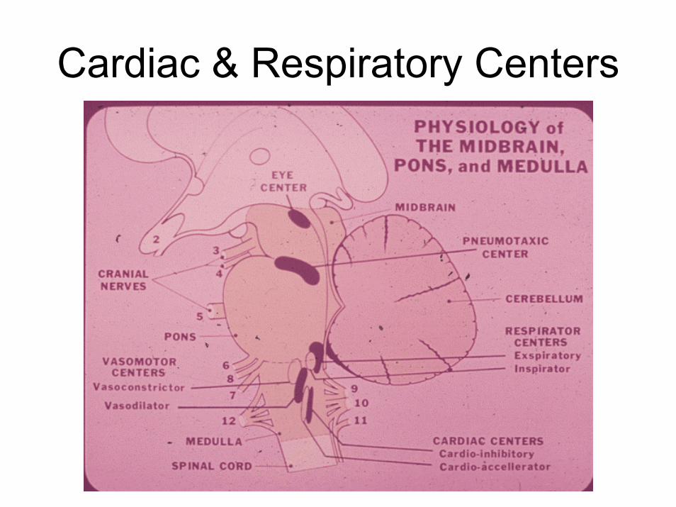
Cardiac & Respiratory Centers
Cranial Nerves
• Will be discussed in detail when the motor system is presented in Chapter 9
• Symptoms of dysfunction of the cranial nerves
• And methods to examine cranial nerve function will be presented at that time.
Cerebellum
• 3 major streams of input into cerebellum
• From Cortex• From Vestibular apparatus• From body via spinal cord

Cerebellum

Functions of Cerebellum• Major part of cerebellum receives input from
cortex = controls skilled movements.
• Other portions receives input from vestibular system = maintain body’s equilibrium.
• Parts that receive input from body senses = postural reflexes & coordinating related muscle groups.

Cerebellar Anatomy
• Surface has many narrow folds called folia
• Cortex of gray matter covers larger area of white matter
• Several nuclei lie within white matter.• Connected to brainstem via 3 major fiber
pathways – inferior, middle & superior cerebellar peduncles.


Effects of Cerebellar Damage
• Damage causes impairments of equilibrium & skilled motor activity & postural defects.
• Smooth movement broken into jerky, sequential components;